Embed Size (px)
Citation preview
Ethosuximide enhances neurogenesis
1
Ethosuximide induces hippocampal neurogenesis and reverses cognitive deficits in amyloid-β toxin
induced Alzheimer’s rat model via PI3K/Akt/Wnt/β-catenin pathway*
Shashi Kant Tiwari#,1,2
, Brashket Seth#,1,2
, Swati Agarwal1,2
, Anuradha Yadav1,2
, Madhumita Karmakar1,
Shailendra Kumar Gupta3, Vinay Choubey
4, Abhay Sharma
5,*, Rajnish Kumar Chaturvedi
1,2,*
1 Developmental Toxicology Laboratory, Systems Toxicology and Health Risk Assessment Group, CSIR-
Indian Institute of Toxicology Research (CSIR-IITR), 80 MG Marg, Lucknow, 226001, India 2Academy of Scientific and Innovative Research (AcSIR), CSIR-IITR Lucknow Campus, Lucknow
3Systems Toxicology and Health Risk Assessment Group, CSIR-IITR Lucknow, India
4Department of Pharmacology; Centre of Excellence for Translational Medicine; University of Tartu;
Tartu, Estonia. 5 CSIR-Institute of Genomics and Integrative Biology (CSIR-IGIB), New Delhi, India.
*Running title: Ethosuximide enhances neurogenesis # These two authors contributed equally
*To whom correspondence should be address: Dr. Rajnish Kumar Chaturvedi, Developmental
Toxicology Laboratory, Systems Toxicology and Health Risk Assessment Group, CSIR-Indian Institute of
Toxicology Research (CSIR-IITR), 80 MG Marg, Lucknow 226001, India, Tel: +91-522-2228227, Fax:
+91-522-2628227, E-mail: [email protected]. Dr. Abhay Sharma, CSIR-Institute of Genomics and
Integrative Biology, Sukhdev Vihar, Mathura Road, New Delhi 110025, India. Email:
Keywords: Neurogenesis, Alzheimer's disease, Ethosuximide, neural stem cells, hippocampus,
neurodeheneration
Background: Neurogenesis, the process of
generation of new neurons, is reduced in Alzheimer
disease (AD).
Results: Ethosuximide (ETH), an anticonvulsant
drug, increased neurogenesis, reduced
neurodegeneration, and reversed cognitive
impairments in rat model of AD like phenotypes.
Conclusion: ETH induces neurogenesis, thus
reverses AD like phenotypes.
Significance: ETH could be used as a therapeutic
molecule to enhance neurogenesis.
ABSTRACT
Neurogenesis involves generation of new neurons
through finely tuned multistep processes such as
neural stem cell’s (NSC) proliferation, migration,
differentiation, and integration into existing
neuronal circuitry in the dentate gyrus of the
hippocampus and sub-ventricular zone (SVZ). Adult
hippocampal neurogenesis is involved in cognitive
functions and altered in various neurodegenerative
disorders including Alzheimer’s disease (AD).
Ethosuximide (ETH), an anticonvulsant drug is used
for the treatment of epileptic seizure. However, the
effects of ETH on adult hippocampal neurogenesis
and underlying cellular and molecular mechanism(s)
are still elusive. Herein, we studied the effects of
ETH on rat multipotent NSC proliferation and
neuronal differentiation, and adult hippocampal
neurogenesis in an amyloid beta (Aβ) toxin induced
rat model of AD like phenotypes. ETH potently
induced NSC proliferation and neuronal
differentiation in the hippocampal derived NSC in
vitro. ETH enhanced NSC proliferation and
neuronal differentiation, and reduced Aβ toxin
mediated toxicity and neurodegeneration, leading to
behavioral recovery in rat AD model. ETH inhibited
Aβ mediated suppression of neurogenic and Akt-
Wnt/β-catenin pathway gene’s expression in the
hippocampus. ETH activated the PI3K/Akt and
Wnt/β-catenin transduction pathways that are known
to be involved in the regulation of neurogenesis.
Inhibition of the PI3K/Akt and Wnt/β-catenin
pathways effectively blocked the mitogenic and
neurogenic effects of ETH. In silico molecular
target prediction docking studies suggest that ETH
interacts with Akt, Dkk-1 and GSK-3β. Our findings
suggest that ETH stimulates NSC proliferation and
differentiation in vitro and adult hippocampal
neurogenesis via the PI3K/Akt and Wnt/β-catenin
signaling.
http://www.jbc.org/cgi/doi/10.1074/jbc.M115.652586The latest version is at JBC Papers in Press. Published on September 29, 2015 as Manuscript M115.652586
Copyright 2015 by The American Society for Biochemistry and Molecular Biology, Inc.
by guest on April 23, 2020
http://ww
w.jbc.org/
Dow
nloaded from
Ethosuximide enhances neurogenesis
2
Introduction
Neurogenesis, the process of generation of new
neurons, occurs throughout the life, with the
multipotent neural stem cells (NSC) in the
neurogenic brain regions such as dentate gyrus (1)
of the hippocampus and sub-ventricular zone (SVZ)
differentiating into neurons (2-4). This involves
proliferation, migration, and differentiation of NSC,
and integration of newly generated neurons into
existing neuronal circuitry (2-4). Neurogenesis plays
an important role in the regulation of learning and
memory processes, and odor discrimination (2,5). It
is negatively modulated by stress, aging, and certain
drugs, while positively affected by neurotrophic
factors, environmental enrichment and exercise (4).
Identifying small molecules that induce
neurogenesis, and understanding the associated
cellular and molecular mechanism(s), may prove
valuable in the development of regenerative
therapies for neurodegenerative and neurological
disorders. Alterations in hippocampal neurogenesis
lead to impaired learning and memory, and other
pathophysiological neurodegenerative disease state.
Several studies have suggested decreased
neurogenesis in neurodegenerative and neurological
disorders such as Parkinson’s disease, Alzheimer’s
disease (AD), Huntington’s disease, depression,
epilepsy, schizophrenia, and other age related
disorders (2,6-14). Early onset of AD is
characterized by the accumulation of amyloid-beta
(Aβ) peptides, hippocampal hyperactivity, and
alteration in neuronal networks, leading to increased
incidence of epileptic seizures and cognitive and
synaptic deficits in patients (15-18). Antiepileptic
drugs (AEDs) like levetiracetam and lamotrigine are
known to reduce overexcitation and synaptic and
cognitive deficits in animal models of AD (15,18).
However, a possible role of AEDs in neurogenesis
and associated functions has not been fully
explored.
Ethosuximide (ETH) is an AED used
commonly in the treatment of absence seizures, a
type of idiopathic generalized epilepsy in children
(19). It effectively attenuates the spike-wave
discharges that characterize absence seizures
(20,21), by blocking T-type voltage-gated Ca2+
channels and reducing low-threshold Ca2+
currents
(22). ETH delays age-related changes and extends
lifespan in C. elegans, suggesting that it may be a
potential therapeutic candidate for
neurodegenerative disorders and age-related
diseases (23). ETH reduces age-dependent toxicity
in C. elegans motor neurons against mutant TAR
DNA-binding protein-43, involved in pathogenesis
of amyotrophic lateral sclerosis (24). It has been
found to reduce infarct volume in ischemic brain
injury rat model (25), and protect neurons in the
hippocampal slice cultures against oxygen and
glucose deprivation (26). Also, it promotes neuronal
differentiation of rat muscle derived stem cells in
vitro (26), and differentiation of neonatal rat
forebrain stem cells to microtubule associated
protein-2 positive neural cell (28). Similarly,
another AED, valproic acid found to enhance
proliferation of neural progenitor cells (NPCs) in the
DG (27,28).
In the present study, we hypothesized that
ETH may affect the process of adult neurogenesis,
and assessed its effects on NSC proliferation, and
neuronal differentiation, and survival in vitro and in
the hippocampal region of the rat brain. We found
that ETH potently induces neurogenesis in vitro and
in adult hippocampus. Furthermore, we found that
the drug reverses learning and memory deficits in an
Aβ induced model of AD like phenotypes by
enhancing neurogenesis. We next elucidated the
molecular mechanisms underlying ETH induced
neurogenesis, and found that the drug acts by
activating the PI3K-Akt/Wnt-β-catenin signaling in
rat brain.
Experimental procedures
Materials: ETH, Aβ, Aβ scramble peptide, Tris,
2,5-diphenyltetrazolium bromide (MTT), alamar
blue, 5-bromo-2-deoxyuridine (BrdU), basic
fibroblast growth factor (bFGF), epidermal growth
factor (EGF), serum free neurobasal medium, N-2
supplement, B-27 supplement, and TRIzol reagent
were obtained from Gibco BRL (Rockville, MD,
USA). Bovine serum albumin (BSA), succinimide
(inactive analog of ETH), poly-L-lysine (PLL),
rabbit anti-cleaved caspase-3, mouse anti-S100-β,
rabbit anti-nestin, mouse anti-S100-β, and rabbit
anti-glial fibriliary acidic protein (GFAP) antibodies
were procured from Sigma Aldrich (29).
Lipofectamine LTX was procured from Invitrogen
(Carlsbad, USA). Cell culture products were
purchased from Gibco-BRL-Life Technologies
(UK). Mouse anti-BrdU primary antibody was
procured from Santa Cruz Biotechnology (29).
Monoclonal mouse anti-neuronal nuclei (NeuN) and
nitrocellulose membrane were obtained from
by guest on April 23, 2020
http://ww
w.jbc.org/
Dow
nloaded from
Ethosuximide enhances neurogenesis
3
Chemicon (Millipore, Billerica, MA, USA). Rabbit
anti-Sox-2, mouse anti-β-actin, rabbit anti-CNPase,
mouse anti-BrdU, rabbit anti-doublecortin (DCX),
rabbit anti-TUJ-1, rabbit anti-phospho-PI3K, rabbit
anti-PI3K, rabbit anti-phospho AKT, rabbit anti-
AKT, mouse anti-phospho-histone-H3, rabbit anti-
GSK3-β, rabbit anti-p-GSK-3α/β, rabbit anti-β-
catenin, and rabbit anti-p-β-catenin primary
antibodies were obtained from Cell Signaling
Technology (Danvers, MA, USA). Alexa Fluor 488
and Alexa Fluor 594 conjugated secondary
antibodies were purchased from Molecular Probes
(Invitrogen, USA). Primers were procured from
Integrated DNA Technologies (IDT; Coralville,
USA) and SYBR Green from Applied Biosystems.
Anti-fade mounting medium with DAPI was
obtained from Vector Labs (Vectashield, Vector
Laboratories, CA, USA). Culture wares were
procured from Nunc (Roskilde, Denmark).
Animals and ETH treatment: Adult Wistar rats
were obtained from Animal Breeding Colony of the
CSIR-Indian Institute of Toxicology Research,
Lucknow and housed under 12h light/dark cycle and
temperature of 25+20C. Animals were provided ad
libitum access to the drinking water and pellet diet
and handled according to the guidelines laid down
by the Institute’s Ethical Committee for Animal
Experiments.
Adult male rats received daily single
intraperitoneal (i.p.) injection of ETH (125 mg/kg
body weight) for consecutive three days from PND
42-45 (acute treatment) or two weeks from PND 42-
56 (chronic treatment). This dose was selected on
the basis of therapeutic or pharmacologic relevant
levels of ETH as described earlier (25,30). Rats in
control group received normal saline as vehicle. To
analyze the effects of ETH on cell proliferation, rats
from both control and ETH treated groups received
daily single i.p. injection of BrdU (50mg/kg of body
weight) for five consecutive days starting from
PND52-56. Animals were sacrificed 4h after last
BrdU injection at PND56, and brains were dissected
for immunohistochemical detection of proliferating
cells. To evaluate the effect on NSC survival and
their differentiation potential in neuron and glial
cells, a set of animals was allowed to survive for
additional 3 weeks (upto PND77) after last BrdU
injection. Animals were sacrificed at PND77 for
double immunofluorescence analysis of BrdU and
neuronal or glial markers.
Preparation of Aβ induced rat model of AD like
phenotypes and ETH treatment: Rat model of Aβ
induced AD like phenotype was prepared as
described earlier (31). Rats were anesthetized with
ketamine (i.p., 100 mg/kg, body weight) and
xylazine (30mg/kg body weight), and fixed in a
stereotaxic apparatus. Rats were given stereotaxic
injection of 2μl of Aβ (1-42) (0.2μg/μl dissolved in
0.9% normal saline), into both side of the
hippocampus at AP -3.3, L 2.0, V 4.0 (coordinates
in mm with respect to bregma), using an auto-
injector pump. Two weeks after Aβ administration,
rats were treated with ETH (125mg/kg body weight
i.p) for two weeks (PND42-56) as per the following
experimental plan.
Experimental groups:
1. Group I (Sham control) : received stereotaxic 2µl
of normal saline as vehicle
2. Group II (Aβ group) : stereotaxic intra
hippocampal 2µl Aβ injection
3. Group III (Aβ+ETH): Aβ injected rats received
ETH
4. Group IV : received ETH
5. Group V (Inactive analog of ETH; succinimide):
received succinimide (125mg/kg body weight
i.p) for two weeks (PND42-56)
6. Group VI (Aβ scrambled peptide): received
stereotaxic intra hippocampal injection of 2µl Aβ
scrambled peptide
7. Group VII (Aβ scrambled peptide+ETH):
received scrambled peptide and ETH
8. Group VIII (succinimide+ETH): received
succinimide and ETH
A set of rats was sacrificed at PND56 for cell
proliferation study, while another at PND77 for cell
survival / neuronal differentiation study in the DG
region and SVZ, as described earlier (31).
Primary hippocampal NSC culture: NSC was
isolated from the hippocampal tissue of embryonic
day 12 (ED12) rat fetuses following our earlier
study (32). Hippocampal tissues were dissected in
HBSS, minced, and incubated in 0.1% trypsin for 30
minutes at 370C. Single cell suspension was
prepared by gentle trituration and cell viability was
determined by trypan blue dye. Cells were plated in
serum free neurobasal medium containing N-2
supplement (1%), B-27 supplement (2%), EGF (10
ng/ml), bFGF (10ng/ml) and 1% antibiotic-
antimycotic solution. The NSC were allowed to
grow as neurospheres.
by guest on April 23, 2020
http://ww
w.jbc.org/
Dow
nloaded from
Ethosuximide enhances neurogenesis
4
BrdU and alamar blue cell proliferation assay:
BrdU and alamar blue cell proliferation assays were
carried out to see the effects of ETH on cell
proliferation. NSC were plated in black bottom 96-
well microplates at a density of 10,000 cells/well.
The cells were treated with different concentrations
of ETH (25, 50, 100, 200 and 400 µM) dissolved in
DMSO for 48h. Vehicle treated cells served as
control. After treatment, alamar blue was added and
cells were incubated at 370C for 2h. Background
florescence was measured by adding 10µl alamar
blue solution/well in medium having no cells.
Alamar blue dye reduction in term of fluorescence
intensity was measured at 530nm excitation and
590nm emission. Values of background florescence
were subtracted from experimental values.
Experiments were performed thrice, with three
replicates per experiment. Relative cell proliferation
= [Absorbancesample /Absorbancecontrol] X 100.
Results were expressed in terms of alamar blue
reduction % control.
Similarly, to assess cell proliferation, control
and ETH treated NSC cultures were labeled with
BrdU (20μM) for 12h before fixing cells with 4%
paraformaldehyde and processing for immuno-
fluorescent analysis.
Neurosphere growth kinetics assay: Neurosphere
growth kinetics assay was carried out to study the
effects of ETH on cell proliferation and neurosphere
formation. Hippocampal single cell suspension was
plated in 12 well plate at a density of 50,000
cells/well in neuro basal medium containing B-27,
N-2, bFGF and EGF. Cultures were treated with
ETH (100µM). The number and size of
neurospheres were analyzed using Nikon Eclipse Ti-
S inverted fluorescent microscope equipped with
Nikon Digital Sight Ds-Ri1 CCD camera and NIS
Elements BR imaging software (Nikon, Japan) as
described earlier (31).
Immunocyto/histochemical analysis of
proliferating cells in NSC cultures and in the
brain: Rats were sacrificed at PND56 by
transcardial perfusion, and every 6th
serial coronal
brain sections of 40μm thickness beginning at
bregma -3.14 to -5.20mm through the dorsal
hippocampus (dentate gyrus) and 0.70 to -0.80mm
through the SVZ were taken for
immunohistochemical analysis of proliferating cells
as described earlier (31). The sections /cultures were
treated with 2N HCl for 30min, followed by
neutralization with borate buffer. The endogenous
peroxidase activity was inhibited by H2O2 and
nonspecific binding sites were blocked with 3%
NGS, 0.5% BSA and 0.1% Triton X-100. The
sections were subsequently incubated with mouse
anti-BrdU primary antibody (1:500) followed by
incubation in peroxidase linked secondary antibody
(1:200). Color was developed with 3-3'
diaminobenzidine and visualized under light
microscope. For immunofluorescence analysis,
paraformaldehyde fixed sections/cultures were
incubated with mouse anti-phospho-histone-H3
(1:500) and anti-BrdU primary antibodies (1:500)
followed by Alexa Fluor conjugated secondary
antibody, and visualized under inverted fluorescent
microscope.
Neuronal differentiation and cell fate analysis by
double co-immunofluorescence in the brain and
NSC cultures: The effects of ETH on neuronal and
glial differentiation and cell phenotype of BrdU+
proliferating cells in the hippocampus and SVZ (at
PND77) and NSC cultures were studied by double
immunofluorescence analysis following earlier
published method (31). NSC cultures /coronal
sections were co-labeled with BrdU/nestin (NSC
marker), BrdU/DCX (immature neuron marker),
BrdU/NeuN (mature neuron marker), BrdU/GFAP
(glial cell marker), Sox-2/GFAP (glial and neural
progenitor cell markers), S100-β (glial cell marker),
CNPase (oligodendrocyte marker). Sections were
incubated in mouse anti-BrdU (1:500), rabbit anti-
DCX (1:200), rat anti-NeuN (1:500), mouse anti-
nestin (1:500), rabbit anti-GFAP (1:100), and mouse
anti-Phospho-histone-H3 primary antibodies
(1:500), rabbit anti-Sox-2 (1:200), mouse anti-
S100-β (1:200), rabbit anti-CNPase (1:250) for 24h
at 40C. Secondary antibodies used were anti-mouse
and anti-rabbit Alexa Fluor 488 (1:200), and anti-
rabbit, anti-mouse, and anti-rat Alexa Fluor 594
(1:200). Sections were mounted with DAPI
containing Hard SetTM
anti-fade mounting medium
(Vectashield, Vector Laboratories, CA, USA).
Slides/cultures were analyzed for fluorescence co-
labeling under Nikon Eclipse Ti-S inverted
fluorescent microscope equipped with Nikon Digital
Sight Ds-Ri1 CCD camera and NIS Elements BR
imaging software (Nikon, Japan).
by guest on April 23, 2020
http://ww
w.jbc.org/
Dow
nloaded from
Ethosuximide enhances neurogenesis
5
Cell Quantification: Unbiased quantification of
proliferating and differentiating cells in the DG
region of the hippocampus and SVZ was carried out
by a person blind to the experimental groups
(1,32,33). These brain regions were identified at low
magnification (10X) and a contour was drawn.
Quantification was done in every sixth section, apart
by at least 240μm in 1/6 series, with total six
sections per rat were analyzed as described earlier
(1,32,33). Each and every BrdU/ phospho-histone-
H3+, BrdU/DCX
+, BrdU/NeuN, BrdU/GFAP,
BrdU/DCX, BrdU/nestin and Sox-2/GFAP and
nestin/phospho-histone-H3 co-labeled cells were
counted in each section. BrdU+ cells were counted
for BrdU/NeuN, BrdU/GFAP, BrdU/DCX,
BrdU/nestin and Sox-2/GFAP, nestin/phospho-
histone-H3 co-labeling at magnification of 600X in
the DG and SVZ. The number of labeled cells was
determined by bilaterally counting cells in total six
sections and averaged for each rat. The total number
of labeled cells was determined by multiplying the
total number of BrdU+ cells from six sections by the
section periodicity i.e 6, and reported as the total
number of BrdU+
cells co-labeled with phenotype
marker per rat.
Gene expression analysis by qRT-PCR:
In order to study the expression of genes involved in
neurogenesis and PI3K/Akt/Wnt/β-catenin
pathways, qRT-PCR analysis was carried out as
described earlier (31,34,35). The sequences for
primers are available on request.
Western immunoblot analysis:
After respective treatments, NSC cultures were
lysed with cell lytic MT Mammalian Tissue
Lysis/Extraction Reagent. Western blot analysis was
performed as described earlier (31,36). Membranes
were blocked and incubated with p-PI3K (1:1000),
PI3K (1:1000), p-Akt (1:500), Akt (1:1000), GSK3-
β (1:1000), p-GSK-3α/β (1:500), β-catenin (1:1000),
p-β-catenin (1:1000) and -actin (1:10,000) primary
antibodies. Protein bands were quantified using
Scion Image for Windows (NIH, USA).
Treatment of NSC with Wnt/β-catenin pathway
inhibitor and PI3K/Akt inhibitor in the presence
and absence of ETH: To evaluate the specific
involvement of ETH induced activation of the
PI3K/Akt/Wnt pathways in NSC proliferation and
neuronal differentiation, hippocampus-derived NSC
cultures were treated with potent Wnt antagonist
recombinant human protein Dkk-1 (100nM) and
PI3K/Akt inhibitor LY294002 (10nM) in the
presence and absence of ETH. After respective
treatments, NSC proliferation was assessed by BrdU
incorporation assay and neuronal differentiation was
assessed by immunocytochemistry. Further, western
blotting was performed to study protein levels of
GSK-3α/β, p-GSK-3α/β, β-catenin and p- β-catenin.
In vivo and in vitro β-catenin knockdown: β-
catenin siRNA was stereotaxically injected into the
hippocampus. Knockdown was done with pool of
target specific siRNA sequences for β-catenin and
scramble sequences. After knockdown, rats were
treated with ETH, and effects on neuronal
differentiation were assessed using
immunohistochemistry.
Similarly, transient transfection with β-
catenin siRNA was carried out in NSC cultures.
Transient transfection was performed with 10nM β-
catenin siRNA at a cell confluence of approximately
70% using Lipofectamine LTX transfection reagent.
After knockdown, effects on neuronal
differentiation were assessed using
immunohisto/cytochemistry.
Conditioned avoidance response (CAR): The
effect of ETH on learning and memory ability in Aβ
induced rat model of AD like phenotypes was
measured following assessment of two-way
conditioned avoidance behavior using shuttle box as
described earlier (37,38).
In silico target prediction studies:
To study the mechanism of the Akt/Wnt/β-catenin
pathway mediated regulation of hippocampal
neurogenesis, we performed molecular docking
studies with key regulatory enzymes of these
pathways as described earlier (31). The structure of
ETH was retrieved from the PubChem server and
submitted into the PharmMapper for potential
human target identification. Protein Data Bank IDs
of all the targets were converted into UniProtKB
IDs using UniProt ID Mapping tool. In order to map
the molecular targets and predict the pathway
components associated with the proteins targeted by
ETH, pathway component analysis was carried out
using PANTHER (Protein ANalysis THrough
Evolutionary Relationship). Molecular docking
studies were conducted to identify the ETH
by guest on April 23, 2020
http://ww
w.jbc.org/
Dow
nloaded from
Ethosuximide enhances neurogenesis
6
orientation within the binding pockets of the human
targets involved in various signaling pathways
related to cellular differentiation in the brain.
Docking study was performed using the CDOCKER
module available in Discovery Studio molecular
simulation package (version 3.5; Accelrys, San
Diego, CA). The virtual hits were selected based on
two scoring functions. Scoring is made by adding
the usual intra-ligand energy terms (bonded and
non-bonded contributions) and protein-ligand non-
bonded interactions. Each of the compounds was
minimized with CharmM force field to allow for
generation and analysis of a wide range of
molecular simulations.
Statistical analysis: The mean significant
difference in the experimental groups was
determined using one-way analysis of variance.
Comparison of least significant differences among
two groups was determined by taking t-values for
error and keeping degrees of freedom at the 5%
level of significance and 95% confidence interval.
P-values of 0.05 were considered to be statistically
significant.
Results
ETH enhances hippocampal derived NSC
proliferation and neurospheres formation in
vitro: We carried out alamar blue cell proliferation
assay, MTT assay, and neurosphere growth kinetics,
in order to assess the effects of ETH on proliferation
and viability of NSC derived from the hippocampus.
NSC cultures were incubated with different
concentrations of ETH (25, 50, 100, 150, 200 and
400µM) for 48h. ETH significantly enhanced NSC
proliferation at 50, 100, and 150µM in alamar blue
assay (Fig. 1A). Among the concentrations used,
100µM ETH showed highest NSC proliferation,
while 25µM had no effect on proliferation. The
higher doses (200 and 400µM) of ETH caused
significantly decreased NSC proliferation. Similarly,
MTT assay also suggested that lower concentrations
of ETH (50 and 100µM) significantly increased
NSC proliferation/ viability, whereas higher doses
(>150µM) caused a decrease in NSC proliferation
(Fig. 1B). The effects of ETH on NSC proliferation
were further confirmed by BrdU
immunocytochemistry. Quantification of BrdU+
cells suggested significant increase in NSC
proliferation by 100µM ETH (Fig. 1C), while 200
and 400µM ETH decreased cell proliferation (data
not shown). These findings showed that the low
concentration of ETH induces proliferation of NSC,
while higher concentrations of ETH are cytotoxic.
Further, we performed a neurospheres formation
assay, in order to assess, if ETH has any effect on
the number of the hippocampal multipotent NSC.
Neurospheres are free floating spherical clusters of
several multipotent NSC, formed in the presence of
specific mitotic growth factors in vitro. The number
and size of clonal neurospheres in vitro is the
measure of absolute putative NSC in vivo. Thus, we
analyzed the gross morphology, size and the number
of neurospheres in control and ETH treated NSC
cultures (Fig. 1D). ETH significantly increased the
number and size of primary and secondary clonal
neurospheres as compared to control (Fig. 1D).
These results suggest that ETH treatment increases
the number of multipotent NSC and hence
neurosphere formation.
We next studied co-expression of BrdU
with nestin in NSC culture. Nestin, an intermediate
filament protein is a molecular marker for
multipotent NSC. Nestin is required for proliferation
and self-renewal of NSC (39). The number of BrdU
and nestin co-labeled cells was significantly up-
regulated by ETH treatment in NSC culture (Fig.
1E). ETH significantly enhanced the mRNA
expression of nestin as compared with control (Fig
1F). These findings demonstrated NSC proliferation
enhancing potential of ETH in vitro.
ETH enhances NSCs proliferation and neuronal
differentiation in the hippocampus of the adult
rats: In order to study the effects of chronic ETH
treatment on NSC proliferation, we counted BrdU+
cells in the SVZ and DG region of the hippocampus.
The BrdU labeled proliferating cells were visualized
by immunohistochemistry. BrdU+
cells were of
varying shape, densely stained, and mostly located
in the small groups of two or more in the DG region
of the hippocampus and SVZ (Fig 2A-B). BrdU+
cells were found located in granular cell layer
(GCL) and hilus regions of the DG. The number of
BrdU+
cells in the DG and SVZ was significantly
increased in rats chronically treated with ETH for
two weeks, compared to control (Fig. 2A-C). The
total number of BrdU+
cells in the DG and SVZ was
not significantly altered by acute treatment of ETH
for 3 days, compared to control (data not shown).
Next, we studied the effects of ETH on
phosphorylation of histone-H3, a nucleosomal core
by guest on April 23, 2020
http://ww
w.jbc.org/
Dow
nloaded from
Ethosuximide enhances neurogenesis
7
protein. Phosphorylation of histone-H3 (Ser10) is a
crucial mitotic event, and a marker of proliferating
cells (40). Thus, we carried out immuno co-labeling
of nestin with phospho-histone. We found that the
number of nestin/phospho-histone-H3+ cells in the
DG region was significantly enhanced by chronic
ETH treatment, suggesting the presence of cells
undergoing mitosis (Fig 2D-E). ETH also
significantly enhanced Nestin mRNA expression in
the hippocampus (data not shown). These studies
suggested that ETH treatment increases cell
proliferation in rat brain.
Further, the effects of ETH on pool of NSCs
and its fate/ phenotypes were studied by co-labeling
of neuronal marker (NeuN) with BrdU, NPCs
marker (Sox-2) with GFAP. ETH treatment
enhanced the number of Sox-2+ cells, however, no
significant difference observed in Sox-2/GFAP+ co-
labeled cells population (Fig 3A-B). These results
suggest that ETH significantly induced population
of NPCs, while showed no effect on glial
population. Effects of ETH on population of neuron
(NeuN), glial cells (S100-β) and oligodendrocytes
(CNPase) were studied. The number of
BrdU/NeuN+ cells was enhanced by ETH treatment
(Fig 3C-D), but no significant changes were
observed in the number of S100-β+
(Fig 3E-F) and
CNPase+
cells (Fig 3G-H) in the hippocampus.
These results suggest that ETH treatment
significantly enhanced neuronal differentiation, but
had no significant effects on glial and
oligodendrocytes differentiation of proliferating
cells.
ETH induces neuronal differentiation, and
reverses Aβ mediated inhibitory effects on the
hippocampal neurogenesis: BrdU+ proliferating
new born cells in the DG have different fates, where
they may differentiate into neurons or glia. Thus,
we studied the effects of ETH treatment on cell fate
/ phenotypes of newly born cells in the DG, by co-
labeling cells with BrdU/DCX (a marker for
immature newborn neurons), BrdU/NeuN (marker
of mature neurons) and BrdU/GFAP (marker for
glial cells). Several studies have implicated reduced
adult hippocampal neurogenesis in the pathogenesis
of AD. We and others have previously shown that
the pathogenic protein in AD brain, Aβ reduces
adult hippocampal neurogenesis in rodents
(9,31,41). Next, we studied the effect of ETH
treatment on Aβ mediated inhibitory effects on the
hippocampal neurogenesis in an Aβ induced rat
model of AD like phenotypes. We found that
stereotaxic intrahippocampal injection of Aβ caused
significantly decreased proliferation of NSC in the
DG (Fig. 4A-B). The number of BrdU labeled cells
was significantly reduced by Aβ in the
hippocampus. ETH treatment significantly enhanced
the number of BrdU+
cells (Fig. 4A-B).
Interestingly, Aβ scrambled peptide and
succinimide; an inactive analog of ETH showed no
significant effect on the number of BrdU labeled
cells (Fig. 4A-B). Further, we studied the effects of
ETH on neuronal differentiation in the
hippocampus. We found that most of the
BrdU/DCX co-localized cells were present in the
GCL of the DG in all the groups. A significant
inducing effect of ETH treatment on newborn
neuron population was observed (data not shown).
ETH significantly increased the number of
BrdU/DCX co-labeled cells as compared to control
rats (data not shown). These results indicated that
ETH increases neuronal differentiation of new born
proliferating cells. Interestingly, we observed that
intrahippocampal injection of Aβ significantly
inhibited neuronal differentiation (data not shown).
The number of BrdU/DCX co-labeled cells was
significantly reduced in Aβ injected rats as
compared with control group. ETH treatment caused
significantly increased neuronal differentiation, as
evident from enhanced number of BrdU/DCX co-
labeled cells in the DG in rat model of AD like
phenotypes (data not shown). Thus, ETH treatment
was found to significantly enhance neuronal
differentiation and reduced Aβ mediated inhibitory
effects on neurogenesis.
To study the effects of ETH treatment on
long term survival of new born neurons, we carried
out co-localization of BrdU with NeuN. We found
that most of the BrdU+
cells were co-localized with
NeuN mainly in the GCL (Fig 4C-D). The number
of BrdU/NeuN co-labeled cells was significantly
increased in ETH treated rats as compared to control
rats, suggesting that most of the cells that
incorporated BrdU in the GCL differentiated into
mature neurons (Fig 4C-D). Aβ significantly
reduced the number of BrdU/NeuN positive cells,
which was prevented by ETH treatment (Fig 4C-D).
Aβ scrambled peptide and succinimide showed no
significant effect on neuronal differentiation (Fig.
4C-D). These results suggested that ETH treatment
by guest on April 23, 2020
http://ww
w.jbc.org/
Dow
nloaded from
Ethosuximide enhances neurogenesis
8
induces neuronal differentiation and long term
survival of new born neurons.
ETH reduces Aβ induced neuronal degeneration
in the hippocampus: Next, we studied the effects
of ETH on Aβ induced hippocampal neuronal
degeneration/apoptosis by fluoro-jade B staining
and activated caspase-3 (CC-3) co-labeling with
BrdU. ETH significantly reduced the number of Aβ
mediated enhanced fluoro-jade B+ cells in the
hippocampus (Fig. 5A-B). Further, ETH also
reduced the number of Aβ mediated enhanced
apoptotic cells co-labeled with BrdU (Fig. 5C-D).
More interestingly, we found that scrambled peptide
of Aβ and inactive analog of ETH (succinimide) had
no significant effects on fluoro-jade B+ and
BrdU/CC3+cells (Fig. 5A-D). These results suggest
that ETH ameliorates Aβ induced neuronal
degeneration and apoptosis in the hippocampus.
ETH enhances expression of neurogenic and the
Wnt/β-catenin pathway genes in the
hippocampus of rat model of AD like phenotypes:
The Wnt/β-catenin signaling plays an important role
in the regulation of adult hippocampal neurogenesis
and self-renewal of NSC/progenitor cells (42-44).
The canonical Wnt/β-catenin signaling has been
implicated in pathophysiology of AD and other
neurodegenerative diseases (31). We assessed the
effects of ETH treatment on mRNA expression of
neurogenic and the Wnt pathway genes in the
hippocampus of Aβ injected rats, by qRT-PCR.
Several neurogenic genes and transcription factors,
such as DCX, β-Tubulin, Nestin, Neurogenin,
Neuroligin, Pax-6, Stat-3 and Neuro-D1 regulate
neurogenesis (31). ETH treatment significantly up-
regulated the expression of these genes in the
hippocampus (Fig. 6A). Interestingly, Aβ caused a
decrease in the expression of these neurogenic
genes, and this effect of Aβ was reversed by ETH
(Fig. 6A).
Next, we assessed the mRNA expression of
the regulatory molecules, receptors and transcription
factors of the Wnt pathway. ETH enhanced the
expression of Wnt1, Wnt3, Dishevelled, Wnt
receptors (Frizzled 1 and LRP-6), Axin-1, LEF-1, β-
catenin, APC, and cyclin-D1, as compared to control
(Fig. 6B). However, the expression of Wnt5 was not
affected (data not shown). On the other hand, ETH
caused significantly decreased expression of
negative regulatory genes of the Wnt pathway, Dkk-
1 and GSK-3β, while the expression of Wif-1 was
not changed. Dkk-1 is a negative regulator of the
Wnt pathway and acts through inhibition of LRP-
5/6. Aβ treatment was found to inhibit the
expression of the genes involved in regulation of the
Wnt pathway in the hippocampus (Fig 6B).
Interestingly, ETH significantly blocked the Aβ
mediated inhibitory effects on the expression of
these genes in the hippocampus. These results
suggest that ETH inhibits the Aβ mediated
alterations in the expression of the Wnt pathway
genes.
ETH induces phosphorylation of GSK-3β
reduces β-catenin phosphorylation, and
inhibition of the Wnt pathway blocks ETH
induced NSC proliferation and differentiation:
The β-catenin and GSK-3β are critical regulator of
the Wnt/β-catenin canonical pathway. In the absence
of Wnt ligands, GSK-3β phosphorylates
cytoplasmic β-catenin for ubiquitin mediated
proteasomal degradation. In the presence of Wnt
ligands, cell surface receptors get activated, leading
to inhibition of GSK-3β, and decreased
phosphorylation of cytoplasmic β-catenin.
Decreased phosphorylation of β-catenin results in its
increased nuclear translocation and activation of the
Wnt pathway. We found that non cytotoxic
concentration of ETH (100µM) significantly
increased GSK-3β phosphorylation and inhibited β-
catenin phosphorylation in time dependent manner
in hippocampal derived NSC culture (Fig 7A-D).
Inhibition of the Wnt pathway through Dkk-1
(inhibits interaction of Wnt with LRP-5/6) reversed
ETH induced effects on GSK-3β and β-catenin
phosphorylation (Fig 8A-C).
We next studied the role of the Wnt/β-
catenin pathway inhibition in ETH mediated
increase in the proliferation and neuronal
differentiation of hippocampal derived NSC in vitro.
The NSC cultures were treated with Dkk-1 protein
in the presence and absence of ETH, and the effects
on proliferation and differentiation were studied
through immunocytochemistry. Dkk-1 significantly
reduced the number of BrdU positive cells and β-
tubulin positive neurons in cultures, suggesting
reduced NSC proliferation (Fig. 8D-E), and
neuronal differentiation (Fig. 8F-G). Interestingly,
co-treatment of Dkk-1 significantly inhibited NSC
proliferation and neuronal differentiation enhancing
effects of ETH via the Wnt/β-catenin pathway.
by guest on April 23, 2020
http://ww
w.jbc.org/
Dow
nloaded from
Ethosuximide enhances neurogenesis
9
ETH activates the PI3K/Akt signal transduction
pathway in adult hippocampal NSC in vitro: The
PI3K/Akt pathway is involved in the regulation of
neurogenesis in the hippocampus and other regions
of the brain (45-47). Further, GSK-3β inhibition and
the Wnt/β-catenin pathway activation through
PI3K/Akt is very well established (45,48).
Therefore, to determine, if ETH induces
hippocampal neurogenesis through the PI3K/Akt
pathway, mRNA expression and phosphorylation of
PI3K (Tyr 458) and Akt (Thr 308) were analyzed in
adult hippocampal NSC culture in vitro. ETH
significantly enhanced mRNA expression of PI3K
and Akt (Fig. 9A). Next, the basal levels of PI3K
and Akt, and their phosphorylation were evaluated
following ETH treatment. Basal levels of PI3K and
Akt were not altered by ETH in cultured
hippocampal NSC (Fig 9B-D). Interestingly, levels
of phosphorylated PI3K and Akt were significantly
up-regulated in NSC following ETH treatment.
These results suggested that the PI3K/Akt pathway
is activated by ETH in cultured hippocampal NSC
(Fig 9B-D).
Blockade of the PI3K/Akt pathway inhibits ETH
induced hippocampal NSC neuronal
differentiation: We further studied, whether
specific activation of the PI3K/Akt signaling
pathway mediates the action of ETH on neuronal
differentiation in hippocampal NSC in culture. NSC
cultures were treated with ETH in the presence and
absence of LY294002, an inhibitor of the PI3K/Akt
pathway. ETH enhanced the number of Tuj-1
positive neurons in NSC cultures (Fig. 9E-F). Pre-
treatment with the Akt inhibitor resulted in
significantly reduced number of Tuj-1 positive
neurons. Interestingly, Akt inhibitor significantly
blocked neuronal differentiation enhancing potential
of ETH, by reducing the number of Tuj-1 positive
neurons (Fig.9E-F). Further, ETH significantly
enhanced the phosphorylation of GSK-3β, which
was significantly blocked by LY294002 (Fig. 9G-
H). These results suggested that the PI3K/Akt
pathway is involved in the stimulatory effects of
ETH on neural differentiation of NSC.
Knockdown of β-catenin in the hippocampus and
in NSC culture reduces ETH induced neuronal
differentiation: To examine ETH mediated
neuronal differentiation via activation and specific
involvement of Wnt/β-catenin pathway, genetic
knockdown of β-catenin was carried out both in
vitro and in vivo. β-catenin mediated Wnt signaling
is involved in NSC proliferation, fate determination
and neuronal differentiation during development
(49,50). After stereotaxic injection of β-catenin
siRNA in the hippocampus, fate of NSC was studied
immunohistochemically by co-labelling of BrdU
with NeuN (Fig. 10A). We found that knockdown of
β-catenin caused an inhibitory effect on ETH
induced increase in the number of BrdU/NeuN co-
lebelled cells (Fig. 10B). Similarly, in vitro studies
indicated that β-catenin knockdown suppresses
neuronal differentiation in ETH treated NSC
cultures (Fig. 10C-D). Scramble β-catenin siRNA
showed no significant effect on neuronal
differentiation. Together, the in vitro and in vivo
results suggested that ETH promotes neuronal
differentiation via the β-catenin mediated signaling.
ETH reverses Aβ-mediated reduced hippocampal
neurogenesis and learning and memory in the rat
model of AD like phenotypes: Prior studies have
suggested that impaired neurogenesis in the
hippocampus plays an important role in the
pathogenesis of AD (2,9,10). Hippocampal
neurogenesis is functionally involved in learning
and memory processes throughout the life (2). We
observed that ETH stimulates hippocampal
neurogenesis via activation of the PI3K/Akt/Wnt/β-
catenin pathways. Further, we found that
intrahippocampal stereotaxic injection of Aβ
significantly inhibited NSC proliferation (Fig. 4A-
B) and neuronal differentiation (Fig. 4C-D). As
neurogenesis in the hippocampus regulates learning
and memory, we assessed the cognitive functions in
the rat model of AD like phenotypes model
following ETH treatment (Fig. 10E). Whereas, Aβ
alone caused significantly decreased learning and
memory abilities, treatment with ETH resulted in
reversal of Aβ-induced cognitive dysfunction in the
rats (Fig. 10E). These observations suggested that
ETH improves Aβ-mediated reduced neurogenesis
and cognitive deficits.
In-silico prediction of molecular targets of ETH;
ETH interacts with Akt, Dkk-1 and GSK-3β: Our
experimental studies suggested that ETH up-
regulates mRNA expression of genes involved in
Akt-Wnt-β-catenin pathway. To understand the
molecular mechanism underlying activation of these
pathways by ETH, we performed detailed
by guest on April 23, 2020
http://ww
w.jbc.org/
Dow
nloaded from
Ethosuximide enhances neurogenesis
10
computational molecular docking studies with
various key regulatory enzymes in the Akt-Wnt-β-
catenin pathway. PharmMapper analysis identified
potential target candidates for the given ligand ETH
using pharmacophore mapping approach. Different
model receptors from Protein Data Bank (PDB)
were extracted based on the fixed score matrix to
measure its score level among all the scores of the
pharmacophore. Out of 300 potential human targets,
molecular targets of ETH involved in the Wnt
signaling pathways were Akt, GSK-3β, and Dkk-1
with PDBIDs 3CQW, 1Q5K, and 3S8V respectively
(Fig 11). We also found plausible binding modes of
ETH in the active sites of these molecular targets
(Fig11). However, no interaction of ETH was
observed in the functional sites of Axin, β- catenin
and ICAT (data not shown).
Akt regulates many processes including
metabolism, proliferation, cell survival, growth and
neurogenesis. In Akt and ETH interaction, a binding
sphere with the radius ~14.5 Å was defined around
Akt1 molecule binding site from the 3D coordinate
file obtained from PDB. We observed the
interaction of ETH with Leu156 amino acid residue,
which is involved in the hydrogen bond formation.
The best binding pose of Akt1 and ETH complex
has the CDOCKER energy -14.17 kcal/mol and
CDOCKER interaction energy -20.51 kcal/mol.
Figure 9 shows ETH in the functional site of Akt1.
Next, we studied the binding efficiency of
ETH with GSK-3β. We defined a binding sphere
with radius ~15 Å around the lipid-binding pocket
of GSK-3β. ETH showed binding affinity with
GSK-3β with CDOCKER energy: -20.20 Kcal/mol
and CDOCKER energy: -20.55 Kcal/mol in the
GSK-3β binding cavity. Fig. 11 shows ETH
interaction in the functional site of GSK-3β. In the
best binding pose, selected on the basis of
CDOCKER energy, hydrogen bonds were formed
between ETH and Val135.
Dkk-1 is a secreted glycoprotein that binds
with LRP6 and inhibits the Wnt signaling by
blocking Wnt mediated Frizzled-LRP complex
formation. In our study, we found up-regulation of
Wnt, Frizzled and LRP-5/6 mRNA expression
following ETH treatment. These findings motivated
us to investigate if the up-regulation of the Wnt
signaling cascade is mediated by inhibition of Dkk-1
activity resulting from binding of ETH to its LRP-
5/6 binding domain. A binding sphere with the
radius of ~12.8 Å was defined around the Dkk-1 and
LRP6 binding site. We observed the interaction of
ETH with Thr221 and Arg236 amino acid residues
of Dkk-1, which are also involved in the hydrogen
bond formation with ETH (Fig. 11). The
CDOCKER energy and CDOCKER interaction
energy for the ETH interaction pose was - 10.9408
Kcal/mol and -17.2298 Kcal/mol respectively.
Taken together, these observations may suggest that
ETH interacts with Akt, Dkk-1 and GSK-3β, and
thereby alters PI3K/Akt/Wnt/β-Catenin signaling
pathway.
On the basis of these experimental and in
silico studies, we propose a schematic representation
illustrating the possible mechanism(s) of ETH
induced alterations of NSC proliferation and
differentiation through the PI3K/Akt/Wnt/β-catenin
signaling pathway (Fig. 12).
Discussion
ETH is an AED that is selectively used in the
treatment of absence epilepsy (51). It is of potential
therapeutic value in other neuropsychiatric disorders
(25,52). In the present study, we demonstrated that
ETH promotes adult hippocampal neurogenesis both
in vitro and in vivo. Further, we found that it
enhances NSC pool and neuronal differentiation,
and ameliorates cognitive deficits in Aβ induced rat
model of AD like phenotypes. NSC possess the
abilities of self-renewal and potential for
differentiation towards neurons, astrocytes and
oligodendrocytes (53). We found that ETH induced
neurogenesis mainly due to increased NSC
proliferation and neuronal differentiation. Several
studies have previously suggested that multiple
intricate cell signaling cascades including PI3K/Akt
(54-57), Wnt/β-catenin (58), and Notch (59) are
involved in NSC proliferation, differentiation and
survival of (60). Herein, we demonstrated that ETH
induces NSC proliferation and differentiation
through recruitment of the PI3K/Akt/Wnt/β-Catenin
signaling pathway. Inhibition of these signaling
pathways resulted in attenuation of the action of
ETH on proliferation and differentiation of the
hippocampal derived NSC. This suggested
involvement of the PI3K/Akt/Wnt/β-catenin
pathway activation in ETH induced neurogenesis.
We performed in vitro studies to understand
the effect of ETH on rat hippocampal NSC
proliferation and differentiation. We found that
therapeutic concentrations of ETH (<100μM) are
non-cytotoxic, whereas higher concentrations
by guest on April 23, 2020
http://ww
w.jbc.org/
Dow
nloaded from
Ethosuximide enhances neurogenesis
11
adversely affect NSC viability. Neurospheres
growth kinetics assay suggested that non-cytotoxic
concentration of ETH enhances the number and size
of neurospheres. Neurospheres are considered as
free floating bunch of NSC, thus increased number
and size of neurospheres following ETH treatment
suggested that the drug enhances the pool of NSC.
Similarly, the number of nestin/BrdU+ cells were
increased after ETH treatment, suggesting an
increased NSC pool. Whereas the drug enhanced the
number of β-tubulin+ cells, it was not found to alter
the number of GFAP+ cells and their morphology.
Cumulatively, these results showed that ETH
induces proliferation and neuronal differentiation of
NSC in vitro.
We further examined the effects of chronic
ETH treatment (125mg/kg body weight i.p) on NSC
proliferation and differentiation in the DG region of
the hippocampus and in SVZ. The ETH therapeutic
plasma concentration range in the human is 25-
120mg/L (61-64). The dose of 125mg/kg body
weight in the present study was selected on the
basis of therapeutic or pharmacologic relevant levels
of ETH as described earlier (61-65), and dose used
in rodent studies (25,66-68). Earlier
pharmacokinetic study in rats at the dose of 50 and
200mg/kg body weight i.p resulted in maximum
plasma concentration of 33.3 and 121.5 µg/ml
respectively (66). Therefore, it is evident that at in
vivo dose of 125mg/kg body weight i.p in rats would
yield in vivo concentration more than the in vitro
effective concentration of ETH (100µM or
14.1µg/ml). Keeping in mind the free concentration
differences in vitro and in vivo systems the dose
selected in our study is optimum to achieve the
effective in vivo ETH concentration after i.p
injection. It was found that ETH induces NSC
proliferation in both DG as well as SVZ. NSC
proliferation was assessed by the differential change
in the total number of BrdU+ cells in the DG and
SVZ after ETH treatment. This temporal profile of
the action of ETH on NSC proliferation is similar to
that reported previously for different classes of
antidepressant drugs (69,70). Lamotrigine, an AED
with mood-stabilizing and antidepressant potential,
has also been found to increase the number of
newborn neurons in rat hippocampus (71). Our
observation that the expression of nestin, an NSC
marker, is up-regulated in ETH treated animals
suggested that the drug enhances NSC proliferation
in the hippocampus and SVZ of rat brain.
We carried out further analysis, using cell
type-specific markers for neurons, astrocytes,
oligodendrocytes, and NSC to identify which cells
are specifically affected by ETH. We found ETH
increased neuronal differentiation and NSC pool
(increased nestin and SOX-2+ cells) in the
hippocampus. On the other hand, glial (S100-β and
GFAP+ cells) and oligodendrocytes (CNPase
+ cells)
population were not altered by ETH treatment.
Interestingly, we observed ETH had no effect on
GFAP+ cells, but did influence nestin-lableled cells.
GFAP also labels adult NSC, thus discrepancy
between observed increase in nestin positive cells
and no change in GFAP+ cells could be explained on
the basis of an earlier study (72). In the brain two
distinct subpopulations (type I and type II) of nestin-
positive cells exist. Type I nestin positive cells and
their radial processes are GFAP positive, on the
other hand, type II nestin-positive cells are
polysialylated neural cell adhesion molecule
positive (72). Thus, it is possible that ETH
specifically increased the type-II population, which
resulted in increased nestin positive cells and no
alteration in GFAP positive cells. However, ETH
had no effect on S100-β positive cells, suggesting no
alteration of astrocyte population.
In our study, the majority of newly
proliferated cells in the DG region following ETH
treatment differentiated into neurons, as represented
by BrdU/NeuN+ and BrdU/DCX
+ co-labeled cells.
The expression of genes like β-tubulin and DCX
were also up-regulated following ETH treatment.
Thus, ETH also causes neuronal differentiation in
the hippocampus region of rat brain. It has
previously been shown that established AEDs such
as topiramate, valproate and carbamazepine enhance
neurogenesis in adult rodent hippocampus
(27,28,73-75). Similarly, commonly used new
generation AEDs like lamotrigine and topiramate
have been found to promote the survival of new
born neurons in the hippocampus region in
experimental temporal lobe epilepsy (74). Recently
it was demonstrated that valproate is capable of
inducing neurogenesis in the rat forebrain stem cells
(76). It enhances neuronal differentiation of NSC,
and increases neurite outgrowth and the number of
GABAergic neurons (76). Further, valproate has
been shown to suppress inhibitory effect of
dexamethasone on the proliferation of adult DG-
derived NPCs via GSK-3β and β-catenin pathway
(27). Mood stabilizing drugs enhance self-renewal
by guest on April 23, 2020
http://ww
w.jbc.org/
Dow
nloaded from
Ethosuximide enhances neurogenesis
12
capacity of mouse NSC in vitro, and NSC pool in
the adult brain, through the activation of the notch
signaling (59). Similar to the action of various drugs
reported in the above studies, we found that ETH
also enhances NSC proliferation, neuronal
differentiation, and survival in the hippocampus
region.
In addition, ETH enhanced NSC pool and
neuronal differentiation in the hippocampus of an
Aβ induced phenotypic model of AD, and reversed
the learning and memory impairment. However, this
model has a caveat that Aβ alone only mimics some
of the phenotypes in AD, and indeed this limitation
has hampered effective therapy development for
AD. In addition, local injection of Aβ peptide causes
acute neurodegeneration, rather a stepwise process
of neurodegeneration involving plaque and tangle
formation in AD. In future, these studies need to
carry out in transgenic animals of AD, which
resembles AD more closely in the human. In Aβ
induced rat model of AD like phenotypes, we found
decreased number of BrdU+ cells and neuronal
differentiation in the DG region, which was reversed
by ETH treatment. Thus, ETH maintained the NSC
pool in the DG by increasing their number, and
reversed the learning and memory deficits induced
by Aβ treatment. In order to assess whether the
increased NSC proliferation and neuronal
differentiation in ETH treated group was ETH
specific, we treated rats with inactive structural
analog of ETH, i.e succinimide. Interestingly, we
found that succinimide did not induce NSC
proliferation and neuronal differentiation in the
hippocampus, suggesting specific role of ETH on
neurogenesis. Similarly, we also assessed the effect
of scrambled Aβ peptide on hippocampal
neurogenesis, as this is of particular importance
because short-term effects of Aβ injection are
considered. Injection of scrambled Aβ peptide
showed no significant effect on hippocampal
neurogenesis.
Next, we investigated the cellular signaling
mechanism underlying action of ETH on NSC
proliferation and differentiation. Activation of the
PI3K-Akt, and Wnt/β-catenin pathways were
examined by assessing phosphorylation of PI3K,
Akt, GSK-3β and β-catenin in the hippocampal NSC
culture after ETH treatment. We found increased
phosphorylation of PI3K, Akt and GSK-3β, and
decreased phosphorylation of β-catenin. From these
result, we hypothesized that PI3K-Akt, Wnt/β-
catenin signaling pathways may mediate ETH
induced NSC proliferation and neuronal
differentiation. Involvement of these pathways was
confirmed using inhibitors and activators of
PI3K/Akt and Wnt/β-catenin. Our results indicated
that inhibition of these pathways attenuated NSC
proliferation and neuronal differentiation induced by
the drug. PI3K-Akt and Wnt/β-catenin transduce
intracellular signals that control adult hippocampal
NSC proliferation and differentiation (77,78). Our
results are consistent with the previous finding that
valproate enhances NPC proliferation in DG via
inhibiting GSK-3β by its phosphorylation and
increasing the levels of β-catenin (27).
Hippocampal neurogenesis plays a major
role in neuronal plasticity and maintenance of
cognitive function (2,3). In various
neurodegenerative disorders including AD, NSC
proliferation and neuronal differentiation are
impaired in neurogenic brain regions, leading to
learning and memory deficits (8,9,31). Importantly,
we found that ETH induced hippocampal
neurogenesis, reduced neuronal degeneration, and
reversed Aβ induced cognitive deficits in rat model
of AD like phenotypes via activation of
PI3K/Akt/Wnt/β-catenin pathway. It is notable that
two other AEDs, levetiracetam and lamotrigine,
have previously been found to ameliorate synaptic
and cognitive deficits in transgenic mouse model of
AD, human amyloid precursor protein or presenilin-
1 mice (17,18).
Finally, in our in silico analysis, we found
that ETH interacts with different molecules of
PI3K/Akt/Wnt/β-catenin signaling pathways that
regulate neurogenesis. Thus, it is possible that its
interaction with Akt, Dkk-1 and GSK-3β leads to
the observed activation of these pathways.
Conclusions:
Our results demonstrate that ETH induces
proliferation and neuronal differentiation in vitro of
NSCs derived from rat hippocampus, promotes
hippocampal neurogenesis in adult rats, and reverses
the loss of hippocampal NSCs and the learning and
memory deficits characterizing Aβ rat model of AD
like phenotypes. Evidence provided here suggests
that ETH possibly acts through PI3K/AKT/Wnt/β-
catenin signaling pathway to induce neurogenesis.
Given these findings, it is tempting to speculate that
ETH may promote regeneration of NSC pool,
by guest on April 23, 2020
http://ww
w.jbc.org/
Dow
nloaded from
Ethosuximide enhances neurogenesis
13
induce neurogenesis, and provide therapeutic
benefits in neurodegenerative disorders.
Acknowledgments:
This work was supported by the Council of
Scientific and Industrial Research (CSIR) - Network
grant UNDO (BSC0103) and Lady Tata Memorial
Young Scientist Grant to RKC. We are thankful to
Prof. Alok Dhawan, the Director, CSIR-IITR for
constant support during this study. SA and BS are
recipients of Senior Research Fellowship from
CSIR, New Delhi. SKT is recipient of Senior
Research Fellowship from University Grants
Commission, New Delhi. CSIR-IITR Manuscript
Communication number-3308
Conflict of Interest: The authors declare no
competing financial interests.
Author contribution: SKT, AS, and RKC conceived
and coordinated the study, performed experiments, analyzed data, and wrote the paper. SKT, BS, SA, AY, and VC designed, performed and analyzed the experiments shown in Figures 1-10. SKT, SKG and MK designed, performed and analyzed the experiments shown in Figure 11. All authors reviewed the results and approved the final version of the manuscript.
References:
1. Wennstrom, M., Hellsten, J., Ekstrand, J., Lindgren, H., and Tingstrom, A. (2006) Corticosterone-
induced inhibition of gliogenesis in rat hippocampus is counteracted by electroconvulsive seizures. Biol
Psychiatry 59, 178-186
2. Zhao, C., Deng, W., and Gage, F. H. (2008) Mechanisms and functional implications of adult
neurogenesis. Cell 132, 645-660
3. Ming, G. L., and Song, H. (2005) Adult neurogenesis in the mammalian central nervous system. Annu
Rev Neurosci 28, 223-250
4. van Praag, H., Schinder, A. F., Christie, B. R., Toni, N., Palmer, T. D., and Gage, F. H. (2002)
Functional neurogenesis in the adult hippocampus. Nature 415, 1030-1034
5. Clelland, C. D., Choi, M., Romberg, C., Clemenson, G. D., Jr., Fragniere, A., Tyers, P., Jessberger, S.,
Saksida, L. M., Barker, R. A., Gage, F. H., and Bussey, T. J. (2009) A functional role for adult
hippocampal neurogenesis in spatial pattern separation. Science (New York, N.Y 325, 210-213
6. Marxreiter, F., Regensburger, M., and Winkler, J. (2013) Adult neurogenesis in Parkinson's disease.
Cell Mol Life Sci 70, 459-473
7. Lazarov, O., Mattson, M. P., Peterson, D. A., Pimplikar, S. W., and van Praag, H. (2010) When
neurogenesis encounters aging and disease. Trends in neurosciences 33, 569-579
8. Winner, B., Kohl, Z., and Gage, F. H. (2011) Neurodegenerative disease and adult neurogenesis. The
European journal of neuroscience 33, 1139-1151
9. Mu, Y., and Gage, F. H. (2011) Adult hippocampal neurogenesis and its role in Alzheimer's disease.
Molecular neurodegeneration 6, 85
10. Steiner, B., Wolf, S., and Kempermann, G. (2006) Adult neurogenesis and neurodegenerative disease.
Regenerative medicine 1, 15-28
11. Snyder, J. S., Soumier, A., Brewer, M., Pickel, J., and Cameron, H. A. (2011) Adult hippocampal
neurogenesis buffers stress responses and depressive behaviour. Nature 476, 458-461
12. Drew, M. R., and Hen, R. (2007) Adult hippocampal neurogenesis as target for the treatment of
depression. CNS & neurological disorders drug targets 6, 205-218
13. Mathern, G. W., Leiphart, J. L., De Vera, A., Adelson, P. D., Seki, T., Neder, L., and Leite, J. P. (2002)
Seizures decrease postnatal neurogenesis and granule cell development in the human fascia dentata.
Epilepsia 43 Suppl 5, 68-73
14. DeCarolis, N. A., and Eisch, A. J. (2010) Hippocampal neurogenesis as a target for the treatment of
mental illness: a critical evaluation. Neuropharmacology 58, 884-893
15. Bakker, A., Krauss, G. L., Albert, M. S., Speck, C. L., Jones, L. R., Stark, C. E., Yassa, M. A., Bassett,
S. S., Shelton, A. L., and Gallagher, M. (2012) Reduction of hippocampal hyperactivity improves
cognition in amnestic mild cognitive impairment. Neuron 74, 467-474
16. Tse, M. T. (2012) Neurodegenerative diseases: Anti-epileptic drug shows benefit in AD mouse model.
Nat Rev Drug Discov 11, 748-749
by guest on April 23, 2020
http://ww
w.jbc.org/
Dow
nloaded from
Ethosuximide enhances neurogenesis
14
17. Sanchez, P. E., Zhu, L., Verret, L., Vossel, K. A., Orr, A. G., Cirrito, J. R., Devidze, N., Ho, K., Yu, G.
Q., Palop, J. J., and Mucke, L. (2012) Levetiracetam suppresses neuronal network dysfunction and
reverses synaptic and cognitive deficits in an Alzheimer's disease model. Proc Natl Acad Sci U S A 109,
E2895-2903
18. Zhang, M. Y., Zheng, C. Y., Zou, M. M., Zhu, J. W., Zhang, Y., Wang, J., Liu, C. F., Li, Q. F., Xiao, Z.
C., Li, S., Ma, Q. H., and Xu, R. X. (2014) Lamotrigine attenuates deficits in synaptic plasticity and
accumulation of amyloid plaques in APP/PS1 transgenic mice. Neurobiology of aging 35, 2713-2725
19. Hwang, H., Kim, H., Kim, S. H., Kim, S. H., Lim, B. C., Chae, J. H., Choi, J. E., Kim, K. J., and
Hwang, Y. S. (2012) Long-term effectiveness of ethosuximide, valproic acid, and lamotrigine in
childhood absence epilepsy. Brain & development 34, 344-348
20. Coulter, D. A., Huguenard, J. R., and Prince, D. A. (1989) Specific petit mal anticonvulsants reduce
calcium currents in thalamic neurons. Neuroscience letters 98, 74-78
21. Coulter, D. A., Huguenard, J. R., and Prince, D. A. (1989) Characterization of ethosuximide reduction
of low-threshold calcium current in thalamic neurons. Annals of neurology 25, 582-593
22. Kostyuk, P. G., Molokanova, E. A., Pronchuk, N. F., Savchenko, A. N., and Verkhratsky, A. N. (1992)
Different action of ethosuximide on low- and high-threshold calcium currents in rat sensory neurons.
Neuroscience 51, 755-758
23. Collins, J. J., Evason, K., Pickett, C. L., Schneider, D. L., and Kornfeld, K. (2008) The anticonvulsant
ethosuximide disrupts sensory function to extend C. elegans lifespan. PLoS genetics 4, e1000230
24. Tauffenberger, A., Julien, C., and Parker, J. A. (2013) Evaluation of longevity enhancing compounds
against transactive response DNA-binding protein-43 neuronal toxicity. Neurobiology of aging 34,
2175-2182
25. Williams, A. J., Bautista, C. C., Chen, R. W., Dave, J. R., Lu, X. C., Tortella, F. C., and Hartings, J. A.
(2006) Evaluation of gabapentin and ethosuximide for treatment of acute nonconvulsive seizures
following ischemic brain injury in rats. The Journal of pharmacology and experimental therapeutics
318, 947-955
26. Kang, M. L., Kwon, J. S., and Kim, M. S. (2013) Induction of neuronal differentiation of rat muscle-
derived stem cells in vitro using basic fibroblast growth factor and ethosuximide. International journal
of molecular sciences 14, 6614-6623
27. Boku, S., Nakagawa, S., Masuda, T., Nishikawa, H., Kato, A., Takamura, N., Omiya, Y., Kitaichi, Y.,
Inoue, T., and Kusumi, I. (2014) Valproate recovers the inhibitory effect of dexamethasone on the
proliferation of the adult dentate gyrus-derived neural precursor cells via GSK-3beta and beta-catenin
pathway. Eur J Pharmacol 723, 425-430
28. Boku, S., Nakagawa, S., Masuda, T., Nishikawa, H., Kato, A., Toda, H., Song, N., Kitaichi, Y., Inoue,
T., and Koyama, T. (2011) Effects of mood stabilizers on adult dentate gyrus-derived neural precursor
cells. Prog Neuropsychopharmacol Biol Psychiatry 35, 111-117
29. Ponnusamy, R., and Pradhan, N. (2006) The effects of chronic administration of ethosuximide on
learning and memory: a behavioral and biochemical study on nonepileptic rats. Behav Pharmacol 17,
573-580
30. Polack, P. O., and Charpier, S. (2009) Ethosuximide converts ictogenic neurons initiating absence
seizures into normal neurons in a genetic model. Epilepsia 50, 1816-1820
31. Tiwari, S. K., Agarwal, S., Seth, B., Yadav, A., Nair, S., Bhatnagar, P., Karmakar, M., Kumari, M.,
Chauhan, L. K., Patel, D. K., Srivastava, V., Singh, D., Gupta, S. K., Tripathi, A., Chaturvedi, R. K.,
and Gupta, K. C. (2014) Curcumin-loaded nanoparticles potently induce adult neurogenesis and reverse
cognitive deficits in Alzheimer's disease model via canonical Wnt/beta-catenin pathway. ACS nano 8,
76-103
32. Mishra, D., Tiwari, S. K., Agarwal, S., Sharma, V. P., and Chaturvedi, R. K. (2012) Prenatal carbofuran
exposure inhibits hippocampal neurogenesis and causes learning and memory deficits in offspring.
Toxicol Sci 127, 84-100
33. Martinez-Canabal, A., Akers, K. G., Josselyn, S. A., and Frankland, P. W. (2013) Age-dependent
effects of hippocampal neurogenesis suppression on spatial learning. Hippocampus 23, 66-74
by guest on April 23, 2020
http://ww
w.jbc.org/
Dow
nloaded from
Ethosuximide enhances neurogenesis
15
34. Chaturvedi, R. K., Hennessey, T., Johri, A., Tiwari, S. K., Mishra, D., Agarwal, S., Kim, Y. S., and
Beal, M. F. (2012) Transducer of regulated CREB-binding proteins (TORCs) transcription and function
is impaired in Huntington's disease. Human molecular genetics 21, 3474-3488
35. Chaturvedi, R. K., Calingasan, N. Y., Yang, L., Hennessey, T., Johri, A., and Beal, M. F. (2010)
Impairment of PGC-1alpha expression, neuropathology and hepatic steatosis in a transgenic mouse
model of Huntington's disease following chronic energy deprivation. Human molecular genetics 19,
3190-3205
36. Chaturvedi, R. K., Adhihetty, P., Shukla, S., Hennessy, T., Calingasan, N., Yang, L., Starkov, A., Kiaei,
M., Cannella, M., Sassone, J., Ciammola, A., Squitieri, F., and Beal, M. F. (2009) Impaired PGC-1alpha
function in muscle in Huntington's disease. Human molecular genetics 18, 3048-3065
37. Tiwari, S. K., Agarwal, S., Chauhan, L. K., Mishra, V. N., and Chaturvedi, R. K. (2014) Bisphenol-A
Impairs Myelination Potential During Development in the Hippocampus of the Rat Brain. Mol
Neurobiol
38. Sinha, C., Seth, K., Islam, F., Chaturvedi, R. K., Shukla, S., Mathur, N., Srivastava, N., and Agrawal, A.
K. (2006) Behavioral and neurochemical effects induced by pyrethroid-based mosquito repellent
exposure in rat offsprings during prenatal and early postnatal period. Neurotoxicology and teratology
28, 472-481
39. Park, D., Xiang, A. P., Mao, F. F., Zhang, L., Di, C. G., Liu, X. M., Shao, Y., Ma, B. F., Lee, J. H., Ha,
K. S., Walton, N., and Lahn, B. T. (2010) Nestin is required for the proper self-renewal of neural stem
cells. Stem Cells 28, 2162-2171
40. Sawicka, A., and Seiser, C. (2012) Histone H3 phosphorylation - a versatile chromatin modification for
different occasions. Biochimie 94, 2193-2201
41. Zheng, M., Liu, J., Ruan, Z., Tian, S., Ma, Y., Zhu, J., and Li, G. (2013) Intrahippocampal injection of
Abeta1-42 inhibits neurogenesis and down-regulates IFN-gamma and NF-kappaB expression in
hippocampus of adult mouse brain. Amyloid 20, 13-20
42. Lie, D. C., Colamarino, S. A., Song, H. J., Desire, L., Mira, H., Consiglio, A., Lein, E. S., Jessberger, S.,
Lansford, H., Dearie, A. R., and Gage, F. H. (2005) Wnt signalling regulates adult hippocampal
neurogenesis. Nature 437, 1370-1375
43. Kuwabara, T., Hsieh, J., Muotri, A., Yeo, G., Warashina, M., Lie, D. C., Moore, L., Nakashima, K.,
Asashima, M., and Gage, F. H. (2009) Wnt-mediated activation of NeuroD1 and retro-elements during
adult neurogenesis. Nature neuroscience 12, 1097-1105
44. Kalani, M. Y., Cheshier, S. H., Cord, B. J., Bababeygy, S. R., Vogel, H., Weissman, I. L., Palmer, T. D.,
and Nusse, R. (2008) Wnt-mediated self-renewal of neural stem/progenitor cells. Proceedings of the
National Academy of Sciences of the United States of America 105, 16970-16975
45. Ahn, Y. J., Park, S. J., Woo, H., Lee, H. E., Kim, H. J., Kwon, G., Gao, Q., Jang, D. S., and Ryu, J. H.
(2014) Effects of allantoin on cognitive function and hippocampal neurogenesis. Food Chem Toxicol
64, 210-216
46. Dong, C., Rovnaghi, C. R., and Anand, K. J. (2014) Ketamine affects the neurogenesis of rat fetal
neural stem progenitor cells via the PI3K/Akt-p27 signaling pathway. Birth defects research 101, 355-
363
47. Bruel-Jungerman, E., Veyrac, A., Dufour, F., Horwood, J., Laroche, S., and Davis, S. (2009) Inhibition
of PI3K-Akt signaling blocks exercise-mediated enhancement of adult neurogenesis and synaptic
plasticity in the dentate gyrus. PLoS One 4, e7901
48. Endo, H., Nito, C., Kamada, H., Nishi, T., and Chan, P. H. (2006) Activation of the Akt/GSK3beta
signaling pathway mediates survival of vulnerable hippocampal neurons after transient global cerebral
ischemia in rats. J Cereb Blood Flow Metab 26, 1479-1489
49. Otero, J. J., Fu, W., Kan, L., Cuadra, A. E., and Kessler, J. A. (2004) Beta-catenin signaling is required
for neural differentiation of embryonic stem cells. Development 131, 3545-3557
50. Gulacsi, A. A., and Anderson, S. A. (2008) Beta-catenin-mediated Wnt signaling regulates neurogenesis
in the ventral telencephalon. Nat Neurosci 11, 1383-1391
51. Goren, M. Z., and Onat, F. (2007) Ethosuximide: from bench to bedside. CNS Drug Rev 13, 224-239
by guest on April 23, 2020
http://ww
w.jbc.org/
Dow
nloaded from
Ethosuximide enhances neurogenesis
16
52. Kaufman, K. R. (2011) Antiepileptic drugs in the treatment of psychiatric disorders. Epilepsy Behav 21,
1-11
53. Gobbel, G. T., Choi, S. J., Beier, S., and Niranjan, A. (2003) Long-term cultivation of multipotential
neural stem cells from adult rat subependyma. Brain research 980, 221-232
54. Fournier, N. M., Lee, B., Banasr, M., Elsayed, M., and Duman, R. S. (2012) Vascular endothelial
growth factor regulates adult hippocampal cell proliferation through MEK/ERK- and PI3K/Akt-
dependent signaling. Neuropharmacology 63, 642-652
55. Zhang, Q., Liu, G., Wu, Y., Sha, H., Zhang, P., and Jia, J. (2011) BDNF promotes EGF-induced
proliferation and migration of human fetal neural stem/progenitor cells via the PI3K/Akt pathway.
Molecules (Basel, Switzerland) 16, 10146-10156
56. Le Belle, J. E., Orozco, N. M., Paucar, A. A., Saxe, J. P., Mottahedeh, J., Pyle, A. D., Wu, H., and
Kornblum, H. I. (2011) Proliferative neural stem cells have high endogenous ROS levels that regulate
self-renewal and neurogenesis in a PI3K/Akt-dependant manner. Cell stem cell 8, 59-71
57. Peng, Y., Jiang, B. H., Yang, P. H., Cao, Z., Shi, X., Lin, M. C., He, M. L., and Kung, H. F. (2004)
Phosphatidylinositol 3-kinase signaling is involved in neurogenesis during Xenopus embryonic
development. The Journal of biological chemistry 279, 28509-28514
58. Ortiz-Matamoros, A., Salcedo-Tello, P., Avila-Munoz, E., Zepeda, A., and Arias, C. (2013) Role of wnt
signaling in the control of adult hippocampal functioning in health and disease: therapeutic implications.
Current neuropharmacology 11, 465-476
59. Higashi, M., Maruta, N., Bernstein, A., Ikenaka, K., and Hitoshi, S. (2008) Mood stabilizing drugs
expand the neural stem cell pool in the adult brain through activation of notch signaling. Stem cells
(Dayton, Ohio) 26, 1758-1767
60. Shioda, N., Han, F., and Fukunaga, K. (2009) Role of Akt and ERK signaling in the neurogenesis
following brain ischemia. International review of neurobiology 85, 375-387
61. Sherwin, A. L., Robb, J. P., and Lechter, M. (1973) Improved control of epilepsy by monitoring plasma
ethosuximide. Archives of neurology 28, 178-181
62. Serrano, P. A., Hava-Kuri, I., Chavez-Lara, B., Cervantes-Osorio, L., and Zaldivar, H. M. (1978)
[Hypotension, mental disorders and changes in catecholamine excretion observed during the
administration of disulfiram in a case of pheochromocytoma]. Archivos del Instituto de Cardiologia de
Mexico 48, 437-443
63. Browne, T. R., Dreifuss, F. E., Dyken, P. R., Goode, D. J., Penry, J. K., Porter, R. J., White, B. G., and
White, P. T. (1975) Ethosuximide in the treatment of absence (peptit mal) seizures. Neurology 25, 515-
524
64. Bentue-Ferrer, D., Tribut, O., and Verdier, M. C. (2012) [Therapeutic drug monitoring of
ethosuximide]. Therapie 67, 391-396
65. Callaghan, N., O'Hare, J., O'Driscoll, D., O'Neill, B., and Daly, M. (1982) Comparative study of
ethosuximide and sodium valproate in the treatment of typical absence seizures (petit mal).
Developmental medicine and child neurology 24, 830-836
66. Mifsud, J., Collier, P. S., and Millership, J. S. (2001) The pharmacokinetics of ethosuximide
enantiomers in the rat. Biopharmaceutics & drug disposition 22, 83-89
67. Mares, P. (1998) Ontogeny of ethosuximide action against two seizure models in rats is different. Life
sciences 63, PL51-57
68. Mares, P., Kubova, H., and Mockova, M. (1997) Two models of epileptic spike-and-wave rhythm in
rats are differently influenced by ethosuximide. Physiological research / Academia Scientiarum
Bohemoslovaca 46, 397-402
69. Santarelli, L., Saxe, M., Gross, C., Surget, A., Battaglia, F., Dulawa, S., Weisstaub, N., Lee, J., Duman,
R., Arancio, O., Belzung, C., and Hen, R. (2003) Requirement of hippocampal neurogenesis for the
behavioral effects of antidepressants. Science (New York, N.Y 301, 805-809
70. Nakagawa, S., Kim, J. E., Lee, R., Malberg, J. E., Chen, J., Steffen, C., Zhang, Y. J., Nestler, E. J., and
Duman, R. S. (2002) Regulation of neurogenesis in adult mouse hippocampus by cAMP and the cAMP
response element-binding protein. J Neurosci 22, 3673-3682
by guest on April 23, 2020
http://ww
w.jbc.org/
Dow
nloaded from
Ethosuximide enhances neurogenesis
17
71. Kondziella, D., Strandberg, J., Lindquist, C., and Asztely, F. (2011) Lamotrigine increases the number
of BrdU-labeled cells in the rat hippocampus. Neuroreport 22, 97-100
72. Fukuda, S., Kato, F., Tozuka, Y., Yamaguchi, M., Miyamoto, Y., and Hisatsune, T. (2003) Two distinct
subpopulations of nestin-positive cells in adult mouse dentate gyrus. J Neurosci 23, 9357-9366
73. Dozawa, M., Kono, H., Sato, Y., Ito, Y., Tanaka, H., and Ohshima, T. (2014) Valproic acid, a histone
deacetylase inhibitor, regulates cell proliferation in the adult zebrafish optic tectum. Dev Dyn 243, 1401-
1415
74. Chen, J., Quan, Q. Y., Yang, F., Wang, Y., Wang, J. C., Zhao, G., and Jiang, W. (2010) Effects of
lamotrigine and topiramate on hippocampal neurogenesis in experimental temporal-lobe epilepsy. Brain
research 1313, 270-282
75. Shankaran, M., King, C., Lee, J., Busch, R., Wolff, M., and Hellerstein, M. K. (2006) Discovery of
novel hippocampal neurogenic agents by using an in vivo stable isotope labeling technique. The Journal
of pharmacology and experimental therapeutics 319, 1172-1181
76. Laeng, P., Pitts, R. L., Lemire, A. L., Drabik, C. E., Weiner, A., Tang, H., Thyagarajan, R., Mallon, B.
S., and Altar, C. A. (2004) The mood stabilizer valproic acid stimulates GABA neurogenesis from rat
forebrain stem cells. Journal of neurochemistry 91, 238-251
77. Wang, L., Zhang, Z. G., Zhang, R. L., Jiao, Z. X., Wang, Y., Pourabdollah-Nejad, D. S., LeTourneau,
Y., Gregg, S. R., and Chopp, M. (2006) Neurogenin 1 mediates erythropoietin enhanced differentiation
of adult neural progenitor cells. J Cereb Blood Flow Metab 26, 556-564
78. Peltier, J., O'Neill, A., and Schaffer, D. V. (2007) PI3K/Akt and CREB regulate adult neural
hippocampal progenitor proliferation and differentiation. Developmental neurobiology 67, 1348-1361
Figures legends:
Figure 1. ETH enhances proliferation of the hippocampus-derived NSC, and increases the number
and size of primary neurospheres.
(A-B) NSC derived from the hippocampus were cultured and treated with the various concentrations of
ETH for 48h and cell proliferation was studied. Values are expressed as mean±SEM (n=3). *p<0.05 vs
control.
(C) BrdU immunocytochemistry in NSC cultures and BrdU immunopositive cell quantification in the
NSC cultures treated with ETH. Graphical representation of number of BrdU+ cells labeled with the
nuclear stain DAPI. Scale bar=20μm. Values are expressed as mean ±SEM (n=3). *p<0.05 vs control.
(D) Representative phase-contrast photomicrographs of primary neurospheres derived from the
hippocampus NSC treated with 100μM of ETH. Quantification of the number of neurospheres and area of
neurospheres. Values are expressed as mean±SEM (n=3). *p<0.05 vs control.
(E) Representative immunofluorescence images of hippocampal derived NSCs co-labeled with BrdU and
nestin (neural stem cell marker). Graphical representation of number of nestin/BrdU+ co-labeled cells
with nuclear stain DAPI. Values are expressed as mean±SEM (n=3). *p<0.05 vs control. Scale bar=20μm
(F) Quantitative real-time PCR analysis was performed for relative mRNA expression of Nestin, and
normalized to β-actin. Values are expressed as mean±SEM (n=3). *p<0.05 vs control.
Figure 2. ETH enhances proliferation and self-renewal of NSC in the hippocampus and SVZ of
adult rats. (A) Photomicrographs showing immunostaining of BrdU
+ cells in the dentate gyrus (1) region of the
hippocampus. BrdU+ cells were mostly evident in the hilus and granular cell layer (GCL) regions, while a
few BrdU+ cells were also present in the molecular layer (ML) of the DG in both control and ETH treated
groups. Arrows indicate BrdU+ cells. Inset shows typical morphology of darkly stained, irregular shaped
and compact BrdU+ nuclei. SPB = suprapyramidal blade, IPB = infrapyramidal blade. Scale bar = 100μm,
10μm in inset.
(B) BrdU+ cells in the SVZ of control and ETH treated rats. ETH treatment increased the number of
BrdU+ cells and thickening of the SVZ lining containing NSC.
by guest on April 23, 2020
http://ww
w.jbc.org/
Dow
nloaded from
Ethosuximide enhances neurogenesis
18
(C) Quantitative analysis of the number of BrdU+ cells in the hippocampus and SVZ. Values are
expressed as mean±SEM (n=6 rats/group). *p < 0.05 vs control.
(D) Photomicrographs showing immune co-labeling of NSCs marker (green: nestin) with mitosis-specific
marker (red: phospho-histone-H3) and DAPI in the hippocampus. Scale bar=100μm
(E) Quantitative analysis of hippocampal sections suggested a significant increase of nestin/phospho-
histone-H3+ cells in ETH treated rats than control. Values are expressed as mean±SEM (n=6 rats/group).
*p < 0.05 vs control.
Figure 3. ETH enhances NPCs pool and neuronal differentiation in the hippocampus of adult rats.
(A) Photomicrographs showing immunostaining of Sox-2+ cells (red) co-labeled with GFAP (green) and
DAPI (blue) in the dentate gyrus region of the hippocampus. Most of the Sox-2/GFAP+ cells found in
hilus and granular cell layer (GCL) regions, while a few Sox-2/GFAP+ cells were also present in the
molecular layer (ML) of the DG. Arrows indicate Sox-2/GFAP+ co-labeled cells. Inset shows higher
magnification of Sox-2+ and Sox-2/GFAP co-labeled cells. Scale bar = 100μm, 10μm in inset.
(B) Quantitative analysis of the number of Sox-2+ cells and Sox-2/GFAP
+ co-labeled cells. Values are
expressed as mean±SEM (n=6 rats/group). *p < 0.05 vs control.
(C) Double immunofluorescence images of matured neurons co-labeled with NeuN (red: mature neuronal
nuclei marker) and BrdU (green) in the DG region of the hippocampus. Inset showing higher
magnification images. Scale bar = 100 μm.
(D) The quantitative analysis of NeuN/BrdU co-labeled cells in the hippocampus.
(E) Images showing immunostaining of astrocyte specific marker (S100-β) in the hippocampus. Scale
bar=100 μm.
(F) The quantitative analysis of S100-β+ cells in the hippocampus.
(G) Images showing immunostaining of oligodendrocyte specific marker (CNPase) in the hippocampus.
Scale bar=100 μm.
(H) The quantitative analysis of CNPase+ cells in the hippocampus.
Figure 4. ETH ameliorates Aβ-mediated impaired hippocampal neurogenesis.
(A) Photomicrographs showing immunostaining of BrdU+ cells in the DG region of the hippocampus.
Rats were given a single stereotaxic injection of Aβ in the hippocampus. After one week of Aβ injection,
rats were treated with ETH. DG = dentate gyrus, SGZ = subgranular zone, GCL = granular cell layer, ML
= molecular layer. Scale bar=100μm
(B) Quantification of BrdU+ cells in the hippocampus. Values are expressed as mean±SEM (n=6
rats/group). *p < 0.05 vs control.
(C) Double immunofluorescence analysis of immature neurons co-labeled with doublecortin; DCX (red:
marker for immature neurons) and BrdU (green) in the DG region of the hippocampus Scale bar = 20 μm.
(D) Quantitative analysis of DCX/BrdU co-labeled cells suggests that ETH significantly enhanced
neuronal differentiation in Aβ+ETH group, which was inhibited by Aβ. Values are expressed as
mean±SEM (n=6 rats/group). *p < 0.05 vs control.
(E) Double immunofluorescence analysis of matured neurons co-labeled with NeuN (red: mature
neuronal nuclei marker) and BrdU (green) in the DG region of the hippocampus Scale bar = 100 μm.
(F) The quantitative analysis of NeuN/BrdU co-labeled cells in the hippocampus. Values are expressed as
mean±SEM (n=6 rats/group). *p < 0.05 vs control.
Figure 5. ETH provides neuroprotection against Aβ-induced neurodegeneration.
(A) Photomicrographs showing labeling of degenerating neuronal cells by fluoro-jadeB (green) in the DG
region of the hippocampus. Rats were given a single stereotaxic injection of Aβ and scrambled Aβ in the
hippocampus. After two week of Aβ injection, rats were treated with ETH and inactive analog of ETH
(succinimide). DG = dentate gyrus, SGZ = subgranular zone, GCL = granular cell layer, ML = molecular
layer. Scale bar=20μm
by guest on April 23, 2020
http://ww
w.jbc.org/
Dow
nloaded from
Ethosuximide enhances neurogenesis
19
(B) Quantification of fluoro-jade B+ cells in the hippocampus. Values are expressed as mean±SEM (n=6
rats/group). *p < 0.05 vs control.
(C) Double immunofluorescence analysis of apoptotic cells co-labeled with activated cleaved caspase-3;
CC-3 (red: marker for apoptotic cells) and BrdU (green) in the hippocampus. Scale bar = 20 μm.
(D) Quantitative analysis of BrdU/CC-3 co-labeled cells suggests that ETH significantly reduced
apoptotic cells that enhanced due to Aβ.
Figure 6. ETH increases the expression of neurogenic and Wnt/β-catenin pathway genes in the
hippocampus region of Aβ induced neurodegeneration.
(A-B) Quantitative real-time PCR analysis was performed for relative mRNA expression of neurogenic
genes and Wnt/β-catenin pathway genes in the hippocampus. β-Actin served as housekeeping gene for
normalization. Values are expressed as mean±SEM (n=6 rats/group). *p<0.05 vs control.
Figure 7: ETH activates the Wnt/β-catenin pathway time dependently in NSCs.
(A-D) After treatment of non cytotoxic concentration of ETH (100µM) to NSC culture at various time
points (0h, 3h, 6h, 12h, 24h, 48h and 72h) protein levels of GSK-3β, p-GSK-3β, β-catenin and p-β-
catenin were studied by western blot. Values are expressed as mean±SEM (n=3). *p<0.05 vs control.
Figure 8. Pharmacological inhibition of the Wnt pathway reduces ETH mediated stimulatory
effects on NSCs proliferation and neuronal differentiation in culture.
NSC cultures were treated with Wnt pathway inhibitor Dkk-1 in the presence and absence of ETH.
(A-C) Protein levels of β-catenin, p-β-catenin, GSK-3α/β and p-GSK-3α/β in the hippocampus. Values
are expressed as mean±SEM (n=3). *p<0.05 vs control.
(D-E) Dkk-1 protein reduced the number of BrdU+ cells, thus inhibited NSCs proliferation that was
induced due to ETH treatment.
(F-G) Dkk-1 protein blocked neuronal differentiation (β-tubulin+ cells) enhancing potential of ETH in the
hippocampal-derived NSC culture. Values are expressed as mean±SEM (n=3). *p<0.05 vs control.
Figure 9: ETH activates the PI3K and Akt pathway and enhances NSC proliferation and
differentiation.
(A) ETH treatment enhanced the mRNA expression of PI3K and Akt genes in NSC cultures. Values are
expressed as mean±SEM (n=3). *p<0.05 vs control.
(B-D) Western blot analysis indicated that ETH enhanced the phosphorylation of PI3K and Akt as
compared to control. Values are expressed as mean±SEM (n=3). *p<0.05 vs control.
(64) NSC cultures were treated with PI3K/Akt inhibitor (LY294002) in the presence and absence of ETH.
Graphical representation of number of Tuj-1 positive neuronal cells co-labeled with nuclear stain DAPI.
LY294002 inhibited the ETH induced neuronal differentiation. Scale bar=20μm, values are expressed as
mean±SEM (n=3). *p<0.05 vs control
(G-H) Western blot analysis of GSK-3α/β and p-GSK-3α/β protein levels. Values are expressed as
mean±SEM (n=3). *p<0.05 vs control.
Figure 10. Genetic inhibition of the Wnt pathway reduces ETH mediated stimulatory effects of
neuronal differentiation in the hippocampus and NSC cultures and ETH reverses learning and
memory deficits in an Aβ induced model of AD like phenotypes.
(A-B) β-catenin was knockdown by stereotaxic injection of β-catenin siRNA directly into the
hippocampus of control and ETH treated rats. β-catenin knockdown significantly reduced the number of
BrdU/NeuN+ cells in ETH treated rats. Scale bar=100μm. Values are expressed as mean±SEM (n=6).
*p<0.05 vs control.
(C-D) NSC cultures were transiently transfected with β-catenin siRNA and then immune co-labeled with
BrdU/β-tubulin. Scale bar=100μm. Values are expressed as mean±SEM (n=3). *p<0.05 vs control.
by guest on April 23, 2020
http://ww
w.jbc.org/
Dow
nloaded from
Ethosuximide enhances neurogenesis
20
(E) The cognitive ability (learning and memory) of the control, ETH, Aβ, and Aβ+ETH treated rats was
measured following assessment of two-way conditioned avoidance behavior. ETH significantly reversed
the Aβ-induced deficits in learning and memory as compared to control rats. Values are expressed as
mean±SEM (n=6). *p<0.05 vs control.
Figure 11. In silico prediction of ETH targets in the Akt/Wnt/β-catenin pathways. In silico molecular
docking studies suggest several targets of ETH in the Akt/Wnt/β-catenin pathways.
Figure 12. Proposed schematic representation showing mechanism of ETH and its effect on
neurogenesis:
On the basis of our experimental and in silico studies, we found that ETH may induce the
PI3K/Akt/Wnt/β-catenin signaling. Binding of Wnt ligands with Frizzled receptor and phosphorylated co-
receptor low-density lipoprotein (LRP-5/6), leads to activation of cytoplasmic dishevelled (Dvl) protein.
Activated Dvl then binds with destruction complex Axin/APC/GSK-3β and inhibits GSK-3β by its
phosphorylation, and activates the levels of β-catenin. Inhibition of GSK-3β leads to accumulation of
cytoplasmic β-catenin and its translocation into the nucleus. In the nucleus, β-catenin interacts with the
Tcf/Lef promoter complex, leading to activation of their target genes, that play an important role in NSC
proliferation and differentiation. ETH activates PI3K/Akt, which in turn phosphorylates and inactivates
GSK-3β and activates β-catenin. ETH enhances expression of Wnt pathway genes. The blockage of the
Wnt/β-catenin signaling (β-catenin siRNA and Dkk-1) and PI3K/Akt pathway (LY294002) results in
inhibition of ETH induced cell proliferation and neuronal differentiation. ETH also blocks Aβ-mediated
inhibition of neurogenesis through activation of PI3K/Akt/Wnt/β-catenin signaling.
by guest on April 23, 2020
http://ww
w.jbc.org/
Dow
nloaded from
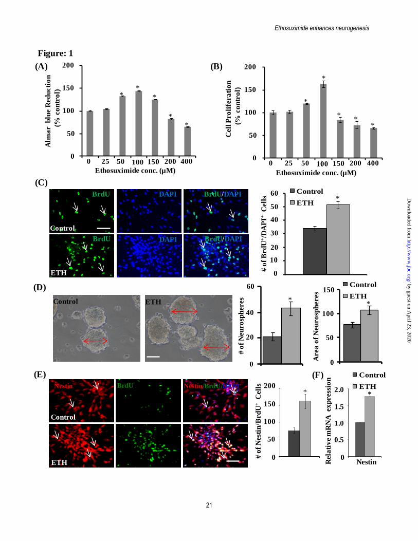
Ethosuximide enhances neurogenesis
21
Figure: 1
Ethosuximide conc. (µM)
Control ETH
(A) (B)
(C)
(D)
(E)
Control
ETH
BrdU DAPI
BrdU DAPI
BrdU/DAPI
BrdU/DAPI
# o
f B
rd
U+/D
AP
I+C
ell
s
0
10
20
30
40
50
60*
Nestin BrdU Nestin/BrdU/DAPI
Control
ETH
Control ETH
150
50
100A
rea
of
Neu
ro
sph
eres
0
*
60
0
20
40
# o
f N
eu
ro
sph
eres
Control
ETH
*
Control
ETH
Control
ETH
0
50
100
150
200
0 25 50 100 150 200 400
**
*
*
*
Alm
ar
blu
e R
ed
ucti
on
(% c
on
tro
l)
0
50
100
150
200
0 25 50 100 150 200 400
*
*
*
**
Ethosuximide conc. (µM)
# o
f N
est
in/B
rd
U+
Cell
s
0
50
100
150
200*
0
0.5
1.0
1.5
2.0
Rela
tiv
e m
RN
A ex
press
ion
Nestin
*
Control
ETH
(F)
Cell
Pro
life
ra
tio
n
(% c
on
tro
l)
by guest on April 23, 2020
http://ww
w.jbc.org/
Dow
nloaded from
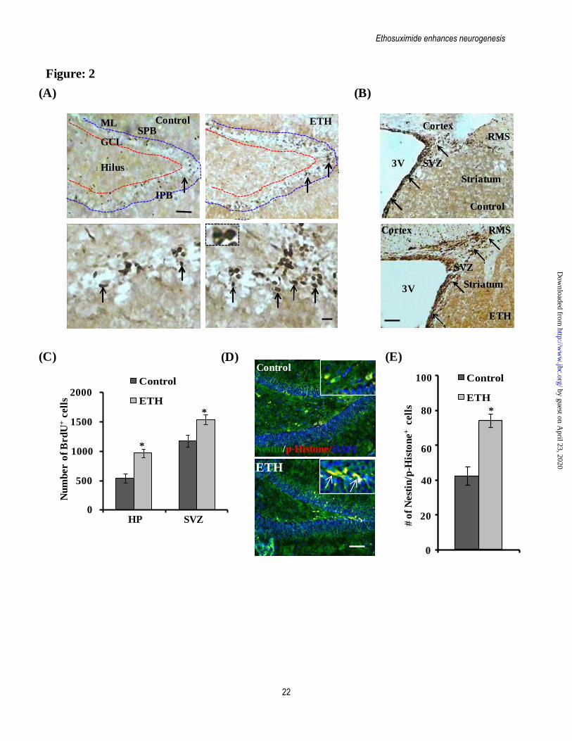
Ethosuximide enhances neurogenesis
22
Figure: 2
(A) (B)
(C) (D)
3V SVZ
CortexRMS
Striatum
3V
SVZ
Cortex RMS
Striatum
Control
ETH
ETHControlSPB
IPB
Hilus
GCL
ML
Nu
mb
er o
f B
rd
U+
cell
s
0
500
1000
1500
2000
SVZHP
Control
ETH
*
*
Control
ETHControl
ETH
0
20
60
80
40
# o
f N
est
in/p
-His
ton
e+
cell
s
100
*
(E)
Control
ETH
Nestin/p-Histone/DAPI
Control
ETH
by guest on April 23, 2020
http://ww
w.jbc.org/
Dow
nloaded from
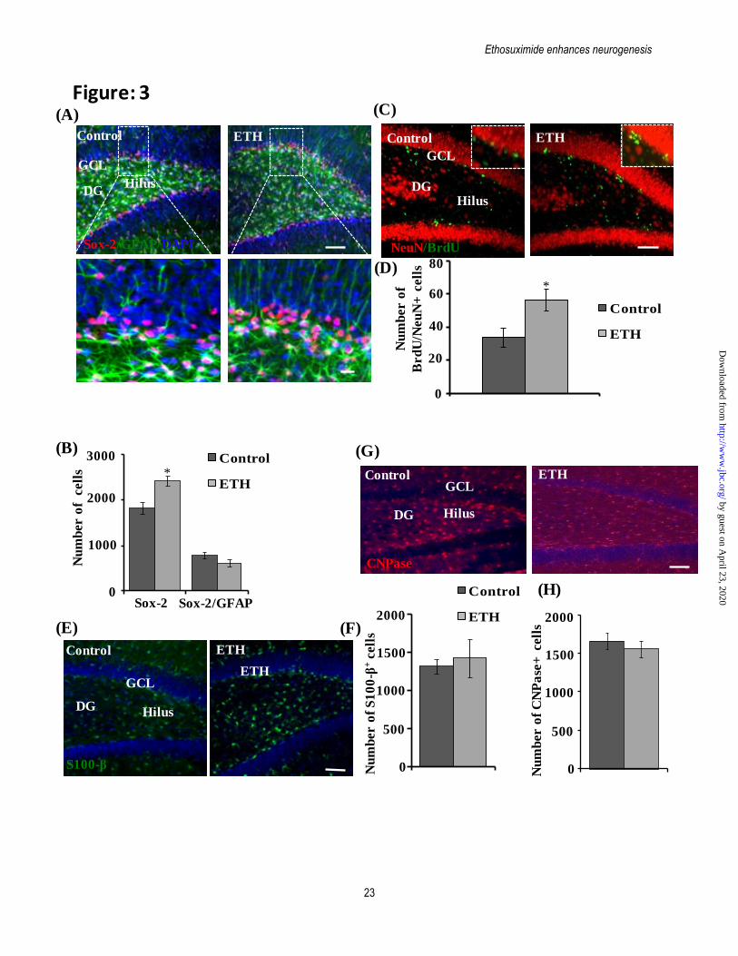
Ethosuximide enhances neurogenesis
23
Figure: 3
NeuN/BrdU
DGHilus
GCL
S100-β
DG Hilus
GCL
Control
CNPase
DG Hilus
GCLContol
ETH
Nu
mb
er o
f cell
s
0
1000
2000
3000
Sox-2 Sox-2/GFAP
Contol
ETH
Nu
mb
er o
f
Brd
U/N
eu
N+
cell
s
0
20
40
60
80
Control
ETH
Nu
mb
er o
f S
10
0-β
+cell
s
0
500
1000
1500
Control
ETH2000
Nu
mb
er o
f C
NP
ase
+ cell
s
0
500
1000
1500
2000
*
*
(A)
(B)
(C)
(D)
(G)
(E) (F)
(H)
Control ETH
Control ETH
Control ETH
Sox-2/GFAP/DAPI
DGHilus
GCL
Control ETH
ETH
Control ETH
Control
ETH
by guest on April 23, 2020
http://ww
w.jbc.org/
Dow
nloaded from
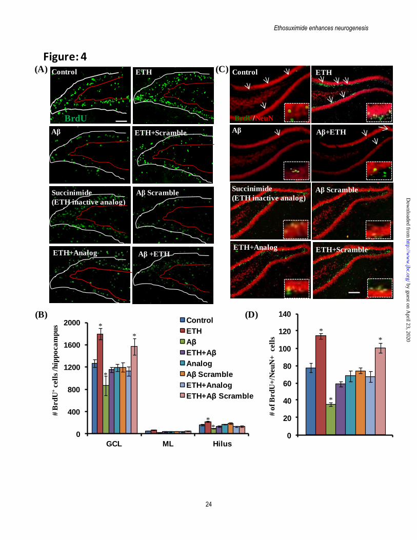
Ethosuximide enhances neurogenesis
24
ETH
Aβ
ETH+Analog
Control
BrdU
Succinimide
(ETH inactive analog)
ETH+Scramble
Aβ Scramble
Aβ +ETH
Control ETH
Aβ Aβ+ETH
BrdU/NeuN
Aβ Scramble
ETH+Analog ETH+Scramble
Succinimide
(ETH inactive analog)
Figure: 4
0
400
800
1200
1600
2000
GCL ML Hilus
Control
ETH
Aβ
ETH+Aβ
Analog
Aβ Scramble
ETH+Analog
ETH+Aβ Scramble
# B
rd
U+
cell
s /h
ipp
oca
mp
us *
*
*
# o
f B
rd
U+
/Neu
N+
cell
s
0
20
40
60
80
100
120
140
*
*
*
**
(A)
(B)
(C)
(D)
by guest on April 23, 2020
http://ww
w.jbc.org/
Dow
nloaded from
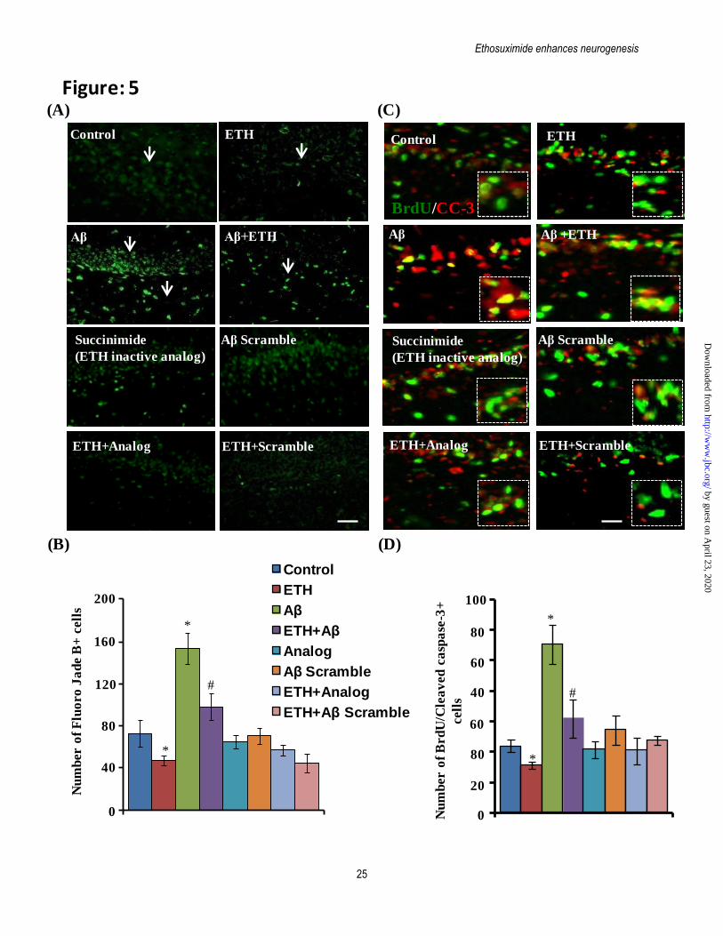
Ethosuximide enhances neurogenesis
25
Aβ Scramble
Control ETH
Aβ
ETH+Analog
Succinimide
(ETH inactive analog)
ETH+Scramble
Aβ Scramble
Nu
mb
er o
f F
luo
ro
Ja
de B
+ c
ell
s
0
40
80
120
160
200
*
*
#
Nu
mb
er o
f B
rd
U/C
lea
ved
ca
spa
se-3
+
cell
s
0
20
80
60
40
60
*
*
#
0
400
800
1200
1600
2000
GCL ML Hilus
Control
ETH
Aβ
ETH+Aβ
Analog
Aβ Scramble
ETH+Analog
ETH+Aβ Scramble
Figure: 5
ETHControl
Aβ +ETH
BrdU/CC-3
Aβ
ETH+Analog
Succinimide
(ETH inactive analog)
ETH+Scramble
Aβ Scramble
Aβ
(A)
(B)
(C)
(D)
80
100
Aβ+ETH
by guest on April 23, 2020
http://ww
w.jbc.org/
Dow
nloaded from
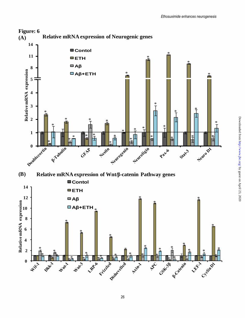
Ethosuximide enhances neurogenesis
26
0
1
2
3
4
5
8
11
14
0
2
10
Rela
tiv
e m
RN
A ex
press
ion
4
6
8
12
14
Relative mRNA expression of Neurogenic genes
Rela
tiv
e m
RN
A ex
press
ion
Relative mRNA expression of Wnt/β-catenin Pathway genes
Figure: 6
(A)
(B)
*
*
**
** *
*
*
*
*
**
*
*
*
*
*
*
*
*
*
*
*
*
* * *
*
*
*
**
*
*
*
*** *
*
*
**
**
*
*
*
**
**
*
*
*
*
**
**
*
**
**
*
Contol
ETH
Aβ
Aβ+ETH
Contol
ETH
Aβ
Aβ+ETH
by guest on April 23, 2020
http://ww
w.jbc.org/
Dow
nloaded from
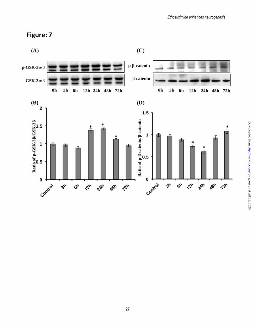
Ethosuximide enhances neurogenesis
27
0h 3h 6h 12h 24h 48h 72h
GSK-3α/β
p-GSK-3α/β
β-catenin
p-β-catenin
Ra
tio
of
p-G
SK
-3β
/GS
K-3
β
Ra
tio
of
p-β
-ca
ten
in/β
-ca
ten
in
Figure: 7
(B)
(A)
(D)
(C)
0
0.5
1
1.5
**
*
0
0.5
1
1.5
2
* *
*
0h 3h 6h 12h 24h 48h 72h
by guest on April 23, 2020
http://ww
w.jbc.org/
Dow
nloaded from
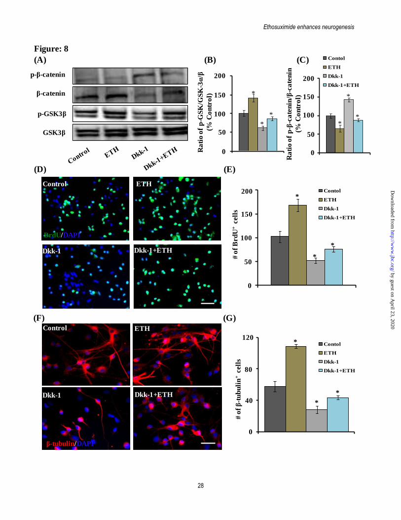
Ethosuximide enhances neurogenesis
28
0
40
80
120
# o
f β
-tu
bu
lin
+cell
s
*
*
*
BrdU/DAPI
Control ETH
Dkk-1 Dkk-1+ETH
Contol
ETH
Dkk-1
Dkk-1+ETH
0
50
100
200
# o
f B
rd
U+
cell
s
150
*
*
*
Control ETH
Dkk-1 Dkk-1+ETH
(D)
Figure: 8
(E)
(F) (G)
(A) (B) (C)
0
50
100
150
200
Ra
tio
of
p-G
SK
/GS
K-3
α/β
(% C
on
tro
l) Control
ETH
Dkk-1
ETH+Dkk-1
0
50
100
150
200
Ra
tio
of
p-β
-ca
ten
in/β
-ca
ten
in
(% C
on
tro
l)
*
*
*
*
*
*
β-catenin
p-β-catenin
GSK3β
p-GSK3β
Contol
ETH
Dkk-1
Dkk-1+ETH
β-tubulin/DAPI
Contol
ETH
Dkk-1
Dkk-1+ETH
Contol
ETH
Dkk-1
Dkk-1+ETH
by guest on April 23, 2020
http://ww
w.jbc.org/
Dow
nloaded from
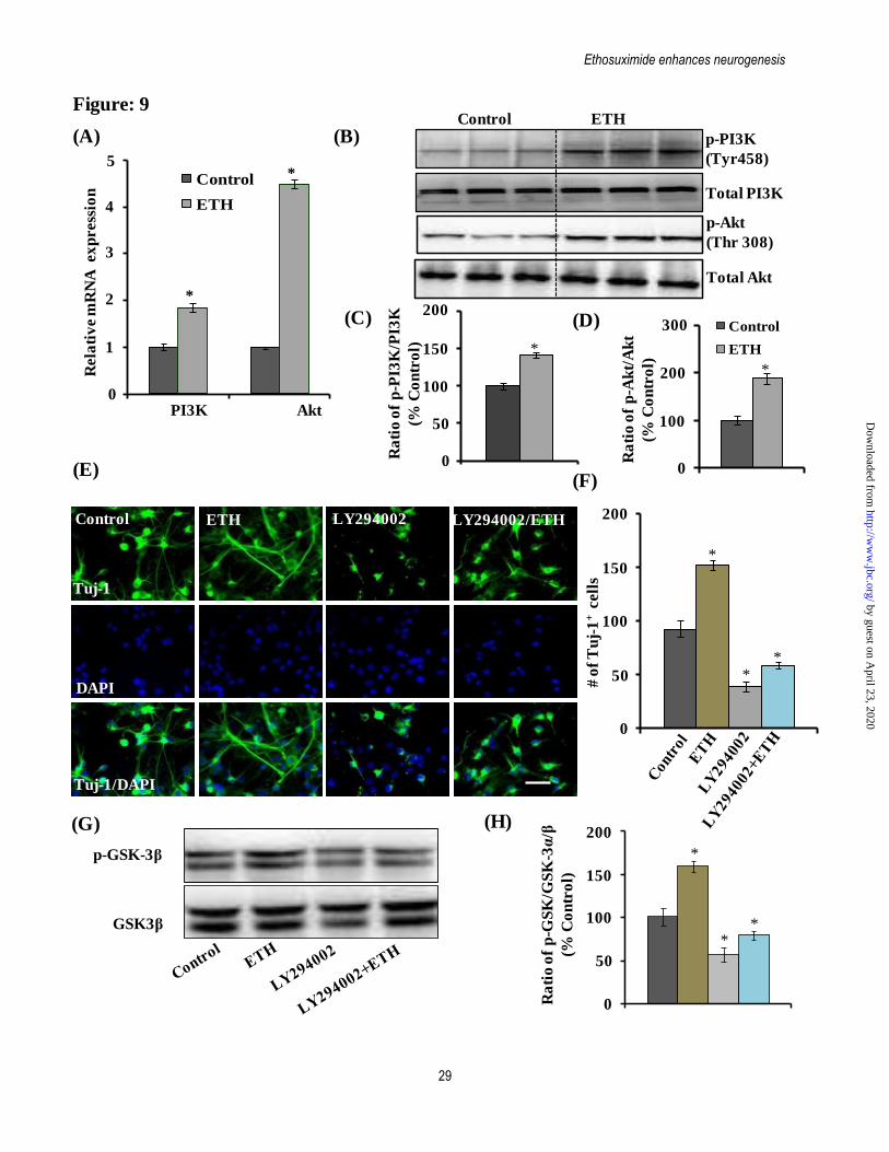
Ethosuximide enhances neurogenesis
29
Control ETH LY294002 LY294002/ETH
Tuj-1
DAPI
Tuj-1/DAPI
0
50
100
150
200
# o
f T
uj-
1+
cell
s
(A)
Figure: 9
(B)
(C)
(E)(F)
(G) (H)
0
50
100
150
200
Ra
tio
of
p-G
SK
/GS
K-3
α/β
(% C
on
tro
l)
*
*
*
*
*
*
AktPI3K0
1
2
3
4
5
Rela
tiv
e m
RN
A ex
press
ion
Control
ETH
*
*
Control
ETH
Total Akt
p-Akt
(Thr 308)
Control ETH
p-PI3K
(Tyr458)
Total PI3K
50
100
150
200
Ra
tio
of
p-P
I3K
/PI3
K
(% C
on
tro
l)
00
100
200
Ra
tio
of
p-A
kt/
Ak
t
(% C
on
tro
l) Control
ETH
300
*
*
(D) Control
ETH
GSK3β
p-GSK-3β
by guest on April 23, 2020
http://ww
w.jbc.org/
Dow
nloaded from
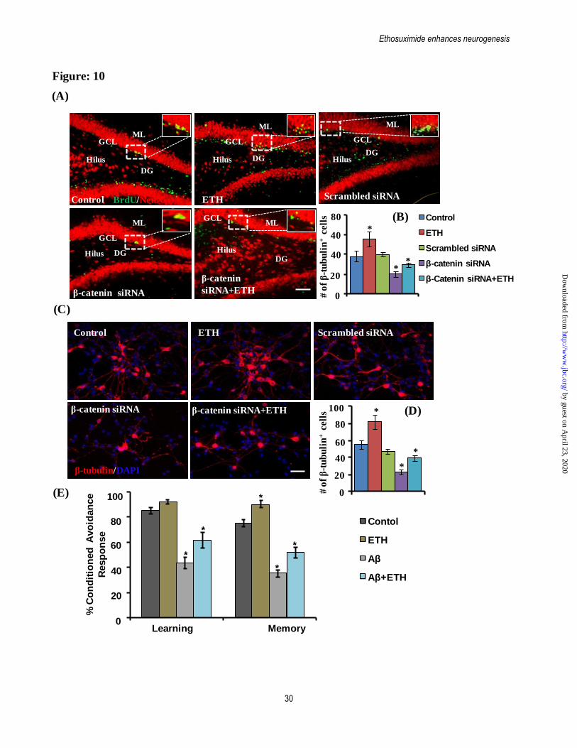
Ethosuximide enhances neurogenesis
30
(A)
Figure: 10
(C)
DGHilus
GCLML
DGHilus
GCL
ML
DG
Hilus
GCLML
BrdU/NeuN
DGHilus
GCL
ML
Control ETH
β-catenin siRNA
β-catenin
siRNA+ETH
Scrambled siRNA
DGHilus
GCL
ML
Control
ETH
Scrambled siRNA
β-catenin siRNA
β-Catenin siRNA+ETH
Control
ETH
Scrambled siRNA
β-catenin siRNA
β-Catenin siRNA+ETH0
20
80
40
# o
f β
-tu
bu
lin
+cell
s
40*
**
Control ETH
β-catenin siRNA β-catenin siRNA+ETH
Scrambled siRNA
0
20
60
80
100
40
# o
f β
-tu
bu
lin
+cell
s *
*
*
(B)
(D)
0
20
40
60
80
100
Learning Memory
% C
on
dit
ion
ed
Avo
idan
ce
Resp
on
se
*
*
*
*
*
(E)
Contol
ETH
Aβ
Aβ+ETH
β-tubulin/DAPI
by guest on April 23, 2020
http://ww
w.jbc.org/
Dow
nloaded from
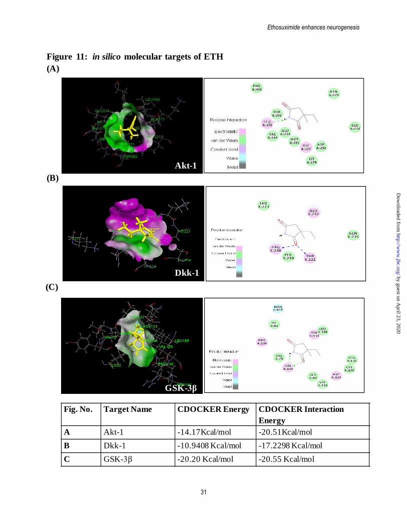
Ethosuximide enhances neurogenesis
31
(A)
(B)
(C)
Figure 11: in silico molecular targets of ETH
Fig. No. Target Name CDOCKER Energy CDOCKER Interaction
Energy
A Akt-1 -14.17Kcal/mol -20.51Kcal/mol
B Dkk-1 -10.9408 Kcal/mol -17.2298 Kcal/mol
C GSK-3β -20.20 Kcal/mol -20.55 Kcal/mol
Akt-1
Dkk-1
GSK-3β
by guest on April 23, 2020
http://ww
w.jbc.org/
Dow
nloaded from
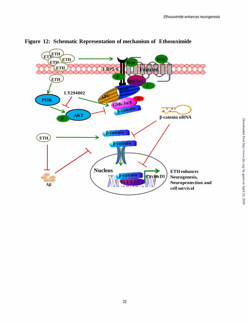
Ethosuximide enhances neurogenesis
32
Figure 12: Schematic Representation of mechanism of Ethosuximide
Aβ
PI3K
AKT
ETH
ETX
ETH
ETH
ETH
ETH
FrizzledLRP5/6
WntWnt
Dsh/Dvl
β-catenin siRNA
TCF/LEF
P
P
P
P
NucleusCyclin D1
ETH enhances
Neurogenesis,
Neuroprotection and
cell survival
ETH
LY294002
by guest on April 23, 2020
http://ww
w.jbc.org/
Dow
nloaded from
ChaturvediKarmakar, Shailendra Kumar Gupta, Vinay Choubey, Abhay Sharma and Rajnish Kumar
Shashi Kant Tiwari, Brashket Seth, Swati Agarwal, Anuradha Yadav, Madhumita-catenin pathwayβ toxin induced Alzheimer's rat model via PI3K/Akt/Wnt/βamyloid-
Ethosuximide induces hippocampal neurogenesis and reverses cognitive deficits in
published online September 29, 2015J. Biol. Chem.
10.1074/jbc.M115.652586Access the most updated version of this article at doi:
Alerts:
When a correction for this article is posted•
When this article is cited•
to choose from all of JBC's e-mail alertsClick here
by guest on April 23, 2020
http://ww
w.jbc.org/
Dow
nloaded from