Embed Size (px)
Citation preview

肺动脉栓塞的诊治
制作XGHRH
敬请指正

基本概念肺栓塞是以各种栓子阻塞肺动脉系统为
其发病原因的一组疾病或临床综合征的总称,包括肺血栓栓塞症,脂肪栓塞综合征,羊水栓塞,空气栓塞等。
肺血栓栓塞症为来自静脉系统或右心的血栓阻塞肺动脉或其分支所致疾病。
肺梗死为肺动脉发生栓塞后,其支配区的肺组织因血流受阻或中断而发生坏死。
肺栓塞的现状
发病率高:仅次于 CAD 和 HBP 。
易漏诊及误诊:警惕性不高,漏诊率高。
不经治疗死亡率高:达 20%-30% 。
明确诊疗者死亡率明显下降:可降至 2-8% 。
Epidemiology
There is no accurate data for pulmonary embolism because we has limit knowledge of it.
In the United States, it is responsible for about 2.3 new cases per 10,000 persons and 50,000 deaths every year.
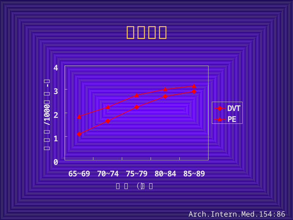
流行病学
0
1
2
3
4
65~69 70~74 75~79 80~84 85~89
年龄(岁)
/100
0-
发生率
患者年
DVTPE
Arch.Intern.Med.154:861,1994
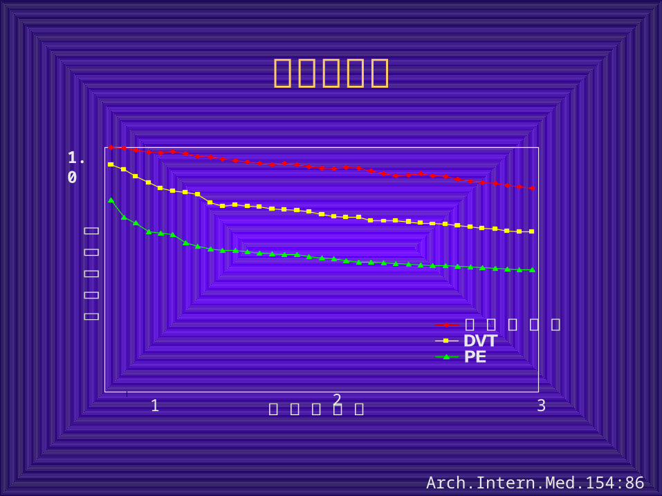
生存率比较
Arch.Intern.Med.154:861,1994
随访的年数
生存的可能性
匹配的样本DVTPE
1.0
1 2 3
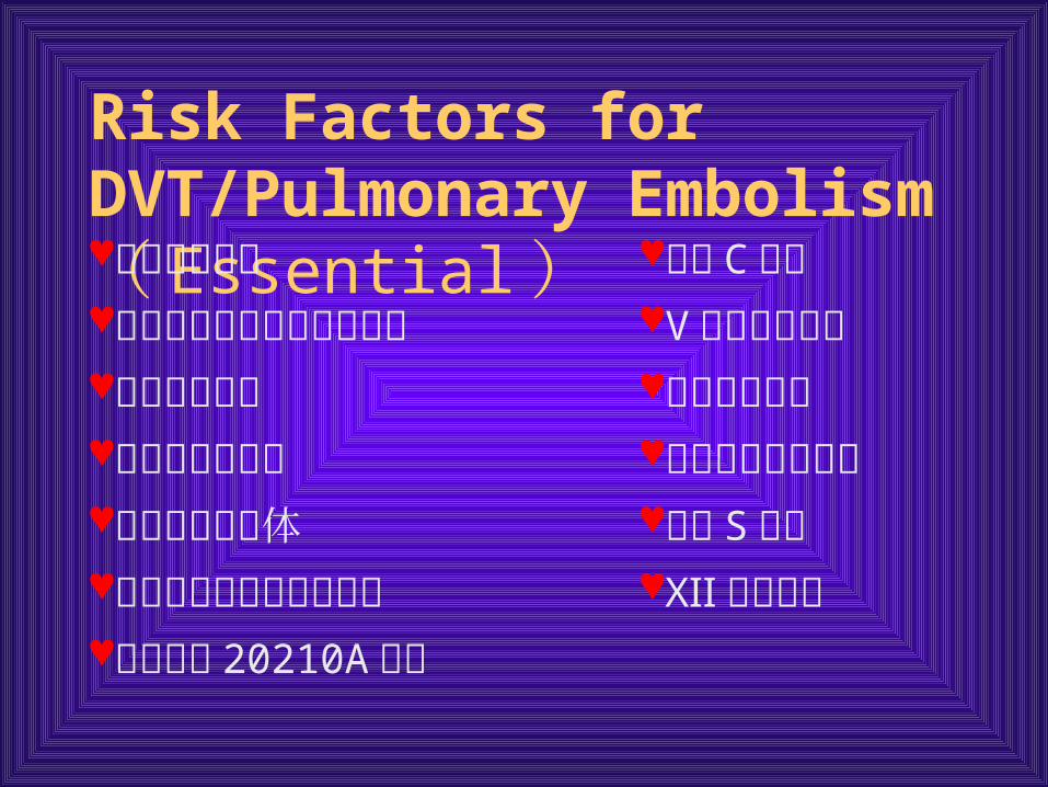
Risk Factors for DVT/Pulmonary Embolism ( Essential )抗凝血酶缺乏 蛋白 C 缺乏先天性异常纤维蛋白原血症 V 因子基因突变血栓调节蛋白 纤溶酶原缺乏高半胱氨酸血症 异常纤溶酶原血症抗心肌碱脂抗体 蛋白 S 缺乏纤溶酶原激活抑制剂过量 Ⅻ 因子缺乏前凝血酶 20210A 突变
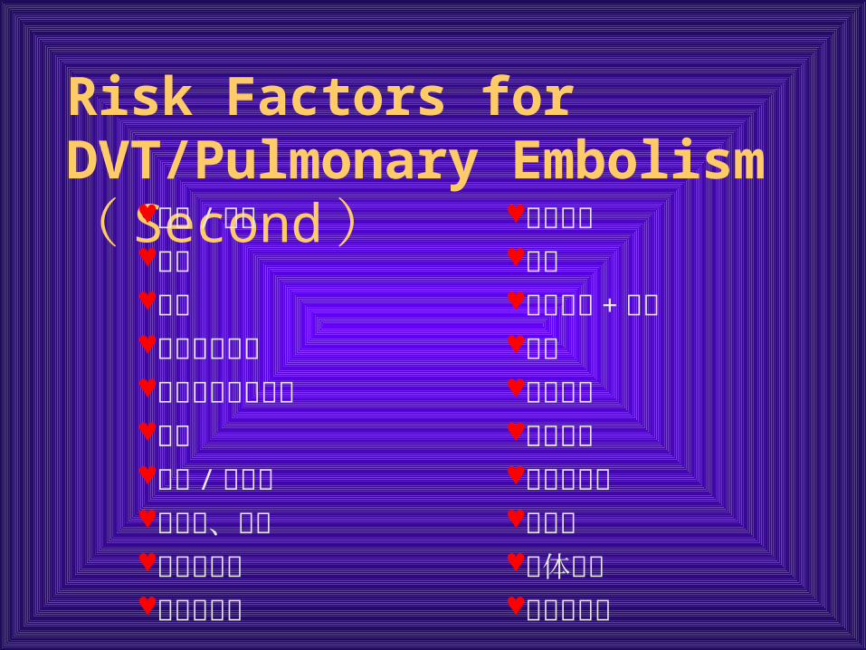
Risk Factors for DVT/Pulmonary Embolism ( Second )
创伤 / 骨折 外科手术卒中 制动高龄 恶性肿瘤 + 化疗中心静脉导管 肥胖慢性静脉机能不全 心力衰竭吸烟 长途旅行妊娠 / 产后期 口服避孕药克隆病、狼疮 抗凝剂肾病综合征 假体表面粘滞性过高 血小板异常
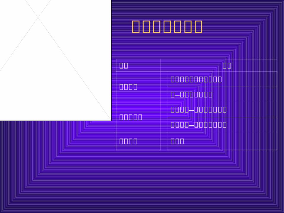
深静脉血栓形成
原因 分类
血流滞缓小腿肌肉静脉丛血栓形成
髂—股静脉血栓形成
静脉壁损伤
原发性髂—肌静脉血栓形成
继发性髂—股静脉血栓形成
高凝状态 股青肿
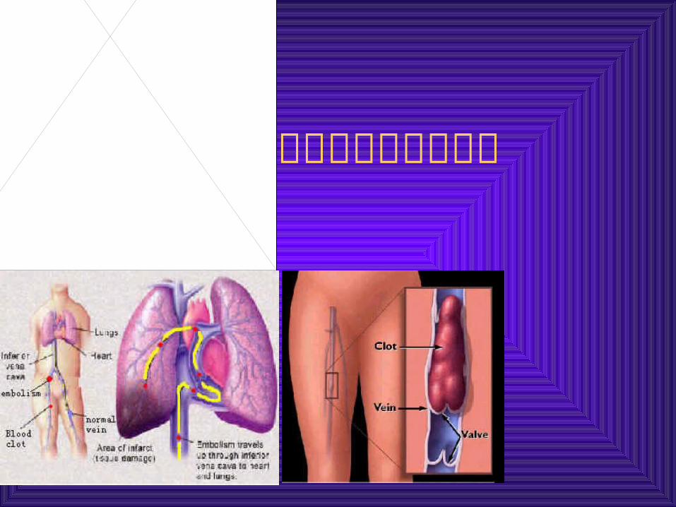
肺血栓与深静脉血栓
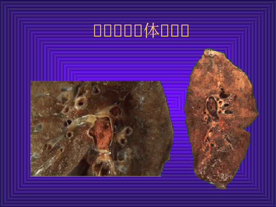
肺栓塞的大体解剖观
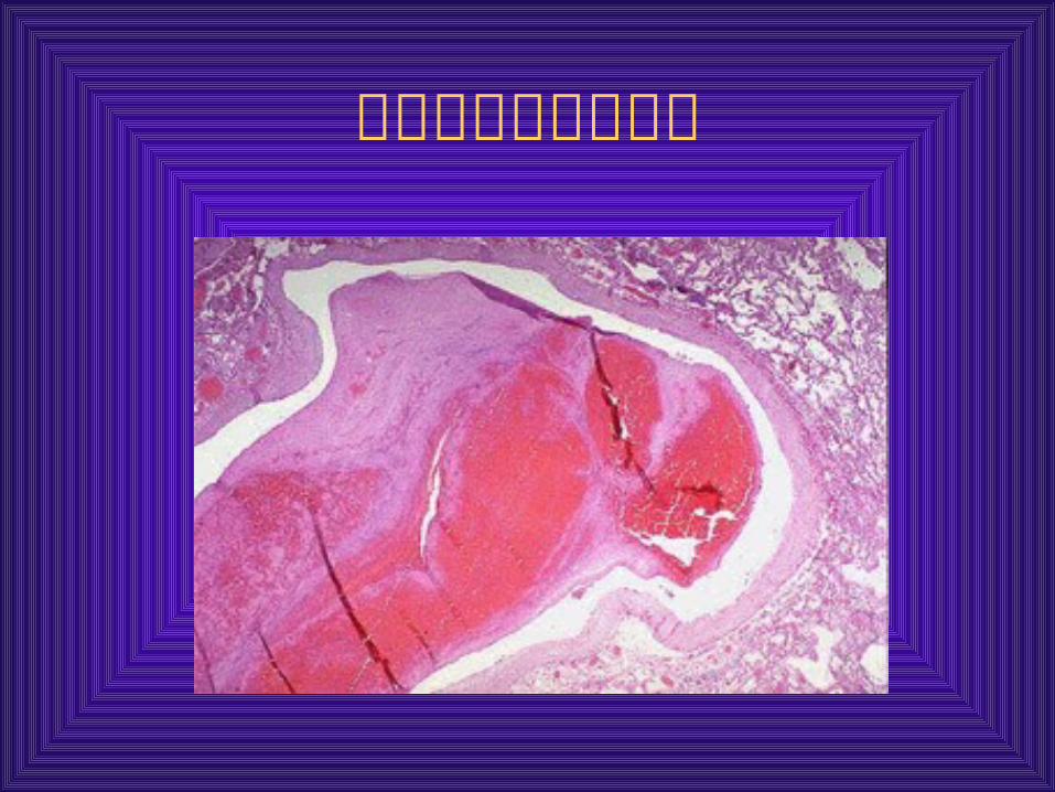
肺栓塞的显微镜下观

肺栓塞的病理生理
肺血管阻塞,神经体液因素或肺动脉压力感受器的作用,引起肺血管阻力增加;
肺血管阻塞→肺泡死腔↑→气体交换↓→肺泡通气↓→低氧血症→ V/Q 单位↓→气体交换面积↓→二氧化碳↑
刺激性受体反射性兴奋(过度换气) 支气管收缩,气道阻力增加 肺水肿、肺出血、肺泡表面活性物质减少,肺顺应性降低。
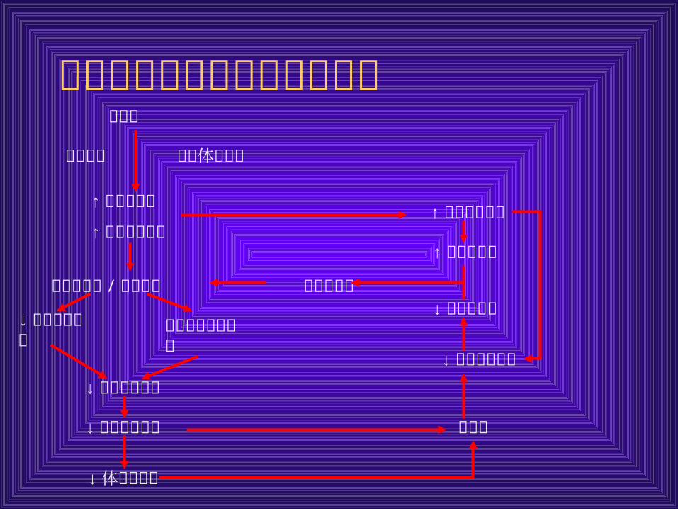
肺栓塞后右心功能不全的病生肺栓塞
↓冠状动脉灌注
↑ 右心室氧需
↑ 右心室壁张力
↓ 右心室排血量
↓ 右心室氧供
↓左心室排血量
↑ 肺动脉压力
↑ 右心室后负荷
解剖阻塞 神经体液作用
右心室扩张 /功能不全 右心室缺血
室间隔移向左心室
低血压
↓ 体循环灌注
↓左心室前负荷
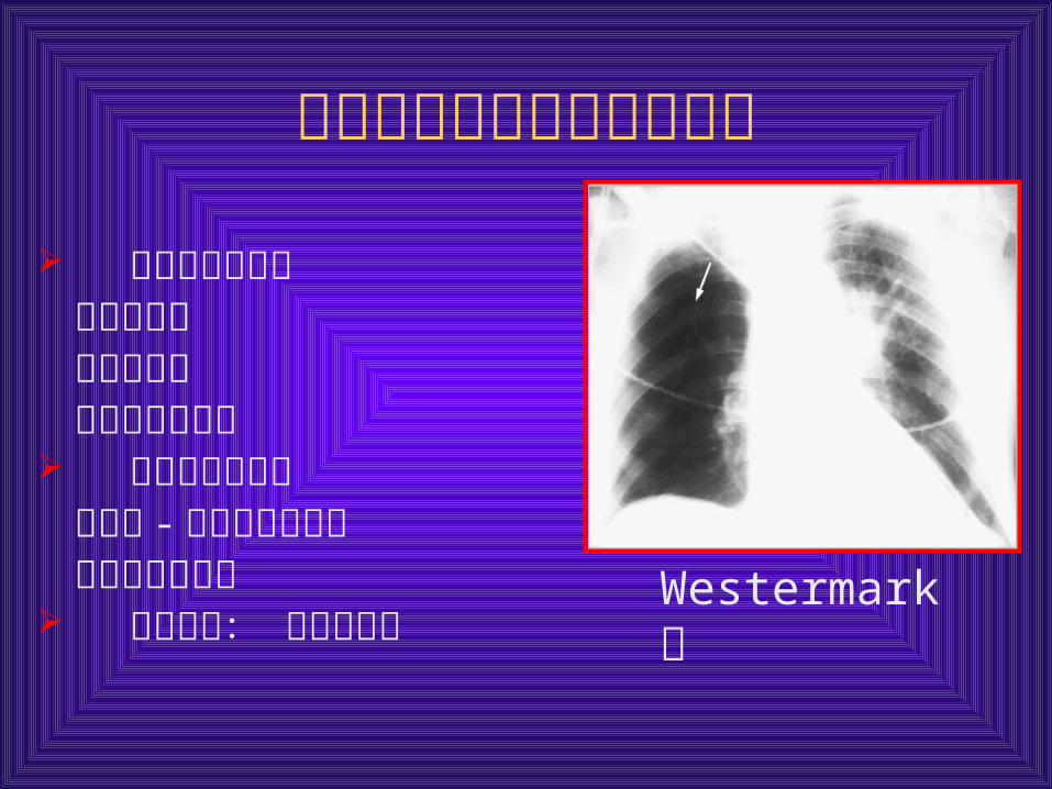
肺栓塞后肺血流动力学变化
前毛细血管高压 血管床减少 支气管收缩 小动脉血管收缩
侧支血管的形成支气管 - 肺动脉吻合形成 肺内动静脉分流
血流改变: 血流重分布 Westermark征

呼吸动力学改变
过度通气: 肺动脉高压
顺应性下降
肺不张 气道阻力增加 :
局限性低碳酸血症
化学介质
临床分型
大面积 PE(massive PE) :休克和低血压;动脉收缩压 <90mmHg或下降幅度≥ 40mmHg ,持续 15min 以上;除外其他原因所致血压下降。
次大面积 PE (submassive PE)亚型超声心动图示右心室运动功能减弱右心功能不全表现。
非大面积 PE(non-massive FE) :不符合以上大面积 PE标准的 PE 。
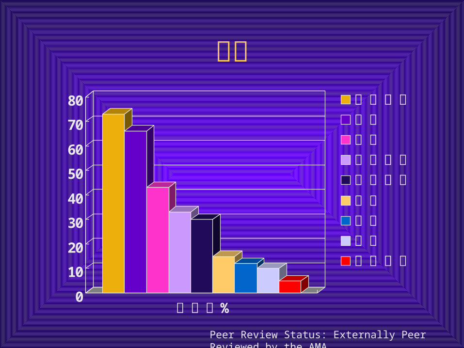
症状
Peer Review Status: Externally Peer Reviewed by the AMA
0
10
20
30
40
50
60
70
80
%发生率
呼吸困难胸痛咳嗽下肢水肿下肢疼痛咳血心悸喘息类心绞痛

体征
0
10
20
30
40
50
60
70呼吸急促
罗音
心动过速
第四心音
肺动脉第二心音亢进临床明显的深静脉血栓形成大汗
>38. 5C体温
喘息
Homans征
右室抬举
胸膜磨擦音
第三心音
紫绀
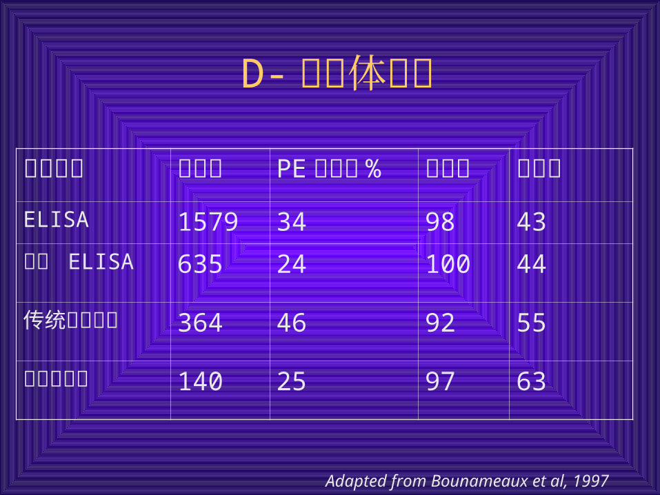
D- 二聚体分析
检验方法 病人数 PE 发生率%
敏感性 特异性
ELISA 1579 34 98 43
快速 ELISA 635 24 100 44
传统乳胶试验 364 46 92 55
血乳胶试验 140 25 97 63
Adapted from Bounameaux et al, 1997
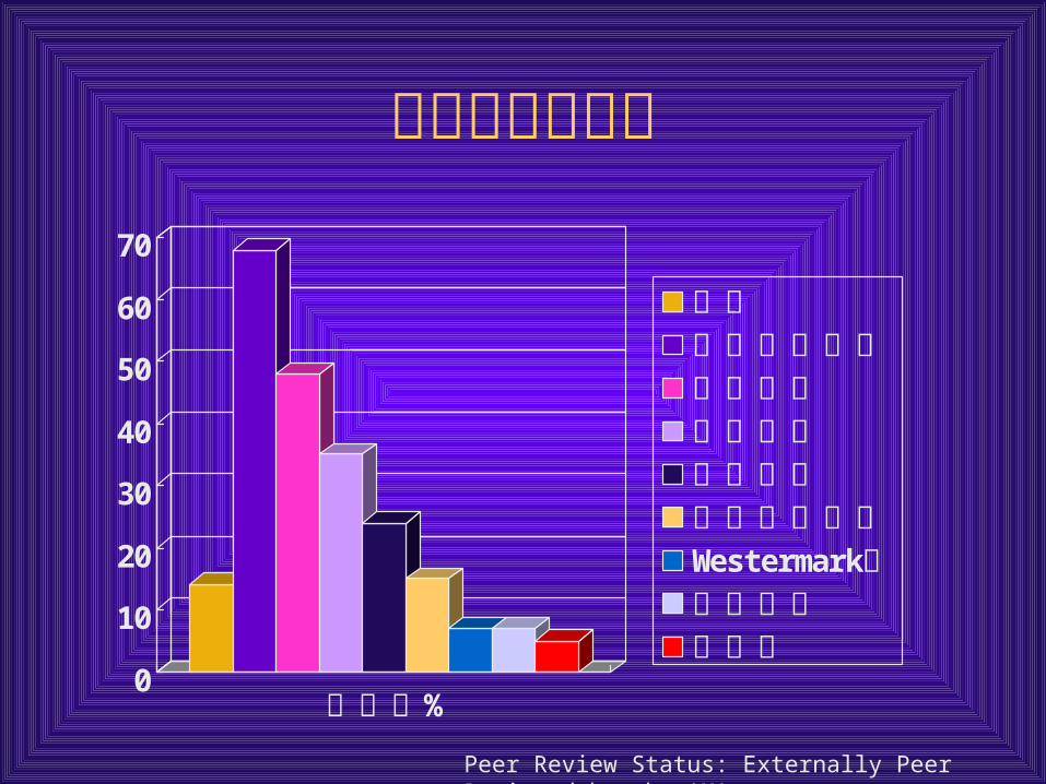
肺栓塞胸片检查
0
10
20
30
40
50
60
70
%发生率
正常肺不张或实变胸腔积液胸膜肥厚纵隔上抬肺动脉搏增宽Westermark征心脏增大肺水肿
Peer Review Status: Externally Peer Reviewed by the AMA
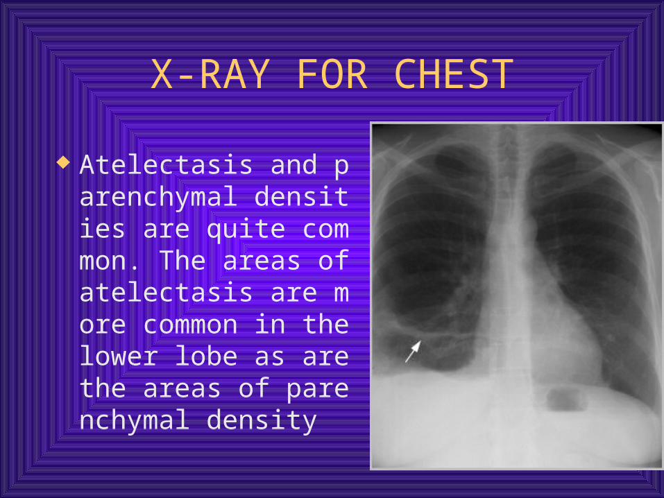
X-RAY FOR CHEST
Atelectasis and parenchymal densities are quite common. The areas of atelectasis are more common in the lower lobe as are the areas of parenchymal density
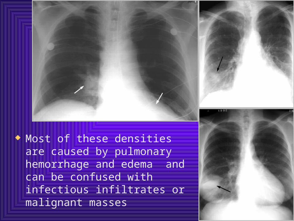
Most of these densities are caused by pulmonary hemorrhage and edema and can be confused with infectious infiltrates or malignant masses
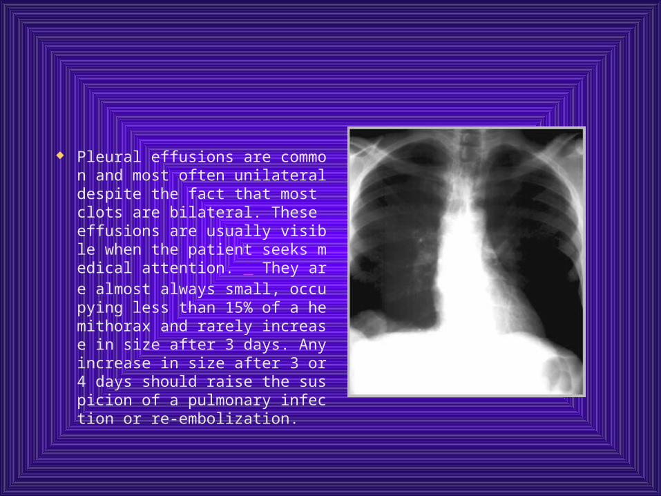
Pleural effusions are common and most often unilateral despite the fact that most clots are bilateral. These effusions are usually visible when the patient seeks medical attention. They are almost always small, occupying less than 15% of a hemithorax and rarely increase in size after 3 days. Any increase in size after 3 or 4 days should raise the suspicion of a pulmonary infection or re-embolization.
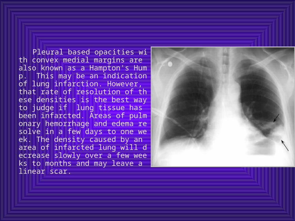
Pleural based opacities with convex medial margins are also known as a Hampton's Hump. This may be an indication of lung infarction. However, that rate of resolution of these densities is the best way to judge if lung tissue has been infarcted. Areas of pulmonary hemorrhage and edema resolve in a few days to one week. The density caused by an area of infarcted lung will decrease slowly over a few weeks to months and may leave a linear scar.
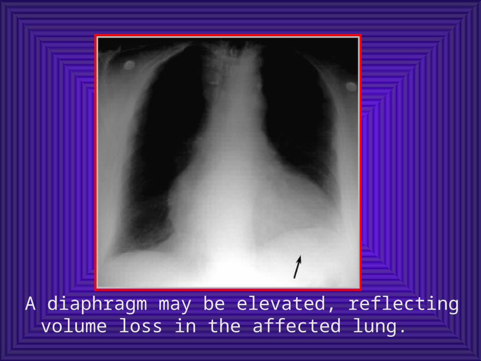
A diaphragm may be elevated, reflecting volume loss in the affected lung.
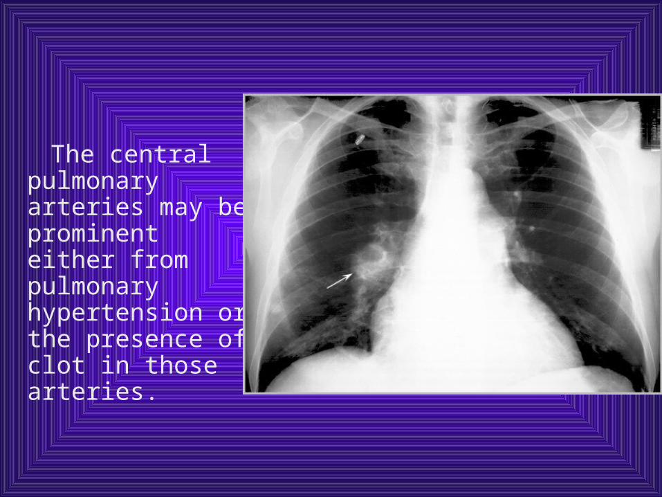
The central pulmonary arteries may be prominent either from pulmonary hypertension or the presence of clot in those arteries.
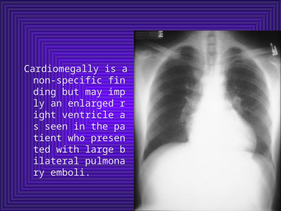
Cardiomegally is a non-specific finding but may imply an enlarged right ventricle as seen in the patient who presented with large bilateral pulmonary emboli.
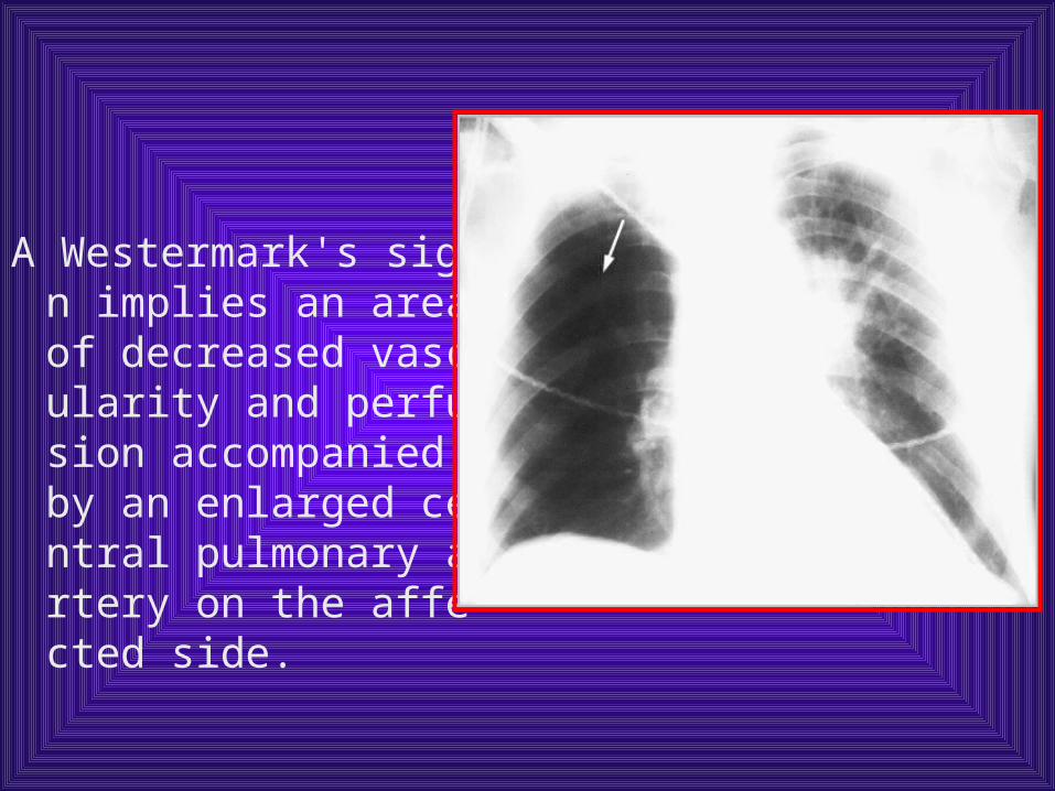
A Westermark's sign implies an area of decreased vascularity and perfusion accompanied by an enlarged central pulmonary artery on the affected side.
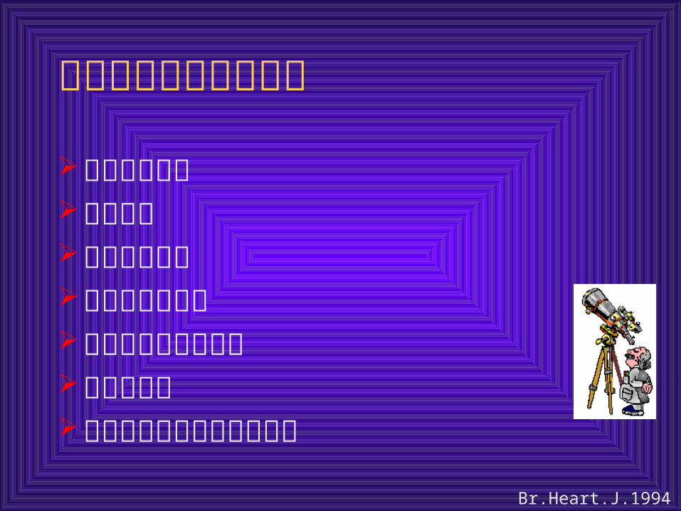
肺栓塞的心动超声征象
直接看到血栓右室扩张右室活动减弱室间隔异常活动三尖瓣反流速度增快肺动脉扩张无吸气性下腔静脉塌陷减弱
Br.Heart.J.1994,72:52
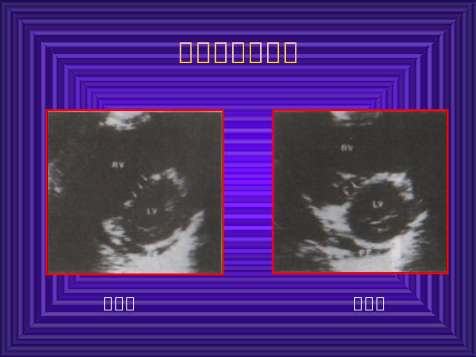
室间隔异常活动
舒张期 收缩期
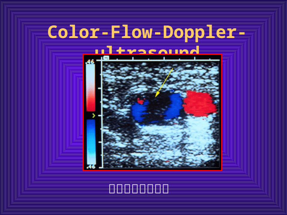
Color-Flow-Doppler-ultrasound
非挤压性充盈缺损
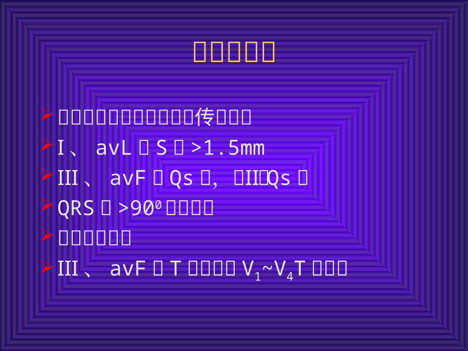
心电图表现
不完全性或完全性右束支传导阻滞Ⅰ、 avL 的 S波 >1.5mmⅢ、 avF有 Qs波,但Ⅱ无 Qs波QRS轴 >900 或不确定肢导联低电压Ⅲ、 avF 的 T波倒置或 V1~V4T波倒置
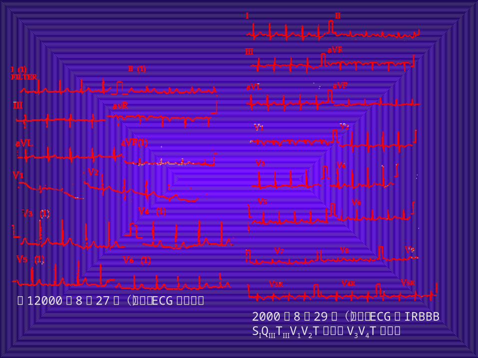
图 12000年 8月 27日(急诊) ECG 大致正常 2000年 8月 29日(门诊) ECG示 IR
BBB SⅠQⅢTⅢV1V2T波倒置 V3V4T波双向
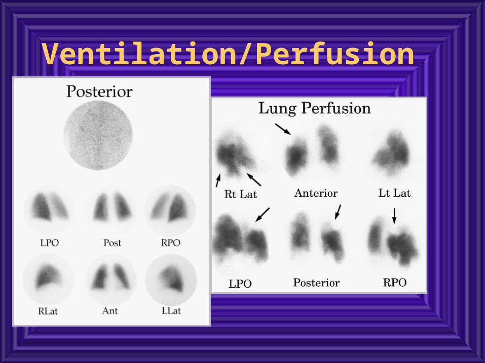
Ventilation/Perfusion Lung Scan
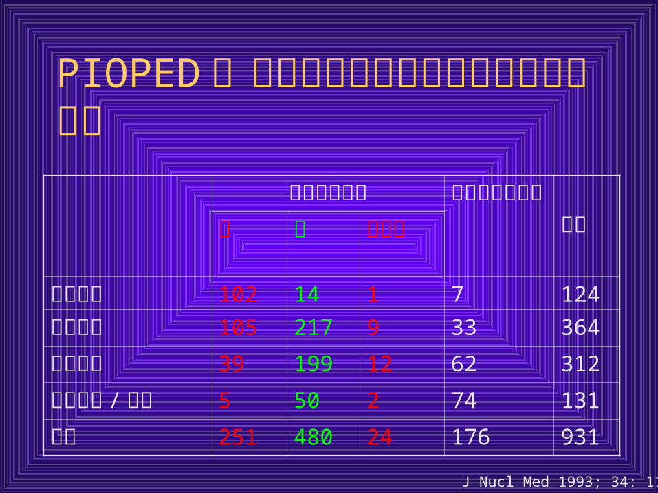
PIOPED :肺扫描分类与肺动脉造影结果的比较
肺扫描肺栓塞 肺动脉造影阴性 总数有 无 不肯定
高度可疑 102 14 1 7 124
中度可疑 105 217 9 33 364
低度可疑 39 199 12 62 312
接近正常 /正常 5 50 2 74 131
总计 251 480 24 176 931
J Nucl Med 1993; 34: 1119
肺扫描肺扫描
怀疑 PE 的患者约 25%可因肺灌注正常而否定诊断,而且不用抗凝治疗可能是安全的
怀疑 PE 的患者约 25%具有高度的肺扫描结果,他们可能需要行抗凝治疗
其余的患者需要进一步的诊断性检查,而这些检查是更广泛的诊断策略
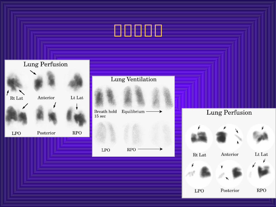
典型肺栓塞
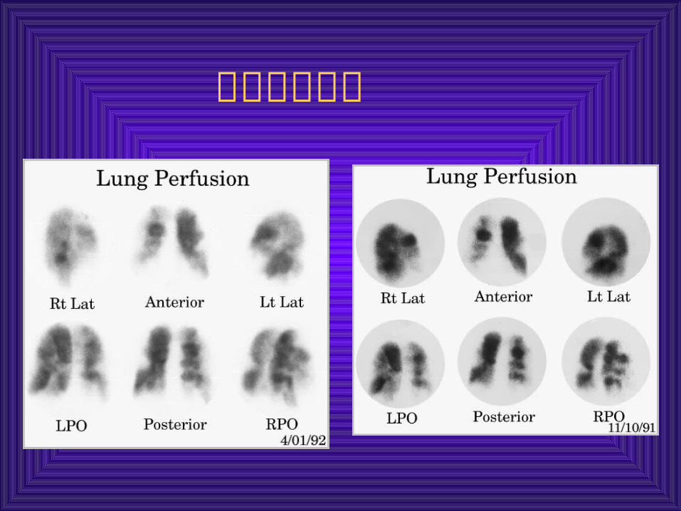
不典型肺栓塞
It is high sensitivity but low specificity
The differential diagnosis for a ventilation perfusion mismatch includes:
acute pulmonary embolus
previous pulmonary embolus
congenital vascular abnormalities
vasculitis,
bronchogenic carcinoma,
radiation therapy,et al.
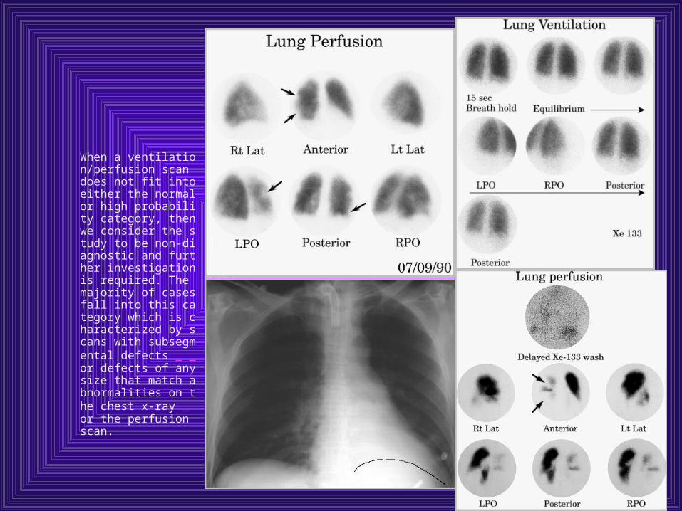
When a ventilation/perfusion scan does not fit into either the normal or high probability category, then we consider the study to be non-diagnostic and further investigation is required. The majority of cases fall into this category which is characterized by scans with subsegmental defects or defects of any size that match abnormalities on the chest x-ray or the perfusion scan.
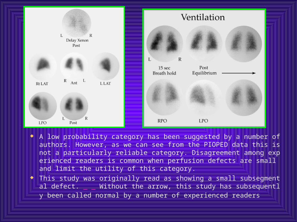
A low probability category has been suggested by a number of authors. However, as we can see from the PIOPED data this is not a particularly reliable category. Disagreement among experienced readers is common when perfusion defects are small and limit the utility of this category.
This study was originally read as showing a small subsegmental defect. Without the arrow, this study has subsequently been called normal by a number of experienced readers
Conclusion
Lung scans are sensitive exams that essentially rule out the diagnosis of pulmonary embolus when they are normal. Patients with high probability lungs can often be treated without further workup. Those patients with non-diagnostic studies require further diagnostic investigation.
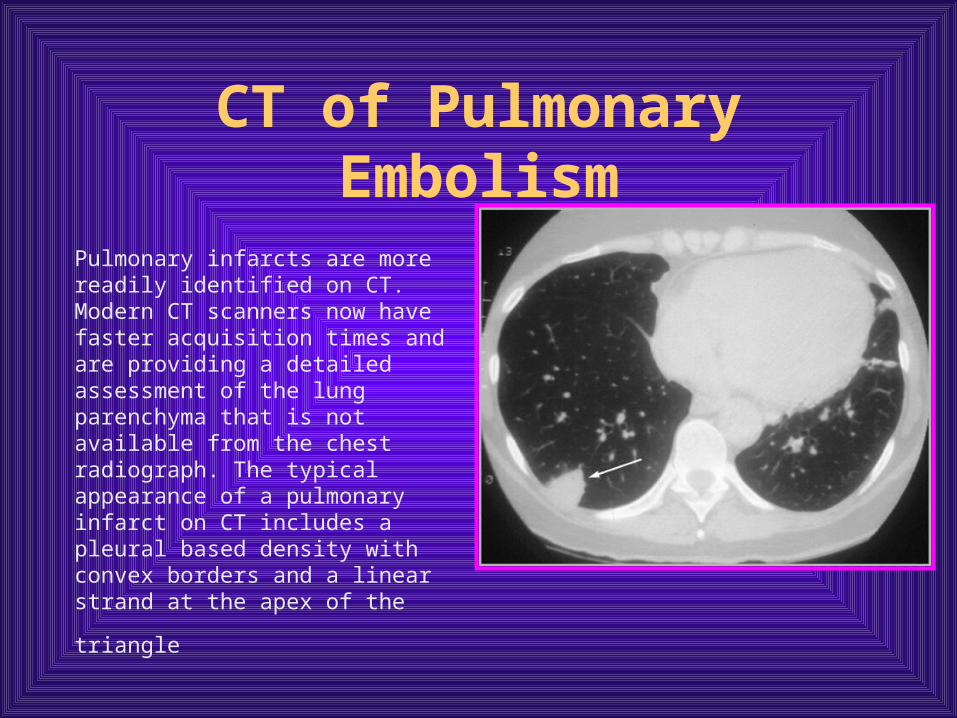
CT of Pulmonary Embolism
Pulmonary infarcts are more readily identified on CT. Modern CT scanners now have faster acquisition times and are providing a detailed assessment of the lung parenchyma that is not available from the chest radiograph. The typical appearance of a pulmonary infarct on CT includes a pleural based density with convex borders and a
linear strand at the apex of the triangle
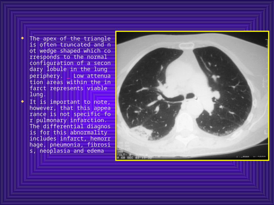
The apex of the triangle is often truncated and not wedge shaped which corresponds to the normal configuration of a secondary lobule in the lung periphery. Low attenuation areas within the infarct represents viable lung.
It is important to note, however, that this appearance is not specific for pulmonary infarction. The differential diagnosis for this abnormality includes infarct, hemorrhage, pneumonia, fibrosis, neoplasia and edema
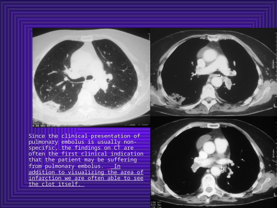
Since the clinical presentation of pulmonary embolus is usually non-specific, the findings on CT are often the first clinical indication that the patient may be suffering from pulmonary embolus. In addition to visualizing the area of infarction we are often able to see the clot itself.
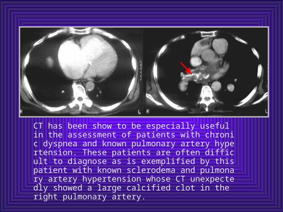
CT has been show to be especially useful in the assessment of patients with chronic dyspnea and known pulmonary artery hypertension. These patients are often difficult to diagnose as is exemplified by this patient with known sclerodema and pulmonary artery hypertension whose CT unexpectedly showed a large calcified clot in the right pulmonary artery.

肺动脉造影
正常肺动脉
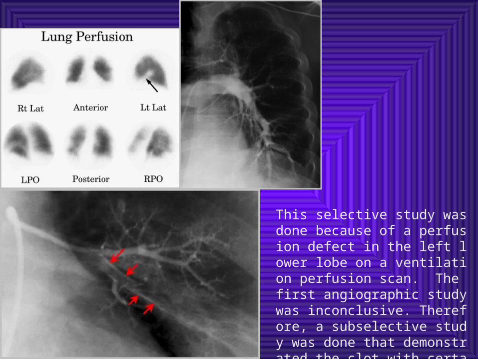
This selective study was done because of a perfusion defect in the left lower lobe on a ventilation perfusion scan. The first angiographic study was inconclusive. Therefore, a subselective study was done that demonstrated the clot with certainty.
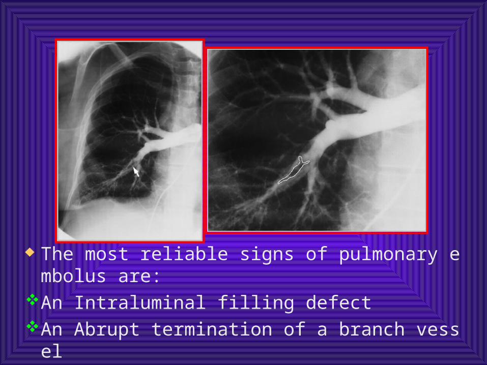
The most reliable signs of pulmonary embolus are:
An Intraluminal filling defect An Abrupt termination of a branch vessel
Conclusion
Angiography is most accurate in segmental and larger sized arteries. The reproducibility of readings is subsegmental and smaller vessels is poor.
Angiography is a safe procedure that is most accurate when imaging emboli that lodge in segmental or larger arteries.
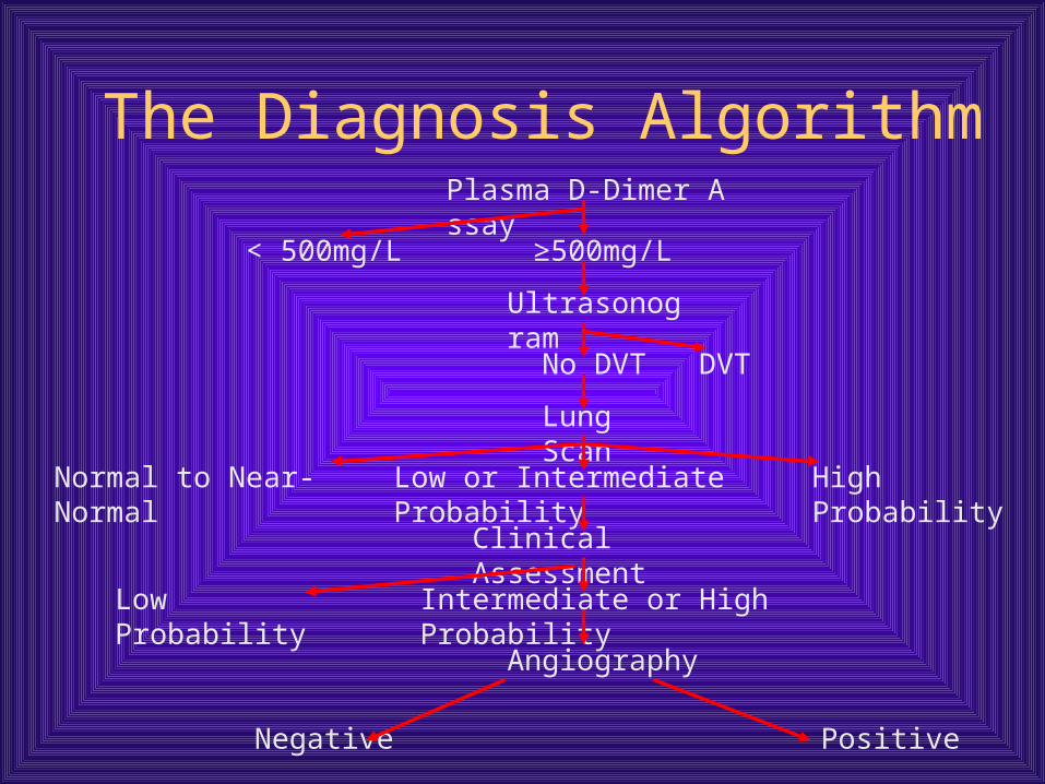
The Diagnosis Algorithm Plasma D-Dimer Assay
Normal to Near-Normal Low or Intermediate Probability High Probability
Clinical Assessment
Low Probability Intermediate or High Probability
Angiography
PositiveNegative
< 500mg/L ≥500mg/L
Ultrasonogram
No DVT DVT
Lung Scan
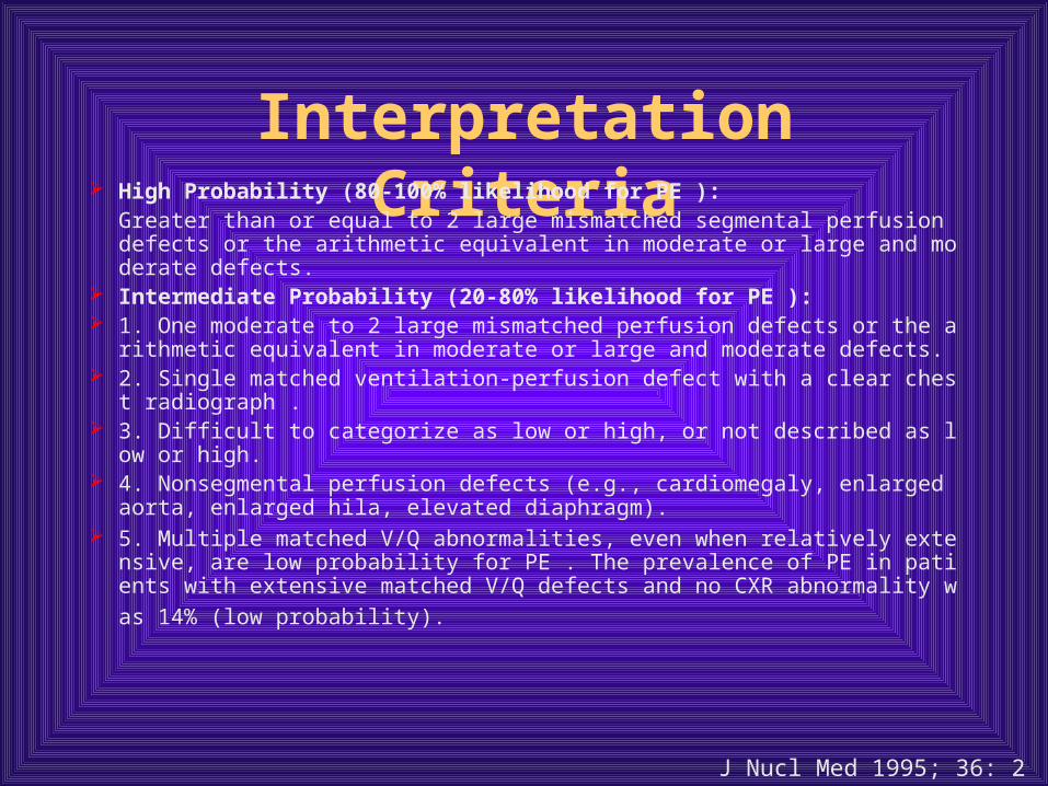
Interpretation Criteria High Probability (80-100% likelihood for PE ):
Greater than or equal to 2 large mismatched segmental perfusion defects or the arithmetic equivalent in moderate or large and moderate defects.
Intermediate Probability (20-80% likelihood for PE ): 1. One moderate to 2 large mismatched perfusion defects or the arithmetic
equivalent in moderate or large and moderate defects. 2. Single matched ventilation-perfusion defect with a clear chest radiograp
h . 3. Difficult to categorize as low or high, or not described as low or high. 4. Nonsegmental perfusion defects (e.g., cardiomegaly, enlarged aorta, enl
arged hila, elevated diaphragm). 5. Multiple matched V/Q abnormalities, even when relatively extensive, ar
e low probability for PE . The prevalence of PE in patients with extensive
matched V/Q defects and no CXR abnormality was 14% (low probability).
J Nucl Med 1995; 36: 2380-2387
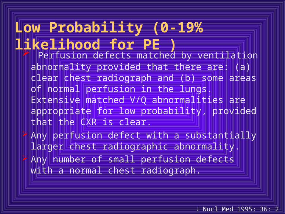
Low Probability (0-19% likelihood for PE )
Perfusion defects matched by ventilation abnormality provided that there are: (a) clear chest radiograph and (b) some areas of normal perfusion in the lungs. Extensive matched V/Q abnormalities are appropriate for low probability, provided that the CXR is clear.
Any perfusion defect with a substantially larger chest radiographic abnormality.
Any number of small perfusion defects with a normal chest radiograph.
J Nucl Med 1995; 36: 2380-2387
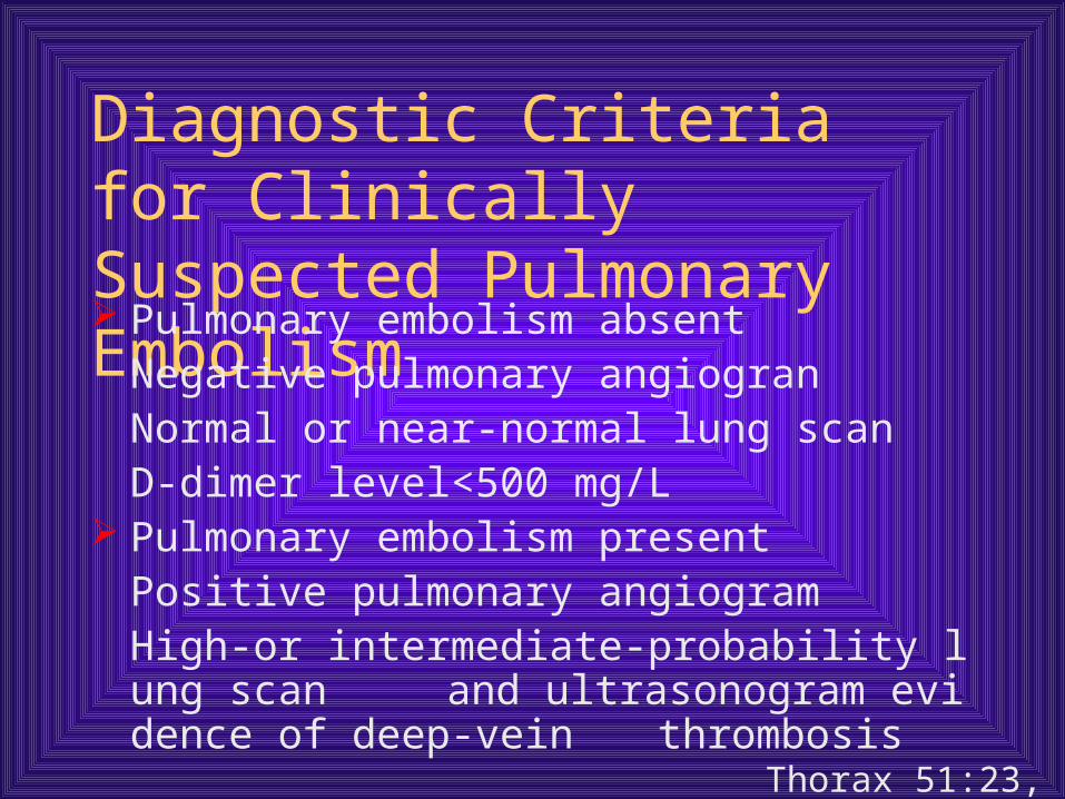
Diagnostic Criteria for Clinically Suspected Pulmonary Embolism
Pulmonary embolism absentNegative pulmonary angiogranNormal or near-normal lung scanD-dimer level<500 mg/L
Pulmonary embolism presentPositive pulmonary angiogramHigh-or intermediate-probability lung scan and ultrasonogram evidence of deep-vein thrombosis
Thorax 51:23, 1996

鉴别诊断
呼吸困难、咳嗽、咯血、呼吸频率增快等呼吸系统表现为主的患者多被诊断为其它的胸肺疾病如肺炎、胸膜炎、肺不张等
以胸痛、心悸、心脏杂音、肺动脉高压等循环系统表现为主的患者易衩诊断为其它的心脏疾病如冠心病、风心病等
以晕厥、惊恐等表现为主的患者有时被诊断为其它心脏或神经及精神系统疾病如心律失常、脑血管意外、癫痫等

原发性肺动脉高压与肺栓塞复发
相似点:症状:疲乏,活动时呼吸困难最常见,胸痛、昏厥、咯血、紫绀也较常见
临床经过:进行性呼吸困难,右心衰竭血流动力学:右心室压力升高、肺毛细
血管嵌压正常治疗:包含抗凝治疗
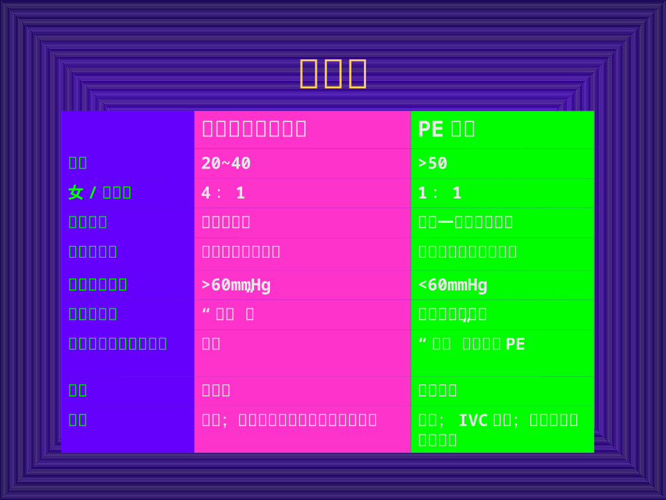
区别点原发性肺动脉高压 PE复发
年龄 20~40 >50
女 /男比例 4: 1 1: 1
临床经过 进行性恶化 稳定一段时间后恶化肺灌注扫描 无节段性灌注缺损 节段性或大片灌注缺损
肺动脉收缩压 >60mmHg <60mmHg
肺动脉造影 “修剪”征 管腔内充盈缺损肺动脉造影混淆的问题
血栓 “修剪”征也提示 PE
确诊 肺活检 肺血管镜治疗 抗凝;大剂量硝苯地平及静
注前列环素抗凝; IVC中断;血栓动脉内膜切除术

急性 PE 的治疗
一般处理:
送入监护病房,加强生命体征的监护
防止栓子脱落,绝对卧床
情感支持
对症治疗:如咳嗽、发热等
急性 PE

呼吸循环支持治疗
一般患者均采用经鼻导管或面罩吸氧治疗低氧
血症
无创伤性或经气管插管机械通气治疗呼吸衰竭,
避免气管切开。
尽量减少正压通气对循环的不种影响。
急性 PE

溶栓治疗的适应证
栓塞面积超过 2个肺叶血管者
合并休克或低血压者
合并右心功能不全者
排除禁忌证者
急性 PE
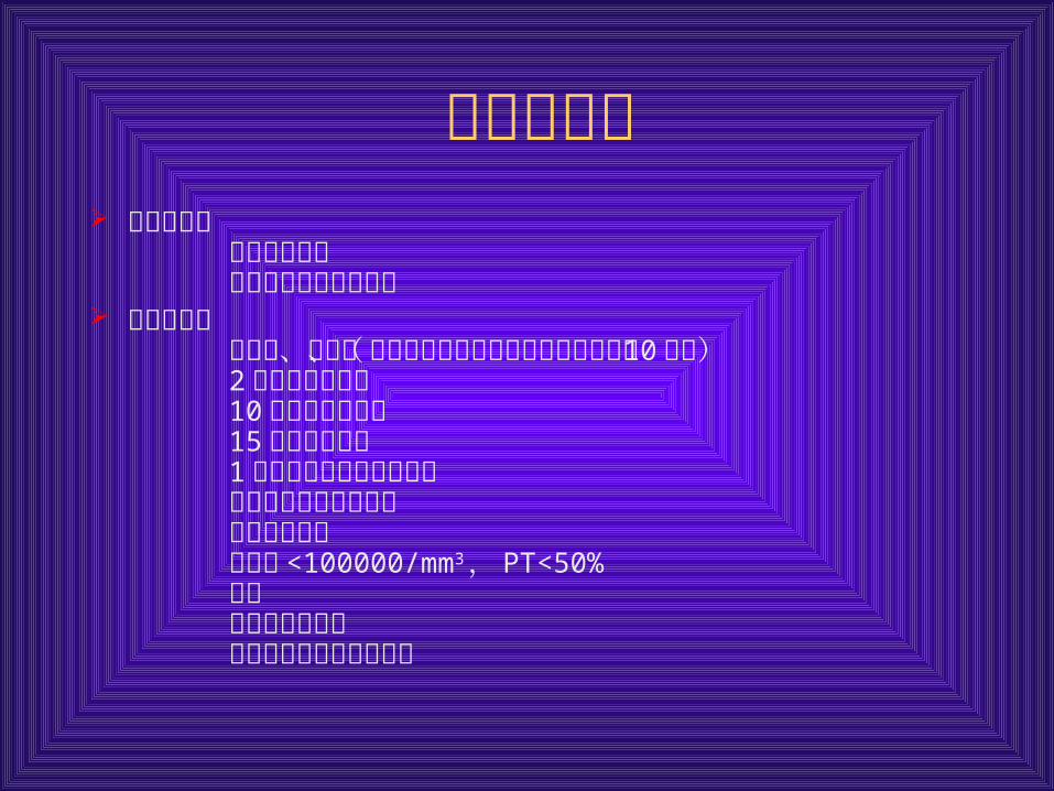
溶栓禁忌证 绝对禁忌证
活动性内出血近期的自发性颅内出血
相对禁忌证大手术、分娩、器官活检或不能压迫的血管穿刺史( 10 天
内)2月内缺血性中风10 天内胃肠道出血15 天内严重外伤1月内神经外科或眼科手术控制不好的重度高血压近期心肺复苏血小板 <100000/mm3 , PT<50%怀孕细菌性心内膜炎糖尿病出血性视网膜病变
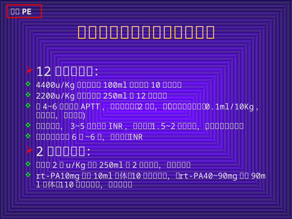
肺动脉栓塞的溶栓及抗凝治疗
12 小时溶栓法: 4400u/Kg尿激酶溶于 100ml 于不少于 10 分钟静推 2200u/Kg尿激酶溶于 250ml 用 12 小时维持 每 4~6 小时监测 APTT ,当其降到正常 2倍时,加用低分子肝素钙( 0.1ml/10Kg ,每天二次,皮下注射 )
同用华法令, 3~5 天后监测 INR ,当重复为 1.5~2倍二天时,停用低分子肝素,
维持剂量华法令 6周 ~6月,同时监测 INR
2 小时溶栓法: 尿激酶 2万 u/Kg 溶于 250ml 用 2 小时静泵,余治疗同上 rt-PA10mg 加入 10ml 液体中 10 分钟内静推,后 rt-PA40~90mg 加入 90ml 液体中 110 分钟内静滴,余治疗同上
急性 PE
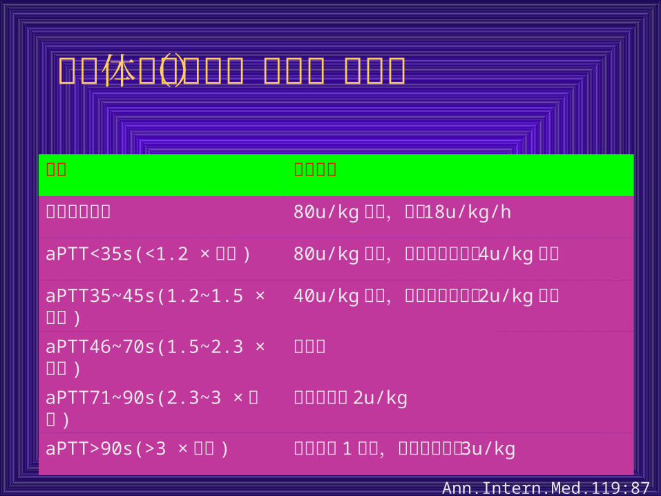
依据体重的肝素(普通)计算图
变量 肝素剂量
肝素起始剂量 80u/kg推注,然后 18u/kg/h
aPTT<35s(<1.2 ×对照 ) 80u/kg推注,然后每小时增加 4u/kg 静滴
aPTT35~45s(1.2~1.5 ×对照 ) 40u/kg推注,然后每小时增加 2u/kg 静滴
aPTT46~70s(1.5~2.3 ×对照 ) 不改变
aPTT71~90s(2.3~3 ×对照 ) 每小时减少 2u/kg
aPTT>90s(>3 ×对照 ) 保持静滴 1 小时,然后滴速减慢 3u/kg
Ann.Intern.Med.119:874,1993
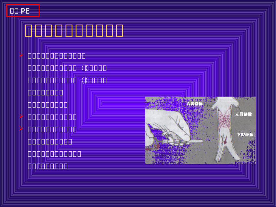
下腔静脉过滤网适应证 抗凝治疗禁忌而肺栓塞已证实
活动性出血可能引起贫血(如胃肠道)担心可能是灾难性的出血(如开颅术)现存的抗凝并发症计划强化的癌症化疗
尽管治疗充分但抗凝失败 在高危病人中预防性使用
广泛的进展性静脉血栓和导管或外科肺去栓术并用严重肺高压或肺心病
急性 PE

肺动脉血栓摘除术
大面积 PTE ,适合手术且无固定肺动脉
高压者者。
有溶栓禁忌证者。
经溶栓和其他积极的内科治疗无效者。
急性 PE
经静脉导管碎解和抽吸血栓
肺动脉主干或主要分支大面积 PTE 者
溶栓和抗凝治疗禁忌
经溶栓或积极的内科治疗无效者
缺乏手术条件
急性 PE

慢性栓塞性肺动脉高压的治疗
手术治疗:严重肺动脉高压。介入治疗:球囊扩张肺动脉成型术。抗凝治疗:华法令。下腔静脉滤器:反复深静脉血栓脱落者。降低肺动脉压力:血管扩张剂治疗心衰

预防策略
机械措施:分级加压长筒袜间歇性序贯充气泵下腔静脉波器
药理学制剂普通肝素低分子肝素华法令
情感支持
对高危人群: