Embed Size (px)
Citation preview
1
Mycobacteria
• Aerobic• Acid fast – bind phenol based dyes (carbol fuchsin) and resist acid alcohol
decoloration (Ziehl-Neelsen stain).
• Non spore forming
• Non motile, rods with varying sizes (1-10μm)
• Gram positive – do not stain well with Gram stain
• Catalase positive
• Many produce pigments on culture
• Relatively simple growth requirements
• Rapid (<7 days) or slow growing (weeks or months)
• Most pathogens slow growing
• Unique cell walls – Lipid rich - acid fastness related to presence of peptidoglycan but particularly glycolipids
• Lipids in cell wall related to pathogenicity particularly survival in phagolysosome of macrophages, resists drying, extreme pH and other stresses
2
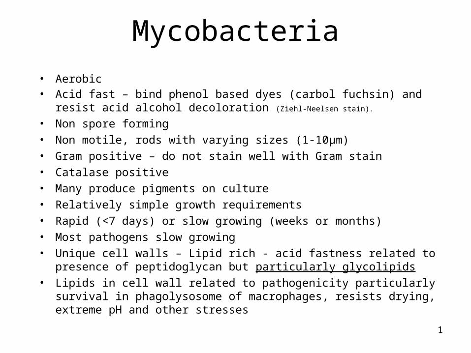
Acid fast (Ziehl-Neelsen) staining of Mycobacteria
3
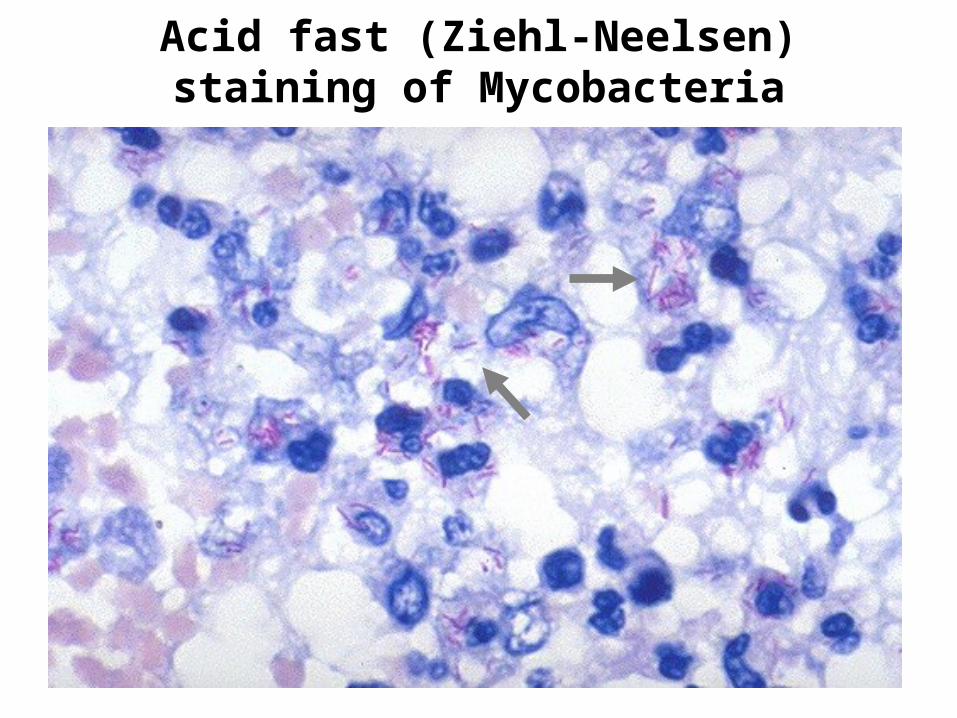
Mycobacterial (acid-fast) cell wall
4
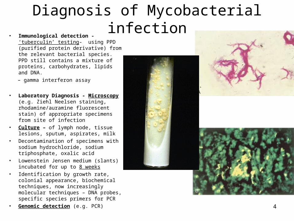
Diagnosis of Mycobacterial infection• Immunological detection - ‘tuberculin’
testing- using PPD (purified protein derivative) from the relevant bacterial species. PPD still contains a mixture of proteins, carbohydrates, lipids and DNA.
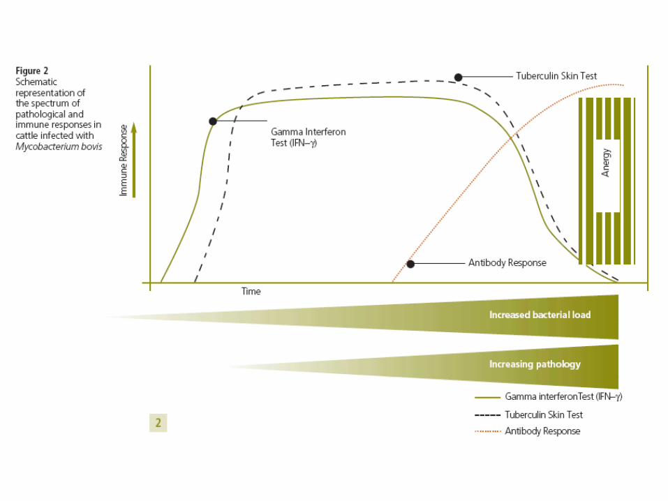
– gamma interferon assay
• Laboratory Diagnosis - Microscopy (e.g. Ziehl Neelsen staining, rhodamine/auramine fluorescent stain) of appropriate specimens from site of infection
• Culture – of lymph node, tissue lesions, sputum, aspirates, milk
• Decontamination of specimens with sodium hydrochloride, sodium triphosphate, oxalic acid
• Lowenstein Jensen medium (slants) incubated for up to 8 weeks
• Identification by growth rate, colonial appearance, biochemical techniques, now increasingly molecular techniques – DNA probes, specific species primers for PCR
• Genomic detection (e.g. PCR)

5
The Comparative intradermal test
1. The tuberculin test is carried out at 1,2,3, or 4 year intervals depending on the frequency of TB in the area. National average 2.7% dairy farms.
2. Animal identified and two sites prepared on the side of the neck, approx.13 cm apart. Hair clipped 2 cm radius, and the skin fold measured.
3. Inject PPD, usually the M. avium preparation in the upper site.
4. Re-measure fold after 72 hrs. Reaction to M. bovis PPD is 5 mm greaterthan to the M. avium then defined a reactor. If 1-4 mm then retested within 40-60 days.
5. Rest of the herd analysed using ‘severe interpretation’ which is 3 mm.

7
Mycobacteria
• Diseases of great importance – Tuberculosis (TB) (Mycobacterium tuberculosis, Mycobacterium bovis) and Leprosy (Mycobacterium leprae)
• Economic and social effects perhaps unparalleled in the history of medicine
• Many species – consisting of major pathogens, opportunist pathogens, harmless saprophytes (live in environment – water, vegetation)
• Pathogenic Mycobacteria produce granulomatous lesions in tissues of a wide range of domestic and wild animals and humans
• Development of Mycobacterial disease in a host depends on the ability of the Mycobacteria to survive and multiply within the macrophages
8
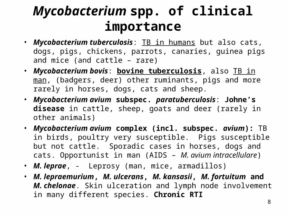
Mycobacterium spp. of clinical importance
• Mycobacterium tuberculosis: TB in humans but also cats, dogs, pigs, chickens, parrots, canaries, guinea pigs and mice (and cattle – rare)
• Mycobacterium bovis: bovine tuberculosis, also TB in man, (badgers, deer) other ruminants, pigs and more rarely in horses, dogs, cats and sheep.
• Mycobacterium avium subspec. paratuberculosis: Johne’s disease in cattle, sheep, goats and deer (rarely in other animals)
• Mycobacterium avium complex (incl. subspec. avium): TB in birds, poultry very susceptible. Pigs susceptible but not cattle. Sporadic cases in horses, dogs and cats. Opportunist in man (AIDS – M. avium intracellulare)
• M. leprae, - Leprosy (man, mice, armadillos)• M. lepraemurium, M. ulcerans, M. kansasii, M. fortuitum and M.
chelonae. Skin ulceration and lymph node involvement in many different species. Chronic RTI

9
Virulence factors of Mycobacteria
Cell wall components
Mycolic acids – resist phagocytic digestion.
Sulfatides – prevent phagocyte activation and phagosome-lysosome fusion.
Trehalose di-mycolate (cord factor) – Inhibits phagocyte chemotaxis,activation, phagosome-lysosome fusions and digesion.
Lipoarabinomannan (LAM) – prevents phagocyte activation and digestion within the phagocyte.
Mycosides – prevent intracellular killing and digestion
Cell wall antigens in general induce DTH
Other factors include SOD (superoxide dismutase) and heat shock proteins.
10
•About 8 million new cases of active disease arise each year, with a global incidence of approximately 160 cases per 100 000 population.
• Worldwide, tuberculosis continues to kill more than 2 million people per year
• Tuberculosis is a leading cause of death in AIDS, and HIV-related tuberculosis deaths are attributed to AIDS, not tuberculosis.
Mycobacterium tuberculosis

11
Natural history of tuberculosis (man)
TB can affect any organ system in man, lungs, meninges (TB meningitis), bones, joints, skin, kidneys etc. Most infectious when open lung lesions containing tubercle bacilli coughed into atmosphere
12
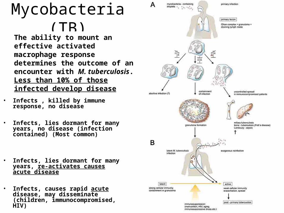
Mycobacteria (TB)
• Infects , killed by immune response, no disease
• Infects, lies dormant for many years, no disease (infection contained) (Most common)
• Infects, lies dormant for many years, re-activates causes acute disease
• Infects, causes rapid acute disease, may disseminate (children, immunocompromised, HIV)
The ability to mount an effective activated macrophage response determines the outcome of an encounter with M. tuberculosis. Less than 10% of those infected develop disease
13
Immune responses to Mycobacterial infections
• Humoral response irrelevant to protection. A bias towards a Th2 response exacerbates the condition. Th1 (CMI) required to limit the disease and provide protection
• Immune status of the animal important. Active response results in lymphocyte infiltration, central necrosis in the lesion, tubercule maybe limited by a fibrin capsule. Response may be strong enough to kill the bacteria but often the response is only able to restrict the disease. Reactivation occurs with stress/immunosuppression.
• IFN gamma from CD4 lymphocytes activates macrophages to kill intracellular mycobacteria. CD8 lymphocytes become cytotoxic killing mycobacterial infected cells. CD1 restricted T cells recognise glycolipids
• Exposure to environmental Mycobacteria provides some cross-protection which may limit the disease caused by virulent species (also complicates hypersensitivity testing).

14
15
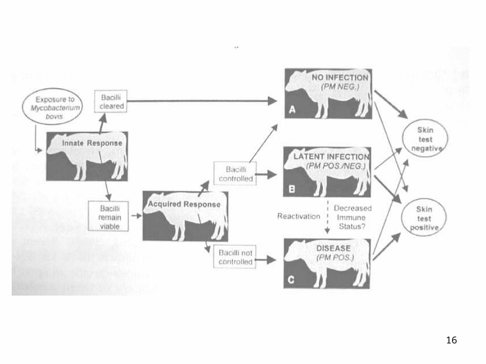
Bovine TuberculosisMycobacterium bovis: control measures have led to a greatly reduced prevalence in Europe. Spread is promoted by high densities of animals and immune suppression.
Generally a primary respiratory infection leads to tubercules in the lung and associated lymph nodes (bronchial and retropharyngeal).
Closed or open lesions
Spread to intestine (via sputum) and serosal surfaces. Pleural lesions (Pearls disease).
Further spread (usually haematogenous) to liver, spleen, kidney, brain etc. Vertical transmission is possible after spread to mammary glands and uterus.
Antibiotic treatments are long term and very expensive for animals. Consequently tuberculin testing and culling of exposed animals.
Prevent cattle movement
16
17
Epidemiology of bovine TB
• Cattle transmit infection to cattle via infected respiratory droplets – respiratory route
• Badgers transmit M. bovis between themselves by the respiratory route and by biting. Mums transmit to cubs but not by milk
• Cattle may get M. bovis from badgers via grazing on pasture contaminated with badger urine, faeces and bronchial pus or badgers urinate and defecate in cattle feeders.
• Aerosol transmission via coughing may be possible or via dried badger saliva in cattle houses
• This may apply to cattle to badger transmission
18
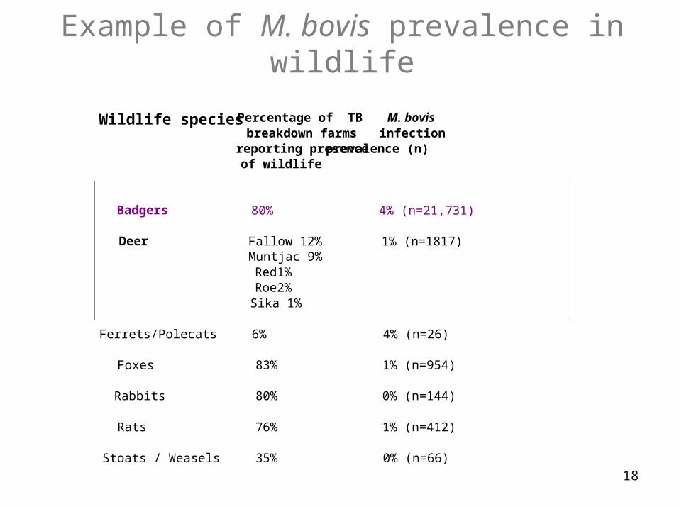
Example of M. bovis prevalence in wildlife
Wildlife species Percentage of TB M. bovis breakdown farms infection
reporting presence prevalence (n) of wildlife
Badgers 80% 4% (n=21,731)
Deer Fallow 12% 1% (n=1817)
Muntjac 9% Red1% Roe2% Sika 1%
Ferrets/Polecats 6% 4% (n=26) Foxes 83% 1% (n=954) Rabbits 80% 0% (n=144) Rats 76% 1% (n=412) Stoats / Weasels 35% 0% (n=66)
19
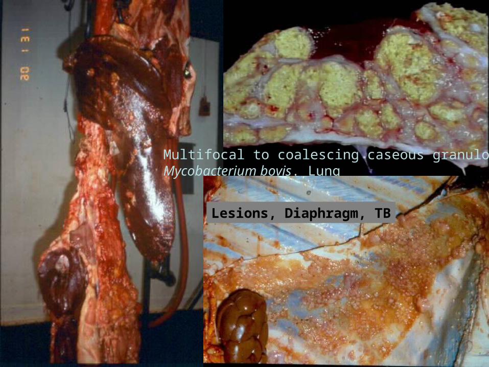
Multifocal to coalescing caseous granulomas. Mycobacterium bovis. Lung
Lesions, Diaphragm, TB
20
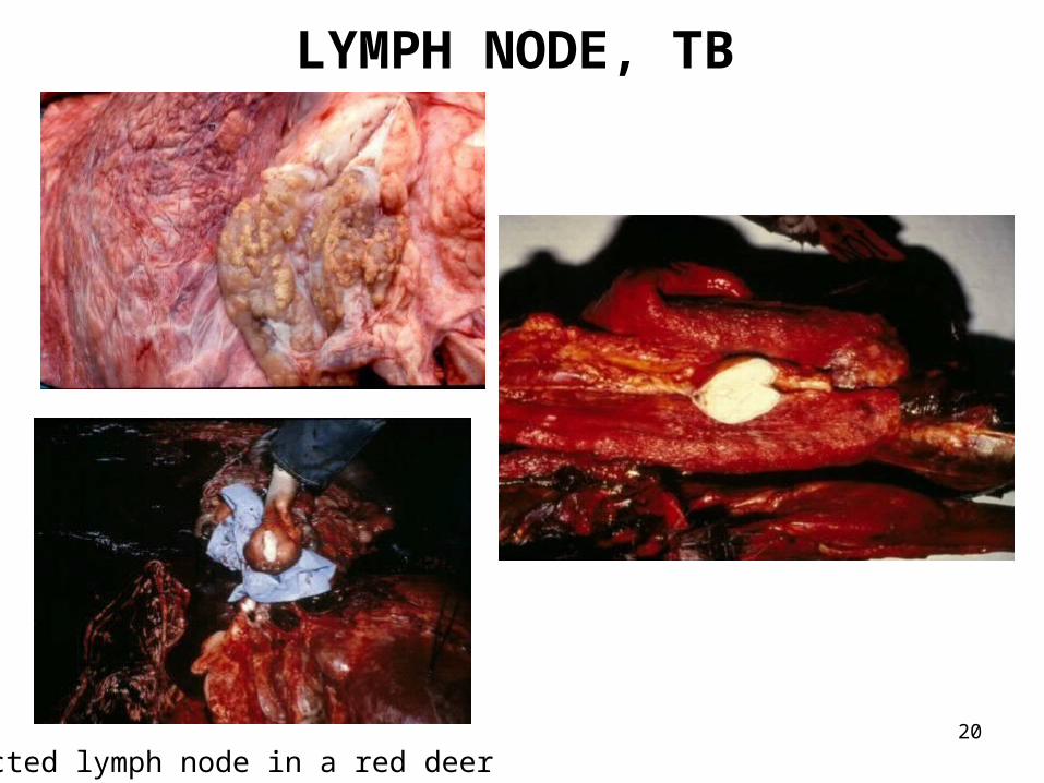
LYMPH NODE, TB
Infected lymph node in a red deer
21
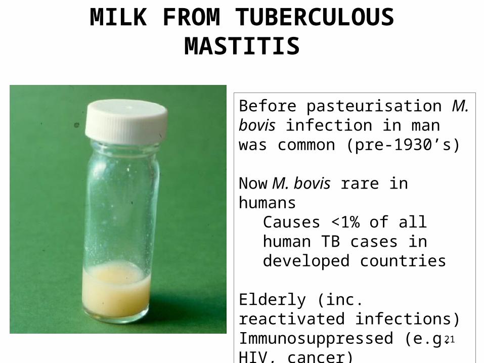
MILK FROM TUBERCULOUS MASTITIS
Before pasteurisation M. bovis infection in man was common (pre-1930’s)
Now M. bovis rare in humansCauses <1% of all human TB cases in developed countries
Elderly (inc. reactivated infections)Immunosuppressed (e.g. HIV, cancer)Foreign travellers
22
Mycobacterium avium
• M. avium subspecies avium and the taxonomically closely related M. intracellulare (both organisms referred to as the M. avium complex)
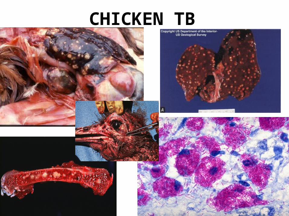
• Widest host range among Mycobacteria• M. avium serovars 1, 2 and 3 isolated from tuberculous
lesions in avian species (avian TB – progressive disease)• Other M. avium serovars produce minimal disease
(microscopic foci in liver and spleen) in chickens• Non human primates, cattle and pigs infection by M. avium
ss avium is confined to lymph node infection (Mycobacteriosis in pigs)
• M. avium-intracellulare causes disseminated disease in HIV/AIDS patients
23
CHICKEN TB
24
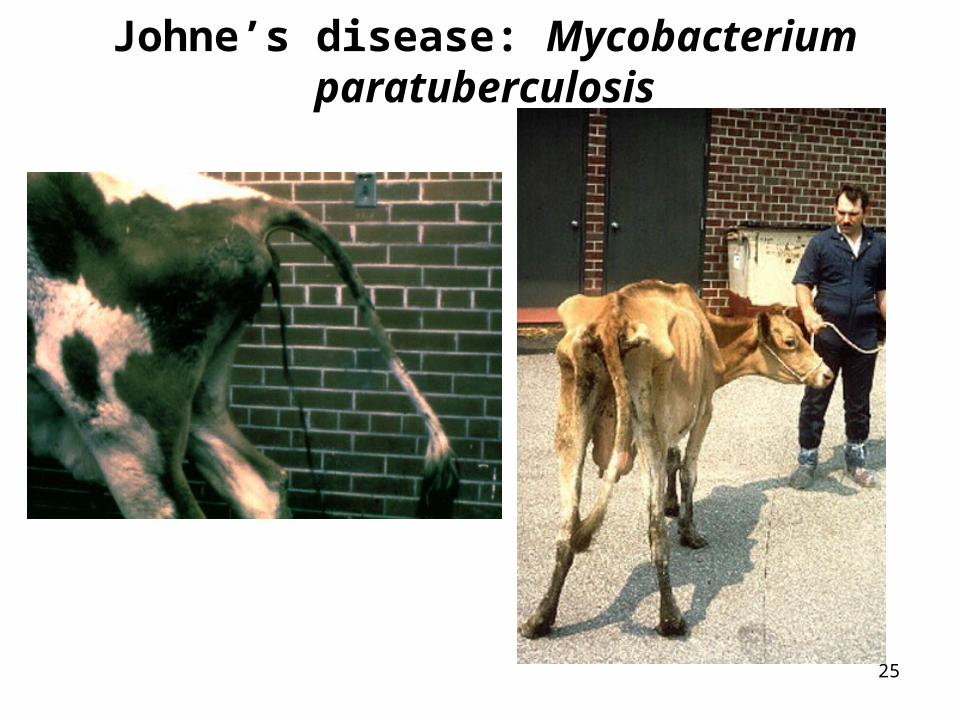
M. avium sub spec. paratuberculosis
This organism causes a transmissible chronic and progressive enteritis in cattle sheep and goats, but not swine or horses.
First observed by Johne and Frothingham in 1895 – Johne’s disease.
Infection usually occurs within the first month but may take 6 months to 5 years to become apparent. Clinical course (1-4 months) starts with general signs of illness (weight loss, int. diarrhoea), followed by severe diarrhoea, emaciation and death.
Impaired intestinal function due to chronic inflammation. Evidenceof diffuse granulomatous changes. Accumulation of lymphocytesand epitheloid cells in the lamina propria and submucosa.
25
Johne’s disease: Mycobacterium paratuberculosis
26
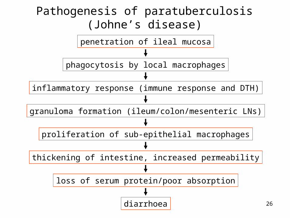
Pathogenesis of paratuberculosis (Johne’s disease)
penetration of ileal mucosa
phagocytosis by local macrophages
inflammatory response (immune response and DTH)
granuloma formation (ileum/colon/mesenteric LNs)
proliferation of sub-epithelial macrophages
thickening of intestine, increased permeability
loss of serum protein/poor absorption
diarrhoea
27
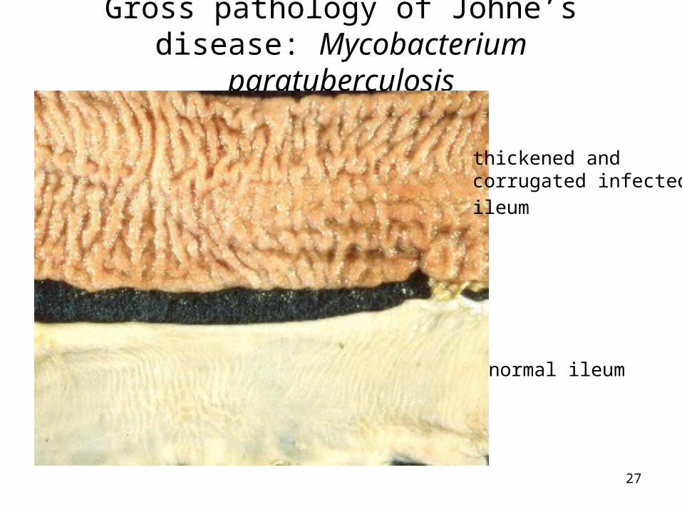
Gross pathology of Johne’s disease: Mycobacterium paratuberculosis
normal ileum
thickened and corrugated infected
ileum
28
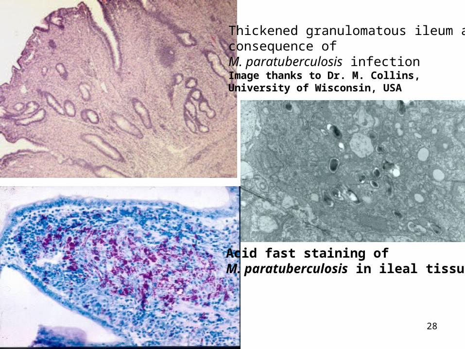
Thickened granulomatous ileum as a consequence of M. paratuberculosis infectionImage thanks to Dr. M. Collins, University of Wisconsin, USA
Acid fast staining of M. paratuberculosis in ileal tissue
29
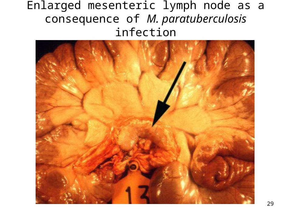
Enlarged mesenteric lymph node as a consequence of M. paratuberculosis infection
30
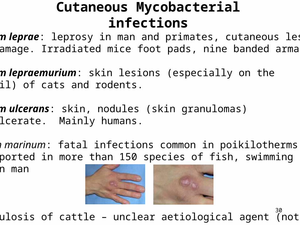
Cutaneous Mycobacterial infections
Mycobacterium leprae: leprosy in man and primates, cutaneous lesionsand nerve damage. Irradiated mice foot pads, nine banded armadillos
Mycobacterium lepraemurium: skin lesions (especially on the head and tail) of cats and rodents.
Mycobacterium ulcerans: skin, nodules (skin granulomas) which can ulcerate. Mainly humans.
Mycobacterium marinum: fatal infections common in poikilotherms (frogs), reported in more than 150 species of fish, swimming pool granuloma in man
Skin tuberculosis of cattle – unclear aetiological agent (not cultured)
31
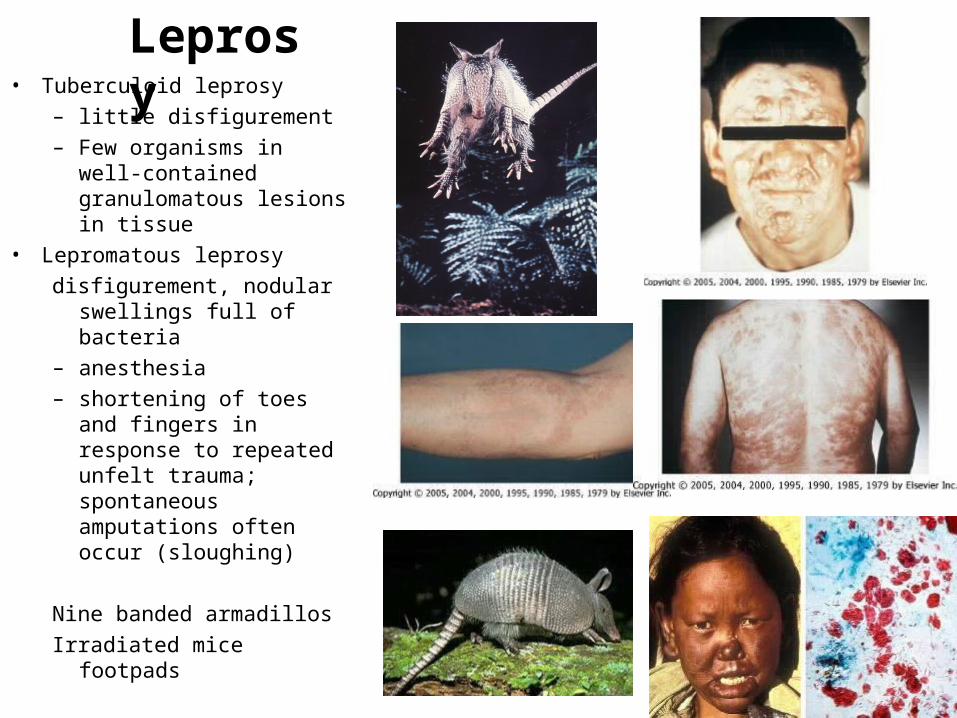
Leprosy• Tuberculoid leprosy
– little disfigurement
– Few organisms in well-contained granulomatous lesions in tissue
• Lepromatous leprosy
disfigurement, nodular swellings full of bacteria
– anesthesia
– shortening of toes and fingers in response to repeated unfelt trauma; spontaneous amputations often occur (sloughing)
Nine banded armadillos
Irradiated mice footpads
32
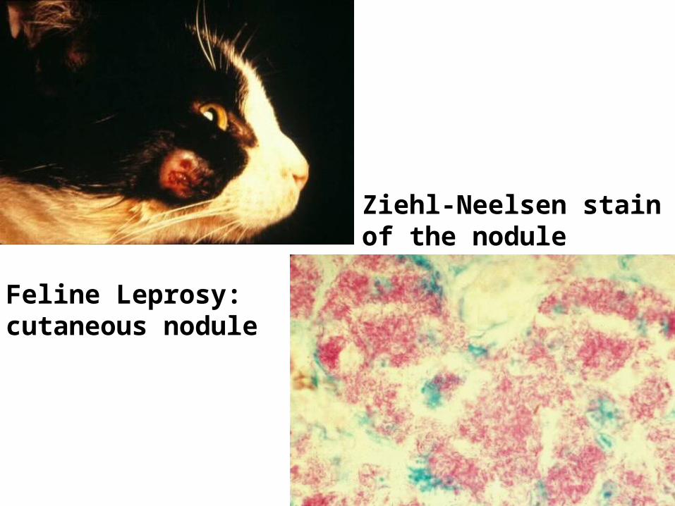
Ziehl-Neelsen stain of the nodule
Feline Leprosy:cutaneous nodule