Embed Size (px)
Citation preview
2. Bioenergetics. Mitochondrial pathology. Mitochondrial medicine.
Lecturer Alexander Koval, PhD
General, bioorganic chemistry and biochemistry dept.
10/29/2019 Koval A.N. (C), 2019 1
Contents • Modern ideas about biological oxidation.
– Oxygen utilization pathways in the body (mitochondrial, microsomal and peroxide), their comparative characteristics.
• Mitochondrial pathology. – Integrative and regulatory functions of mitochondria. Heterogeneity of the
intracellular mitochondrial population. – Structure and function of mitochondria. Comparative characteristics of
mitochondrial membranes. Enzyme composition of various compartments. – Mitochondrial genome: features of organization and functioning. – Mitochondria as apoptosis trigger mechanism.
• Mitochondrial diseases. – Mitochondrial medicine, a brief historical background. Mitochondrial diseases.
Classification. Types. Clinical manifestations. – Diagnosis of mitochondrial diseases. – Defects of mitochondrial DNA (mtDNA). Kearns-Sayre syndrome (KSS), progressive
external ophthalmoplegia (PEO), Pearson syndromes, MERRF, MELAS. – Defects of nuclear DNA. – Defects of the linkage between nuclear and mitochondrial DNA. – The linkage of mitochondrial pathologies with aging and manifestations of
Parkinson's disease, Alzheimer's disease, diabetes mellitus. 10/29/2019 Koval A.N. (C), 2019 2
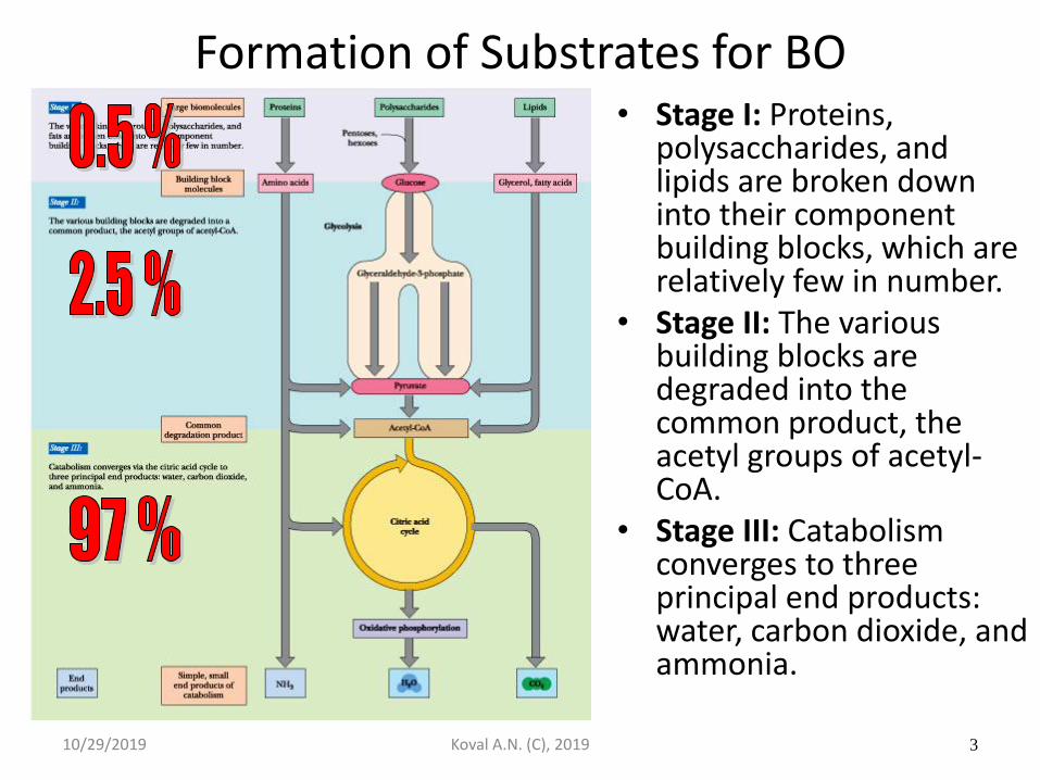
Formation of Substrates for BO
• Stage I: Proteins, polysaccharides, and lipids are broken down into their component building blocks, which are relatively few in number.
• Stage II: The various building blocks are degraded into the common product, the acetyl groups of acetyl-CoA.
• Stage III: Catabolism converges to three principal end products: water, carbon dioxide, and ammonia.
10/29/2019 Koval A.N. (C), 2019 3
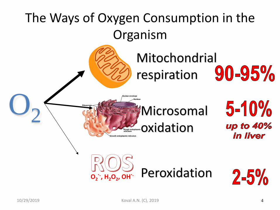
The Ways of Oxygen Consumption in the Organism
O2
Mitochondrial respiration
Microsomal oxidation
Peroxidation
10/29/2019 Koval A.N. (C), 2019 4
O2˙-, H2O2, OH˙-
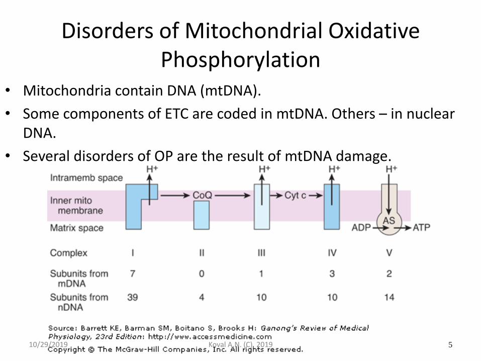
Disorders of Mitochondrial Oxidative Phosphorylation
• Mitochondria contain DNA (mtDNA).
• Some components of ETC are coded in mtDNA. Others – in nuclear DNA.
• Several disorders of OP are the result of mtDNA damage.
10/29/2019 Koval A.N. (C), 2019 5
Mitochondria
• Animal mitochondrial genomes are 13-18 kb in size.
• Fungal mitochondrial genomes are ~75 kb.
• Higher plant mitochondrial genomes are 300-500 kb.
• Each mitochondrion has 5-20 copies of the mitochondrial chromosomes.
10/29/2019 Koval A.N. (C), 2019 6
Mitochondria
• Human cells have a range of numbers of mitochondria:
– Liver cells have 1000 mitochondria per cell.
– Skin cells have 100.
– Egg cells have up to 10 million.
Human mitochondria have 37 genes.
10/29/2019 Koval A.N. (C), 2019 7
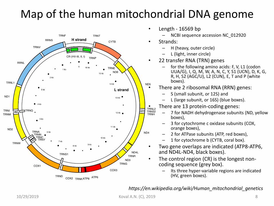
Map of the human mitochondrial DNA genome • Length - 16569 bp
– NCBI sequence accession NC_012920
• Strands: – H (heavy, outer circle) – L (light, inner circle)
• 22 transfer RNA (TRN) genes – for the following amino acids: F, V, L1 (codon
UUA/G), I, Q, M, W, A, N, C, Y, S1 (UCN), D, K, G, R, H, S2 (AGC/U), L2 (CUN), E, T and P (white boxes).
• There are 2 ribosomal RNA (RRN) genes: – S (small subunit, or 12S) and – L (large subunit, or 16S) (blue boxes).
• There are 13 protein-coding genes: – 7 for NADH dehydrogenase subunits (ND, yellow
boxes), – 3 for cytochrome c oxidase subunits (COX,
orange boxes), – 2 for ATPase subunits (ATP, red boxes), – 1 for cytochrome b (CYTB, coral box).
• Two gene overlaps are indicated (ATP8-ATP6, and ND4L-ND4, black boxes).
• The control region (CR) is the longest non-coding sequence (grey box). – Its three hyper-variable regions are indicated
(HV, green boxes).
10/29/2019 Koval A.N. (C), 2019 8
https://en.wikipedia.org/wiki/Human_mitochondrial_genetics
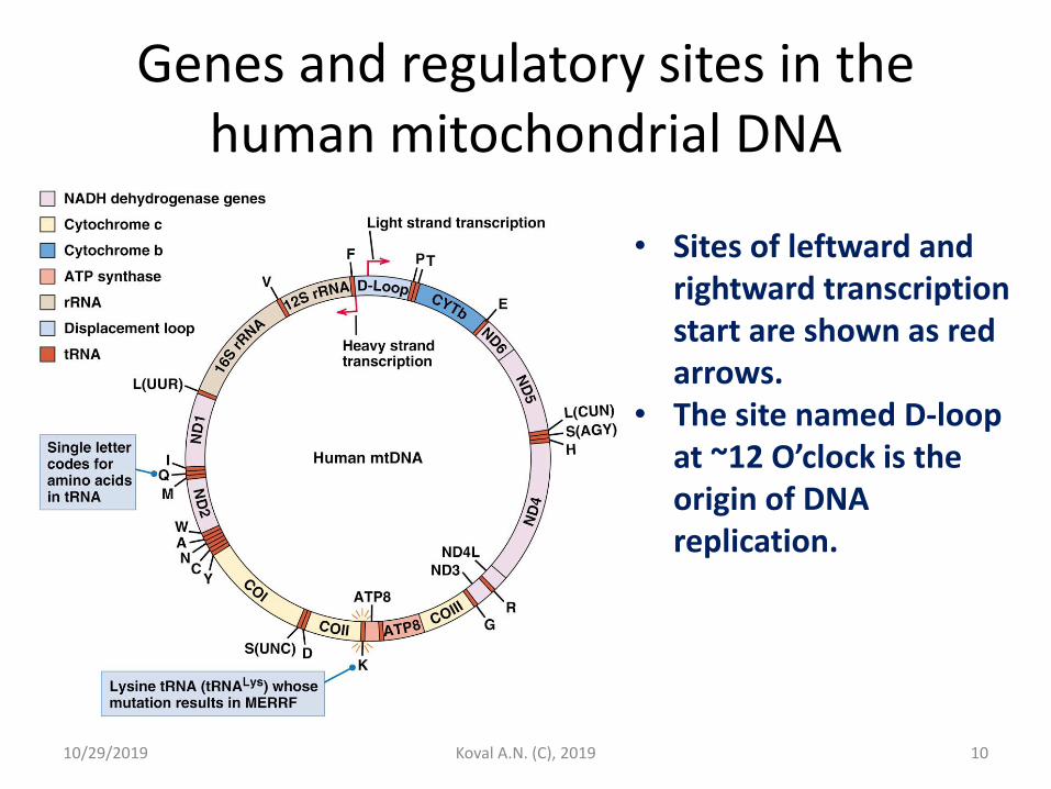
Genes and regulatory sites in the human mitochondrial DNA
• Sites of leftward and rightward transcription start are shown as red arrows.
• The site named D-loop at ~12 O’clock is the origin of DNA replication.
10/29/2019 Koval A.N. (C), 2019 10
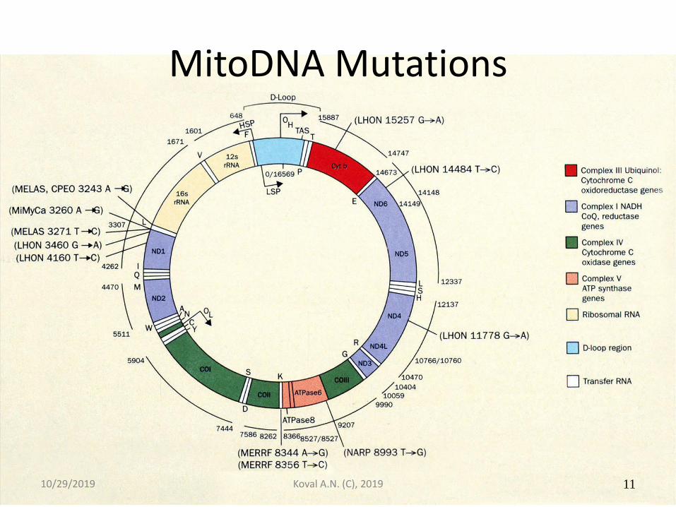
MitoDNA Mutations
10/29/2019 Koval A.N. (C), 2019 11
Clinical Manifestation and Treatment of Mito Disorders
• Manifestation
– Muscle cramping and weakness,
– Fatigue,
– Lactic acidosis,
– CNS dysfunction,
– Vision problems.
• Treatment
– Is difficult and often unsuccesfull
– In some cases can be helpful ubiquinone, vitamin C, menadione.
10/29/2019 Koval A.N. (C), 2019 12
Some Mitochondrial Diseases • The names of mitochondrial diseases are often complex and
usually are described by abbreviations. – LHON, Lebers hereditary optical neuropathy; – MERRF, myoclonic epilepsy and ragged-red-fiber disease; – MELAS, mitochondrial encephalomyopathy, lactic acidosis, and
strokelike episodes; – NARP, neurological muscle weakness,ataxia, and retinitis pigmentosa; – Leigh disease. – SNE, subacute necrotizing encephalomyelopathy; – KSS, Kearns–Sayre syndrome; – CPEO, chronic progressive external ophthalmoplegia.
• LHON is a hereditary disease that often leads to sudden blindness from death of the optic nerve especially among males. Any one of several point mutations in subunits ND1, 2, 4, 5, and 6 of NADH dehydrogenase.
10/29/2019 Koval A.N. (C), 2019 13
Kearns-Sayre syndrome • Most patients with Kearns-Sayre syndrome have a
single, large deletion of mitochondrial DNA ranging from 1,000 to 10,000 nucleotides. – the most common deletion is 4,997 nucleotides.
• Kearns-Sayre syndrome mainly affects the eyes, causing weakness of the eye muscles (ophthalmoplegia) and retina pathology (retinopathy).
• The mitochondrial DNA deletions result in the decreased cellular energy production. – Till now it is not clear how these deletions lead to the
specific signs and symptoms of Kearns-Sayre syndrome, probably related to a lack of cellular energy.
10/29/2019 Koval A.N. (C), 2019 14
Progressive External Ophthalmoplegia (PEO) • This disorder weakens the muscles for eye movement and
causes the drooping of eyelids (ptosis). • Some people with PEO have a single large deletion of
mtDNA. – The most common deletion is 4,997 nucleotides, as in KSS. – Other patients have a mutation in the mitochondrial gene MT-
TL1. • This gene encodes tRNALeu(UUR). • This tRNA is found only in mitochondria.
• The A3243G mutation, often found in MELAS patients, also can cause of PEO. – It is unclear how the same MT-TL1 gene mutation can result in
different signs and symptoms. – MT-TL1 gene mutations impair the ability of mitochondria to
make proteins, use oxygen, and produce energy. – not clear how these mutations associated with PEO symptoms.
10/29/2019 Koval A.N. (C), 2019 15
Pearson Marrow-pancreas Syndrome • Deletion of mtDNA causes Pearson marrow-pancreas syndrome.
– Like in KSS. • Affects the development of blood cells and the function of the
pancreas and other organs; – often fatal in infancy, early childhood.
• The size and location of mitochondrial DNA deletions vary, usually ranging from 1,000 to 10,000 nucleotides. – About 20 % of patients have a deletion of 4,997 nucleotides;
• Also common in KSS.
• Impairs oxidative phosphorylation, reduces the energy available to cells. – It is unclear how mtDNA deletions lead to the symptoms of Pearson
marrow-pancreas syndrome. • Unclear: the same deletion results in different signs and symptoms.
– Some patients with Pearson marrow-pancreas syndrome who survive past early childhood develop signs and symptoms of KSS later in life.
10/29/2019 Koval A.N. (C), 2019 16
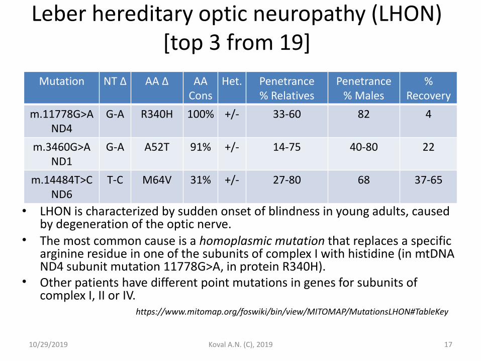
Leber hereditary optic neuropathy (LHON) [top 3 from 19]
• LHON is characterized by sudden onset of blindness in young adults, caused by degeneration of the optic nerve.
• The most common cause is a homoplasmic mutation that replaces a specific arginine residue in one of the subunits of complex I with histidine (in mtDNA ND4 subunit mutation 11778G>A, in protein R340H).
• Other patients have different point mutations in genes for subunits of complex I, II or IV.
10/29/2019 Koval A.N. (C), 2019 17
Mutation NT Δ AA Δ AA Cons
Het. Penetrance
% Relatives Penetrance
% Males %
Recovery
m.11778G>A ND4
G-A R340H 100% +/- 33-60 82 4
m.3460G>A ND1
G-A A52T 91% +/- 14-75 40-80 22
m.14484T>C ND6
T-C M64V 31% +/- 27-80 68 37-65
https://www.mitomap.org/foswiki/bin/view/MITOMAP/MutationsLHON#TableKey
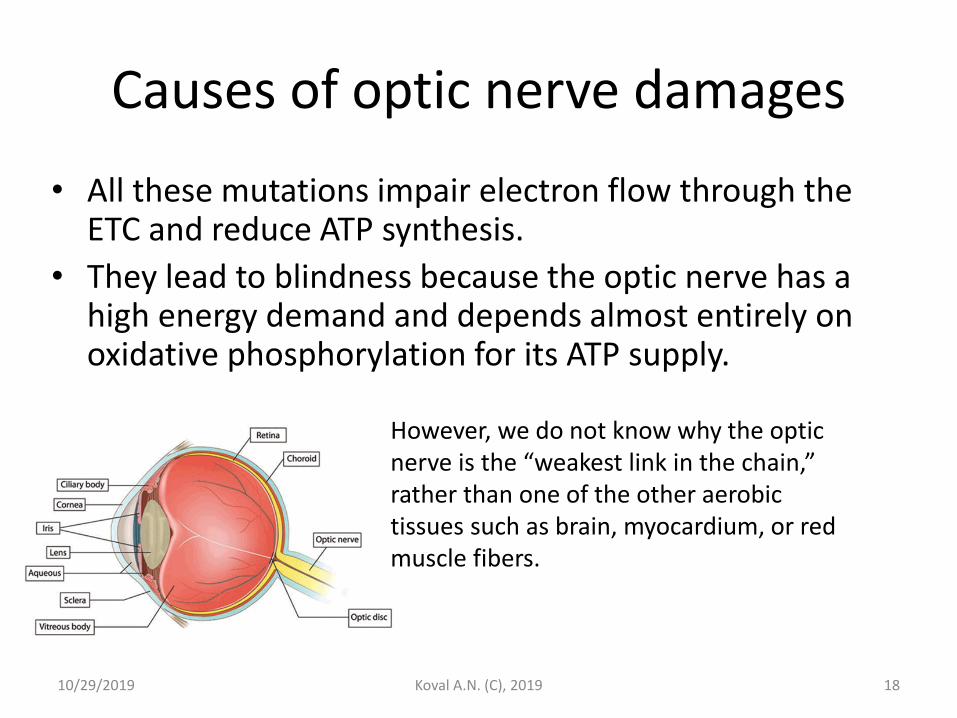
Causes of optic nerve damages
10/29/2019 Koval A.N. (C), 2019 18
• All these mutations impair electron flow through the ETC and reduce ATP synthesis.
• They lead to blindness because the optic nerve has a high energy demand and depends almost entirely on oxidative phosphorylation for its ATP supply.
However, we do not know why the optic nerve is the “weakest link in the chain,” rather than one of the other aerobic tissues such as brain, myocardium, or red muscle fibers.
Leigh Syndrome
• A group of metabolic disorders primarily of infancy characterized by the subacute onset of psychomotor retardation, hypotonia, ataxia, weakness, vision loss, eye movement abnormalities, seizures, dysphagia, and lactic acidosis.
• Pathological features include spongy degeneration of the neuropile of the basal ganglia, thalamus, brain stem, and spinal cord.
• Patterns of inheritance include X-linked recessive, autosomal recessive, and mitochondrial.
• Leigh disease has been associated with mutations in genes for the – pyruvate dehydrogenase complex; – cytochrome-c oxidase; – ATP synthase subunit 6; and – subunits of mitochondrial complex I.
[From Menkes, Textbook of Child Neurology, 5th ed, p850]
10/29/2019 Koval A.N. (C), 2019 19
Leigh Syndrome: symptoms and genetics
• Genetic defects that lead to complex I deficiency present most frequently as neurological degeneration. – Most affected children have normal early development but present
with neurological abnormalities in late infancy or early childhood. – Symptoms are related to dysfunction of the basal ganglia and other
brain regions and include hypotonia and ataxia. – Developmental regression is common, meaning that children lose
abilities that they had acquired earlier. – Characteristic histopathological lesions are spongiosis, neuronal loss,
astrocytosis, and capillary proliferation.
• Mutations in at least 75 genes (mostly nuclear genome) have been associated with the common pathologies of Leigh syndrome; – the most common mtDNA mutations are in the MT-ATP6 gene; – MT-ATP6 encodes the A subunit of the F0 protein of the mitochondrial
ATP synthase complex (often called complex V) of oxidative phosphorylation
10/29/2019 Koval A.N. (C), 2019 20
Genetic Heterogeneity of Leigh Syndrome • Mutations in complex I genes include
– mitochondrial-encoded MTND2, MTND3, MTND5, and MTND6,
– the nuclear-encoded NDUFS1, NDUFS3, NDUFS4, NDUFS7, NDUFS8, NDUFA2, NDUFA9, NDUFA10, NDUFA12, NDUFAF6, and NDUFAF5. • Mutation in the MTFMT gene (involved in mitochondrial translation), has also been reported
with complex I deficiency.
– A mutation has been found in a complex III gene: BCS1L, which is involved in the assembly of complex III.
– Mutations in complex IV genes include mitochondrial-encoded MTCO3 and nuclear-encoded COX10, COX15, SCO2, SURF1, which is involved in the assembly of complex IV, TACO1, and PET100.
– A mutation has been found in a complex V gene: the mitochondrial-encoded MTATP6.
– Mutations in genes encoding mitochondrial tRNA proteins have also been identified in patients with Leigh syndrome: see MTTV, MTTK, MTTW, and MTTL1. • Leigh syndrome may also be caused by mutations in components of the pyruvate
dehydrogenase complex (PDC). • The French Canadian (or Saguenay-Lac-Saint-Jean) type of Leigh syndrome with COX deficiency (LSFC) is
caused by mutation in the LRPPRC gene.
• Deficiency of coenzyme Q10 can present as Leigh syndrome.
10/29/2019 Koval A.N. (C), 2019 21
Can Mitochondrial Diseases be Treated?
• Attempts are being made to improve the function of impaired mitochondria by adding large amounts of ubiquinone, vitamin K, thiamin, riboflavin, and succinate to the diet.
– One report suggests that mitochondrial decay during aging can be reversed by administration of N-acetylcarnitine.
10/29/2019 Koval A.N. (C), 2019 23
Mitochondria as trigger of apoptosis
• The process of apoptosis or programmed cell death may be initiated through the intrinsic (mitochondrial-mediated) pathway by the formation of pores in the outer mitochondrial membrane.
• These pores allow cytochrome c to leave the intermembrane space and enter the cytosol.
• Once in the cytosol, cytochrome c, in association with proapoptotic factors, activates a family of proteolytic enzymes (the caspases), causing cleavage of key proteins and resulting in the morphologic and biochemical changes characteristic of apoptosis.
10/29/2019 Koval A.N. (C), 2019 24

10/29/2019 Koval A.N. (C), 2019 25

10/29/2019 Koval A.N. (C), 2019 26

10/29/2019 Koval A.N. (C), 2019 27

10/29/2019 Koval A.N. (C), 2019 28

10/29/2019 Koval A.N. (C), 2019 29

10/29/2019 Koval A.N. (C), 2019 30

10/29/2019 Koval A.N. (C), 2019 31

10/29/2019 Koval A.N. (C), 2019 32

10/29/2019 Koval A.N. (C), 2019 33