Embed Size (px)
Citation preview
Int. J. Mol. Sci. 2015, 16, 3095-3115; doi:10.3390/ijms16023095
International Journal of
Molecular Sciences ISSN 1422-0067
www.mdpi.com/journal/ijms
Review
Neuron Membrane Trafficking and Protein Kinases Involved in Autism and ADHD
Yasuko Kitagishi †, Akari Minami †, Atsuko Nakanishi †, Yasunori Ogura and Satoru Matsuda †,*
Department of Food Science and Nutrition, Nara Women’s University, Kita-Uoya Nishimachi,
Nara 630-8506, Japan; E-Mails: [email protected] (Y.K.); [email protected] (A.M.);
[email protected] (A.N.); [email protected] (Y.O.)
† These authors contributed equally to this work.
* Author to whom correspondence should be addressed; E-Mail: [email protected];
Tel./Fax: +81-742-20-3451.
Academic Editor: Xiaofeng Jia
Received: 28 October 2014 / Accepted: 19 January 2015 / Published: 30 January 2015
Abstract: A brain-enriched multi-domain scaffolding protein, neurobeachin has been
identified as a candidate gene for autism patients. Mutations in the synaptic adhesion
protein cell adhesion molecule 1 (CADM1) are also associated with autism spectrum
disorder, a neurodevelopmental disorder of uncertain molecular origin. Potential roles of
neurobeachin and CADM1 have been suggested to a function of vesicle transport in endosomal
trafficking. It seems that protein kinase B (AKT) and cyclic adenosine monophosphate
(cAMP)-dependent protein kinase A (PKA) have key roles in the neuron membrane trafficking
involved in the pathogenesis of autism. Attention deficit hyperactivity disorder (ADHD) is
documented to dopaminergic insufficiencies, which is attributed to synaptic dysfunction of
dopamine transporter (DAT). AKT is also essential for the DAT cell-surface redistribution.
In the present paper, we summarize and discuss the importance of several protein kinases
that regulate the membrane trafficking involved in autism and ADHD, suggesting new targets
for therapeutic intervention.
Keywords: autism; attention deficit hyperactivity disorder; neurobeachin; CADM1;
dopamine transporter; membrane trafficking
OPEN ACCESS
Int. J. Mol. Sci. 2015, 16 3096
1. Introduction
Cell polarity and vesicle sorting are important processes that influence normal cell functions such as
migration, adhesion, and neurotransmission [1]. In particular, the progression of membranes of
the secretory and endocytic pathway is predominantly organized. Maintenance of the endomembrane
physical design needs a stability of lipid flows of the various compartments. In addition, proteins destined
for various organelles must be correctly sorted. Endosomes can bud inwardly from the membranes to form
the vesicles, which receive cargo from the cell surface via endocytosis and biosynthetic cargo from
the late Golgi complex [2]. Spatiotemporally, the endosomal membrane trafficking is regulated,
which confirms appropriate delivery of cargo via the pathway. Membrane trafficking is indispensable
for a wide range of developmental processes, which also require the interplay of several proteins and
lipids [3,4]. The precise coordination of membrane trafficking is controlled by protein phosphorylation.
In particular, phosphatidylinositol 3,4,5 trisphosphate (PIP3) is crucial for the membrane trafficking of
early endosomes [5]. Compatibly, disruption of the PIP3 synthesis by wortmannin, a Phosphoinositide
3-kinase (PI3K) inhibitor, severely affects the formation of internal vesicles and the maturation of
endosomes [6]. In addition, membrane trafficking can be induced by cAMP dependent PKA activity [7].
Changes in the activity of PKA provoke a variety of effects on the intracellular membrane dynamics
including membrane sorting and trafficking [8].
In neurons, the machineries for membrane trafficking must be complex. Because of the long
cell extensions, neurons have to form and keep a large membrane area, which is organized into
the axonal macrodomains with specific protein compositions at synaptic sites [9]. Understandably,
the assembly of central nervous synapse requires the polarized targeting of numerous proteins to
synaptic compartments [10]. Establishment of the asymmetric organization of cellular components,
called cell polarity, involves various processes containing membrane trafficking events and cytoskeletal
dynamics [11], which is implicated in morphogenesis of various cellular organisms [12]. Cell polarization
is essential for a cell to function properly. For example, the presence of an axon in neuronal cells
determines the directional flow of the signal. Dysregulation of the cell polarity can cause developmental
disorders. In recent years, there has been accumulating genetic evidence that links the components of
membrane trafficking of intracellular vesicles to a variety of neurological conditions including autism
and attention deficit hyperactivity disorder (ADHD) [13,14]. Symptoms of autism and ADHD often
co-occur [15]. As neurodevelopmental disorders, both autism and ADHD share some phenotypic
similarities, but are characterized by distinct diagnostic criteria [15], imposing a major impediment to
childhood development and a significant burden on society. A recent study provided new insight on
mechanisms of the disorders and opens up new possibilities for therapeutic intervention [16]. Here,
we summarize evidence for the importance of several protein kinases that regulate the membrane
trafficking involved in autism and ADHD.
2. Relationship between Autisms and Neurobeachin
Autism and autism spectrum disorder (autisms) are a prevalent developmental disorder characterized by
severe and sustained impairments in social interaction, repetitive restricted communication and fixed
behavior. The neuropathology of autisms seems to be a defect in neurogenesis and/or its dysplastic changes
Int. J. Mol. Sci. 2015, 16 3097
in the central nervous system [17]. Although several susceptible gene loci related to autisms have been
expansively studied, the molecular pathogenesis of autisms has not been well understood. The genetic
structure of autisms may be heterogeneous. However, there is increasing interest in the signaling
pathways associating autism-pathology with cellular functions, such as neurite outgrowth of evolving
neurons and the synaptic functions [18]. So far, it has been found that many genes that are mutated,
disrupted, and/or deleted in autism patients [19] may be involved in certain function and signal
transduction of autisms-related cellular biology. Autism patients with a monoallelic deletion of the gene
have also been reported [20]. Among the candidate genes, the neurobeachin gene was identified in
a patient with a genetic disorder [20,21]. A single nucleotide polymorphism (SNP) of neurobeachin gene
has also been found to associate with autisms [21]. The neurobeachin gene encodes a multidomain
neuron-specific protein that is principally expressed in brain [22,23]. The protein is a member of
the BEACH protein family implicated in membrane trafficking [22,23], in which the BEACH domain is
headed by an unusual pleckstrin homology (PH) domain, and followed by a tryptophan-aspartic acid
repeat (WD40) repeat domain [24] (Figure 1). The neurobeachin protein may be a negative regulator
of notch function associated with the synaptic plasma membrane and involved in endosomal
trafficking [25]. In addition, a function for neurobeachin in altering the actin cytoskeleton has been
suggested [26]. This scaffolding protein has been suggested to be involved in neuronal trans-Golgi
membrane traffic [20,22]. Actually, neurobeachin in Drosophila has been linked to the membrane
trafficking of growth factor receptors [27]. Neurobeachin concentrates near the trans-Golgi network,
suggesting a functional association to the post-Golgi sorting of membrane trafficking proteins [22]. High
expression of neurobeachin seems to be limited to neuronal cells and endocrine cells [28]. It has been
shown that knockout of the neurobeachin gene in two independent mouse models prevents an activity
in synaptic function with neurotransmitter release [20]. Similarly, knockdown of neurobeachin in
a neuroendocrine cell line (βTC3 cells) has shown a role as negative regulator of secretion of vesicles [20].
Insufficiency of the neurobeachin function results in dense granules with an aberrant morphology [20].
New understandings in the function of neurobeachin may support identifying novel molecular pathways
affected in neurons with autistic patients [29–31].
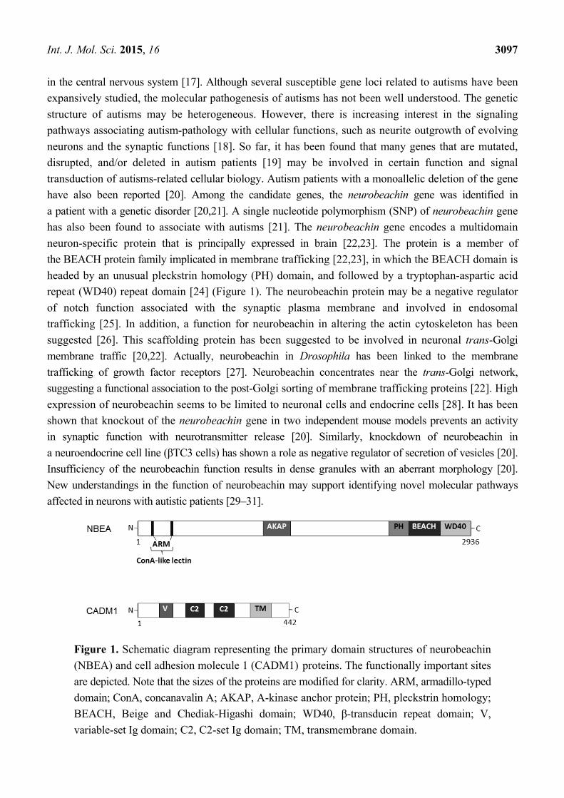
Figure 1. Schematic diagram representing the primary domain structures of neurobeachin
(NBEA) and cell adhesion molecule 1 (CADM1) proteins. The functionally important sites
are depicted. Note that the sizes of the proteins are modified for clarity. ARM, armadillo-typed
domain; ConA, concanavalin A; AKAP, A-kinase anchor protein; PH, pleckstrin homology;
BEACH, Beige and Chediak-Higashi domain; WD40, β-transducin repeat domain; V,
variable-set Ig domain; C2, C2-set Ig domain; TM, transmembrane domain.

Int. J. Mol. Sci. 2015, 16 3098
3. Relationship between Autisms and Cell Adhesion Molecule 1 (CADM1)
Although mutations in genes encoding neurobeachin have been shown in autism patients,
the consistent idea on the molecular pathogenesis of autisms is still unknown. Cell-adhesion molecule
1 (CADM1, TSLC1/SynCAM1) is a member of the immunoglobulin (Ig) superfamily containing
extracellular Ig-like loops, a single transmembrane domain, and a small intracellular carboxy-terminal
tail, is another synaptic cell adhesion molecule [32,33] (Figure 1). CADM1 mRNA is expressed diffusely
in the lateral membrane of cell-cell attachment sites in polarized epithelia, and is also expressed
on rod photoreceptors in a developmentally regulated manner [32,33]. In addition, the CADM1 is
expressed not only in various regions of the cerebrum but also in the developing cerebellum [34–36].
Mutations in CADM1 are associated with autisms [34–36]. The mutated CADM1 shows morphological
abnormalities including impaired synaptogenesis in mice model neurons [35]. CADM1 co-localizes
with alpha-bungarotoxin at the neuromuscular junctions and interacts with the multiple PDZ domain
protein Mupp1, a scaffold protein containing PDZ domains [37]. In addition, CADM1 localizes on
the dendrites in molecular layers of developing cerebellum as well as on the dendrites of hippocampal
neurons [35]. Accordingly, CADM1 synaptic receptor complex may be associated with autisms pathogenesis
locating on the dendrites of neuron cells. Cerebellar aberrations including Purkinje cell damage have
been shown in autisms patients [38]. Furthermore, the autism-related mutations of CADM1 may bring
defective membrane trafficking at the mouse neuronal cell surface [39], suggesting that a link between
impaired synaptogenesis and the molecular pathogenesis of autisms [39]. In fact, the CADM1-knock out
mice exhibit small cerebellums with decreased numbers of synapses with Purkinje neuron cells, which
show some similar behaviors associated with autisms [36]. The mutated CADM1 also exhibits defective
membrane trafficking and greater susceptibility to the cleavage and/or degradation [39], which is
essential for trans-active molecular interaction [39]. In addition, CADM1 is localized to the thalamus
cortical afferent pathway in the cerebrum. Mutations in CADM1 may increase its susceptibility to
processing errors and the accumulation of some CADM1 degradation products in the endoplasmic
reticulum [40], which may diminish CADM1 function in cell adhesion and result in synaptic disorders
in neurons. Impaired synaptogenesis then underlies the pathogenesis of autisms. Actually, CADM1 has
homo-dimer aggregation activity when introduced into Madin-Darby canine kidney cells (MDCK)
cells lacking endogenous CADM1 expression in a Ca2+/Mg2+ independent manner [41], indicating that
CADM1 is involved in cell adhesion through homophilic trans-interaction [41]. However, the
cytoplasmic signaling pathways started by CADM1 have not been fully elucidated. Epigenetic factors may
also complicate the understanding of pathogenesis in autisms. The example of exposure to valproate
provides a good illustration of epigenetic mechanisms involved in autisms [42].
4. Relationship between Attention Deficit/Hyperactivity Disorder (ADHD) and Dopamine
Transporter (DAT)
Attention deficit/hyperactivity disorder (ADHD) is associated to dopaminergic insufficiencies in
prefrontal cortex [43], which is a heterogeneous disorder typically diagnosed in school-age children.
ADHD is characterized by hyperactivity, impulsivity, and inappropriate levels of inattention. Although
studies suggest a contribution of altered dopamine signaling in ADHD brains, evidences of signaling
disturbances contributing to the risk of ADHD may be often conditional [14,44]. On the other hand,

Int. J. Mol. Sci. 2015, 16 3099
some studies have pointed to a contribution of variation in the genes encoding dopamine transporter
(DAT), dopamine receptors, and/or catechol-O-methyl transferase (COMT) as influencing risk for
ADHD [45,46]. Remarkably, all of these are involved in signal transduction at the neuronal synapse.
In particular, a link between DAT function and ADHD is mostly suggested from the therapeutic utility
of DAT-interacting psychostimulants such as amphetamine (AMPH) and methylphenidate. ADHD is
attributed to dysfunction of DAT in the prefrontal cortex [47]. Removal of DAT expression in animal
models decrease presynaptic dopamine stores [48] and produces hyperactivity [49,50]. Altered DAT
expression also affects age-related changes in dopaminergic function [50]. ADHD is associated with
increased DAT expression in striatum [49,50], and with specific polymorphisms in the DAT gene [46,51].
Furthermore, methylphenidate is a standard successful treatment for ADHD, which is an inhibitor of DAT
and norepinephrine transporters [52]. In a well-established animal model of ADHD, spontaneously
hypertensive rat (SHR), methylphenidate recovers the abnormal behaviors including attention-deficit
and hyperactivity to a certain extent [53]. Methylphenidate may act as an inhibitor of striatal and
prefrontal cortical DAT function, increasing extracellular dopamine concentrations [54,55]. Dopamine
receptor-blockade could be considered as a novel treatment approach for symptoms observed in
ADHD [54]. The presynaptic AMPH-sensitive DAT restrains dopamine availability at the synaptic
receptors following vesicular release [14]. While mutants of DAT proteins show insensitivity to
the endocytic effects of AMPH, phosphorylation of DAT may be involved in sorting DAT in regulated
pathways [14]. Re-uptake of dopamine through presynaptic DAT is a chief mechanism for dismissing
dopamine action at synaptic receptors. DAT is also a molecular target for therapeutic treatment used in
mental disorders such as depression and Parkinson’s disease [56]. The DAT-mediated re-uptake system
controls the intensity as well as the duration of dopamine actions at synaptic receptors, which provides
critical modulatory influences over attention and behaviors [57]. Therefore, dopamine signaling is
a crucial risk factor for ADHD. DAT may be critically involved in the dopaminergic dysfunction
associated with ADHD. Importantly, the intra-cellular signaling of DAT may go through the PKA and
AKT pathways (Figure 2).
Figure 2. Schematic illustration of intracellular dopamine transporter (DAT) signaling with
PKA and AKT pathways has been shown. Arrowhead means stimulation whereas hammerhead
represents inhibition. Note that some critical pathways have been omitted for clarity.

Int. J. Mol. Sci. 2015, 16 3100
5. Neuronal Membrane Trafficking Involved in Autisms and ADHD Regulated by Several
Protein Kinases
The amino-terminal region of neurobeachin with a concanavalin A-like domain fringed by armadillo
repeats may play an important role in intracellular membrane trafficking. Distal from these regions,
an A-kinase anchoring protein (AKAP) domain functions to recruit cAMP-dependent protein kinase A
(PKA) by binding to its regulatory RIIα subunit (Figure 1). PKA is a collective term for an enzyme
family containing three catalytic subunit isoforms and four regulatory subunit isoforms. Neurobeachin
belongs to the AKAP family of proteins, which is known to scaffold PKA near its target proteins in a
subcellular compartment. In neurobeachin haplo-insufficiency mice, the level of brain-derived neurotrophic
factor (BDNF) is increased, which is one of the targets of cAMP response element-binding protein
(CREB) transcription [58]. Similarly, after knockdown of the neurobeachin expression, PKA-mediated
phosphorylation of CREB is increased in a neuronal cell line. The modified PKA phosphorylation of
different proteins affected by neurobeachin could be explained by the effect on AKAP function, an
altered PKA-mediated phosphorylation of target proteins depending on its subcellular localization [59].
Neurobeachin appears to be important for the formation and composition of central synapses [59]. There is
genetic evidence for the involvement of AKAP function to integrate signaling cascades in the etiology of
autisms [26]. Pleiotropic effects of alterations in PKA activity due to neurobeachin were demonstrated,
with an important function of the AKAP domain limiting PKA activity [26], suggesting a role for
neurobeachin in remodeling the actin cytoskeleton [26]. PKA is regulated through the cAMP second
messenger in response to a variety of extracellular signals, following activation of different intracellular
pathways including that of membrane trafficking [60]. Regulation of PKA action is thought to be
facilitated in part by AKAPs [61], which cause an increase in cAMP, then activates catalytic subunits
of the PKA inactive enzymes. AKAPs are linked to synaptic sites and microfilaments [62,63], which
are implicated in the PKA-regulation of certain physiological synaptic events, including modulation of
neurotransmitter receptors and the exocytosis of synaptic vesicles. For example, dendritic spine
formation requires neurobeachin [64]. As actin is a spine-associated protein, a role emerges for
neurobeachin in trafficking cargo to synaptic compartments [64].
CADM1 is also linked to the actin cytoskeleton [65]. In addition, it has been shown that CADM1
associates with members of a group of scaffolding proteins and/or membrane-associated guanylate
kinase homologs [66]. Several membrane-associated guanylate kinase homologs are localized at
the synaptic regions, working on a synaptic plasticity through the clustering of receptors [67].
Cytoplasmic CADM1domain recruits PI3K to the juxta-membrane region in order to induce actin
reorganization by activating AKT, which then results in cell spreading [65]. When CADM1 is activated,
the AKT may be a key molecule downstream of the signaling [65,68]. Consistently, some of PI3K and
AKT inhibitors show an activity of inhibiting the cell spreading [68]. So, the PI3K/AKT pathway
seems to be important for the signals mediated by CADM1, which may also represent a novel mechanism
for regulating dopamine efflux induced by AMPH through DAT modulation [69,70]. AKT is essential for
the DAT cell-surface redistribution [69]. Likewise, insulin regulates dopamine clearance through the
PI3K/AKT signaling by DAT membrane expression [71]. Several PI3K/AKT kinase modulators may
exert principal effects on DAT cellular distribution [72]. In addition, inhibition of PI3K decreases the
DAT on cell surface expression [73]. High dopamine concentrations reduce uptake velocities in the

Int. J. Mol. Sci. 2015, 16 3101
presence of LY294002, a well-known PI3K/AKT inhibitor, suggesting that PI3K/AKT mediates
substantial effects on DAT function [74]. Synaptic dopamine signaling may also be altered through a
reduction of the available cell surface DAT via the modulation of PI3K/AKT activity.
6. Interplay of the Kinases Involved in Autisms and ADHD
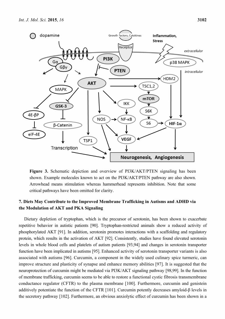
AKT is a central player in signal transduction activated in response to several growth factors, which is
thought to contribute many important cellular functions, including cell growth, apoptosis, nutrient
metabolism, and modulating the activity of various transcription factors [75] (Figure 3). AKT is subjected
to phosphorylation-regulation by phosphoinositide-dependent kinase 1 (PDK1) at Thr308. Full activation
of AKT requires further phosphorylation of its Ser473 at the carboxyl-terminus by kinases such as PDK2
and the mTOR complex 2 (mTORC2) [76]. The PKA signaling is involved in affecting the GSK-3β
phosphorylation status at phospho-GSK-3β (Ser9) [77], which is also a downstream target of PI3K/AKT
signaling. GSK-3 contains α and β isoforms, which is an important kinase involved in the regulation
of a group of transcription factors [78]. Evidence suggests that lithium causes its neuro-protective
effects predominantly through inhibition of the GSK-3 [79]. Similarly, the development of GSK-3
isoform-specific inhibitors seems to be warranted for treating GSK-3-mediated pathology [79]. Lithium
may also modify GSK-3 activity through phosphorylation both of GSK-3α and GSK-3β by various
mechanisms including the activation of PKA and AKT [80,81], indicating that lithium exerts its potentiating
and inhibiting bidirectional cellular actions on GSK-3 activity. In addition, inhibition of GSK-3 seems
to be involved in the antagonistic effects of lithium on depressant and manic properties [82]. In mouse
photoreceptor-derived 661W cells, bFGF signaling inactivates GSK-3β by phosphorylation at Ser9, which
is dependent on PKA activation [83]. A pharmacological inhibitor of PKA can antagonize the GSK-3β
Ser9 phosphorylation [84], supporting its potential use in chemotherapeutic options.
PKA and AKT have been shown to establish complexes with AKAP150, which may act as a key
regulator to control AKT phosphorylation. PKA activation leads to a reduction of AKT phosphorylation.
In diverse neuronal processes ranging from neuronal survival to synaptic plasticity, cAMP-dependent
signaling is tightly connected with the AKT signaling pathway [85]. In addition, a crosstalk between
PKA and mammalian target of rapamycin (mTOR) pathway in apoptosis resistance signaling has been
reported [86]. mTOR, a conserved serine/threonine kinase within cells, is a key molecule in controlling
protein synthesis and cell growth, and also involved in neurological disorders including autisms and
ADHD [87]. To achieve a better quality of life for those patients, therapy approaches are directed
at restoring dysregulated mTOR signaling [87]. mTOR complex 1 (mTORC1) and mTOR complex 2
(mTORC2) are two distinct complexes with mTOR, which are also involved in autisms and
ADHD [88]. Loss of mTORC2 signaling in the cortex independent of mTORC1 might disrupt normal
brain development and its function [88]. The mTORC1 pathway is activated by the PKA signaling,
leading to increased cell survival, which is correlated with BAD hyper-phosphorylation. Furthermore,
PKA and mTOR signaling cascades are important even for the development of follicular thyroid
carcinogenesis, suggesting new targets for therapeutic intervention [89].
Int. J. Mol. Sci. 2015, 16 3102
Figure 3. Schematic depiction and overview of PI3K/AKT/PTEN signaling has been
shown. Example molecules known to act on the PI3K/AKT/PTEN pathway are also shown.
Arrowhead means stimulation whereas hammerhead represents inhibition. Note that some
critical pathways have been omitted for clarity.
7. Diets May Contribute to the Improved Membrane Trafficking in Autisms and ADHD via
the Modulation of AKT and PKA Signaling
Dietary depletion of tryptophan, which is the precursor of serotonin, has been shown to exacerbate
repetitive behavior in autistic patients [90]. Tryptophan-restricted animals show a reduced activity of
phosphorylated AKT [91]. In addition, serotonin promotes interactions with a scaffolding and regulatory
protein, which results in the activation of AKT [92]. Consistently, studies have found elevated serotonin
levels in whole blood cells and platelets of autism patients [93,94] and changes in serotonin transporter
function have been implicated in autisms [95]. Enhanced activity of serotonin transporter variants is also
associated with autisms [96]. Curcumin, a component in the widely used culinary spice turmeric, can
improve structure and plasticity of synapse and enhance memory abilities [97]. It is suggested that the
neuroprotection of curcumin might be mediated via PI3K/AKT signaling pathway [98,99]. In the function
of membrane trafficking, curcumin seems to be able to restore a functional cystic fibrosis transmembrane
conductance regulator (CFTR) to the plasma membrane [100]. Furthermore, curcumin and genistein
additively potentiate the function of the CFTR [101]. Curcumin potently decreases amyloid-β levels in
the secretory pathway [102]. Furthermore, an obvious anxiolytic effect of curcumin has been shown in a
Int. J. Mol. Sci. 2015, 16 3103
lead-induced anxiety animal model, possibly resulted from modulation of central neuronal serotonin
neurotransmission [103]. Recently, omega-3 (ω-3) long-chain polyunsaturated fatty acids (PUFAs)
have become a focus of interest. Especially, docosahexaenonic acids (DHA) are essential for brain
development and physical health. The symptoms of ADHD have been suggested to be associated with
a deficiency of the ω-3 PUFAs [104]. In addition, low blood ω-3 PUFAs have been reported in children
with ADHD and related learning difficulties, suggesting benefits from dietary supplementation [105].
Fish oil administration was reported to protect hippocampal neurons and improves cognitive deficit by
increasing AKT phosphorylation [106]. In addition, neuroprotecton could be performed by certain
diets involved in the PI3K/AKT pathway [107,108]. Several fruits may be promising. Kaempferol is
a flavonol that is present in various plants including grapefruit and some edible berries, which
also induces the activation of PI3K and AKT [109]. On the contrary, the biological activity of
the isothiocyanates, rich in certain vegetables such as broccoli, has been shown to suppress AKT
phosphorylation [110]. However, despite these experimental observations, the precise mechanisms for
these food ingredients remain elusive further for the clinical uses. Additionally, it seems important to
exploit the potential benefits of optimal treatment and/or combination with these PI3K/AKT modulators.
Overexpression of phosphatase and tensin homologue deleted on chromosome 10 (PTEN) has been
shown to have inhibitory effects on serotonin signaling via decreased AKT activity [111]. PTEN
negatively regulates activity of the PI3K/AKT pathway, which is a dual-specificity phosphatase
acting as both protein phosphatase and lipid phosphatase that suppresses PI3K activity through
converting PIP3 to PIP2 [112]. Honokiol, a chemical compound in traditional eastern herbal medicines,
can attenuate the PI3K/AKT signaling by up-regulation of PTEN expression [113]. Dietary and/or
therapeutic interventions to counteract the reduction of PTEN expression could contribute to the prevention
of the diseases and/or decrease the rate of its development. Accordingly, the culinary herb sage
(Salvia officinalis) may be unhelpful for autism patients [114]. However, PTEN indirectly promotes
serotonin synthesis and secretion via inhibiting the signaling [115]. In addition, there is a crosstalk
between PTEN and the serotonin receptor [116]. It has been shown that docosahexaenonic acids
(DHA) and eicosapentaenoic acid (EPA) increase the level of PTEN in breast cancer cells, providing
a mechanism for the beneficial effects of fish oils even on cancer cell growth [117,118]. Since DHA
and EPA are ligands of PPARγ, both of the ω-3 PUFAs exert anti-proliferative effects by inducing PTEN
via the activation of the PPARγ [119]. Controversially, phosphorylated AKT may be down-regulated
by treatment with curcumin due to the activation of PTEN. In addition, the most attractive target for
phytoestrogen with regard to PTEN transcription seems to be PPARγ [120]. Both genistein and quercetin
also have an effect on PPARγ activation, which has been shown to up-regulate PTEN transcription,
then, suppresses the PI3K/AKT pathway [121]. Dietary exposure to the soy isoflavones at physiologically
relevant concentrations induces PTEN expression [122]. Generally, phytoestrogen exposure may result
in an increase in PTEN expression. In addition, a high-fat diet raises circulating fatty acids, which
significantly alters PTEN expression [123]. Interestingly, rosemary extract was reported to repress PTEN
expression in K562 leukemic culture cells [124]. Again, indole-3-carbinol is a promising cancer-preventive
phytochemical found in some vegetables such as broccoli. Dietary intake of the indole-3-carbinol
up-regulates PTEN in the mouse model [125].
Int. J. Mol. Sci. 2015, 16 3104
The PKA pathway regulates cell growth and division in response to nutrient status [126]. PKA is
activated by the ω-3 PUFAs EPA [127]. Bitter melon seed oil (BMSO), which is rich in the isomers of
conjugated linolenic acid, increases phosphorylation and activation of PKA [128]. In addition, genistein
directly activates the cAMP/PKA cascade [129]. Very low protein diets result in a desensitization of cAMP
signaling, which is characterized by a loss of PKA activity [130], suggesting that dietary protein and
energy restriction may modulate PKA activity. It seems that both activation and inhibition of those
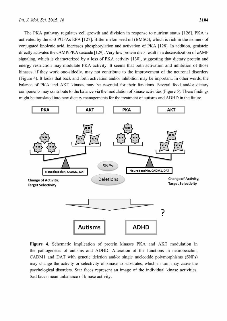
kinases, if they work one-sidedly, may not contribute to the improvement of the neuronal disorders
(Figure 4). It looks that back and forth activation and/or inhibition may be important. In other words, the
balance of PKA and AKT kinases may be essential for their functions. Several food and/or dietary
components may contribute to the balance via the modulation of kinase activities (Figure 5). These findings
might be translated into new dietary managements for the treatment of autisms and ADHD in the future.
Figure 4. Schematic implication of protein kinases PKA and AKT modulation in
the pathogenesis of autisms and ADHD. Alteration of the functions in neurobeachin,
CADM1 and DAT with genetic deletion and/or single nucleotide polymorphisms (SNPs)
may change the activity or selectivity of kinase to substrates, which in turn may cause the
psychological disorders. Star faces represent an image of the individual kinase activities.
Sad faces mean unbalance of kinase activity.
Int. J. Mol. Sci. 2015, 16 3105
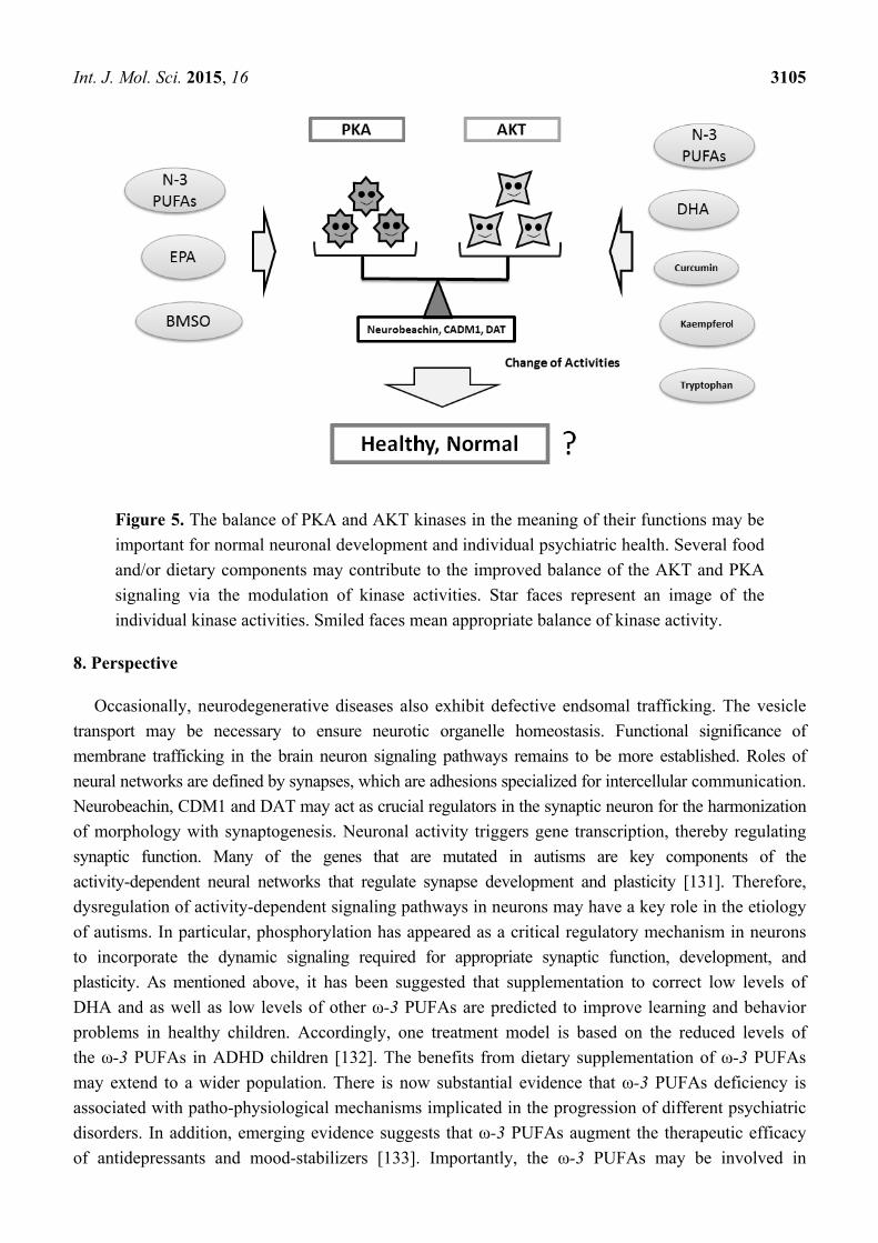
Figure 5. The balance of PKA and AKT kinases in the meaning of their functions may be
important for normal neuronal development and individual psychiatric health. Several food
and/or dietary components may contribute to the improved balance of the AKT and PKA
signaling via the modulation of kinase activities. Star faces represent an image of the
individual kinase activities. Smiled faces mean appropriate balance of kinase activity.
8. Perspective
Occasionally, neurodegenerative diseases also exhibit defective endsomal trafficking. The vesicle
transport may be necessary to ensure neurotic organelle homeostasis. Functional significance of
membrane trafficking in the brain neuron signaling pathways remains to be more established. Roles of
neural networks are defined by synapses, which are adhesions specialized for intercellular communication.
Neurobeachin, CDM1 and DAT may act as crucial regulators in the synaptic neuron for the harmonization
of morphology with synaptogenesis. Neuronal activity triggers gene transcription, thereby regulating
synaptic function. Many of the genes that are mutated in autisms are key components of the
activity-dependent neural networks that regulate synapse development and plasticity [131]. Therefore,
dysregulation of activity-dependent signaling pathways in neurons may have a key role in the etiology
of autisms. In particular, phosphorylation has appeared as a critical regulatory mechanism in neurons
to incorporate the dynamic signaling required for appropriate synaptic function, development, and
plasticity. As mentioned above, it has been suggested that supplementation to correct low levels of
DHA and as well as low levels of other ω-3 PUFAs are predicted to improve learning and behavior
problems in healthy children. Accordingly, one treatment model is based on the reduced levels of
the ω-3 PUFAs in ADHD children [132]. The benefits from dietary supplementation of ω-3 PUFAs
may extend to a wider population. There is now substantial evidence that ω-3 PUFAs deficiency is
associated with patho-physiological mechanisms implicated in the progression of different psychiatric
disorders. In addition, emerging evidence suggests that ω-3 PUFAs augment the therapeutic efficacy
of antidepressants and mood-stabilizers [133]. Importantly, the ω-3 PUFAs may be involved in
Int. J. Mol. Sci. 2015, 16 3106
membrane trafficking via the modulation of PI3K/AKT activity. Uptake of the PUFAs into endo-
membranes may alter the rate of trafficking of molecules [134]. Membrane modification of neuronal
development appears to be an important mechanism of the antipsychotic effects by the PUFAs. In
addition, the ω-3 PUFAs have an established long-term safety, and the total cost-benefit ratio provides a
validation for the psychiatric treatment-protocols. Deciphering the precise mechanisms of the pathology
will offer new insight into the physiological roles in regulating membrane trafficking. It is expected that
future studies will address this area to gain a better understanding of the potential and specific signaling
molecules involved in autisms and ADHD.
Acknowledgments
This work was supported by JSPS KAKENHI Grant Number 25560050, 26-12035, 24240098.
Author Contributions
All authors contributed comprehensively to the work presented in this paper.
Abbreviation
ADHD: Attention deficit/hyperactivity disorder; AMPH: Amphetamine; AKAPs: A-kinase anchor
proteins; AKT: Protein kinase B; BDNF: Brain-derived neurotrophic factor; bFGF: Basic fibroblast
growth factor; BEACH: BEige and Chediak-Higashi; BMSO: Bitter melon seed oil; CADM1: Cell
adhesion molecule 1; CFTR: Functional cystic fibrosis transmembrane conductance regulator; cAMP:
Cyclic adenosine monophosphate; COMT: Catechol-O-methyl transferase; CREB: cAMP response
element-binding protein; DAT: Dopamine transporter; DHA: Docosahexaenonic acids; EPA:
Eicosapentaenoic acid; GSK-3: Glycogen synthase kinase 3; Ig: Immunoglobulin; mTOR: Mammalian
target of rapamycin; mTORC1: mTOR complex 1; mTORC2: mTOR complex 2; NBEA:
Neurobeachin; PDK1: Phosphoinositide-dependent kinase 1; PDK2: Phosphoinositide-dependent kinase 2;
PDZ: PSD-95/Dlg/ZO-1; PH: plekstrin homology; PIP3: Phosphatidylinositol 3,4,5-triphosphate; PI3K:
Phosphatidylinositol-3 kinase; PKA: Protein kinase A; PPARγ: Peroxisome proliferator-activated
receptor; PTEN: Phosphatase and tensin homologue deleted on chromosome 10; PUFAs: Polyunsaturated
fatty acids; SHR: Spontaneously hypertensive rat; SNP: Single nucleotide polymorphism; WD40:
tryptophan-aspartic acid repeat.
Conflicts of Interest
The authors declare no conflicts of interest.
References
1. Huang, C.; Rajfur, Z.; Yousefi, N.; Chen, Z.; Jacobson, K.; Ginsberg, M.H. Talin phosphorylation
by Cdk5 regulates Smurf1-mediated talin head ubiquitylation and cell migration. Nat. Cell Biol.
2009, 11, 624–630.
2. Bishop, N.E. Dynamics of endosomal sorting. Int. Rev. Cytol. 2003, 232, 1–57.
Int. J. Mol. Sci. 2015, 16 3107
3. Mizutani, R.; Yamauchi, J.; Kusakawa, S.; Nakamura, K.; Sanbe, A.; Torii, T.; Miyamoto, Y.;
Tanoue, A. Sorting nexin 3, a protein up-regulated by lithium, contains a novel
phosphatidylinositol-binding sequence and mediates neurite outgrowth in N1E-115 cells.
Cell Signal. 2009, 21, 1586–1594.
4. Koutelou, E.; Sato, S.; Tomomori-Sato, C.; Florens, L.; Swanson, S.K.; Washburn, M.P.;
Kokkinaki, M.; Conaway, R.C.; Conaway, J.W.; Moschonas, N.K. Neuralized-like 1 (Neurl1)
targeted to the plasma membrane by N-myristoylation regulates the Notch ligand Jagged1.
J. Biol. Chem. 2008, 283, 3846–3853.
5. Kanamarlapudi, V. Centaurin-α1 and KIF13B kinesin motor protein interaction in ARF6
signalling. Biochem. Soc. Trans. 2005, 33, 1279–1281.
6. Venkateswarlu, K.; Gunn-Moore, F.; Tavaré, J.M.; Cullen, P.J. EGF-and NGF-stimulated
translocation of cytohesin-1 to the plasma membrane of PC12 cells requires PI 3-kinase
activation and a functional cytohesin-1 PH domain. J. Cell Sci. 1999, 112, 1957–1965.
7. Woo, J.; Chae, Y.K.; Jang, S.J.; Kim, M.S.; Baek, J.H.; Park, J.C.; Trink, B.; Ratovitski, E.; Lee, T.;
Park, B.; et al. Membrane trafficking of AQP5 and cAMP dependent phosphorylation in
bronchial epithelium. Biochem. Biophys. Res. Commun. 2008, 366, 321–327.
8. Wojtal, K.A.; Hoekstra, D.; van Ijzendoorn, S.C. cAMP-dependent protein kinase A and the
dynamics of epithelial cell surface domains: Moving membranes to keep in shape. Bioessays 2008,
30, 146–155.
9. Foletti, D.L.; Prekeris, R.; Scheller, R.H. Generation and maintenance of neuronal polarity:
Mechanisms of transport and targeting. Neuron 1999, 23, 641–644.
10. Horton, A.C.; Ehlers, M.D. Neuronal polarity and trafficking. Neuron 2003, 40, 277–295.
11. Tang, D.; Wang, Y. Cell cycle regulation of Golgi membrane dynamics. Trends Cell Biol. 2013,
23, 296–304.
12. Lecuit, T. Regulation of membrane dynamics in developing epithelia. Curr. Opin. Genet. Dev.
2003, 13, 351–357.
13. Bowton, E.; Saunders, C.; Reddy, I.A.; Campbell, N.G.; Hamilton, P.J.; Henry, L.K.; Coon, H.;
Sakrikar, D.; Veenstra-VanderWeele, J.M.; Blakely, R.D.; et al. SLC6A3 coding variant Ala559Val
found in two autism probands alters dopamine transporter function and trafficking. Transl. Psychiatry
2014, 4, e464.
14. Sakrikar, D.; Mazei-Robison, M.S.; Mergy, M.A.; Richtand, N.W.; Han, Q.; Hamilton, P.J.;
Bowton, E.; Galli, A.; Veenstra-Vanderweele, J.; Gill, M.; et al. Attention deficit/hyperactivity
disorder-derived coding variation in the dopamine transporter disrupts microdomain targeting
and trafficking regulation. J. Neurosci. 2012, 32, 5385–5397.
15. Leitner Y. The co-occurrence of autism and attention deficit hyperactivity disorder in
children—What do we know? Front. Hum. Neurosci. 2014, 8, 268.
16. Kondapalli, K.C.; Prasad, H.; Rao, R. An inside job: How endosomal Na+/H+ exchangers link to
autism and neurological disease. Front. Cell Neurosci. 2014, 8, 172.
17. Wegiel, J.; Kuchna, I.; Nowicki, K.; Imaki, H.; Wegiel, J.; Marchi, E.; Ma, S.Y.; Chauhan, A.;
Chauhan, V.; Bobrowicz, T.W.; et al. The neuropathology of autism: Defects of neurogenesis
and neuronal migration, and dysplastic changes. Acta Neuropathol. 2010, 119, 755–770.
Int. J. Mol. Sci. 2015, 16 3108
18. Zuko, A.; Kleijer, K.T.; Oguro-Ando, A.; Kas, M.J.; van Daalen, E.; van der Zwaag, B.;
Burbach, J.P. Contactins in the neurobiology of autism. Eur. J. Pharmacol. 2013, 719, 63–74.
19. Gu, W.; Lupski, J.R. CNV and nervous system diseases—What’s new? Cytogenet. Genome Res.
2008, 123, 54–64.
20. Volders, K.; Nuytens, K.; Creemers, J.W. The autism candidate gene Neurobeachin encodes
a scaffolding protein implicated in membrane trafficking and signaling. Curr. Mol. Med. 2011,
11, 204–217.
21. Olszewski, P.K.; Rozman, J.; Jacobsson, J.A.; Rathkolb, B.; Strömberg, S.; Hans, W.; Klockars, A.;
Alsiö, J.; Risérus, U.; Becker, L.; et al. Neurobeachin, a regulator of synaptic protein targeting,
is associated with body fat mass and feeding behavior in mice and body-mass index in humans.
PLoS Genet. 2012, 8, e1002568.
22. Wang, X.; Herberg, F.W.; Laue, M.M.; Wullner, C.; Hu, B.; Petrasch-Parwez, E.; Kilimann, M.W.
Neurobeachin: A protein kinase A-anchoring, beige/Chediak-higashi protein homolog implicated
in neuronal membrane traffic. J. Neurosci. 2000, 20, 8551–8565.
23. Savelyeva, L.; Sagulenko, E.; Schmitt, J.G.; Schwab, M. The neurobeachin gene spans the common
fragile site FRA13A. Hum. Genet. 2006, 118, 551–558.
24. De Lozanne, A. The role of BEACH proteins in Dictyostelium. Traffic 2003, 4, 6–12.
25. Vaccari, T.; Bilder, D. The Drosophila tumor suppressor vps25 prevents nonautonomous
overproliferation by regulating notch trafficking. Dev. Cell 2005, 9, 687–698.
26. Nuytens, K.; Tuand, K.; di Michele, M.; Boonen, K.; Waelkens, E.; Freson, K.; Creemers, J.W.
Platelets of mice heterozygous for neurobeachin, a candidate gene for autism spectrum disorder,
display protein changes related to aberrant protein kinase A activity. Mol. Autism 2013, 4, 43.
27. Shamloula, H.K.; Mbogho, M.P.; Pimentel, A.C.; Chrzanowska-Lightowlers, Z.M.; Hyatt, V.;
Okano, H.; Venkatesh, T.R. rugose (rg), a Drosophila A kinase anchor protein, is required for
retinal pattern formation and interacts genetically with multiple signaling pathways. Genetics
2002, 161, 693–710.
28. Wang, K.; Hackett, J.T.; Cox, M.E.; van Hoek, M.; Lindstrom, J.M.; Parsons, S.J. Regulation of
the neuronal nicotinic acetylcholine receptor by SRC family tyrosine kinases. J. Biol. Chem.
2004, 279, 8779–8786.
29. Miller, A.C.; Voelker, L.H.; Shah, A.N.; Moens, C.B. Neurobeachin is required postsynaptically
for electrical and chemical synapse formation. Curr. Biol. 2015, 25, 16–28.
30. Chen, C.K.; Bregere, C.; Paluch, J.; Lu, J.F.; Dickman, D.K.; Chang, K.T. Activity-dependent
facilitation of Synaptojanin and synaptic vesicle recycling by the Minibrain kinase. Nat. Commun.
2014, 5, 4246.
31. Ebert, D.H.; Greenberg, M.E. Activity-dependent neuronal signalling and autism spectrum
disorder. Nature 2013, 493, 327–337.
32. Ribic, A.; Liu, X.; Crair, M.C.; Biederer, T. Structural organization and function of mouse
photoreceptor ribbon synapses involve the immunoglobulin protein synaptic cell adhesion
molecule 1. J. Comp. Neurol. 2014, 522, 900–920.
33. Masuda, M.; Yageta, M.; Fukuhara, H.; Kuramochi, M.; Maruyama, T.; Nomoto, A.; Murakami, Y.
The tumor suppressor protein TSLC1 is involved in cell-cell adhesion. J. Biol. Chem. 2002, 277,
31014–31019.

Int. J. Mol. Sci. 2015, 16 3109
34. Fujita, E.; Tanabe, Y.; Imhof, B.A.; Momoi, M.Y.; Momoi, T. A complex of synaptic adhesion
molecule CADM1, a molecule related to autism spectrum disorder, with MUPP1 in the cerebellum.
J. Neurochem. 2012, 123, 886–894.
35. Fujita, E.; Dai, H.; Tanabe, Y.; Zhiling, Y.; Yamagata, T.; Miyakawa, T.; Tanokura, M.;
Momoi, M.Y.; Momoi, T. Autism spectrum disorder is related to endoplasmic reticulum stress
induced by mutations in the synaptic cell adhesion molecule, CADM1. Cell Death Dis. 2010, 1, e47.
36. Patiño-Lopez, G.; Hevezi, P.; Lee, J.; Willhite, D.; Verge, G.M.; Lechner, S.M.; Ortiz-Navarrete, V.;
Zlotnik, A. Human class-I restricted T cell associated molecule is highly expressed in the cerebellum
and is a marker for activated NKT and CD8+ T lymphocytes. J. Neuroimmunol. 2006, 171, 145–155.
37. Tanabe, Y.; Fujita, E.; Hayashi, Y.K.; Zhu, X.; Lubbert, H.; Mezaki, Y.; Senoo, H.; Momoi, T.
Synaptic adhesion molecules in Cadm family at the neuromuscular junction. Cell Biol. Int. 2013,
37, 731–736.
38. Murphy, C.M.; Christakou, A.; Daly, E.M.; Ecker, C.; Giampietro, V.; Brammer, M.; Smith, A.B.;
Johnston, P.; Robertson, D.M.; MRC AIMS Consortium; et al. Abnormal functional activation
and maturation of fronto-striato-temporal and cerebellar regions during sustained attention in
autism spectrum disorder. Am. J. Psychiatry 2014, 171, 1107–1116.
39. Zhiling, Y.; Fujita, E.; Tanabe, Y.; Yamagata, T.; Momoi, T.; Momoi, M.Y. Mutations in
the gene encoding CADM1 are associated with autism spectrum disorder. Biochem. Biophys.
Res. Commun. 2008, 377, 926–929.
40. Momoi, T.; Fujita, E.; Senoo, H.; Momoi, M. Genetic factors and epigenetic factors for autism:
Endoplasmic reticulum stress and impaired synaptic function. Cell Biol. Int. 2009, 34, 13–19.
41. Williams, Y.N.; Masuda, M.; Sakurai-Yageta, M.; Maruyama, T.; Shibuya, M.; Murakami, Y.
Cell adhesion and prostate tumor-suppressor activity of TSLL2/IGSF4C, an immunoglobulin
superfamily molecule homologous to TSLC1/IGSF4. Oncogene 2006, 25, 1446–1453.
42. Tordjman, S.; Somogyi, E.; Coulon, N.; Kermarrec, S.; Cohen, D.; Bronsard, G.; Bonnot, O.;
Weismann-Arcache, C.; Botbol, M.; Lauth, B.; et al. Gene × Environment interactions in autism
spectrum disorders: Role of epigenetic mechanisms. Front. Psychiatry 2014, 5, 53.
43. Hauser, T.U.; Iannaccone, R.; Ball, J.; Mathys, C.; Brandeis, D.; Walitza, S.; Brem, S. Role of
the medial prefrontal cortex in impaired decision making in juvenile attention-deficit/hyperactivity
disorder. JAMA Psychiatry 2014, 71, 1165–1173.
44. Pearlman, D.M.; Vora, H.S.; Marquis, B.G.; Najjar, S.; Dudley, L.A. Anti-basal ganglia
antibodies in primary obsessive-compulsive disorder: Systematic review and meta-analysis.
Br. J. Psychiatry 2014, 205, 8–16.
45. Kirley, A.; Hawi, Z.; Daly, G.; McCarron, M.; Mullins, C.; Millar, N.; Waldman, I.; Fitzgerald, M.;
Gill, M. Dopaminergic system genes in ADHD: Toward a biological hypothesis.
Neuropsychopharmacology 2002, 27, 607–619.
46. Grant, P.; Kuepper, Y.; Wielpuetz, C.; Hennig, J. Differential associations of dopamine-related
polymorphisms with discrete components of reaction time variability: Relevance for attention
deficit/hyperactivity disorder. Neuropsychobiology 2014, 69, 220–226.
47. Somkuwar, S.S.; Darna, M.; Kantak, K.M.; Dwoskin, L.P. Adolescence methylphenidate
treatment in a rodent model of attention deficit/hyperactivity disorder: Dopamine transporter
function and cellular distribution in adulthood. Biochem. Pharmacol. 2013, 86, 309–316.
Int. J. Mol. Sci. 2015, 16 3110
48. Patel, J.; Mooslehner, K.A.; Chan, P.M.; Emson, P.C.; Stamford, J.A. Presynaptic control of
striatal dopamine neurotransmission in adult vesicular monoamine transporter 2 (VMAT2)
mutant mice. J. Neurochem. 2003, 85, 898–910.
49. Kim, P.; Choi, C.S.; Park, J.H.; Joo, S.H.; Kim, S.Y.; Ko, H.M.; Kim, K.C.; Jeon, S.J.; Park, S.H.;
Han, S.H.; et al. Chronic exposure to ethanol of male mice before mating produces attention
deficit hyperactivity disorder-like phenotype along with epigenetic dysregulation of dopamine
transporter expression in mouse offspring. J. Neurosci. Res. 2014, 92, 658–670.
50. Hall, F.S.; Itokawa, K.; Schmitt, A.; Moessner, R.; Sora, I.; Lesch, K.P.; Uhl, G.R. Decreased
vesicular monoamine transporter 2 (VMAT2) and dopamine transporter (DAT) function in
knockout mice affects aging of dopaminergic systems. Neuropharmacology 2014, 76, 146–155.
51. Greenwood, T.A.; Joo, E.J.; Shekhtman, T.; Sadovnick, A.D.; Remick, R.A.; Keck, P.E.;
McElroy, S.L.; Kelsoe, J.R. Association of dopamine transporter gene variants with childhood
ADHD features in bipolar disorder. Am. J. Med. Genet. B 2013, 162B, 137–145.
52. Fosi, T.; Lax-Pericall, M.T.; Scott, R.C.; Neville, B.G.; Aylett, S.E. Methylphenidate treatment
of attention deficit hyperactivity disorder in young people with learning disability and difficult-to-
treat epilepsy: Evidence of clinical benefit. Epilepsia 2013, 54, 2071–2081.
53. Meneses, A.; Perez-Garcia, G.; Ponce-Lopez, T.; Tellez, R.; Gallegos-Cari, A.; Castillo, C.
Spontaneously hypertensive rat (SHR) as an animal model for ADHD: A short overview.
Rev. Neurosci. 2011, 22, 365–371.
54. Barth, V.; Need, A.B.; Tzavara, E.T.; Giros, B.; Overshiner, C.; Gleason, S.D.; Wade, M.;
Johansson, A.M.; Perry, K.; Nomikos, G.G.; et al. In vivo occupancy of dopamine D3 receptors
by antagonists produces neurochemical and behavioral effects of potential relevance to
attention-deficit-hyperactivity disorder. J. Pharmacol. Exp. Ther. 2013, 344, 501–510.
55. Volkow, N.D.; Wang, G.J.; Fowler, J.S.; Logan, J.; Franceschi, D.; Maynard, L.; Ding, Y.S.;
Gatley, S.J.; Gifford, A.; Zhu, W.; et al. Relationship between blockade of dopamine transporters
by oral methylphenidate and the increases in extracellular dopamine: Therapeutic implications.
Synapse 2002, 43, 181–187.
56. Torres, G.E. The dopamine transporter proteome. J. Neurochem. 2006, 97, 3–10.
57. Robbins, T.W. Dopamine and cognition. Curr. Opin. Neurol. 2003, 16, S1–S2.
58. Nuytens, K.; Gantois, I.; Stijnen, P.; Iscru, E.; Laeremans, A.; Serneels, L.; van Eylen, L.;
Liebhaber, S.A.; Devriendt, K.; Balschun, D.; et al. Haploinsufficiency of the autism candidate
gene Neurobeachin induces autism-like behaviors and affects cellular and molecular processes of
synaptic plasticity in mice. Neurobiol. Dis. 2013, 51, 144–151.
59. Su, Y.; Balice-Gordon, R.J.; Hess, D.M.; Landsman, D.S.; Minarcik, J.; Golden, J.; Hurwitz, I.;
Liebhaber, S.A.; Cooke, N.E. Neurobeachin is essential for neuromuscular synaptic transmission.
J. Neurosci. 2004, 24, 3627–3636.
60. Ohashi, M.; Huttner, W.B. An elevation of cytosolic protein phosphorylation modulates trimeric
G-protein regulation of secretory vesicle formation from the trans-Golgi network. J. Biol. Chem.
1994, 269, 24897–24905.
61. Colledge, M.; Scott, J.D. AKAPs: From structure to function. Trends Cell Biol. 1999, 9, 216–221.
62. Dell'Acqua, M.L.; Smith, K.E.; Gorski, J.A.; Horne, E.A.; Gibson, E.S.; Gomez, L.L. Regulation of
neuronal PKA signaling through AKAP targeting dynamics. Eur. J. Cell Biol. 2006, 85, 627–633.
Int. J. Mol. Sci. 2015, 16 3111
63. Röder, I.V.; Choi, K.R.; Reischl, M.; Petersen, Y.; Diefenbacher, M.E.; Zaccolo, M.; Pozzan, T.;
Rudolf, R. Myosin Va cooperates with PKA RIα to mediate maintenance of the endplate
in vivo. Proc. Natl. Acad. Sci. USA 2010, 107, 2031–2036.
64. Niesmann, K.; Breuer, D.; Brockhaus, J.; Born, G.; Wolff, I.; Reissner, C.; Kilimann, M.W.;
Rohlmann, A.; Missler, M. Dendritic spine formation and synaptic function require neurobeachin.
Nat. Commun. 2011, 2, 557.
65. Murakami, S.; Sakurai-Yageta, M.; Maruyama, T.; Murakami, Y. Trans-homophilic interaction
of CADM1 activates PI3K by forming a complex with MAGuK-family proteins MPP3 and Dlg.
PLoS One 2014, 9, e82894.
66. Masuda, M.; Maruyama, T.; Ohta, T.; Ito, A.; Hayashi, T.; Tsukasaki, K.; Kamihira, S.; Yamaoka, S.;
Hoshino, H.; Yoshida, T.; et al. CADM1 interacts with Tiam1 and promotes invasive phenotype of
human T-cell leukemia virus type I-transformed cells and adult T-cell leukemia cells. J. Biol. Chem.
2010, 285, 15511–15522.
67. Montgomery, J.M.; Zamorano, P.L.; Garner, C.C. AGUKs in synapse assembly and function:
An emerging view. Cell Mol. Life Sci. 2004, 61, 911–929.
68. Liegl, R.; Wertheimer, C.; Kernt, M.; Docheva, D.; Kampik, A.; Eibl-Lindner, K.H. Attenuation of
human lens epithelial cell spreading, migration and contraction via downregulation of the PI3K/Akt
pathway. Graefes. Arch. Clin. Exp. Ophthalmol. 2014, 252, 285–292.
69. Garcia, B.G.; Wei, Y.; Moron, J.A.; Lin, R.Z.; Javitch, J.A.; Galli, A. Akt is essential for insulin
modulation of amphetamine-induced human dopamine transporter cell-surface redistribution.
Mol. Pharmacol. 2005, 68, 102–109.
70. Wei, Y.; Williams, J.M.; Dipace, C.; Sung, U.; Javitch, J.A.; Galli, A.; Saunders, C.
Dopamine transporter activity mediates amphetamine-induced inhibition of Akt through a
Ca2+/calmodulin-dependent kinase II-dependent mechanism. Mol. Pharmacol. 2007, 71, 835–842.
71. Bourque, M.; Liu, B.; Dluzen, D.E.; di Paolo, T. Sex differences in methamphetamine toxicity in
mice: Effect on brain dopamine signaling pathways. Psychoneuroendocrinology 2011, 36, 955–969.
72. Ugrumov, M.V. Non-dopaminergic neurons partly expressing dopaminergic phenotype: Distribution
in the brain, development and functional significance. J. Chem. Neuroanat. 2009, 38, 241–256.
73. Lute, B.J.; Khoshbouei, H.; Saunders, C.; Sen, N.; Lin, R.Z.; Javitch, J.A.; Galli, A. PI3K signaling
supports amphetamine-induced dopamine efflux. Biochem. Biophys. Res. Commun. 2008, 372, 656–661.
74. Carvelli, L.; Morón, J.A.; Kahlig, K.M.; Ferrer, J.V.; Sen, N.; Lechleiter, J.D.; Leeb-Lundberg, L.M.;
Merrill, G.; Lafer, E.M.; Ballou, L.M.; et al. PI 3-kinase regulation of dopamine uptake.
J. Neurochem. 2002, 81, 859–869.
75. Woodgett, J.R. Recent advances in the protein kinase B signaling pathway. Curr. Opin. Cell Biol.
2005, 17, 150–157.
76. Sarbassov, D.D.; Ali, S.M.; Sabatini, D.M. Growing roles for the mTOR pathway. Curr. Opin.
Cell Biol. 2005, 17, 596–603.
77. Khattak, M.N.; Buchfelder, M.; Kleindienst, A.; Schöfl, C.; Kremenevskaja, N. CRH and SRIF
have opposite effects on the Wnt/β-catenin signalling pathway through PKA/GSK-3β in
corticotroph pituitary cells. Cancer Investig. 2010, 28, 797–805.
78. Grimes, C.A.; Jope, R.S. The multifaceted roles of glycogen synthase kinase 3β in cellular
signaling. Prog. Neurobiol. 2001, 65, 391–426.
Int. J. Mol. Sci. 2015, 16 3112
79. Liang, M.H.; Chuang, D.M. Regulation and function of glycogen synthase kinase-3 isoforms in
neuronal survival. J. Biol. Chem. 2007, 282, 3904–3917.
80. Tian, N.; Kanno, T.; Jin, Y.; Nishizaki, T. Lithium potentiates GSK-3β activity by inhibiting
phosphoinositide 3-kinase-mediated Akt phosphorylation. Biochem. Biophys. Res. Commun.
2014, 450, 746–749.
81. Liang, M.H.; Wendland, J.R.; Chuang, D.M. Lithium inhibits Smad3/4 transactivation via
increased CREB activity induced by enhanced PKA and AKT signaling. Mol. Cell Neurosci.
2008, 37, 440–453.
82. Kirshenboim, N.; Plotkin, B.; Shlomo, S.B.; Kaidanovich-Beilin, O.; Eldar-Finkelman, H.
Lithium-mediated phosphorylation of glycogen synthase kinase-3β involves PI3 kinase-dependent
activation of protein kinase C-α. J. Mol. Neurosci. 2004, 24, 237–245.
83. O’Driscoll, C.; Wallace, D.; Cotter, T.G. bFGF promotes photoreceptor cell survival in vitro by
PKA-mediated inactivation of glycogen synthase kinase 3β and CREB-dependent Bcl-2 up-regulation.
J. Neurochem. 2007, 103, 860–870.
84. Ku, B.M.; Lee, Y.K.; Jeong, J.Y.; Ryu, J.; Choi, J.; Kim, J.S.; Cho, Y.W.; Roh, G.S.; Kim, H.J.;
Cho, G.J.; et al. Caffeine inhibits cell proliferation and regulates PKA/GSK3β pathways in
U87MG human glioma cells. Mol. Cells 2011, 31, 275–279.
85. Nijholt, I.M.; Dolga, A.M.; Ostroveanu, A.; Luiten, P.G.; Schmidt, M.; Eisel, U.L. Neuronal AKAP150
coordinates PKA and Epac-mediated PKB/Akt phosphorylation. Cell Signal. 2008, 20, 1715–1724.
86. De Joussineau, C.; Sahut-Barnola, I.; Tissier, F.; Dumontet, T.; Drelon, C.; Batisse-Lignier, M.;
Tauveron, I.; Pointud, J.C.; Lefrançois-Martinez, A.M.; Stratakis, C.A.; et al. mTOR pathway is
activated by PKA in adrenocortical cells and participates in vivo to apoptosis resistance in primary
pigmented nodular adrenocortical disease (PPNAD). Hum. Mol. Genet. 2014, 23, 5418–5428.
87. Jülich, K.; Sahin, M. Mechanism-based treatment in tuberous sclerosis complex. Pediatr. Neurol.
2014, 50, 290–296.
88. Carson, R.P.; Fu, C.; Winzenburger, P.; Ess, K.C. Deletion of Rictor in neural progenitor cells
reveals contributions of mTORC2 signaling to tuberous sclerosis complex. Hum. Mol. Genet.
2013, 22, 140–152.
89. Pringle, D.R.; Vasko, V.V.; Yu, L.; Manchanda, P.K.; Lee, A.A.; Zhang, X.; Kirschner, J.M.;
Parlow, A.F.; Saji, M.; Jarjoura, D.; et al. Follicular thyroid cancers demonstrate dual activation
of PKA and mTOR as modeled by thyroid-specific deletion of Prkar1a and Pten in mice. J. Clin.
Endocrinol. Metab. 2014, 99, E804–E812.
90. McDougle, C.J.; Naylor, S.T.; Cohen, D.J.; Aghajanian, G.K.; Heninger, G.R.; Price, L.H.
Effects of tryptophan depletion in drug-free adults with autistic disorder. Arch. Gen. Psychiatry 1996,
53, 993–1000.
91. Penedo, L.A.; Oliveira-Silva, P.; Gonzalez, E.M.; Maciel, R.; Jurgilas, P.B.; Melibeu Ada, C.;
Campello-Costa, P.; Serfaty, C.A. Nutritional tryptophan restriction impairs plasticity of retinotectal
axons during the critical period. Exp. Neurol. 2009, 217, 108–115.
92. Schmid, C.L.; Streicher, J.M.; Meltzer, H.Y.; Bohn, L.M. Clozapine acts as an agonist at
serotonin 2A receptors to counter MK-801-induced behaviors through a β-arrestin2-independent
activation of Akt. Neuropsychopharmacology 2014, 39, 1902–1913.
Int. J. Mol. Sci. 2015, 16 3113
93. Spivak, B.; Golubchik, P.; Mozes, T.; Vered, Y.; Nechmad, A.; Weizman, A.; Strous, R.D. Low
platelet-poor plasma levels of serotonin in adult autistic patients. Neuropsychobiology 2004, 50, 157–160.
94. Cross, S.; Kim, S.J.; Weiss, L.A.; Delahanty, R.J.; Sutcliffe, J.S.; Leventhal, B.L.; Cook, E.H., Jr.;
Veenstra-Vanderweele, J. Molecular genetics of the platelet serotonin system in first-degree
relatives of patients with autism. Neuropsychopharmacology 2008, 33, 353–360.
95. Iwata, K.; Matsuzaki, H.; Tachibana, T.; Ohno, K.; Yoshimura, S.; Takamura, H.; Yamada, K.;
Matsuzaki, S.; Nakamura, K.; Tsuchiya, K.J.; et al. N-ethylmaleimide-sensitive factor interacts
with the serotonin transporter and modulates its trafficking: Implications for pathophysiology in
autism. Mol. Autism 2014, 5, 33.
96. Prasad, H.C.; Steiner, J.A.; Sutcliffe, J.S.; Blakely, R.D. Enhanced activity of human serotonin
transporter variants associated with autism. Philos. Trans. R. Soc. Lond. B 2009, 364, 163–173.
97. Aggarwal, B.B.; Yuan, W.; Li, S.; Gupta, S.C. Curcumin-free turmeric exhibits anti-inflammatory
and anticancer activities: Identification of novel components of turmeric. Mol. Nutr. Food Res. 2013,
57, 1529–1542.
98. Wang, R.; Li, Y.H.; Xu, Y.; Li, Y.B.; Wu, H.L.; Guo, H.; Zhang, J.Z.; Zhang, J.J.; Pan, X.Y.; Li, X.J.
Curcumin produces neuroprotective effects via activating brain-derived neurotrophic factor/
TrkB-dependent MAPK and PI-3K cascades in rodent cortical neurons. Prog Neuropsychopharmacol.
Biol. Psychiatry 2010, 34, 147–153.
99. Wu, J.; Li, Q.; Wang, X.; Yu, S.; Li, L.; Wu, X.; Chen, Y.; Zhao, J.; Zhao, Y. Neuroprotection by
curcumin in ischemic brain injury involves the Akt/Nrf2 pathway. PLoS One 2013, 8, e59843.
100. Lipecka, J.; Norez, C.; Bensalem, N.; Baudouin-Legros, M.; Planelles, G.; Becq, F.; Edelman, A.;
Davezac, N. Rescue of ΔF508-CFTR (cystic fibrosis transmembrane conductance regulator) by
curcumin: Involvement of the keratin 18 network. J. Pharmacol. Exp. Ther. 2006, 317, 500–505.
101. Yu, Y.C.; Miki, H.; Nakamura, Y.; Hanyuda, A.; Matsuzaki, Y.; Abe, Y.; Yasui, M.; Tanaka, K.;
Hwang, T.C.; Bompadre, S.G.; et al. Curcumin and genistein additively potentiate G551D-CFTR.
J. Cyst. Fibros. 2011, 10, 243–252.
102. Zhang, C.; Browne, A.; Child, D.; Divito, J.R.; Stevenson, J.A.; Tanzi, R.E. Loss of function
of ATXN1 increases amyloid β-protein levels by potentiating β-secretase processing of
β-amyloid precursor protein. J. Biol. Chem. 2010, 285, 8515–8526.
103. Benammi, H.; el Hiba, O.; Romane, A.; Gamrani, H. A blunted anxiolytic like effect of curcumin
against acute lead induced anxiety in rat: Involvement of serotonin. Acta Histochem. 2014, 116, 920–925.
104. Burgess, J.R.; Stevens, L.; Zhang, W.; Peck, L. Long-chain polyunsaturated fatty acids in
children with attention-deficit hyperactivity disorder. Am. J. Clin. Nutr. 2000, 71, 327S–330S.
105. Montgomery, P.; Burton, J.R.; Sewell, R.P.; Spreckelsen, T.F.; Richardson, A.J. Low blood long
chain ω-3 fatty acids in UK children are associated with poor cognitive performance and
behavior: A cross-sectional analysis from the DOLAB study. PLoS One 2013, 8, e66697.
106. Jia, D.; Heng, L.J.; Yang, R.H.; Gao, G.D. Fish oil improves learning impairments of diabetic
rats by blocking PI3K/AKT/nuclear factor-κB-mediated inflammatory pathways. Neuroscience
2014, 258, 228–237.
107. Kitagishi, Y.; Nakanishi, A.; Ogura, Y.; Matsuda, S. Dietary regulation of PI3K/AKT/GSK-3β
pathway in Alzheimer’s disease. Alzheimers Res. Ther. 2014, 6, 35.
Int. J. Mol. Sci. 2015, 16 3114
108. Kitagishi, Y.; Matsuda, S. Diets involved in PPAR and PI3K/AKT/PTEN pathway may
contribute to neuroprotection in a traumatic brain injury. Alzheimers Res. Ther. 2013, 5, 42.
109. Xie, F.; Su, M.; Qiu, W.; Zhang, M.; Guo, Z.; Su, B.; Liu, J.; Li, X.; Zhou, L. Kaempferol
promotes apoptosis in human bladder cancer cells by inducing the tumor suppressor, PTEN.
Int. J. Mol. Sci. 2013, 14, 21215–21226.
110. Gao, N.; Budhraja, A.; Cheng, S.; Liu, E.H.; Chen, J.; Yang, Z.; Chen, D.; Zhang, Z.; Shi, X.
Phenethyl isothiocyanate exhibits antileukemic activity in vitro and in vivo by inactivation of Akt
and activation of JNK pathways. Cell Death Dis. 2011, 2, e140.
111. Hsiung, S.C.; Adlersberg, M.; Arango, V.; Mann, J.J.; Tamir, H.; Liu, K.P. Attenuated 5-HT1A
receptor signaling in brains of suicide victims: Involvement of adenylyl cyclase, phosphatidylinositol
3-kinase, Akt and mitogen-activated protein kinasze. J. Neurochem. 2003, 87, 182–194.
112. Carnero, A.; Paramio, J.M. The PTEN/PI3K/AKT pathway in vivo, cancer mouse models.
Front. Oncol. 2014, 4, 252.
113. Liu, H.; Zang, C.; Emde, A.; Planas-Silva, M.D.; Rosche, M.; Kühnl, A.; Schulz, C.O.; Elstner, E.;
Possinger, K.; Eucker, J. Anti-tumor effect of honokiol alone and in combination with other
anti-cancer agents in breast cancer. Eur. J. Pharmacol. 2008, 591, 43–51.
114. Hamidpour, M.; Hamidpour, R.; Hamidpour, S.; Shahlari, M. Chemistry, pharmacology, and
medicinal property of sage (Salvia) to prevent and cure Illnesses such as obesity, diabetes, depression,
dementia, lupus, autism, heart disease, and cancer. J. Tradit. Complement. Med. 2014, 4, 82–88.
115. Silva, S.R.; Zaytseva, Y.Y.; Jackson, L.N.; Lee, E.Y.; Weiss, H.L.; Bowen, K.A.;
Townsend, C.M., Jr.; Evers, B.M. The effect of PTEN on serotonin synthesis and secretion from
the carcinoid cell line BON. Anticancer Res. 2011, 31, 1153–1160.
116. Cai, J.; Yi, Z.; Lu, W.; Fang, Y.; Zhang, C. Crosstalk between 5-HT2cR and PTEN signaling
pathway in atypical antipsychotic-induced metabolic syndrome and cognitive dysfunction.
Med. Hypotheses 2013, 80, 486–489.
117. Rovito, D.; Giordano, C.; Vizza, D.; Plastina, P.; Barone, I.; Casaburi, I.; Lanzino, M.; de Amicis, F.;
Sisci, D.; Mauro, L.; et al. Omega-3 PUFA ethanolamides DHEA and EPEA induce autophagy
through PPARγ activation in MCF-7 breast cancer cells. J. Cell Physiol. 2013, 228, 1314–1322.
118. Ghosh-Choudhury, T.; Mandal, C.C.; Woodruff, K.; St Clair, P.; Fernandes, G.; Choudhury, G.G.;
Ghosh-Choudhury, N. Fish oil targets PTEN to regulate NFκB for down-regulation of
anti-apoptotic genes in breast tumor growth. Breast Cancer Res. Treat. 2009, 118, 213–228.
119. Paintlia, A.S.; Paintlia, M.K.; Singh, A.K.; Orak, J.K.; Singh, I. Activation of PPAR-γ and PTEN
cascade participates in lovastatin-mediated accelerated differentiation of oligodendrocyte progenitor
cells. Glia 2010, 58, 1669–1685.
120. Montales, M.T.; Rahal, O.M.; Nakatani, H.; Matsuda, T.; Simmen, R.C. Repression of mammary
adipogenesis by genistein limits mammosphere formation of human MCF-7 cells. J. Endocrinol.
2013, 218, 135–149.
121. Wang, L.; Waltenberger, B.; Pferschy-Wenzig, E.M.; Blunder, M.; Liu, X.; Malainer, C.;
Blazevic, T.; Schwaiger, S.; Rollinger, J.M.; Heiss, E.H.; et al. Natural product agonists of
peroxisome proliferator-activated receptor gamma (PPARγ): A review. Biochem. Pharmacol.
2014, 92, 73–89.
Int. J. Mol. Sci. 2015, 16 3115
122. Dave, B.; Eason, R.R.; Till, S.R.; Geng, Y.; Velarde, M.C.; Badger, T.M.; Simmen, R.C. The soy
isoflavone genistein promotes apoptosis in mammary epithelial cells by inducing the tumor
suppressor PTEN. Carcinogenesis 2005, 26, 1793–1803.
123. La Merrill, M.; Gordon, R.R.; Hunter, K.W.; Threadgill, D.W.; Pomp, D. Dietary fat alters
pulmonary metastasis of mammary cancers through cancer autonomous and non-autonomous
changes in gene expression. Clin. Exp. Metastasis 2010, 27, 107–116.
124. Yoshida, H.; Okumura, N.; Kitagishi, Y.; Nishimura, Y.; Matsuda, S. Ethanol extract of
Rosemary repressed PTEN expression in K562 culture cells. Int. J. Appl. Biol. Pharm. Technol.
2011, 2, 316–322.
125. Qi, M.; Anderson, A.E.; Chen, D.Z.; Sun, S.; Auborn, K.J. Indole-3-carbinol prevents PTEN loss
in cervical cancer in vivo. Mol. Med. 2005, 11, 59–63.
126. Enns, L.C.; Morton, J.F.; Mangalindan, R.S.; McKnight, G.S.; Schwartz, M.W.; Kaeberlein, M.R.;
Kennedy, B.K.; Rabinovitch, P.S.; Ladiges, W.C. Attenuation of age-related metabolic dysfunction
in mice with a targeted disruption of the Cβ subunit of protein kinase A. J. Gerontol. A Biol. Sci.
Med. Sci. 2009, 64, 1221–1231.
127. Szentandrássy, N.; Pérez-Bido, M.R.; Alonzo, E.; Negretti, N.; O’Neill, S.C. Protein kinase A is
activated by the n-3 polyunsaturated fatty acid eicosapentaenoic acid in rat ventricular muscle.
J. Physiol. 2007, 582, 349–358.
128. Chen, P.H.; Chen, G.C.; Yang, M.F.; Hsieh, C.H.; Chuang, S.H.; Yang, H.L.; Kuo, Y.H.;
Chyuan, J.H.; Chao, P.M. Bitter melon seed oil-attenuated body fat accumulation in diet-induced
obese mice is associated with cAMP-dependent protein kinase activation and cell death in white
adipose tissue. J. Nutr. 2012, 142, 1197–1204.
129. Liu, D.; Zhen, W.; Yang, Z.; Carter, J.D.; Si, H.; Reynolds, K.A. Genistein acutely stimulates
insulin secretion in pancreatic β-cells through a cAMP-dependent protein kinase pathway.
Diabetes 2006, 55, 1043–1050.
130. O’Brien, L.J.; Levac, K.D.; Nagy, L.E. Moderate dietary protein and energy restriction modulate
cAMP-dependent protein kinase activity in rat liver. J. Nutr. 1998, 128, 927–933.
131. Castermans, D.; Wilquet, V.; Parthoens, E.; Huysmans, C.; Steyaert, J.; Swinnen, L.; Fryns, J.P.;
van de ven, W.; Devriendt, K. The neurobeachin gene is disrupted by a translocation in a patient
with idiopathic autism. J. Med. Genet. 2003, 40, 352–356.
132. Millichap, J.G.; Yee, M.M. The diet factor in attention-deficit/hyperactivity disorder. Pediatrics
2012, 129, 330–337.
133. Basselin M; Ramadan E; Rapoport SI. Imaging brain signal transduction and metabolism via
arachidonic and docosahexaenoic acid in animals and humans. Brain Res Bull. 2012, 87, 154–171.
134. Shaikh, S.R.; Edidin, M. Polyunsaturated fatty acids, membrane organization, T cells, and antigen
presentation. Am. J. Clin. Nutr. 2006, 84, 1277–1289.
© 2015 by the authors; licensee MDPI, Basel, Switzerland. This article is an open access article
distributed under the terms and conditions of the Creative Commons Attribution license
(http://creativecommons.org/licenses/by/4.0/).