Embed Size (px)
Citation preview
Multiple locus genealogies and phenotypic characters reappraisethe causal agents of apple ring rot in China
Chao Xu & Chunsheng Wang & Liangliang Ju & Rong Zhang &
Alan R. Biggs & Eiji Tanaka & Bingzhi Li & Guangyu Sun
Received: 6 March 2014 /Accepted: 2 September 2014 /Published online: 30 September 2014# School of Science 2014
Abstract Apple ring rot inflicts severe economic losses in themain apple producing areas of East Asia. The causal agent ofthe disease has been variously identified as Macrophomakuwatsukai, Physalospora piricola and Botryosphaeriaberengeriana f. sp. piricola, although B. dothidea is currentlythe most widely accepted pathogen name. The taxonomicuncertainty has delayed research that is needed to manageeffectively this destructive disease. In the present study, gene-alogical concordance phylogenetic species recognition(GCPSR) was applied to pathogenic fungal isolates fromapple and pear from several locations in China, along withseveral reference isolates. Phylogenetic results based on se-quences of four nuclear loci (ITS, EF-1α, HIS and HSP)revealed the existence of two species within the examinedisolates. One includes an ex-epitype isolate of B. dothidea andthe other includes an isolate that was previously designated asB. berengeriana f. sp. piricola. Morphologically, the lattertaxon presented an appressed mycelial mat on PDA whereasB. dothidea displayed columns of aerial mycelia reaching thelids, and conidia of the latter species were longer than
B. dothidea. Botryosphaeria dothidea had a faster growth ratethan the latter taxon under relatively high temperatures. Path-ogenicity tests showed that on pear stems the latter taxoncaused large-scale cankers along with blisters whereasB. dothidea was non-pathogenic, but on apple shoots thetwo fungi induced large and small wart-like prominences,respectively. Overall, this cryptic species demonstrated suffi-cient genetic variations and biological differences fromB. dothidea. As a result of taxonomic study, we describedhere the latter taxon in a new combination, Botryosphaeriakuwatsukai and designate an epitype. Both B. kuwatsukai andB. dothidea are considered to be the main causal agents forapple ring rot in China and Japan.
Key words Botryosphaeriaceae . Pear .Multi-genephylogeny . Pathogenicity . Group I intron . Taxonomy
Introduction
Ring rot has recently become one of the most destructiveapple diseases in China, as well as in several neighboringcountries, including Japan and South Korea (Ogata et al.2000; Park 2005; Tang et al. 2012). Symptoms of the diseaseappear as a soft, light-coloured rot on fruit, especially duringstorage, and extensive cankers and/or warts on branches andtrunks (Chen 1999). Widespread planting of susceptible cul-tivars (e.g., Fuji) over the past several years and reducedfungicide usage due to fruit bagging have likely resulted inthe increased occurrence of ring rot, resulting in serious eco-nomic losses to Chinese apple growers (Kang et al. 2009).
Apple ring rot disease was first reported in Japan in 1907.The pathogen was later described asMacrophoma kuwatsukaiHara (Hara 1930). Soon afterwards, the sexual morph of thepathogen was found and named Physalospora piricola (Nose1933). This taxon had long been known as the causal agent of
C. Xu : C. Wang : L. Ju : R. Zhang :G. Sun (*)Key Laboratory of Crop Stress Biology in Arid Areas, College ofPlant Protection, Northwest A&F University, No.3 Taicheng Road,Yangling 712100, Shaanxi, Chinae-mail: [email protected]
B. Li (*)College of Horticulture, Northwest A&F University, No.3 TaichengRoad, Yangling 712100, Shaanxi, Chinae-mail: [email protected]
A. R. BiggsKearneysville Tree Fruit Research and Education Center, WestVirginia University, P.O. Box 609, Kearneysville, WV 25443, USA
E. TanakaDivision of Environmental Science, Ishikawa Prefectural University,Suematsu 1-308, Nonoichi, Ishikawa 921-8836, Japan
Fungal Diversity (2015) 71:215–231DOI 10.1007/s13225-014-0306-5
apple ring rot in other countries. Koganezawa and Sakuma(1980, 1984) reappraised the sexual morph and tried to equateit with the morphologically identical Botryosphaeriaberengeriana, a fungus causing apple Botryosphaeria canker(and fruit rot) in Japan. However, distinctly different symp-toms, cankers and wart bark, caused by B. berengeriana andby P. piricola, respectively, on branches and trunks resulted inthe authors proposing the name Botryosphaeria berengerianaf. sp. piricola for Physalospora piricola (Koganezawa andSakuma 1984). As Botryosphaeria berengeriana andB. berengeriana f. sp. piricola induced the same apple rotsymptom, diseases caused on fruit were referred to both asapple ring rot (Koganezawa and Sakuma 1984). These twopathogen taxons are generally accepted in Japan, but they arerejected by European and American researchers who considerB. berengeriana to be a synonym of B. dothidea (Jones andAldwinckle 1990; Slippers et al. 2004a). In China, the correcttaxonomy of the apple ring rot pathogen is uncertain, in thatPhysalospora piricola, Botryosphaeria berengeriana,B. berengeriana f. sp. piricola and B. dothidea have beenadopted by different researchers (Qu et al. 2007; Peng et al.2011; Lv et al. 2012; Tang et al. 2012).
The application of molecular techniques to the taxa asso-ciated with apple ring rot has shown that genetic heterogeneityexists among these pathogenic fungal isolates. Ogata et al.(2000), using ITS sequences, divided isolates ofBotryosphaeria causing ring rot on apple fruit into two groupsbased on twig symptoms (warts or blight) and size of conidia.Huang and Liu (2001) showed that there was marked a dif-ference between B. berengeriana and B. berengeriana f. sp.piricola by RAPD analysis, in spite of their close geneticrelationship. Peng et al. (2011) separated isolates from applering rot into two ISSR groups. Lv et al. (2012) proposed thatthe isolates causing apple ring rot with different variable sitesin ITS sequences might behave differently in terms of patho-genicity and some biological characteristics (e.g., the ability tosporulate). Xu et al. (2013) genotyped B. dothidea isolates(including some from apple trees) according to the distributionof ribosomal group I introns. Four genotypes were describedand the authors indicated that there may be a correlationbetween these genotypes and host or geographic origin. Thehigh amount of variation detected by the different approachessuggests that there is a mixture of pathogens rather than asingle pathogenic species that causes apple ring rot.
Accurate identification of etiological factors is the founda-tion of plant disease research. Vague classification and con-fused appellation of the apple ring rot pathogen has led to theinability to compare research results from different countries,regions and investigators. Therefore, the objectives of thisstudy were to: 1) re-assess the Botryosphaeria isolates asso-ciated with symptoms of apple ring rot, including fruit rot,branch canker and wart bark in China, 2) detect the reasons forthe high amount of variation among isolates, and 3) clarify
whether any cryptic fungal species exist within the hypothet-ical pathogenic complex. Our approaches to the study includ-ed genealogical concordance phylogenetic species recognition(GCPSR) of multi-gene loci (Taylor et al. 2000), as well asanalysis of morphology, pathogenicity and growthcharacteristics.
Materials and methods
Fungal isolates
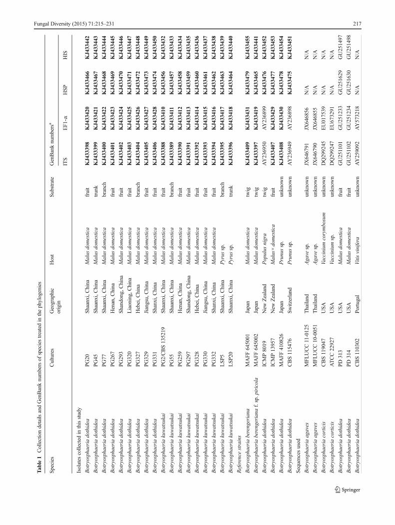
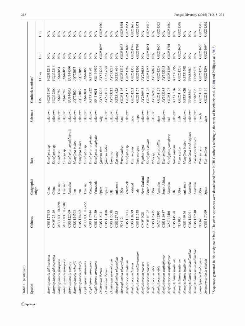
Twenty-four isolates (Table 1) were used in this study, 18 ofwhich were collected in the main apple production areas ofChina (Shaanxi, Henan, Shandong, Liaoning, Jiangsu, Shanxi,Hebei Provinces) during 2008–2011. Of these 18 isolates, 16were either from warts or cankers on apple branches and trunksor apple fruit with rot symptoms; and two were isolated fromthe diseased woody tissue of pear exhibiting canker symptoms.The other six were reference isolates, including one isolate eachof B. dothidea, B. berengeriana and B. berengeriana f. sp.piricola from the Fruit Tree Research Experiment Station ofthe Ministry of Agriculture, Forestry and Fisheries (MAFF),Japan, two isolates of B. dothidea from the International Col-lection of Microorganisms from Plants (ICMP), New Zealand,and one ex-epitype isolate of B. dothidea from theCentraalbureau voor Schimmelcultures (CBS), Netherlands.All the isolates collected in this research were obtained fromeither pycnidia directly or diseased tissue by cultivating onpotato dextrose agar (PDA), and were examined for colonycharacteristics and microscopic morphology to preliminarilyidentify them as Botryosphaeria spp. They were then purifiedby single conidium isolation and stored at −80 °C in College ofPlant Protection, Northwest A&F University, China.
DNA extraction, PCR amplification and sequencing
Single-conidial isolates were grown on PDA and incubatedfor 5 days at 25 °C in the dark. Total genomic DNA wasextracted from fungal mycelium following the modified phe-nol : chloroform DNA extraction method (Smith et al. 2001).Four different gene regions were selected for characterization,including the complete nuclear rDNA internal transcribedspacer (ITS) region (White et al. 1990), partial sequence oftranslation elongation factor 1 alfa (EF-1α), histone H3 (HIS)and heat shock protein (HSP) genes (Inderbitzin et al. 2010).The polymerase chain reaction (PCR) was performed in areaction mixture of 25 μl containing approximately 10–30 ng fungal genomic DNA, 10× Taq buffer with (NH4)2SO4,1.5 mMMgCl2, 0.2 μMof each dNTP, 5 pmol of each primer,1 U Taq polymerase and sterile ultrapure water. The followingthermal protocol for PCR was applied: an initial denaturationat 94 °C for 2 min, followed by 32 amplification cycles of
216 Fungal Diversity (2015) 71:215–231
Tab
le1
Collectiondetails
andGenBanknumbersof
speciestreatedin
thephylogenies
Species
Cultures
Geographic
origin
Host
Substrate
GenBanknumbersa
ITS
EF1-α
HSP
HIS
Isolates
collected
inthisstudy
Botryosphaeriadothidea
PG20
Shaanxi,China
Malus
domestica
fruit
KJ433398
KJ433420
KJ433466
KJ433442
Botryosphaeriadothidea
PG45
Shaanxi,China
Malus
domestica
trunk
KJ433399
KJ433421
KJ433467
KJ433443
Botryosphaeriadothidea
PG77
Shaanxi,China
Malus
domestica
branch
KJ433400
KJ433422
KJ433468
KJ433444
Botryosphaeriadothidea
PG267
Henan,C
hina
Malus
domestica
fruit
KJ433401
KJ433423
KJ433469
KJ433445
Botryosphaeriadothidea
PG293
Shandong,C
hina
Malus
domestica
fruit
KJ433402
KJ433424
KJ433470
KJ433446
Botryosphaeriadothidea
PG320
Liaoning,China
Malus
domestica
fruit
KJ433403
KJ433425
KJ433471
KJ433447
Botryosphaeriadothidea
PG327
Hebei,C
hina
Malus
domestica
branch
KJ433404
KJ433426
KJ433472
KJ433448
Botryosphaeriadothidea
PG329
Jiangsu,China
Malus
domestica
fruit
KJ433405
KJ433427
KJ433473
KJ433449
Botryosphaeriadothidea
PG331
Shanxi,C
hina
Malus
domestica
fruit
KJ433406
KJ433428
KJ433474
KJ433450
Botryosphaeriakuwatsukai
PG2/CBS135219
Shaanxi,China
Malus
domestica
fruit
KJ433388
KJ433410
KJ433456
KJ433432
Botryosphaeriakuwatsukai
PG55
Shaanxi,China
Malus
domestica
branch
KJ433389
KJ433411
KJ433457
KJ433433
Botryosphaeriakuwatsukai
PG259
Henan,C
hina
Malus
domestica
fruit
KJ433390
KJ433412
KJ433458
KJ433434
Botryosphaeriakuwatsukai
PG297
Shandong,C
hina
Malus
domestica
fruit
KJ433391
KJ433413
KJ433459
KJ433435
Botryosphaeriakuwatsukai
PG328
Hebei,C
hina
Malus
domestica
fruit
KJ433392
KJ433414
KJ433460
KJ433436
Botryosphaeriakuwatsukai
PG330
Jiangsu,China
Malus
domestica
fruit
KJ433393
KJ433415
KJ433461
KJ433437
Botryosphaeriakuwatsukai
PG332
Shanxi,C
hina
Malus
domestica
fruit
KJ433394
KJ433416
KJ433462
KJ433438
Botryosphaeriakuwatsukai
LSP5
Shaanxi,China
Pyrus
sp.
branch
KJ433395
KJ433417
KJ433463
KJ433439
Botryosphaeriakuwatsukai
LSP2
0Sh
aanxi,China
Pyrus
sp.
trunk
KJ433396
KJ433418
KJ433464
KJ433440
Reference
strains
Botryosphaeriaberengeriana
MAFF
645001
Japan
Malus
domestica
twig
KJ433409
KJ433431
KJ433479
KJ433455
Botryosphaeriaberengeriana
f.sp. piricola
MAFF
645002
Japan
Malus
domestica
twig
KJ433397
KJ433419
KJ433465
KJ433441
Botryosphaeriadothidea
ICMP8019
New
Zealand
Populus
nigra
twig
AY236950
AY236899
KJ433476
KJ433452
Botryosphaeriadothidea
ICMP13957
New
Zealand
Malus×domestica
fruit
KJ433407
KJ433429
KJ433477
KJ433453
Botryosphaeriadothidea
MAFF
410826
Japan
Prunussp.
unknow
nKJ433408
KJ433430
KJ433478
KJ433454
Botryosphaeriadothidea
CBS115476
Switzerland
Prunussp.
unknow
nAY236949
AY236898
KJ433475
KJ433451
Sequences
used
Botryosphaeriaagaves
MFL
UCC11-0125
Thailand
Agave
sp.
unknow
nJX
646791
JX646856
N/A
N/A
Botryosphaeriaagaves
MFL
UCC10-0051
Thailand
Agave
sp.
unknow
nJX
646790
JX646855
N/A
N/A
Botryosphaeriacorticis
CBS119047
USA
Vacciniumcorymbosum
unknow
nDQ299245
EU017539
N/A
N/A
Botryosphaeriacorticis
ATCC22927
USA
Vacciniumsp.
unknow
nDQ299247
EU673291
N/A
N/A
Botryosphaeriadothidea
PD313
USA
Malus
domestica
fruit
GU251101
GU251233
GU251629
GU251497
Botryosphaeriadothidea
PD314
USA
Malus
domestica
fruit
GU251102
GU251234
GU251630
GU251498
Botryosphaeriadothidea
CBS110302
Portugal
Vitis
vinifera
unknow
nAY259092
AY573218
N/A
N/A
Fungal Diversity (2015) 71:215–231 217
Tab
le1
(contin
ued)
Species
Cultures
Geographic
origin
Host
Substrate
GenBanknumbersa
ITS
EF1-α
HSP
HIS
Botryosphaeriafabicerciana
CBS127193
China
Eucalyptussp.
unknow
nHQ332197
HQ332213
N/A
N/A
Botryosphaeriafabicerciana
CMW
27108
China
Eucalyptussp.
unknow
nHQ332200
HQ332216
N/A
N/A
Botryosphaeriafusispora
MFL
UCC10-0098
Thailand
Entadasp.
unknow
nJX
646789
JX646854
N/A
N/A
Botryosphaeriafusispora
MFL
UCC11-0507
Thailand
Caryota
sp.
unknow
nJX
646788
JX646853
N/A
N/A
Botryosphaeriaramosa
CBS122069
Australia
Eucalyptuscamaldulensis
unknow
nEU144055
EU144070
N/A
N/A
Botryosphaeriascharifii
CBS124703
Iran
Mangifera
indica
unknow
nJQ
772020
JQ772057
N/A
N/A
Botryosphaeriascharifii
CBS124702
Iran
Mangifera
indica
unknow
nJQ
772019
JQ772056
N/A
N/A
Cophinformaatrovirens
MFL
UCC11-0655
Thailand
Eucalyptussp.
unknow
nJX
646801
JX646866
N/A
N/A
Cophinformaatrovirens
CBS117444
Venezuela
Eucalyptusurophylla
unknow
nKF5
31822
KF5
31801
N/A
N/A
Cophinformaatrovirens
CBS117450
Venezuela
Eucalyptusurophylla
unknow
nEF118051
GU134937
N/A
N/A
Dothiorella
iberica
CBS115041
Spain
Quercus
ilex
twig
AY573202
AY573222
GU251696
GU251564
Dothiorella
iberica
CBS113188
Spain
Quercus
suber
unknow
nAY573198
EU673278
N/A
N/A
Dothiorella
sarm
entorum
CBS115038
UK
Ulmus
sp.
unknow
nAY573212
AY573235
N/A
N/A
Macrophom
inaphaseolin
aCBS227.33
unknow
nZea
mays
unknow
nKF5
31825
KF5
31804
N/A
N/A
Macrophom
inaphaseolin
aPD112
USA
Prunusdulcis
band
GU251105
GU251237
GU251633
GU251501
Neofusicoccum
andinum
CBS117453
Venezuela
Eucalyptussp.
unknow
nGU251155
GU251287
GU251683
GU251551
Neofusicoccum
luteum
CBS110299
Portugal
Vitis
vinifera
cane
GU251221
GU251353
GU251749
GU251617
Neofusicoccum
mediterraneum
CBS121558
Italy
Oleaeuropea
drupe
GU251175
GU251307
GU251703
GU251571
Neofusicoccum
parvum
CMW
9081
New
Zealand
Populus
nigra
unknow
nAY236943
AY236888
N/A
N/A
Neofusicoccum
parvum
CMW
10123
SouthAfrica
Eucalyptussm
ithii
unknow
nGU251123
GU251255
GU251651
GU251519
Neofusicoccum
ribis
CBS115475
USA
Ribes
sp.
unknow
nAY236935
AY236877
N/A
N/A
Neofusicoccum
ribis
WAC12395
Australia
Eucalyptuspellita
stem
GU251127
GU251259
GU251655
GU251523
Neofusicoccum
vitifusifo
rme
CBS110887
SouthAfrica
Vitis
vinifera
unknow
nAY343383
AY343343
N/A
N/A
Neofusicoccum
vitifusifo
rme
WAC12401
Australia
Eucalyptuspauciflora
leaf
GU251173
GU251305
GU251701
GU251569
Neoscytalidiumhyalinum
CBS145.78
UK
Hom
osapiens
unknow
nKF5
31816
KF5
31795
N/A
N/A
Neoscytalidiumhyalinum
PD103
USA
Ficus
carica
limb
GU251106
GU251238
GU251634
GU251502
Neoscytalidiumhyalinum
CBS499.66
unknow
nMangifera
indica
unknow
nKF5
31820
KF5
31798
N/A
N/A
Neoscytalidiumnovaehollandiae
CBS122071
Australia
Crotalariamedicaginea
unknow
nEF5
85540
EF5
85580
N/A
N/A
Neoscytalidiumnovaehollandiae
CBS122610
Australia
Acaciasynchronicia
unknow
nEF5
85536
EF5
85578
N/A
N/A
Lasiodiplodiatheobrom
aePD
161
USA
Pistaciavera
branch
GU251122
GU251254
GU251650
GU251518
Spencerm
artin
siaviticola
CBS117009
Spain
Vitis
vinifera
cane
GU251166
GU251298
GU251694
GU251562
aSequencesgeneratedin
thisstudyarein
bold.T
heothersequencesweredownloadedfrom
NCBIGenBankreferringto
theworkof
Inderbitzin
etal.(2010)andPhillips
etal.(2013)
218 Fungal Diversity (2015) 71:215–231
denaturation at 94 °C for 35 s, annealing at their respectivedependent temperatures for 30 s, elongating at 72 °C for 3 minand then one final step at 72 °C for 10 min. All PCR products(5 μl) were electrophoresed on 1 % agarose gel and stainedwith the DNA dye, EZ-Vision One (Amresco, USA), and thenvisualized under UV illumination. PCR products were puri-fied and sequenced using the same forward and reverseprimers at Sangon Biotech (Shanghai, China).
Sequence alignment and phylogenetic analysis
Raw sequences of isolates collected in this study and somereference isolates were obtained from ABI 3730XL DNAAnalyzer (Applied Biosystems, USA) and all the other se-quences used here were downloaded from GenBank (Table 1)following Blast searches or references to published papers(Liu et al. 2012; Hyde et al. 2014). All nucleotide sequenceswere initially aligned with the software Clustal X 2.0, and thenimported into BioEdit 5.0.9.1 for optimizing manually toanalyze their nucleotide polymorphisms (Hall 1999; Larkinet al. 2007). Phylogenetic reconstructions of concatenated andindividual gene-trees were performed using Maximum-parsimony (MP) and Bayesian Markov Chain Monte Carlocriteria. In addition, to determine whether the combined se-quences can be used to construct phylogenetic analyses, sta-tistical congruence was examined by applying a partitionhomogeneity test (PHT) (Farris et al. 1994). The PHT wasperformed in PAUP version 4.0b10 using 1,000 replicates andthe heuristic standard search options (Swofford 2003).
PAUP 4.0b10 was used to separately analyze single genesand combined genes to construct parsimony trees (Swofford2003). Gaps were treated as “missing” and all characters wereunordered and of equal weight. Insertions/deletions (indels),irrespective of their size, were each treated as one evolutionaryevent and weighted as one base substitution. MP trees werefound using the heuristic search function with 1,000 randomaddition replicates and tree bisection and reconstruction (TBR)selected as branch swapping algorithm. Branches of zero lengthwere collapsed and all multiple, equally parsimonious treeswere saved. Branch supports were estimated using 1,000 boot-strap replicates (Felsenstein 1985). Tree length (TL), consisten-cy index (CI), retention index (RI), rescaled consistency index(RC) and homoplasy index (HI) were calculated.
Bayesian inferences were performed using MrBayes 3.1.2for single locus datasets and for the combined dataset ofmultiple loci (Altekar et al. 2004). The optimal nucleotidesubstitutionmodels for each gene region and for the combineddata were estimated using MrModeltest v2.2 software(Nylander 2004). The option for rates was set to invgamma,whereas all the other parameters of the likelihood model weredefault. Four simultaneous Markov chains were run startingfrom a random tree for 5,000,000 generations and trees weresampled every 1,000 generations. The first 1,250 of the 5,000
saved trees were discarded as burn-in, and the consensus treewas based on the remaining 3,750 trees. To determine theconfidence of the tree topologies, values of Bayesian posteriorprobabilities (BPPs) were estimated using MrBayes.
Novel sequence data are deposited in GenBank (Table 1)and the alignment in TreeBASE (http://purl.org/phylo/treebase/phylows/study/TB2: S15479).
Morphological analysis
Colony traits of single-conidium isolates on potato dextroseagar (PDA) medium were noted after a 5-day incubation at25 °C in the dark. To induce sporulation, PDA dishes or maltextract agar (MEA) dishes filled with mycelia were incubatedat 25 °C under alternating 12 h cycles of light/dark using bothfluorescent and UV light for 15 days. In addition to the 18isolates collected from China’s apple-growing regions thatwere used for our phylogenetic analyses, 18 additional sam-ples that were collected similarly were used to make conidialmeasurements (36 total isolates). Conidial measurements weremade using conidia collected from pycnidia on the agar sur-face. Conidia were observed and measured from images takenusing an Olympus BX51 microscope with an Olympus DP72digital camera (Olympus Corp., Japan). Thirty measurementsof conidial lengths and widths were taken for each isolate, andthe ranges and averages as well as length and width ratio werecalculated. SPSS statistical software was used to analyzevariability in conidial lengths and widths among the isolates.
Growth rates testing
To assess colony growth rate, mycelial plugs (5 mm in diam)of all test isolates were taken from the edge of 3-day-oldcolonies and reversely transferred to the centers of 9 cmPDA dishes. Three replicates for each isolate were used.Colony diameters of each isolate were measured after 5 daysof incubation at 25, 35 and 37 °C in the dark, and their averagegrowth rates were calculated and expressed as colony growthrate per 24 h. This experiment was conducted twice. Analysisof variance (ANOVA) of mycelial growth rate was performedusing the ANOVA procedure of SPSS statistical software.
Pathogenicity testing
The pathogenicity of tested isolates was evaluated on shoots orstems of apple (Malus×domestica ‘Fuji’) and pear (Pyruspyrifolia Nakai ‘Suli’). The pathogenicity test was conductedin an orchard (in Yangling, Shaanxi Province) from May toAugust, 2011, and was repeated in 2012. Scions were graftedonMalus micromalus, and then cultivated in the field with 1.5-m×4-m spacing. Test isolates were grown on PDA medium at25 °C in the dark for 3 days before inoculation. The inoculationwas performed on the non-wounded bark surface of 2-year-old
Fungal Diversity (2015) 71:215–231 219
shoots of 4-year-old apple saplings (3 to 3.5 m high and 15 to20 branches) and 2-year-old pear (1 to 1.5 m high) stems. Inmid-May, mycelial plugs (5 mm in diameter) for each isolatewere transferred to the inoculation positions that had beenpreviously disinfected with 70 % ethyl alcohol, and then cov-ered with sterile moist cotton balls and wrapped with Parafilmto maintain high humidity. Five inoculations (10 cm away fromeach other) were made on a tree as one replicate, and threereplicates were used for each isolate; controls were treated in asimilar manner with noncolonized PDA plugs. The inoculatedtrees were arranged in a randomized block design. In mid-August of those two years, the symptoms and sizes of lesionsat the inoculated positions were examined, measured and re-corded. Measurements on the same tree were averaged, andthen data among treatments in each repeated test were com-pared using ANOVA in SPSS statistical software.
Pathogenicity was tested also on detached non-woundedapple fruit in the laboratory. Mature apple (Fuji) fruit werewashed, surface disinfested with 3.5 % NaOCl for 5 min,rinsed twice with sterile-distilled water, and dried in a transferhood. Three mycelial plugs (5 mm in diameter) cut from themargin of a 5-day-old culture were placed on the surface ofeach non-wounded fruit with an equal distance between adja-cent plugs. Three fruits (replicates) were used for each isolate.Fruit inoculated with PDA plugs without fungus were used ascontrols. The incidence of lesions at the inoculated positionswas determined on the second day after inoculation.
Re-isolation of fungi from diseased parts of inoculatedshoots, stems and fruit was performed to complete Koch’spostulates. Warts or edges of the lesions were cleansed with3.5 % NaOCl for surface disinfestation, washed in sterile-distilled water twice, and then kept on PDA media at 25 °Cfor 5 days. Fungi were identified based on morphology andthe sequence of the rDNA ITS region as described previously.
Intron analysis
The rDNA small-subunit (SSU) regions of all tested isolateswere amplified and then sequenced to determine the presenceor absence, distribution and primary structure of group Iintrons with reference to the method of Xu et al. (2013).Sequences of these introns were submitted to GenBank forBLASTN to clarify their attributes.
Results
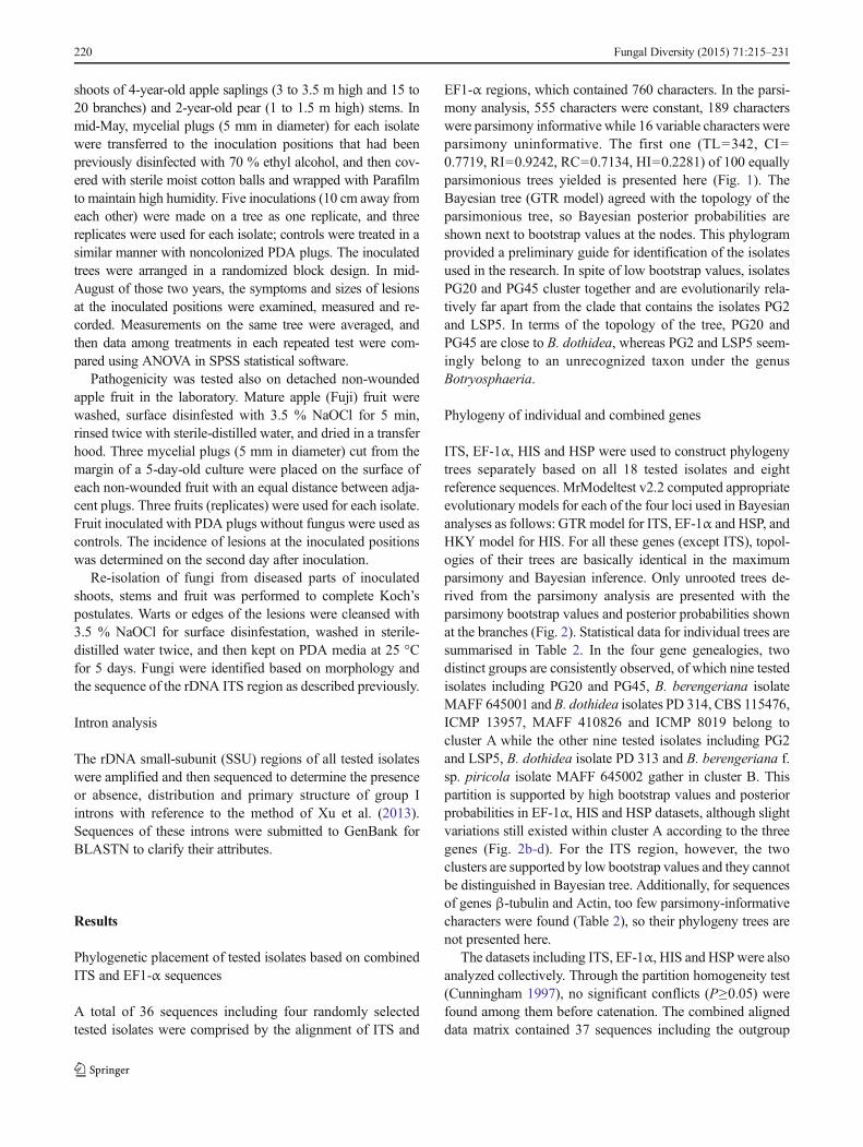
Phylogenetic placement of tested isolates based on combinedITS and EF1-α sequences
A total of 36 sequences including four randomly selectedtested isolates were comprised by the alignment of ITS and
EF1-α regions, which contained 760 characters. In the parsi-mony analysis, 555 characters were constant, 189 characterswere parsimony informative while 16 variable characters wereparsimony uninformative. The first one (TL=342, CI=0.7719, RI=0.9242, RC=0.7134, HI=0.2281) of 100 equallyparsimonious trees yielded is presented here (Fig. 1). TheBayesian tree (GTR model) agreed with the topology of theparsimonious tree, so Bayesian posterior probabilities areshown next to bootstrap values at the nodes. This phylogramprovided a preliminary guide for identification of the isolatesused in the research. In spite of low bootstrap values, isolatesPG20 and PG45 cluster together and are evolutionarily rela-tively far apart from the clade that contains the isolates PG2and LSP5. In terms of the topology of the tree, PG20 andPG45 are close to B. dothidea, whereas PG2 and LSP5 seem-ingly belong to an unrecognized taxon under the genusBotryosphaeria.
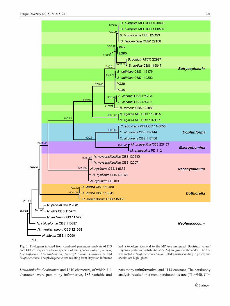
Phylogeny of individual and combined genes
ITS, EF-1α, HIS and HSP were used to construct phylogenytrees separately based on all 18 tested isolates and eightreference sequences. MrModeltest v2.2 computed appropriateevolutionary models for each of the four loci used in Bayesiananalyses as follows: GTRmodel for ITS, EF-1α and HSP, andHKY model for HIS. For all these genes (except ITS), topol-ogies of their trees are basically identical in the maximumparsimony and Bayesian inference. Only unrooted trees de-rived from the parsimony analysis are presented with theparsimony bootstrap values and posterior probabilities shownat the branches (Fig. 2). Statistical data for individual trees aresummarised in Table 2. In the four gene genealogies, twodistinct groups are consistently observed, of which nine testedisolates including PG20 and PG45, B. berengeriana isolateMAFF 645001 and B. dothidea isolates PD 314, CBS 115476,ICMP 13957, MAFF 410826 and ICMP 8019 belong tocluster A while the other nine tested isolates including PG2and LSP5, B. dothidea isolate PD 313 and B. berengeriana f.sp. piricola isolate MAFF 645002 gather in cluster B. Thispartition is supported by high bootstrap values and posteriorprobabilities in EF-1α, HIS and HSP datasets, although slightvariations still existed within cluster A according to the threegenes (Fig. 2b-d). For the ITS region, however, the twoclusters are supported by low bootstrap values and they cannotbe distinguished in Bayesian tree. Additionally, for sequencesof genes β-tubulin and Actin, too few parsimony-informativecharacters were found (Table 2), so their phylogeny trees arenot presented here.
The datasets including ITS, EF-1α, HIS and HSPwere alsoanalyzed collectively. Through the partition homogeneity test(Cunningham 1997), no significant conflicts (P≥0.05) werefound among them before catenation. The combined aligneddata matrix contained 37 sequences including the outgroup
220 Fungal Diversity (2015) 71:215–231
Lasiodiplodia theobromae and 1610 characters, of which 311characters were parsimony informative, 185 variable and
parsimony uninformative, and 1114 constant. The parsimonyanalysis resulted in a most parsimonious tree (TL=940, CI=
Fig. 1 Phylogram inferred from combined parsimony analysis of ITSand EF1-α sequences from species of the genera Botryosphaeria,Cophinforma, Macrophomina, Neoscytalidium, Dothiorella andNeofusicoccum. The phylogenetic tree resulting from Bayesian inference
had a topology identical to the MP tree presented. Bootstrap values/Bayesian posterior probabilities (>50 %) are given at the nodes. The treewas rooted toNeofusicoccum luteum. Clades corresponding to genera andspecies are highlighted
Fungal Diversity (2015) 71:215–231 221
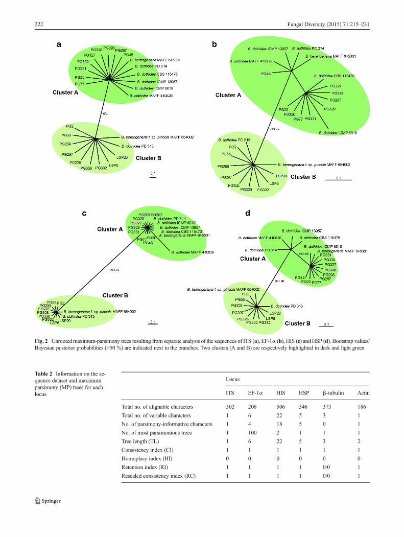
Table 2 Information on the se-quence dataset and maximumparsimony (MP) trees for eachlocus
Locus
ITS EF-1α HIS HSP β-tubulin Actin
Total no. of alignable characters 502 208 506 346 373 186
Total no. of variable characters 1 6 22 5 3 1
No. of parsimony-informative characters 1 4 18 5 0 1
No. of most parsimonious trees 1 100 2 1 1 1
Tree length (TL) 1 6 22 5 3 2
Consistency index (CI) 1 1 1 1 1 1
Homoplasy index (HI) 0 0 0 0 0 0
Retention index (RI) 1 1 1 1 0/0 1
Rescaled consistency index (RC) 1 1 1 1 0/0 1
222 Fungal Diversity (2015) 71:215–231
Fig. 2 Unrootedmaximum-parsimony trees resulting from separate analysis of the sequences of ITS (a), EF-1α (b), HIS (c) and HSP (d). Bootstrap values/Bayesian posterior probabilities (>50 %) are indicated next to the branches. Two clusters (A and B) are respectively highlighted in dark and light green
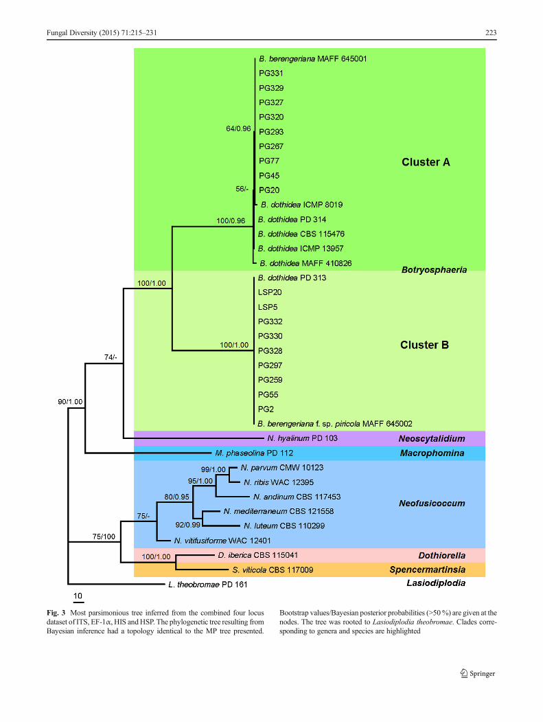
Fig. 3 Most parsimonious tree inferred from the combined four locusdataset of ITS, EF-1α, HIS andHSP. The phylogenetic tree resulting fromBayesian inference had a topology identical to the MP tree presented.
Bootstrap values/Bayesian posterior probabilities (>50%) are given at thenodes. The tree was rooted to Lasiodiplodia theobromae. Clades corre-sponding to genera and species are highlighted
Fungal Diversity (2015) 71:215–231 223
0.6968, HI=0.3032, RI=0.8208, RC=0.5719) (Fig. 3).Bayesian tree (GTR model) with an identical topology to theMP tree was reconstructed, and its posterior probabilities werethus added next to the bootstrap values. In the phylogeneticreconstruction of combined dataset, two deep clades wererecognized and strongly supported with bootstrap valuesequal to 100 % and posterior probabilities of 1.00, whichcompletely corresponded to the two clusters formed in theindividual gene genealogies.
Morphological characteristics
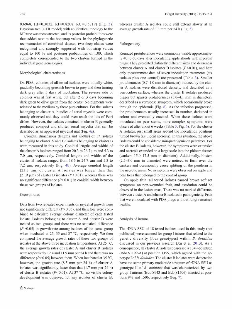
On PDA, colonies of all tested isolates were initially white,gradually becoming greenish brown to grey and then turningdark grey after 5 days of incubation. The reverse side ofcolonies was at first white, but after 2 or 3 days becomingdark green to olive green from the centre. No pigments werereleased to the medium by these pure cultures. For the isolatesbelonging to cluster A, bundles of aerial mycelia were com-monly observed and they could even reach the lids of Petridishes. However, the isolates contained in cluster B generallyproduced compact and shorter aerial mycelia that can bedescribed as an appressed mycelial mat (Fig. 4a).
Conidial dimensions (lengths and widths) of 17 isolatesbelonging to cluster A and 19 isolates belonging to cluster Bwere measured in this study. Conidial lengths and widths ofthe cluster A isolates ranged from 20.3 to 26.7 μm and 5.3 to7.0 μm, respectively. Conidial lengths and widths of thecluster B isolates ranged from 18.6 to 24.7 μm and 5.3 to7.2 μm, respectively (Fig. 4b). Average conidial length(23.3 μm) of cluster A isolates was longer than that(21.9 μm) of cluster B isolates (P<0.01), whereas there wasno significant difference (P>0.01) in conidial width betweenthese two groups of isolates.
Growth rates
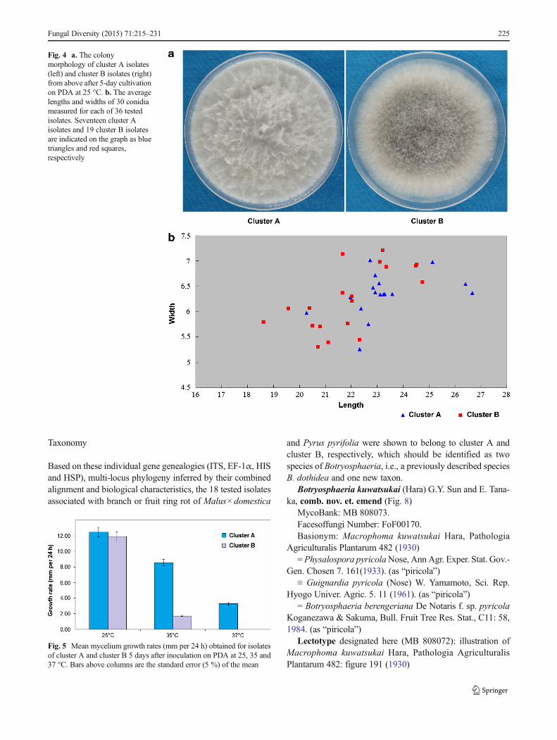
Data from two repeated experiments on mycelial growth werenot significantly different (P>0.05), and therefore were com-bined to calculate average colony diameter of each testedisolate. Isolates belonging to cluster A and cluster B weretreated as two groups and there was no statistical difference(P>0.05) in growth rate among isolates of the same groupwhen incubated at 25, 35 and 37 °C, respectively. We thencompared the average growth rates of these two groups ofisolates at the above three incubation temperatures. At 25 °C,the average growth rates of cluster A and cluster B isolateswere respectively 12.4 and 11.9 mm per 24 h and there was nodifference (P>0.05) between them. When incubated at 35 °C,however, the growth rate (8.5 mm per 24 h) of cluster Aisolates was significantly faster than that (1.7 mm per 24 h)of cluster B isolates (P<0.01). At 37 °C, no visible colonydevelopment was observed for any isolates of cluster B,
whereas cluster A isolates could still extend slowly at anaverage growth rate of 3.3 mm per 24 h (Fig. 5).
Pathogenicity
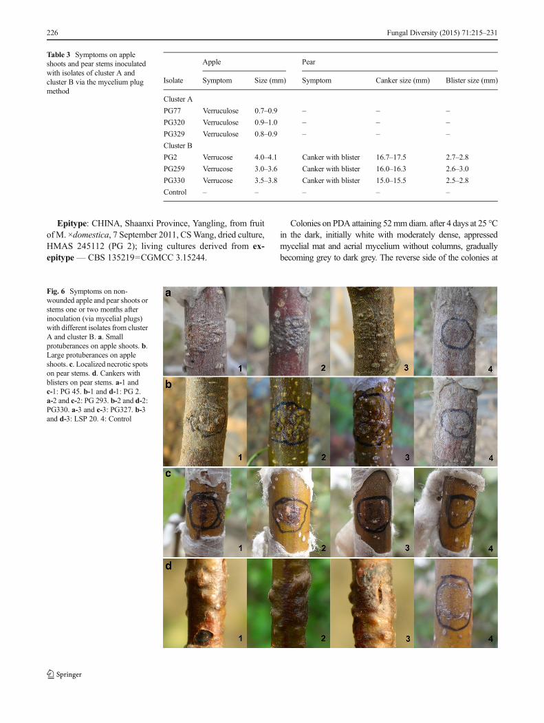
Rounded protuberances were commonly visible approximate-ly 40 to 60 days after inoculating apple shoots with mycelialplugs. They presented distinctly different sizes and densenessbetween cluster A and cluster B isolates (P<0.01), and hereonly measurement data of seven inoculation treatments (sixisolates plus one control) are presented (Table 3). Smallerprotuberances (0.7–1.0 mm in diameter) induced by the clus-ter A isolates were distributed densely, and described as averruculose surface, whereas the cluster B isolates producedbigger but sparser protuberances (3.0–4.1 mm in diameter),described as a verrucose symptom, which occasionally brokethrough the epidermis (Fig. 6). As the infection progressed,the protuberances usually increased in number, darkened incolour and eventually cracked. When these isolates wereinoculated on pear stems, more complex symptoms wereobserved after about 6 weeks (Table 3, Fig. 6). For the clusterA isolates, just small areas around the inoculation positionsturned brown (i.e., local necrosis). In this situation, the aboveisolates could be considered non-pathogenic on pear trees. Forthe cluster B isolates, however, the symptoms were extensiveand necrosis extended on a large scale into the phloem tissues(cankers 15.0–17.5 mm in diameter). Additionally, blisters(2.5–3.0 mm in diameter) were noticed to form over thecankers and occasionally cause splitting of the periderm inthe necrotic areas. No symptoms were observed on apple andpear trees that belonged to the control group
On apple fruit, all tested isolates caused brown soft rotsymptoms on non-wounded fruit, and exudation could beobserved in the lesion areas. There was no marked differencebetween cluster A and cluster B isolates in pathogenicity. Fruitthat were inoculated with PDA plugs without fungi remainedhealthy.
Analysis of introns
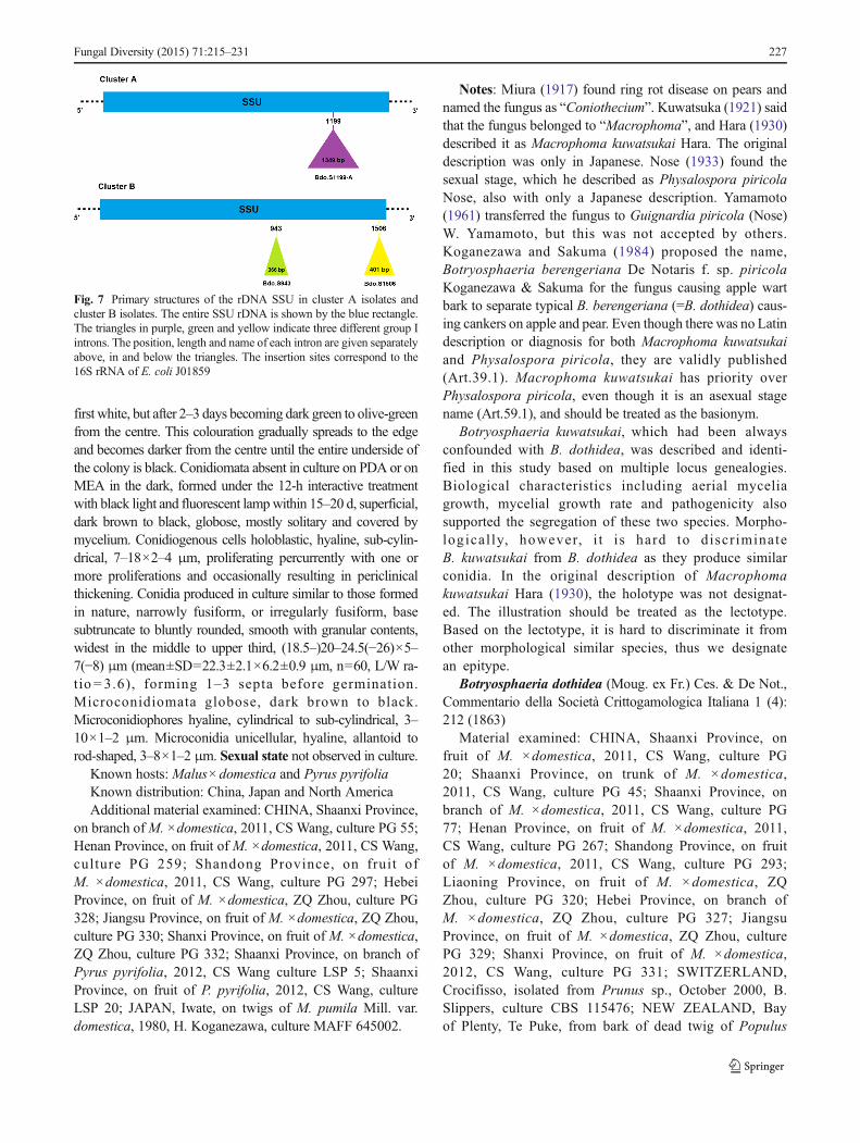
The rDNA SSU of 18 tested isolates used in this study (notpublished) were scanned for group I introns that related to thegenetic diversity (four genotypes) within B. dothideadiscussed in our previous research (Xu et al. 2013). As aconsequence, all cluster A isolates possessed a 1349-bp intron(Bdo.S1199-A) at position 1199, which agreed with the ge-notype I ofB. dothidea. The cluster B isolates were detected tohave the same primary nucleotide structure of rDNA SSU asgenotype II of B. dothidea that was characterized by twogroup I introns (Bdo.S943 and Bdo.S1506) inserted at posi-tions 943 and 1506, respectively (Fig. 7).
224 Fungal Diversity (2015) 71:215–231
Taxonomy
Based on these individual gene genealogies (ITS, EF-1α, HISand HSP), multi-locus phylogeny inferred by their combinedalignment and biological characteristics, the 18 tested isolatesassociated with branch or fruit ring rot of Malus×domestica
and Pyrus pyrifolia were shown to belong to cluster A andcluster B, respectively, which should be identified as twospecies of Botryosphaeria, i.e., a previously described speciesB. dothidea and one new taxon.
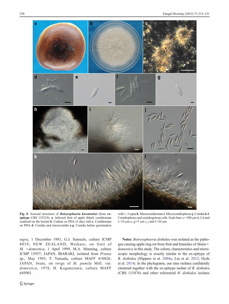
Botryosphaeria kuwatsukai (Hara) G.Y. Sun and E. Tana-ka, comb. nov. et. emend (Fig. 8)
MycoBank: MB 808073.Facesoffungi Number: FoF00170.Basionym: Macrophoma kuwatsukai Hara, Pathologia
Agriculturalis Plantarum 482 (1930)= Physalospora pyricolaNose, AnnAgr. Exper. Stat. Gov.-
Gen. Chosen 7. 161(1933). (as “piricola”)≡ Guignardia pyricola (Nose) W. Yamamoto, Sci. Rep.
Hyogo Univer. Agric. 5. 11 (1961). (as “piricola”)= Botryosphaeria berengeriana De Notaris f. sp. pyricola
Koganezawa & Sakuma, Bull. Fruit Tree Res. Stat., C11: 58,1984. (as “piricola”)
Lectotype designated here (MB 808072): illustration ofMacrophoma kuwatsukai Hara, Pathologia AgriculturalisPlantarum 482: figure 191 (1930)
Fig. 4 a. The colonymorphology of cluster A isolates(left) and cluster B isolates (right)from above after 5-day cultivationon PDA at 25 °C. b. The averagelengths and widths of 30 conidiameasured for each of 36 testedisolates. Seventeen cluster Aisolates and 19 cluster B isolatesare indicated on the graph as bluetriangles and red squares,respectively
Fig. 5 Mean mycelium growth rates (mm per 24 h) obtained for isolatesof cluster A and cluster B 5 days after inoculation on PDA at 25, 35 and37 °C. Bars above columns are the standard error (5 %) of the mean
Fungal Diversity (2015) 71:215–231 225
Epitype: CHINA, Shaanxi Province, Yangling, from fruitofM. ×domestica, 7 September 2011, CSWang, dried culture,HMAS 245112 (PG 2); living cultures derived from ex-epitype — CBS 135219=CGMCC 3.15244.
Colonies on PDA attaining 52mmdiam. after 4 days at 25 °Cin the dark, initially white with moderately dense, appressedmycelial mat and aerial mycelium without columns, graduallybecoming grey to dark grey. The reverse side of the colonies at
Table 3 Symptoms on appleshoots and pear stems inoculatedwith isolates of cluster A andcluster B via the mycelium plugmethod
Apple Pear
Isolate Symptom Size (mm) Symptom Canker size (mm) Blister size (mm)
Cluster A
PG77 Verruculose 0.7–0.9 – – –
PG320 Verruculose 0.9–1.0 – – –
PG329 Verruculose 0.8–0.9 – – –
Cluster B
PG2 Verrucose 4.0–4.1 Canker with blister 16.7–17.5 2.7–2.8
PG259 Verrucose 3.0–3.6 Canker with blister 16.0–16.3 2.6–3.0
PG330 Verrucose 3.5–3.8 Canker with blister 15.0–15.5 2.5–2.8
Control – – – – –
Fig. 6 Symptoms on non-wounded apple and pear shoots orstems one or two months afterinoculation (via mycelial plugs)with different isolates from clusterA and cluster B. a. Smallprotuberances on apple shoots. b.Large protuberances on appleshoots. c. Localized necrotic spotson pear stems. d. Cankers withblisters on pear stems. a-1 andc-1: PG 45. b-1 and d-1: PG 2.a-2 and c-2: PG 293. b-2 and d-2:PG330. a-3 and c-3: PG327. b-3and d-3: LSP 20. 4: Control
226 Fungal Diversity (2015) 71:215–231
first white, but after 2–3 days becoming dark green to olive-greenfrom the centre. This colouration gradually spreads to the edgeand becomes darker from the centre until the entire underside ofthe colony is black. Conidiomata absent in culture on PDA or onMEA in the dark, formed under the 12-h interactive treatmentwith black light and fluorescent lampwithin 15–20 d, superficial,dark brown to black, globose, mostly solitary and covered bymycelium. Conidiogenous cells holoblastic, hyaline, sub-cylin-drical, 7–18×2–4 μm, proliferating percurrently with one ormore proliferations and occasionally resulting in periclinicalthickening. Conidia produced in culture similar to those formedin nature, narrowly fusiform, or irregularly fusiform, basesubtruncate to bluntly rounded, smooth with granular contents,widest in the middle to upper third, (18.5–)20–24.5(−26)×5–7(−8) μm (mean±SD=22.3±2.1×6.2±0.9 μm, n=60, L/W ra-tio = 3.6), forming 1–3 septa before germination.Microconidiomata globose, dark brown to black.Microconidiophores hyaline, cylindrical to sub-cylindrical, 3–10×1–2 μm. Microconidia unicellular, hyaline, allantoid torod-shaped, 3–8×1–2 μm. Sexual state not observed in culture.
Known hosts: Malus×domestica and Pyrus pyrifoliaKnown distribution: China, Japan and North AmericaAdditional material examined: CHINA, Shaanxi Province,
on branch ofM. ×domestica, 2011, CS Wang, culture PG 55;Henan Province, on fruit of M. ×domestica, 2011, CS Wang,culture PG 259; Shandong Province, on fruit ofM. ×domestica, 2011, CS Wang, culture PG 297; HebeiProvince, on fruit of M. ×domestica, ZQ Zhou, culture PG328; Jiangsu Province, on fruit of M. ×domestica, ZQ Zhou,culture PG 330; Shanxi Province, on fruit of M. ×domestica,ZQ Zhou, culture PG 332; Shaanxi Province, on branch ofPyrus pyrifolia, 2012, CS Wang culture LSP 5; ShaanxiProvince, on fruit of P. pyrifolia, 2012, CS Wang, cultureLSP 20; JAPAN, Iwate, on twigs of M. pumila Mill. var.domestica, 1980, H. Koganezawa, culture MAFF 645002.
Notes: Miura (1917) found ring rot disease on pears andnamed the fungus as “Coniothecium”. Kuwatsuka (1921) saidthat the fungus belonged to “Macrophoma”, and Hara (1930)described it as Macrophoma kuwatsukai Hara. The originaldescription was only in Japanese. Nose (1933) found thesexual stage, which he described as Physalospora piricolaNose, also with only a Japanese description. Yamamoto(1961) transferred the fungus to Guignardia piricola (Nose)W. Yamamoto, but this was not accepted by others.Koganezawa and Sakuma (1984) proposed the name,Botryosphaeria berengeriana De Notaris f. sp. piricolaKoganezawa & Sakuma for the fungus causing apple wartbark to separate typical B. berengeriana (=B. dothidea) caus-ing cankers on apple and pear. Even though there was no Latindescription or diagnosis for both Macrophoma kuwatsukaiand Physalospora piricola, they are validly published(Art.39.1). Macrophoma kuwatsukai has priority overPhysalospora piricola, even though it is an asexual stagename (Art.59.1), and should be treated as the basionym.
Botryosphaeria kuwatsukai, which had been alwaysconfounded with B. dothidea, was described and identi-fied in this study based on multiple locus genealogies.Biological characteristics including aerial myceliagrowth, mycelial growth rate and pathogenicity alsosupported the segregation of these two species. Morpho-logical ly, however, i t i s hard to discr iminateB. kuwatsukai from B. dothidea as they produce similarconidia. In the original description of Macrophomakuwatsukai Hara (1930), the holotype was not designat-ed. The illustration should be treated as the lectotype.Based on the lectotype, it is hard to discriminate it fromother morphological similar species, thus we designatean epitype.
Botryosphaeria dothidea (Moug. ex Fr.) Ces. & De Not.,Commentario della Società Crittogamologica Italiana 1 (4):212 (1863)
Material examined: CHINA, Shaanxi Province, onfruit of M. ×domestica, 2011, CS Wang, culture PG20; Shaanxi Province, on trunk of M. ×domestica,2011, CS Wang, culture PG 45; Shaanxi Province, onbranch of M. ×domestica, 2011, CS Wang, culture PG77; Henan Province, on fruit of M. ×domestica, 2011,CS Wang, culture PG 267; Shandong Province, on fruitof M. ×domestica, 2011, CS Wang, culture PG 293;Liaoning Province, on fruit of M. ×domestica, ZQZhou, culture PG 320; Hebei Province, on branch ofM. ×domestica, ZQ Zhou, culture PG 327; JiangsuProvince, on fruit of M. ×domestica, ZQ Zhou, culturePG 329; Shanxi Province, on fruit of M. ×domestica,2012, CS Wang, culture PG 331; SWITZERLAND,Crocifisso, isolated from Prunus sp., October 2000, B.Slippers, culture CBS 115476; NEW ZEALAND, Bayof Plenty, Te Puke, from bark of dead twig of Populus
Fig. 7 Primary structures of the rDNA SSU in cluster A isolates andcluster B isolates. The entire SSU rDNA is shown by the blue rectangle.The triangles in purple, green and yellow indicate three different group Iintrons. The position, length and name of each intron are given separatelyabove, in and below the triangles. The insertion sites correspond to the16S rRNA of E. coli J01859
Fungal Diversity (2015) 71:215–231 227
nigra, 1 December 1981, G.J. Samuels, culture ICMP8019; NEW ZEALAND, Waikato , on frui t ofM. ×domestica, 1 April 1999, M.A. Manning, cultureICMP 13957; JAPAN, IBARAKI, isolated from Prunussp., May 1993, T. Yamada, culture MAFF 410826;JAPAN, Iwate, on twigs of M. pumila Mill. var.domestica, 1978, H. Koganezawa, culture MAFF645001.
Notes: Botryosphaeria dothidea was isolated as the patho-gen causing apple ring rot from fruit and branches ofMalus×domestica in this study. The colony characteristics and micro-scopic morphology is exactly similar to the ex-epitype ofB. dothidea (Slippers et al. 2004a; Liu et al. 2012; Hydeet al. 2014). In the phylograms, our nine isolates confidentlyclustered together with the ex-epitype isolate of B. dothidea(CBS 115476) and other referential B. dothidea isolates
Fig. 8 Asexual structures of Botryosphaeria kuwatsukai (from ex-epitype CBS 135219). a. Infected fruit of apple (black conidiomatascattered on the lesion) b. Culture on PDA (5 days old) c. Conidiomataon PDA d. Conidia and microconidia e-g. Conidia before germination
with 1–3 septa h. Microconidiomata i. Microconidiophores j. Conidia k-l.Conidiophores and conidiogenous cells. Scale bars: c=500 μm d, f, h andi=10 μm e, g=5 μm i, j and l=20 μm
228 Fungal Diversity (2015) 71:215–231
including ICMP 8019, ICMP 13957, MAFF 410826 andMAFF 645001 (Figs. 2, and 3).
Discussion
The theory of multiple gene genealogies has been increasinglyapplied in studies of species boundaries in both human andplant pathogenic fungi, revealing vast numbers of crypticspecies and species complexes in fungal taxa previously iden-tified as one morphospecies (Pringle et al. 2005; Hyde et al.2010, 2014; Liu et al. 2012; Maharachchikumbura et al. 2012;Udayanga et al. 2012; Morgado et al. 2013). InBotryosphaeriaceae, for example, Slippers et al. (2004a)redefined the circumscription of B. dothidea sensu stricto andseparated out B. parva and B. ribis that had previously served assynonyms of B. dothidea. Pavlic et al. (2009) revealed threecryptic species within the Neofusicoccum parvum/N. ribis com-plex isolated from Syzygium cordatum trees in South Africa.Furthermore, Diplodia scrobiculata was described as a sisterspecies of D. pinea (De Wet et al. 2003) and N. eucalypticolaand N. australe were perceived as sister species ofN. eucalyptorum and N. luteum, respectively (Slippers et al.2004b, c). In this study, Botryosphaeria kuwatsukai is describedand identified from pathogenic isolates causing ring rot of appleand pear trees in China, thus proving our hypothesis that theprevious ring rot pathogen, B. dothidea, is a species complex.This cryptic species was recognized primarily based on DNAsequence data of four nuclear genes combined with some phe-notypic characters as supplementary evidence.
According to the phylogram of combined ITS and EF1-αsequences (Fig. 1), we can estimate that the four randomlyselected tested isolates belong to two different taxa (oneprobably equals to B. dothidea) in Botryosphaeria. Furtheranalysis of the genealogies of EF-1α, HIS and HSP genes andtheir combination (Figs. 2, and 3) demonstrated that all testedisolates were subsumed in two genetically well-separatedclusters (cluster A and cluster B), which correspond to theabove two taxa. In cluster A, despite some slight variations,nine tested isolates gathered well with five referenceB. dothidea isolates including the ex-epitype CBS 115476,indicating that they truly belong to B. dothidea. In cluster B,the remaining nine tested isolates and two referenceB. dothidea isolates were re-identified and named asB. kuwatsukai, thus suggesting that previous B. dothidea iden-tified from apple ring rot was a species complex. Two othergenes, β-tubulin and Actin, widely used in phylogenetics,were also analyzed here. They were more inclined to makeall tested isolates fall together in the B. dothidea clade. This isconsistent with the result of Tang et al. (2012), who consideredaccording to the multi-gene sequence data of ITS, β-tubulinand Actin that isolates associated with symptoms of apple and
pear ring rot (warts, cankers and fruit rot) all belong to thesame pathogen, B. dothidea.
In microscopic morphology, B. kuwatsukai and B. dothideaexhibited a statistically significant difference in conidiallength. However, this variation represented a continuumamong isolates of the B. dothidea/B. kuwatsukai complexand, therefore, it was too difficult for us to use this characterto set a clear delimitation of groups, and then to screenB. kuwatsukai out of B. dothidea prior to the application ofsome other effective methods. This indicates that geneticallyisolated species do not necessarily show divergence in somemorphological characters such as conidial morphology, whichis consistent with conclusions of several previous studies(Pavlic et al. 2009; Maharachchikumbura et al. 2012;Udayanga et al. 2012; Muggia et al. 2014). Colony morphol-ogy and different growth patterns of aerial mycelia (i.e.,appressed mycelial mat from B. kuwatsukai and columns ofaerial mycelia from B. dothidea) can be used to preliminarilydistinguish these two species.
When cultured below their optimal temperatures (generally22 to 28 °C) in darkness, both B. kuwatsukai and B. dothideagrew faster on PDA as temperature increased and there was nosignificant difference between their growth rates (unpublisheddata). When cultured above their optimal temperatures, bothspecies grew more slowly as temperature increased; however,the decline in growth rate was more pronounced withB. kuwatsukai. Compared to the demanding requirement fortechniques and equipment used in molecular identification, itis undoubtedly a more expeditious approach to differentiatethe two species by simultaneously cultivating them at rela-tively high temperatures (e.g., 35 °C) and then assessing theircolony diameters after 3–5 days.
Although the host affiliations of isolates collected in thisstudy were limited to apple and pear, host ranges of the twofungi could be inferred according to previous research(Inderbitzin et al. 2010; Marques et al. 2013; Xu et al. 2013).As mentioned above, the isolates of B. kuwatsukai actuallycorrespond to the genotype II of previous B. dothidea, whereasthe other three genotypes (III and IV were not isolated here) arestill considered to be genetically different populations withincurrent B. dothidea. Through investigation and statistics, Xuet al. (2013) found that genotype II isolates of B. dothidea couldbe isolated only from apple and pear, and was the only popu-lation detected on pear, whereas the other three genotypes ofB. dothidea collectively infected dozens of shrubs and trees,except pear. From the above, it is conjectured thatB. kuwatsukaimay have host specificity for apple and pear.
The phylogenetic analyses showed that both species caus-ing apple ring rot belong to the genus Botryosphaeria (Liuet al. 2012; Hyde et al. 2014). The Japanese B. berengerianaisolate MAFF645001 belonged to B. dothidea, which agreeswith the conclusion of Slippers et al. (2004a), whereas anotherJapanese isolate B. berengeriana f. sp. piricolaMAFF645002
Fungal Diversity (2015) 71:215–231 229
and B. dothidea isolate PD313 from the USA were both re-identified as B. kuwatsukai, which suggests that this species isa cosmopolitan fungus, not unique to China. According to themorphological and biological description of B. berengerianaf. sp. piricola byKoganezawa and Sakuma (1984), this fungusis morphologically identical with B. dothidea, grows slowerthan B. dothidea and mainly causes ring rot of apple and pear.All these characters are shared by B. kuwatsukai, supportingour phylogenetic results. Reference isolates PD313 andPD314 (Inderbitzin et al. 2010), which were isolated fromfruit of M. domestica in USA, were once both described asB. dothidea and caused a prevalent disease called apple whiterot (Jones and Aldwinckle 1990; Inderbitzin et al. 2010).However, in this study the two isolates were classified asdifferent species, indicating that apple white rot, which iswidely distributed in the North America, is probably able tobe induced by two pathogens, B. dothidea and/orB. kuwatsukai.
Pathogenicity tests revealed that both B. kuwatsukai andB. dothidea could induce protuberances on the surfaces ofapple shoots, with the two species associated with protuber-ances of different size and density (described as verrucose andverruculose, respectively). Protuberances caused byB. kuwatsukai were nearly four times as larger than those byB. dothidea. Previously, this phenomenon was often attributedto differences in virulence of different B. dothidea isolates andwas applied to the establishment of evaluation criteria for hostresistance (Zhou et al. 2010; Lin et al. 2011). Our results showthat size of protuberances is not related to virulencedifferences and this character should not be applied whenscreening for resistance.
The common canker symptom of apple ring rot on shootswas not observed in the inoculation tests. Koganezawa andSakuma (1984) inoculated mycelial plugs of B. berengeriana(B. dothidea) isolates and B. berengeriana f. sp. piricola(=B. kuwatsukai) isolates on wounded apple trunks, and re-ported the former produced typical cankers while the latterformed rough callus bark on inoculation sites. They alsoinoculated spore suspension of the two pathogens on non-wounded apple trunks, and consequently B. berengerianacaused no symptoms while B. berengeriana f. sp. piricolaproduced typical wart-like protrusions. Tang et al. (2012)performed similar pathogenicity tests with several B. dothideaisolates, in which warts formed only on non-wounded appleshoots whereas cankers often appeared on wounded appleshoots. Therefore, we speculate that different inoculationmethods (non-wounded or wounded and mycelium plugs orspore suspension) lead to different symptoms on apple trunks.B. kuwatsukai tends to infect hosts from the lenticels andcauses wart-like protuberances, whereas B. dothidea tends toinfect hosts from wounds and causes cankers and, in fact,when the inoculum dose is enough (e.g., mycelium plugs),B. dothidea can cause similar symptoms as B. kuwatsukai
(Koganezawa and Sakuma 1984; Zhang et al. 2011). On pearstems, large-scale cankers along with blisters were producedby B. kuwatsukai, whereas B. dothidea just induced localizednecrotic spots, which could be regarded as non-infectious.This agrees with the view of Koganezawa and Sakuma(1984) that B. berengeriana f. sp. piricola (=B. kuwatsukai)is the true and only pathogen of pear ring rot.
Our research provided sufficient evidence to prove thatapple ring rot disease is caused by two different pathogens,B. dothidea and B. kuwatsukai, whereas B. kuwatsukai aloneis the pathogen responsible for pear ring rot. With increasedunderstanding of the etiology of apple ring rot, we can beginto develop targeted management strategies based on sanita-tion, cultural methods, chemical methods and resistancebreeding.
Acknowledgments We are grateful for help in sample collecting byProf. Zengqiang Zhou (Zhengzhou Institute of Pomology, Henan, China)and Prof. Meng Zhang (Henan Agricultural University, Henan, China).We thank Prof Pedro W. Crous (CBS-KNAW Fungal Biodiversity Cen-tre, The Netherlands.) and Dr Eric H.C. McKenzie (Landcare Research,Auckland, New Zealand) for exchanging the authentic cultures andgiving suggestion in nomenclature. This work was supported by NationalNatural Science Foundation of China (31371887, 31171797), the 111Project from Education Ministry of China (B07049), Specialized Re-search Fund for the Doctoral Program of Higher Education(20130204110002) and China Agriculture Research System (CARS-28).
References
Altekar G, Dwarkadas S, Huelsenbeck JP, Ronquist F (2004) Parallelmetropolis coupled Markov chain Monte Carlo for Bayesian phylo-genetic inference. Bioinformatics 20:407–415
Chen C (1999) Advances in the research of apple ring rot. ActaPhytopathol Sinica 29(3):1–7 (in Chinese)
Cunningham CW (1997) Can three incongruence tests predict when datashould be combined? Mol Biol Evol 14:733–740
DeWet J, Burgess T, Slippers B, Preisig O, Wingfield BD, Wingfield MJ(2003) Multiple gene genealogies and microsatellite markers reflectrelationships between morphotypes of Sphaeropsis sapinea anddistinguish a new species of Diplodia. Mycol Res 107:557–566
Farris JS, Källersjö M, Kluge AG, Bult C (1994) Testing significance ofincongruence. Cladistics 10:315–319
Felsenstein J (1985) Confidence limits on phylogenies: an approach usingthe bootstrap. Evolution 39:783–791
Hall TA (1999) BioEdit: a user-friendly biological sequence alignmenteditor and analysis program for Windows 95/98/NT. Nucleic AcidsSymp Ser 41:95–98
Hara K (1930) Pathologia Agriculturalis Plantarum. Yokendo, Tokyo, pp481–483 (in Japanese)
Huang C, Liu K (2001) RAPD analysis of the pathogenic fungi of applering rot and other major related diseases. Acta Phytopathol Sinica31(2):69–74
HydeKD, Chomnunti P, Crous PW, Groenewald JZ, DammU, Ko TWK,Shivas RG, Summerell BA, Tan YP (2010) A case for re-inventoryof Australia’s plant pathogens. Persoonia 25:50–60
Hyde KD, Nilsson RH, Alias SA, Ariyawansa HA, Blair JE, Cai L, deCock AWAM, Dissanayake AJ, Glockling SL, Goonasekara ID,Gorczak M, Hahn M, Jayawardena RS, van Kan JAL, Laurence
230 Fungal Diversity (2015) 71:215–231
MH, Lévesque CA, Li X, Liu JK, Maharachchikumbura SSN,Manamgoda DS, Martin FN, McKenzie EHC, McTaggart AR,Mortimer PE, Nair PVR, Pawłowska J, Rintoul TL, Shivas RG,Spies CFJ, Summerell BA, Taylor PWJ, Terhem RB, Udayanga D,Vaghefi N, Walther G, Wilk M, Wrzosek M, Xu JC, Yan JY, Zhou N(2014) One stop shop: backbones trees for important phytopathogenicgenera: I. Fungal Divers 67:21–125. doi:10.1007/s13225-014-0298-1
Inderbitzin P, Bostock RM, Trouillas FP, Michailides TJ (2010) A sixlocus phy logeny revea l s h igh spec ies d ive r s i ty inBotryosphaeriaceae from California almond. Mycologia 102:1350–1368
Jones AL, Aldwinckle HS (1990) Compendium of apple and pear dis-eases. American Phytopathological Society, St. Paul, Minnesota,USA
Kang L, Hao H, Yang Z, Li X, Kang G (2009) The advances in theresearch of apple ring rot. Chin Agric Sci Bull 25(09):188–191 (inChinese)
Koganezawa H, Sakuma T (1980) Fungi associated with blister cankerand internal bark necrosis of apple trees. Bull Fruit Tree Res StationC (Morioka) 7:83–99
Koganezawa H, Sakuma T (1984) Causal fungi of apple fruit rot. BullFruit Tree Res Station C (Morioka) 11:49–62
Kuwatsuka K (1921) J Okitsu Hortic Soc (Engei no Kenkyu) 17:190–195, in Japanese
Larkin MA, Blackshields G, Brown NP, Chenna R, McGettigan PA,McWilliam H, Valentin F, Wallace IM, Wilm A, Lopez R (2007)Clustal Wand Clustal X version 2.0. Bioinformatics 23(21):2947–2948
Lin Y, Huang L, Suolang L, Gao X, Chen Y, Kang Z (2011) A rapidlaboratory evaluation system for apple ring rot. Acta PhytophylacicaSinica 38(1):37–41 (in Chinese)
Liu JK, Phookamsak R, Doilom M, Wikee S, Li YM, Ariyawansa H,Boonmee S, Chomnunti P, Dai DQ, Bhat JD, Romero AI, ZhuangWY, Monkai J, Jones EBG, Chukeatirote E, Ko Ko TW, Zhao YC,Wang Y, Hyde KD (2012) Towards a natural classification ofBotryosphaeriales. Fungal Divers 57:149–210
Lv D, Zhang J, Zhang Z, Zhou Z, Chen X, Du X, Qu S (2012) Therelationship between rDNA-ITS sequences and biological charac-teristics of the apple ring rot pathogen Botryosphaeria berengerianade Not f. sp. piricola (Nose). Fungal Genom Biol 2:104
Maharachchikumbura SSN, Guo LD, Cai L, Chukeatirote E, Wu WP,SunX, Crous PW, Bhat DJ,McKenzie EHC, Bahkali AH, Hyde KD(2012) A multi-locus backbone tree for Pestalotiopsis, with a poly-phasic characterization of 14 new species. Fungal Divers 56:95–129
Marques MW, Lima NB, de Morais MA, Michereff SJ, Phillips AJL,Câmara MPS (2013) Botryosphaeria , Neofusicoccum ,Neoscytalidium and Pseudofusicoccum species associated withmango in Brazil. Fungal Divers 61:195–208
Miura M (1917) Ringo no Byoki. Shokabo, Tokyo, pp 106–109 (inJapanese)
Morgado LN, Noordeloos ME, Lamoureux Y, Geml J (2013) Multi-genephylogenetic analyses reveal species limits, phylogeographic pat-terns, and evolutionary histories of key morphological traits inEntoloma (Agaricales, Basidiomycota). Persoonia 31:159–178
Muggia L, Prerez-Ortega S, Fryday A, Spribille T, Grube M (2014) Globalassessment of genetic variation and phenotypic plasticity in the lichen-forming species Tephromela atra. Fungal Divers 64:233–251
Nose T (1933)On the ring rot of pears and the causal organism, especiallyon its perfect generation Physalospora piricola. Ann Agric Exp StaChosen 7(2):156–163 (in Japanese)
Nylander JAA (2004) MrModeltest v2. Program Distributed by theAuthor. Uppsala University, Evolutionary Biology Centre
Ogata T, Sano T, Harada Y (2000) Botryosphaeria spp. isolated fromapple and several deciduous fruit trees are divided into three groupsbased on the production of warts on twigs, size of conidia, and
nucleotide sequences of nuclear ribosomal DNA ITS regions.Mycoscience 41:331–337
Park EW (2005) An infection model of apple white rot based on conidialgermination and appressorium formation of Botryosphaeriadothidea. Plant Pathol J 21:322–327
Pavlic D, Slippers B, Coutinho TA, Wingfield MJ (2009) Multiple genegenealogies and phenotypic data reveal cryptic species of theBotryosphaeriaceae: a case study on the Neofusicoccum parvum/N. ribis complex. Mol Phylogenet Evol 51:259–268
Peng B, Liu L, Wu H, Tian L, Zhou Z, Gu Q (2011) The intraspecificgenetic diversity of pathogenic fungi of apple ring rot. Sci Agric Sin44(6):1125–1135 (in Chinese)
Phillips AJL, Alves A, Abdollahzadeh J, Slippers B, Wingfield MJ,Groenewald JZ, Crous PW (2013) The Botryosphaeriaceae: generaand species known from culture. Stud Mycol 76:51–167
Pringle A, Baker DM, Platt JL, Wares JP, Latge JP, Taylor JW (2005)Cryptic speciation in the cosmopolitan and clonal human pathogenicfungus Aspergillus fumigatus. Evolution 59:1886–1899
Qu J, Li X, ZhangY, FanK (2007) Evaluation of fungitoxicity of tebuconazoleagainst Alternaria mali and Physalospora piricola on apple in laboratoryand in field. Chin J Pestic Sci 9(2):149–152 (in Chinese)
Slippers B, Crous PW, Denman S, Coutinho TA, Wingfield BD,Wingfield MJ (2004a) Combined multiple gene genealogies andphenotypic characters differentiate several species previously iden-tified as Botryosphaeria dothidea. Mycologia 96:83–101
Slippers B, Fourie G, Crous PW, Coutinho TA, Wingfield BD, CarnegieAJ, Wingfield MJ (2004b) Speciation and distribution ofBotryosphaeria spp. on native and introduced Eucalyptus trees inAustralia and South Africa. Stud Mycol 50:343–358
Slippers B, Fourie G, Crous PW, Coutinho TA,Wingfield BD, WingfieldMJ (2004c) Multiple gene sequences delimit Botryosphaeriaaustralis sp. nov. from B. lutea. Mycologia 96:1030–1041
Smith H, Crous PW, Wingfield MJ, Coutinho TA, Wingfield BD (2001)Botryosphaeria eucalyptorum sp. nov., a new species in theB. dothidea-complex on Eucalyptus in South Africa. Mycologia93:277–285
Swofford DL (2003) PAUP*. Phylogenetic analysis using parsimony (*and other methods). Version 4. Sinauer Associates, Sunderland,Massachusetts
Tang W, Ding Z, Zhou Z, Wang Y, Guo L (2012) Phylogenetic andpathogenic analyses show that the causal agent of apple ring rot inChina is Botryosphaeria dothidea. Plant Dis 96:486–496
Taylor JW, Jacobson DJ, Kroken S, Kasuga T, Geiser DM, Hibbett DS,Fisher MC (2000) Phylogenetic species recognition and speciesconcepts in fungi. Fungal Genet Biol 31:21–32
Udayanga D, Liu X, Crous PW, McKenzie EHC, Chukeatirote E, HydeKD (2012) A multi-locus phylogenetic evaluation of Diaporthe(Phomopsis). Fungal Divers 56:157–171
White TJ, Bruns T, Lee S, Taylor J (1990) Amplification and directsequencing of fungal ribosomal RNA genes for phylogenetics.PCR Protoc: Guide Methods Appl 18:315–322
Xu C, Wang C, Sun X, Zhang R, Gleason ML, Eiji T, Sun G (2013)Multiple group I introns in the small-subunit rDNA ofBotryosphaeria dothidea: implication for intraspecific genetic diver-sity. PLoS One 8:e67808
Yamamoto W (1961) Species of the genera of Glomerella andGuignardia with special reference to their imperfect stages. SciRep Hyogo Univ Agric 5(1):1–12 (in Japanese)
Zhang G, Li B, Dong X, Wang C, Li G, Guo L (2011) Microanatomyconformation of apple branch tumors caused by Botryosphaeriadothidea. Acta Phytopathol Sinica 41(1):98–101 (in Chinese)
Zhou Z, Hou H, Wang L, Zhu F (2010) Trunk apple ring rot artificialinoculation method and the identification of cultivar resistance. JFruit Sci 27(6):952–955 (in Chinese)
Fungal Diversity (2015) 71:215–231 231