Embed Size (px)
DESCRIPTION
25. The Urinary System: Part A. Urinary System Organs. Kidneys are major excretory organs Urinary bladder is the temporary storage reservoir for urine Ureters transport urine from the kidneys to the bladder Urethra transports urine out of the body. Hepatic veins (cut). Esophagus (cut). - PowerPoint PPT Presentation
Citation preview

PowerPoint® Lecture Slides prepared by Janice Meeking, Mount Royal College
C H A P T E R
Copyright © 2010 Pearson Education, Inc.
25
The Urinary System: Part A

Copyright © 2010 Pearson Education, Inc.
Urinary System Organs
• Kidneys are major excretory organs
• Urinary bladder is the temporary storage reservoir for urine
• Ureters transport urine from the kidneys to the bladder
• Urethra transports urine out of the body

Copyright © 2010 Pearson Education, Inc. Figure 25.1
Esophagus (cut)Inferior vena cava
Adrenal gland
Hepatic veins (cut)
Renal artery
Renal hilum
Renal vein
Iliac crest
Kidney
Ureter
Urinarybladder
Urethra
Aorta
Rectum (cut)
Uterus (part of female reproductive system)

Copyright © 2010 Pearson Education, Inc.
Kidney Functions
• Removal of toxins, metabolic wastes, and excess ions from the blood
• Regulation of blood volume, chemical composition, and pH

Copyright © 2010 Pearson Education, Inc.
Kidney Functions
• Gluconeogenesis during prolonged fasting
• Endocrine functions
• Renin: regulation of blood pressure and kidney function
• Erythropoietin: regulation of RBC production
• Activation of vitamin D
Copyright © 2010 Pearson Education, Inc.
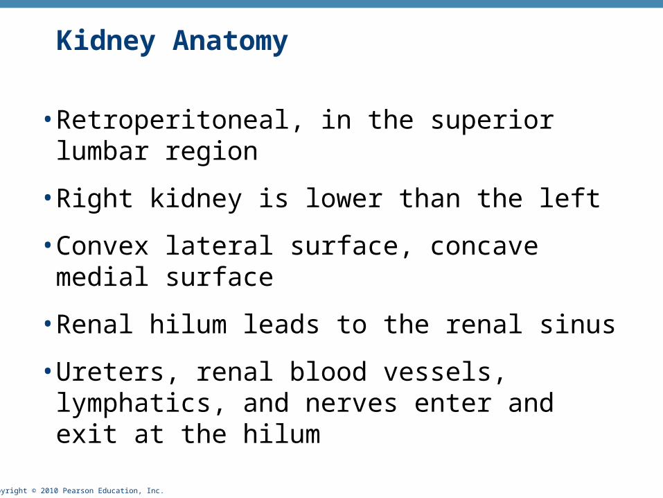
Kidney Anatomy
• Retroperitoneal, in the superior lumbar region
• Right kidney is lower than the left
• Convex lateral surface, concave medial surface
• Renal hilum leads to the renal sinus
• Ureters, renal blood vessels, lymphatics, and nerves enter and exit at the hilum

Copyright © 2010 Pearson Education, Inc.
Kidney Anatomy
• Layers of supportive tissue
1. Renal fascia
• The anchoring outer layer of dense fibrous connective tissue
2. Perirenal fat capsule
• A fatty cushion
3. Fibrous capsule
• Prevents spread of infection to kidney

Copyright © 2010 Pearson Education, Inc. Figure 25.2a
Body wall
• Perirenal fat capsule
Renalartery
Renalvein
Inferior vena cava
Aorta
• Fibrous capsule
• Renal fascia anterior posterior
Supportivetissue layers
Body ofvertebra L2
PeritoneumPeritoneal cavity(organs removed)
Anterior
Posterior(a)

Copyright © 2010 Pearson Education, Inc.
Internal Anatomy
• Renal cortex
• A granular superficial region
• Renal medulla
• The cone-shaped medullary (renal) pyramids separated by renal columns
• Lobe
• A medullary pyramid and its surrounding cortical tissue

Copyright © 2010 Pearson Education, Inc.
Internal Anatomy
• Papilla
• Tip of pyramid; releases urine into minor calyx
• Renal pelvis
• The funnel-shaped tube within the renal sinus

Copyright © 2010 Pearson Education, Inc.
Internal Anatomy
• Major calyces
• The branching channels of the renal pelvis that
• Collect urine from minor calyces
• Empty urine into the pelvis
• Urine flows from the pelvis to ureter

Copyright © 2010 Pearson Education, Inc. Figure 25.3
Renal cortex
Renal medulla
Major calyx
Papilla ofpyramidRenal pelvis
Ureter
Minor calyx
Renal column
Renal pyramid in renal medulla
Fibrous capsule
Renalhilum
(a) Photograph of right kidney, frontal section (b) Diagrammatic view

Copyright © 2010 Pearson Education, Inc.
Blood and Nerve Supply
• Renal arteries deliver ~ 1/4 (1200 ml) of cardiac output to the kidneys each minute
• Arterial flow into and venous flow out of the kidneys follow similar paths
• Nerve supply is via sympathetic fibers from the renal plexus

Copyright © 2010 Pearson Education, Inc. Figure 25.4a
Cortical radiate vein
Cortical radiate artery
Arcuate vein
Arcuate artery
Interlobar vein
Interlobar artery
Segmental arteries
Renal artery
Renal vein
Renal pelvis
Ureter
Renal medulla
Renal cortex
(a) Frontal section illustrating major blood vessels

Copyright © 2010 Pearson Education, Inc. Figure 25.4b
Aorta
Renal artery
Segmental artery
Interlobar artery
Arcuate artery
Cortical radiate artery
Afferent arteriole
Glomerulus (capillaries)
Nephron-associated blood vessels(see Figure 25.7)
Inferior vena cava
Renal vein
Interlobar vein
Arcuate vein
Cortical radiatevein
Peritubularcapillaries
and vasa recta
Efferent arteriole
(b) Path of blood flow through renal blood vessels

Copyright © 2010 Pearson Education, Inc.
Nephrons
• Structural and functional units that form urine
• ~1 million per kidney
• Two main parts
1. Glomerulus: a tuft of capillaries
2. Renal tubule: begins as cup-shaped glomerular (Bowman’s) capsule surrounding the glomerulus

Copyright © 2010 Pearson Education, Inc. Figure 25.5

Copyright © 2010 Pearson Education, Inc.
Nephrons
• Renal corpuscle
• Glomerulus + its glomerular capsule
• Fenestrated glomerular endothelium
• Allows filtrate to pass from plasma into the glomerular capsule
Copyright © 2010 Pearson Education, Inc.
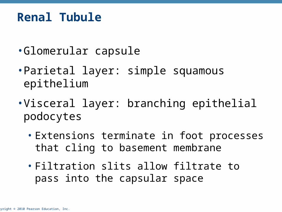
Renal Tubule
• Glomerular capsule
• Parietal layer: simple squamous epithelium
• Visceral layer: branching epithelial podocytes
• Extensions terminate in foot processes that cling to basement membrane
• Filtration slits allow filtrate to pass into the capsular space

Copyright © 2010 Pearson Education, Inc. Figure 25.5
Glomerular capsule: parietal layer

Copyright © 2010 Pearson Education, Inc. Figure 25.5
Fenestratedendotheliumof the glomerulus
Podocyte
Basementmembrane
Glomerular capsule: visceral layer

Copyright © 2010 Pearson Education, Inc.
Renal Tubule
• Proximal convoluted tubule (PCT)
• Cuboidal cells with dense microvilli and large mitochondria
• Functions in reabsorption and secretion
• Confined to the cortex

Copyright © 2010 Pearson Education, Inc. Figure 25.5
Microvilli Mitochondria
Highly infolded plasma membrane
Proximal convoluted tubule cells

Copyright © 2010 Pearson Education, Inc.
Renal Tubule
• Loop of Henle with descending and ascending limbs
• Thin segment usually in descending limb
• Simple squamous epithelium
• Freely permeable to water
• Thick segment of ascending limb
• Cuboidal to columnar cells

Copyright © 2010 Pearson Education, Inc. Figure 25.5
Loop of Henle (thin-segment) cells

Copyright © 2010 Pearson Education, Inc.
Renal Tubule
• Distal convoluted tubule (DCT)
• Cuboidal cells with very few microvilli
• Function more in secretion than reabsorption
• Confined to the cortex

Copyright © 2010 Pearson Education, Inc. Figure 25.5
Distal convoluted tubule cells

Copyright © 2010 Pearson Education, Inc.
Collecting Ducts
• Receive filtrate from many nephrons
• Fuse together to deliver urine through papillae into minor calyces

Copyright © 2010 Pearson Education, Inc.
Collecting Ducts
• Cell types
• Intercalated cells
• Cuboidal cells with microvilli
• Function in maintaining the acid-base balance of the body

Copyright © 2010 Pearson Education, Inc. Figure 25.5
Intercalated cellPrincipal cell
Collecting duct cells

Copyright © 2010 Pearson Education, Inc.
Collecting Ducts
• Principal cells
• Cuboidal cells without microvilli
• Help maintain the body’s water and salt balance

Copyright © 2010 Pearson Education, Inc. Figure 25.5
Fenestratedendotheliumof the glomerulus
Microvilli
Cortex
Medulla
Podocyte
Basementmembrane
Mitochondria
Highly infolded plasmamembrane
Proximalconvolutedtubule
Distalconvolutedtubule
• Descending limbLoop of Henle
• Ascending limb
• Glomerular capsule
Renal corpuscle
• Glomerulus
Thick segment
Collectingduct
Intercalated cellPrincipal cell
Thin segment
Proximal convoluted tubule cells
Glomerular capsule: parietal layer
Glomerular capsule: visceral layer
Distal convoluted tubule cells
Loop of Henle (thin-segment) cells
Collecting duct cells
Renal cortex
Renal medulla
Renal pelvis
Ureter
Kidney

Copyright © 2010 Pearson Education, Inc.
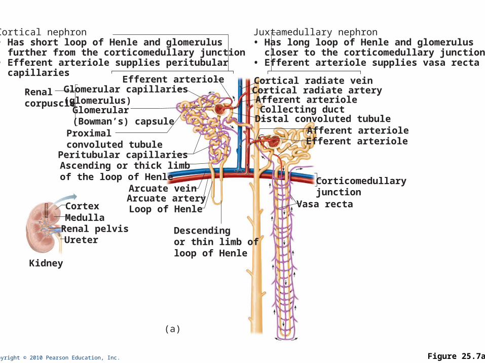
Nephrons
• Cortical nephrons—85% of nephrons; almost entirely in the cortex
• Juxtamedullary nephrons
• Long loops of Henle deeply invade the medulla
• Extensive thin segments
• Important in the production of concentrated urine
Copyright © 2010 Pearson Education, Inc. Figure 25.7a
Cortical nephron• Has short loop of Henle and glomerulus further from the corticomedullary junction• Efferent arteriole supplies peritubular capillaries
Juxtamedullary nephron• Has long loop of Henle and glomerulus closer to the corticomedullary junction• Efferent arteriole supplies vasa recta
Corticomedullaryjunction
UreterRenal pelvis
Kidney
CortexMedulla
(a)
Cortical radiate veinCortical radiate arteryAfferent arteriole
Afferent arteriole
Collecting ductDistal convoluted tubule
Efferent arteriole
Vasa rectaLoop of HenleArcuate arteryArcuate vein
Peritubular capillaries
Glomerular capillaries (glomerulus)Glomerular(Bowman’s) capsule
Renalcorpuscle
Ascending or thick limb of the loop of Henle
Descendingor thin limb of loop of Henle
Efferent arteriole
Proximalconvoluted tubule
Copyright © 2010 Pearson Education, Inc.
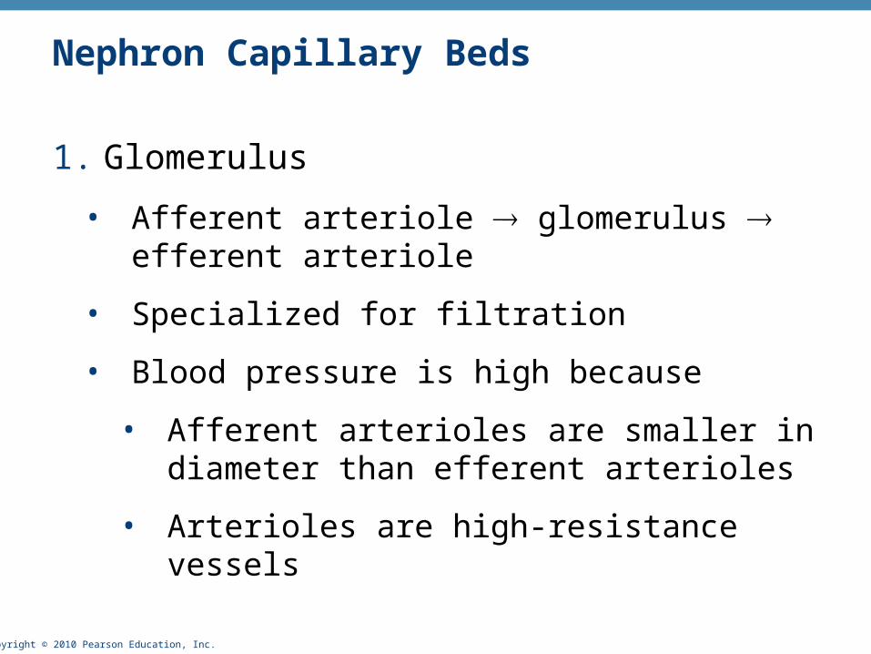
Nephron Capillary Beds
1. Glomerulus
• Afferent arteriole glomerulus efferent arteriole
• Specialized for filtration
• Blood pressure is high because
• Afferent arterioles are smaller in diameter than efferent arterioles
• Arterioles are high-resistance vessels

Copyright © 2010 Pearson Education, Inc.
Nephron Capillary Beds
2. Peritubular capillaries
• Low-pressure, porous capillaries adapted for absorption
• Arise from efferent arterioles
• Cling to adjacent renal tubules in cortex
• Empty into venules

Copyright © 2010 Pearson Education, Inc.
Nephron Capillary Beds
3. Vasa recta
• Long vessels parallel to long loops of Henle
• Arise from efferent arterioles of juxtamedullary nephrons
• Function information of concentrated urine

Copyright © 2010 Pearson Education, Inc. Figure 25.7a
Cortical nephron• Has short loop of Henle and glomerulus further from the corticomedullary junction• Efferent arteriole supplies peritubular capillaries
Juxtamedullary nephron• Has long loop of Henle and glomerulus closer to the corticomedullary junction• Efferent arteriole supplies vasa recta
Corticomedullaryjunction
UreterRenal pelvis
Kidney
CortexMedulla
(a)
Cortical radiate veinCortical radiate arteryAfferent arteriole
Afferent arteriole
Collecting ductDistal convoluted tubule
Efferent arteriole
Vasa rectaLoop of HenleArcuate arteryArcuate vein
Peritubular capillaries
Glomerular capillaries (glomerulus)Glomerular(Bowman’s) capsule
Renalcorpuscle
Ascending or thick limb of the loop of Henle
Descendingor thin limb of loop of Henle
Efferent arteriole
Proximalconvoluted tubule

Copyright © 2010 Pearson Education, Inc.
Vascular Resistance in Microcirculation
• High resistance in afferent and efferent arterioles
• Causes blood pressure to decline from ~95 mm Hg to ~8 mm Hg in kidneys

Copyright © 2010 Pearson Education, Inc.
Vascular Resistance in Microcirculation
• Resistance in afferent arterioles
• Protects glomeruli from fluctuations in systemic blood pressure
• Resistance in efferent arterioles
• Reinforces high glomerular pressure
• Reduces hydrostatic pressure in peritubular capillaries

Copyright © 2010 Pearson Education, Inc.
Juxtaglomerular Apparatus (JGA)
• One per nephron
• Important in regulation of filtrate formation and blood pressure
• Involves modified portions of the
• Distal portion of the ascending limb of the loop of Henle
• Afferent (sometimes efferent) arteriole

Copyright © 2010 Pearson Education, Inc.
Juxtaglomerular Apparatus (JGA)
• Granular cells (juxtaglomerular, or JG cells)
• Enlarged, smooth muscle cells of arteriole
• Secretory granules contain renin
• Act as mechanoreceptors that sense blood pressure

Copyright © 2010 Pearson Education, Inc.
Juxtaglomerular Apparatus (JGA)
• Macula densa
• Tall, closely packed cells of the ascending limb
• Act as chemoreceptors that sense NaCl content of filtrate
• Extraglomerular mesangial cells
• Interconnected with gap junctions
• May pass signals between macula densa and granular cells

Copyright © 2010 Pearson Education, Inc. Figure 25.8
GlomerulusGlomerular capsule
Afferent arteriole
Efferent arteriole
Red blood cell
Podocyte cell body (visceral layer)
Foot processesof podocytesParietal layer
of glomerularcapsule
Proximaltubule cell
Lumens of glomerularcapillaries
Endothelial cellof glomerularcapillary
Efferent arteriole
• Macula densa cells of the ascending limb of loop of Henle
• Granular cells
• Extraglomerular mesangial cells
Afferent arteriole
Capsularspace
Renal corpuscleJuxtaglomerularapparatus
Mesangial cellsbetween capillaries
Juxtaglomerularapparatus

Copyright © 2010 Pearson Education, Inc.
Filtration Membrane
• Porous membrane between the blood and the capsular space
• Consists of
1. Fenestrated endothelium of the glomerular capillaries
2. Visceral membrane of the glomerular capsule (podocytes with foot processes and filtration slits)
3. Gel-like basement membrane (fused basal laminae of the two other layers)

Copyright © 2010 Pearson Education, Inc. Figure 25.9a
Glomerular capillarycovered by podocyte-containing visceral layer of glomerular capsule
Glomerular capillaryendothelium (podocyte covering and basement membrane removed)
Proximal convolutedtubule
Parietal layerof glomerular capsule
Afferentarteriole
Glomerular capsular space
Fenestrations(pores)
Efferentarteriole
Podocytecell body
Foot processesof podocyte
Filtration slits
Cytoplasmic extensionsof podocytes
(a) Glomerular capillaries and the visceral layer of the glomerular capsule

Copyright © 2010 Pearson Education, Inc.
Filtration Membrane
• Allows passage of water and solutes smaller than most plasma proteins
• Fenestrations prevent filtration of blood cells
• Negatively charged basement membrane repels large anions such as plasma proteins
• Slit diaphragms also help to repel macromolecules

Copyright © 2010 Pearson Education, Inc.
Filtration Membrane
• Glomerular mesangial cells
• Engulf and degrade macromolecules
• Can contract to change the total surface area available for filtration

Copyright © 2010 Pearson Education, Inc. Figure 25.9c
(c) Three parts of the filtration membrane
Fenestration(pore)
Filtrate incapsularspace
Foot processesof podocyte
Filtration slit
Slit diaphragm
Capillary
Filtration membrane• Capillary endothelium• Basement membrane• Foot processes of podocyte of glomerular capsule
Plasma

Copyright © 2010 Pearson Education, Inc.
Kidney Physiology: Mechanisms of Urine Formation
• The kidneys filter the body’s entire plasma volume 60 times each day
• Filtrate
• Blood plasma minus proteins
• Urine
• <1% of total filtrate
• Contains metabolic wastes and unneeded substances

Copyright © 2010 Pearson Education, Inc.
Mechanisms of Urine Formation
1. Glomerular filtration
2. Tubular reabsorption
• Returns all glucose and amino acids, 99% of water, salt, and other components to the blood
3. Tubular secretion
• Reverse of reabsoprtion: selective addition to urine

Copyright © 2010 Pearson Education, Inc. Figure 25.10
Corticalradiateartery
Afferent arteriole
Glomerular capillaries
Efferent arteriole
Glomerular capsule
Rest of renal tubulecontaining filtrate
Peritubularcapillary
To cortical radiate vein
Urine
Glomerular filtration
Tubular reabsorption
Tubular secretion
Three majorrenal processes:

Copyright © 2010 Pearson Education, Inc.
Glomerular Filtration
• Passive mechanical process driven by hydrostatic pressure
• The glomerulus is a very efficient filter because
• Its filtration membrane is very permeable and it has a large surface area
• Glomerular blood pressure is higher (55 mm Hg) than other capillaries
• Molecules >5 nm are not filtered (e.g., plasma proteins) and function to maintain colloid osmotic pressure of the blood

Copyright © 2010 Pearson Education, Inc.
Net Filtration Pressure (NFP)
• The pressure responsible for filtrate formation (10 mm Hg)

Copyright © 2010 Pearson Education, Inc.
Net Filtration Pressure (NFP)
• Determined by
• Glomerular hydrostatic pressure (HPg) the chief force
• Two opposing forces:
• Colloid osmotic pressure of glomerular blood (OPg)
• Capsular hydrostatic pressure (HPc)
NFP = HPg – (OPg + HPc)

Copyright © 2010 Pearson Education, Inc. Figure 25.11
Glomerularcapsule
Afferentarteriole
10 mm Hg
Netfiltrationpressure
Glomerular (blood) hydrostatic pressure(HPg = 55 mm Hg)
Blood colloid osmotic pressure(Opg = 30 mm Hg)
Capsular hydrostatic pressure(HPc = 15 mm Hg)

Copyright © 2010 Pearson Education, Inc.
Glomerular Filtration Rate (GFR)
• Volume of filtrate formed per minute by the kidneys (120–125 ml/min)
• Governed by (and directly proportional to)
• Total surface area available for filtration
• Filtration membrane permeability
• NFP

Copyright © 2010 Pearson Education, Inc.
Regulation of Glomerular Filtration
• GFR is tightly controlled by two types of mechanisms
• Intrinsic controls (renal autoregulation)
• Act locally within the kidney
• Extrinsic controls
• Nervous and endocrine mechanisms that maintain blood pressure, but affect kidney function

Copyright © 2010 Pearson Education, Inc.
Intrinsic Controls
• Maintains a nearly constant GFR when MAP is in the range of 80–180 mm Hg
• Two types of renal autoregulation
• Myogenic mechanism (Chapter 19)
• Tubuloglomerular feedback mechanism, which senses changes in the juxtaglomerular apparatus

Copyright © 2010 Pearson Education, Inc.
Intrinsic Controls: Myogenic Mechanism
• BP constriction of afferent arterioles
• Helps maintain normal GFR
• Protects glomeruli from damaging high BP
• BP dilation of afferent arterioles
• Helps maintain normal GFR

Copyright © 2010 Pearson Education, Inc.
Intrinsic Controls: Tubuloglomerular Feedback Mechanism
• Flow-dependent mechanism directed by the macula densa cells
• If GFR increases, filtrate flow rate increases in the tubule
• Filtrate NaCl concentration will be high because of insufficient time for reabsorption

Copyright © 2010 Pearson Education, Inc.
Intrinsic Controls: Tubuloglomerular Feedback Mechanism
• Macula densa cells of the JGA respond to NaCl by releasing a vasoconstricting chemical that acts on the afferent arteriole GFR
• The opposite occurs if GFR decreases.

Copyright © 2010 Pearson Education, Inc.
Extrinsic Controls: Sympathetic Nervous System
• Under normal conditions at rest
• Renal blood vessels are dilated
• Renal autoregulation mechanisms prevail

Copyright © 2010 Pearson Education, Inc.
Extrinsic Controls: Sympathetic Nervous System
• Under extreme stress
• Norepinephrine is released by the sympathetic nervous system
• Epinephrine is released by the adrenal medulla
• Both cause constriction of afferent arterioles, inhibiting filtration and triggering the release of renin
Copyright © 2010 Pearson Education, Inc.
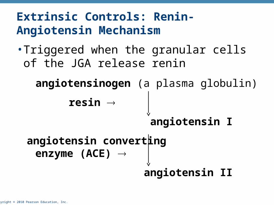
Extrinsic Controls: Renin-Angiotensin Mechanism
• Triggered when the granular cells of the JGA release renin
angiotensinogen (a plasma globulin)
resin
angiotensin I
angiotensin converting enzyme (ACE)
angiotensin II

Copyright © 2010 Pearson Education, Inc.
Effects of Angiotensin II
1. Constricts arteriolar smooth muscle, causing MAP to rise
2. Stimulates the reabsorption of Na+
• Acts directly on the renal tubules
• Triggers adrenal cortex to release aldosterone
3. Stimulates the hypothalamus to release ADH and activates the thirst center

Copyright © 2010 Pearson Education, Inc.
Effects of Angiotensin II
4. Constricts efferent arterioles, decreasing peritubular capillary hydrostatic pressure and increasing fluid reabsorption
5. Causes glomerular mesangial cells to contract, decreasing the surface area available for filtration

Copyright © 2010 Pearson Education, Inc.
Extrinsic Controls: Renin-Angiotensin Mechanism
• Triggers for renin release by granular cells
• Reduced stretch of granular cells (MAP below 80 mm Hg)
• Stimulation of the granular cells by activated macula densa cells
• Direct stimulation of granular cells via 1-adrenergic receptors by renal nerves

Copyright © 2010 Pearson Education, Inc. Figure 25.12
Stretch of smoothmuscle in walls of afferent arterioles
Blood pressure inafferent arterioles; GFR
Vasodilation ofafferent arterioles
GFR
Myogenic mechanismof autoregulation
Release of vasoactive chemical inhibited
Intrinsic mechanisms directly regulate GFR despitemoderate changes in blood pressure (between 80 and 180 mm Hg mean arterial pressure).
Extrinsic mechanisms indirectly regulate GFRby maintaining systemic blood pressure, whichdrives filtration in the kidneys.
Tubuloglomerularmechanism ofautoregulation
Hormonal (renin-angiotensin)mechanism Neural controls
SYSTEMIC BLOOD PRESSURE
GFR
Macula densa cellsof JG apparatus
of kidney
Filtrate flow andNaCl in ascending
limb of Henle’s loop
Targets
Granular cells ofjuxtaglomerular
apparatus of kidney
Angiotensinogen Angiotensin II
Adrenal cortex Systemic arterioles
(+) Renin
Release
Catalyzes cascaderesulting in conversion
(+)
(+)
(+)
Kidney tubules
Aldosterone
Releases
Targets
Vasoconstriction;peripheral resistance
Blood volume
Na+ reabsorption;water follows
Systemicblood pressure
(+)
(+) (–)
IncreaseDecrease
StimulatesInhibits
Baroreceptors inblood vessels of
systemic circulation
Sympatheticnervous system
(+)
(–)
Vasodilation ofafferent arterioles

Copyright © 2010 Pearson Education, Inc.
Other Factors Affecting GRF
• Prostaglandin E2
• Vasodilator that counteracts vasoconstriction by norepinephrine and angiotensin II
• Prevents renal damage when peripheral resistance is increased

Copyright © 2010 Pearson Education, Inc.
Other Factors Affecting GRF
• Intrarenal angiotensin II
• Reinforces the effects of hormonal angiotensin II
• Adenosine
• A vasoconstrictor of renal vasculature