Embed Size (px)
Citation preview
*digital red-free
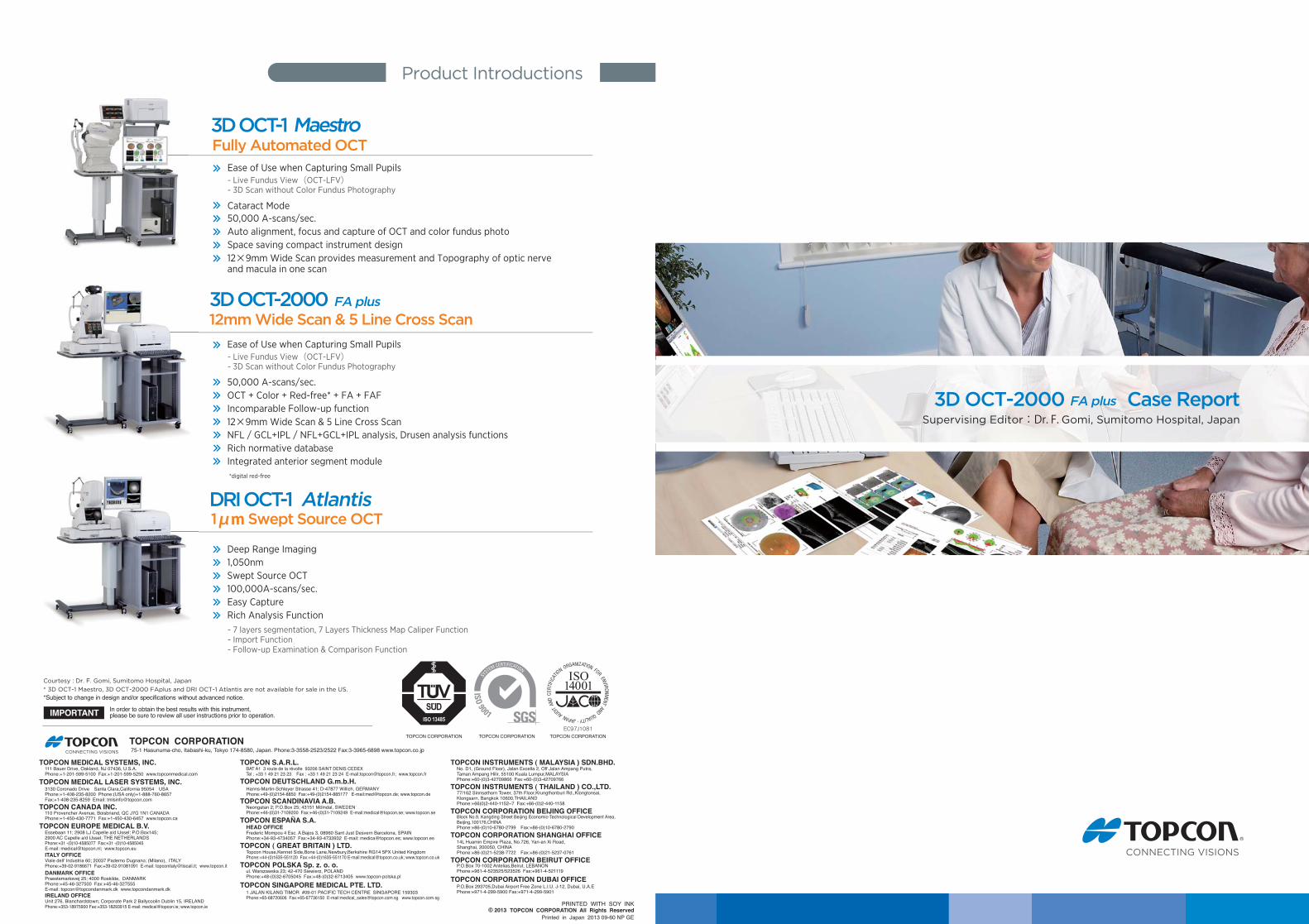
3D OCT-2000 Case Report
1μm Swept Source OCT
3D OCT-2000
DRI OCT-1 Atlantis
Product Introductions
Supervising Editor:Dr. F. Gomi, Sumitomo Hospital, Japan
Courtesy : Dr. F. Gomi, Sumitomo Hospital, Japan* 3D OCT-1 Maestro, 3D OCT-2000 FAplus and DRI OCT-1 Atlantis are not available for sale in the US.
12mm Wide Scan & 5 Line Cross Scan
Fully Automated OCT
Printed in Japan 2013 09-60 NP GE2013
111 Bauer Drive, Oakland, NJ 07436, U.S.A. Phone:+1-201-599-5100 Fax:+1-201-599-5250 www.topconmedical.com
75-1 Hasunuma-cho, Itabashi-ku, Tokyo 174-8580, Japan. Phone:3-3558-2523/2522 Fax:3-3965-6898 www.topcon.co.jp
TOPCON MEDICAL SYSTEMS, INC.
TOPCON MEDICAL LASER SYSTEMS, INC.
Essebaan 11; 2908 LJ Capelle a/d IJssel; P.O.Box145; 2900 AC Capelle a/d IJssel; THE NETHERLANDS Phone:+31 -(0)10-4585077 Fax:+31 -(0)10-4585045 E-mail: [email protected]; www.topcon.eu
TOPCON EUROPE MEDICAL B.V.
Hanns-Martin-Schleyer Strasse 41; D-47877 Willich, GERMANY Phone:+49-(0)2154-8850 Fax:+49-(0)2154-885177 E-mail:[email protected]; www.topcon.de
TOPCON DEUTSCHLAND G.m.b.H.
110 Provencher Avenue, Boisbriand, QC J7G 1N1 CANADA Phone:+1-450-430-7771 Fax:+1-450-430-6457 www.topcon.ca
TOPCON CANADA INC.
TOPCON ESPANA S.A.
Neongatan 2; P.O.Box 25; 43151 Mölndal, SWEDEN Phone:+46-(0)31-7109200 Fax:+46-(0)31-7109249 E-mail:[email protected]; www.topcon.se
TOPCON SCANDINAVIA A.B.
Topcon House,Kennet Side,Bone Lane,Newbury,Berkshire RG14 5PX United Kingdom Phone:+44-(0)1635-551120 Fax:+44-(0)1635-551170 E-mail:[email protected]; www.topcon.co.uk
TOPCON ( GREAT BRITAIN ) LTD.
ul. Warszawska 23; 42-470 Siewierz, POLAND Phone:+48-(0)32-6705045 Fax:+48-(0)32-6713405 www.topcon-polska.pl
TOPCON POLSKA Sp. z. o. o.
1 JALAN KILANG TIMOR #09-01 PACIFIC TECH CENTRE SINGAPORE 159303Phone:+65-68720606 Fax:+65-67736150 E-mail:[email protected] www.topcon.com.sg
TOPCON SINGAPORE MEDICAL PTE. LTD.
No. D1, (Ground Floor), Jalan Excella 2, Off Jalan Ampang Putra, Taman Ampang Hilir, 55100 Kuala Lumpur,MALAYSIA Phone:+60-(0)3-42709866 Fax:+60-(0)3-42709766
TOPCON INSTRUMENTS ( MALAYSIA ) SDN.BHD.
77/162 Sinnsathorn Tower, 37th Floor,Krungthonburi Rd.,Klongtonsai, Klongsarn, Bangkok 10600,THAILAND Phone:+66(0)2-440-1152~7 Fax:+66-(0)2-440-1158
TOPCON INSTRUMENTS ( THAILAND ) CO.,LTD.
Block No.9, Kangding Street Beijing Economic-Technological Development Area, Beijing,100176,CHINA Phone:+86-(0)10-6780-2799 Fax:+86-(0)10-6780-2790
TOPCON CORPORATION BEIJING OFFICE
14L Huamin Empire Plaza, No.726, Yan-an Xi Road, Shanghai, 200050, CHINA Phone:+86-(0)21-5238-7722 Fax:+86-(0)21-5237-0761
TOPCON CORPORATION SHANGHAI OFFICE
P.O.Box 70-1002 Antelias,Beirut, LEBANON Phone:+961-4-523525/523526 Fax:+961-4-521119
TOPCON CORPORATION BEIRUT OFFICE
P.O.Box 293705,Dubai Airport Free Zone L.I.U. J-12, Dubai, U.A.EPhone:+971-4-299-5900 Fax:+971-4-299-5901
TOPCON CORPORATION DUBAI OFFICE
BAT A1 3 route de la révolte 93206 SAINT DENIS CEDEXTel : +33 1 49 21 23 23 Fax : +33 1 49 21 23 24 E-mail:[email protected]; www.topcon.fr
TOPCON S.A.R.L.
ITALY OFFICEViale dell' Industria 60; 20037 Paderno Dugnano; (Milano), ITALY Phone:+39-02-9186671 Fax:+39-02-91081091 E-mail: [email protected]; www.topcon.it
DANMARK OFFICEPraestemarksvej 25; 4000 Roskilde, DANMARK Phone:+45-46-327500 Fax:+45-46-327555 E-mail: [email protected] www.topcondanmark.dkIRELAND OFFICEUnit 276, Blanchardstown; Corporate Park 2 Ballycoolin Dublin 15, IRELAND Phone:+353-18975900 Fax:+353-18293915 E-mail: [email protected]; www.topcon.ie
HEAD OFFICEFrederic Mompou 4 Esc. A Bajos 3, 08960 Sant Just Desvern Barcelona, SPAIN Phone:+34-93-4734057 Fax:+34-93-4733932 E-mail: [email protected]; www.topcon.es
3130 Coronado Drive Santa Clara,California 95054 USA Phone:+1-408-235-8200 Phone:(USA only)+1-888-760-8657 Fax:+1-408-235-8259 Email: [email protected]
3D OCT-1 Maestro
50,000 A-scans/sec.Auto alignment, focus and capture of OCT and color fundus photoSpace saving compact instrument design12×9mm Wide Scan provides measurement and Topography of optic nerve and macula in one scan
50,000 A-scans/sec.OCT + Color + Red-free* + FA + FAFIncomparable Follow-up function12×9mm Wide Scan & 5 Line Cross ScanNFL / GCL+IPL / NFL+GCL+IPL analysis, Drusen analysis functions Rich normative database Integrated anterior segment module
Deep Range Imaging1,050nmSwept Source OCT100,000A-scans/sec.Easy CaptureRich Analysis Function
Ease of Use when Capturing Small Pupils
Cataract Mode
- Live Fundus View(OCT-LFV)- 3D Scan without Color Fundus Photography
Ease of Use when Capturing Small Pupils- Live Fundus View(OCT-LFV)- 3D Scan without Color Fundus Photography
- 7 layers segmentation, 7 Layers Thickness Map Caliper Function- Import Function- Follow-up Examination & Comparison Function
FA plus
FA plus
Case Reports
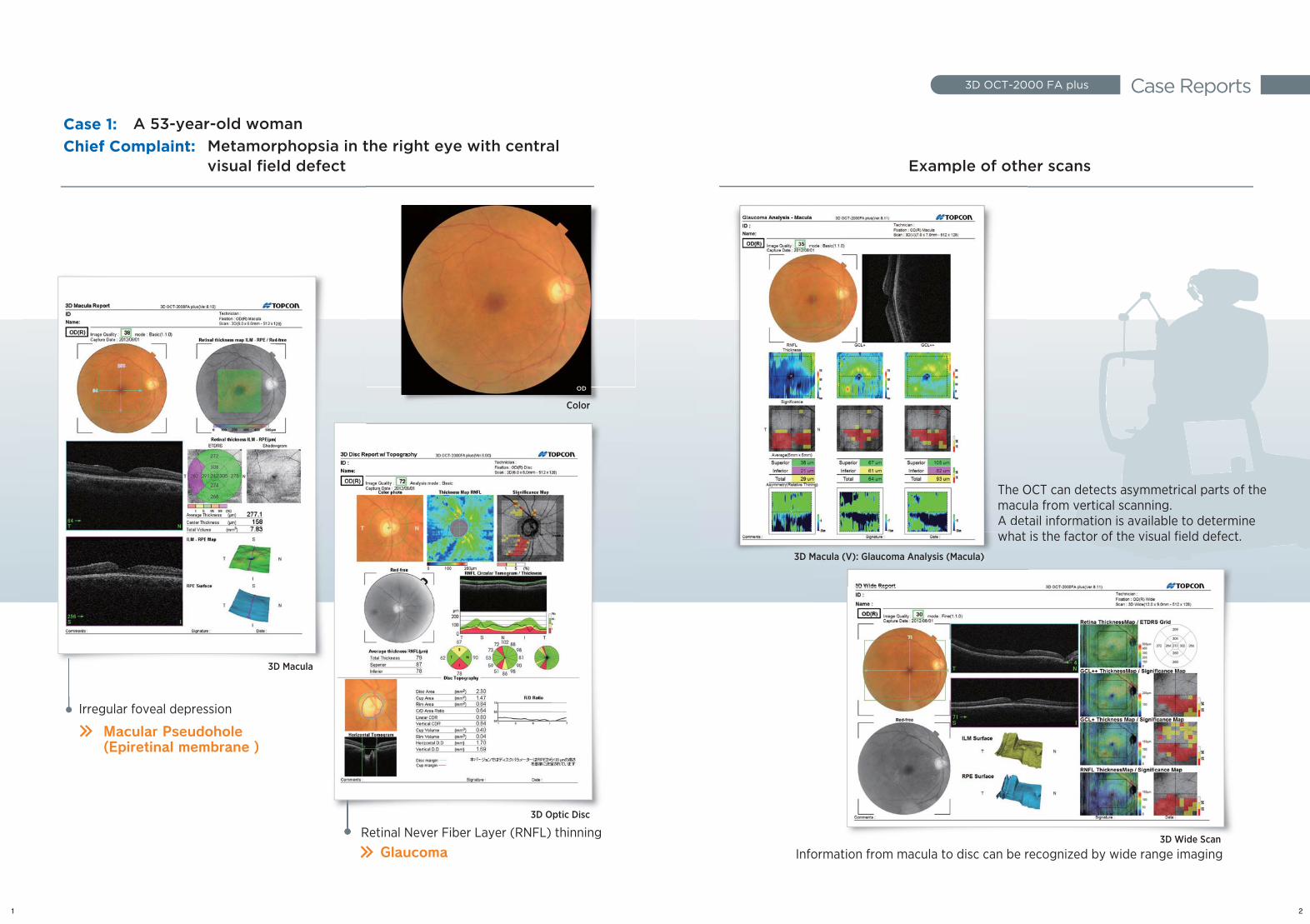
Case 1: Chief Complaint:
Irregular foveal depression
Retinal Never Fiber Layer (RNFL) thinning
Macular Pseudohole(Epiretinal membrane )
Glaucoma
3D Macula
3D Optic Disc
3D OCT-2000 FA plus
Retinal Never Fiber Layer (RNFL) thinning3D Optic Disc
3D Maccula
A 53-year-old womanMetamorphopsia in the right eye with central visual field defect Example of other scans
Color
3D Macula (V): Glaucoma Analysis (Macula)
The OCT can detects asymmetrical parts of the macula from vertical scanning. A detail information is available to determine what is the factor of the visual field defect.
3D Wide Scan
Information from macula to disc can be recognized by wide range imaging
Example of oth
3D MMacula (V): Glaucoma Analysis (Macula)
The OmacuA det
hwhat
1 2
OD
Case Reports3D OCT-2000 FA plus
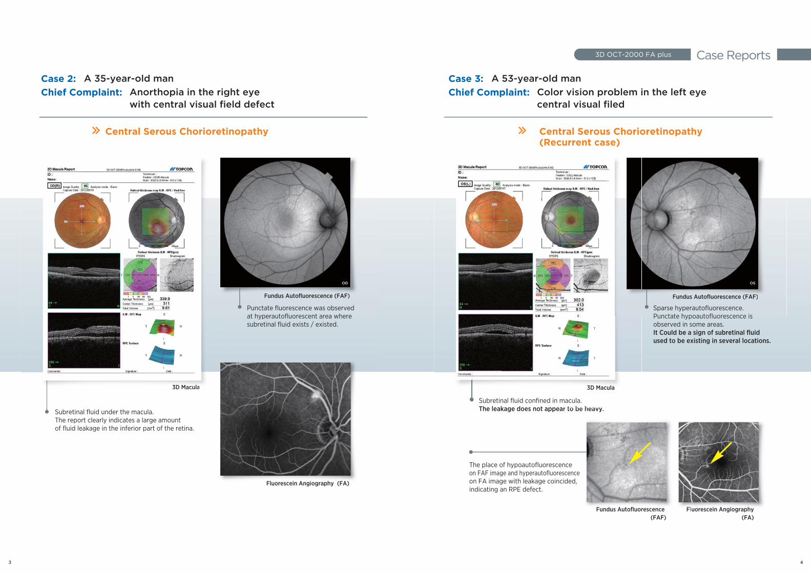
Central Serous Chorioretinopathy Central Serous Chorioretinopathy (Recurrent case)
Subretinal fluid under the macula.The report clearly indicates a large amount of fluid leakage in the inferior part of the retina.
Punctate fluorescence was observed at hyperautofluorescent area where subretinal fluid exists / existed.
Sparse hyperautofluorescence.Punctate hypoautofluorescence is observed in some areas.It Could be a sign of subretinal fluid used to be existing in several locations.
Subretinal fluid confined in macula.The leakage does not appear to be heavy.
The place of hypoautofluorescence on FAF image and hyperautofluorescence on FA image with leakage coincided, indicating an RPE defect.
3D Macula 3D Macula
Fundus Autofluorescence (FAF) Fundus Autofluorescence (FAF)
Fundus Autofluorescence (FAF)
Fluorescein Angiography (FA)
Fluorescein Angiography (FA)
Fundus Autofluorescence (FAF)
p y
Punctate fluorescence was observed at hyperautofluorescent area where
Fundus Autofluorescence (FAF)
Fluorescein Angiography (FA)
p y
Sparse hyperautofluorescence.Punctate hypoautofluorescence is
Fundus Autofluorescence (F(FAFAF))
r to be heavy.
ncescence ided,
Fundus Autofluorescen(F
uorescein Angiogra hphy (FA)
FlFlunce AF)
3D Maculaa
(Recurrent cas
Subretinal fluid confined in macula.The leakage does not appear
3D Macula
se)
macula.r to be heavy
Case 2: Chief Complaint:
A 35-year-old manAnorthopia in the right eye with central visual field defect
Case 3: Chief Complaint:
A 53-year-old manColor vision problem in the left eye central visual filed
OD OS
3 4
Case Reports3D OCT-2000 FA plus
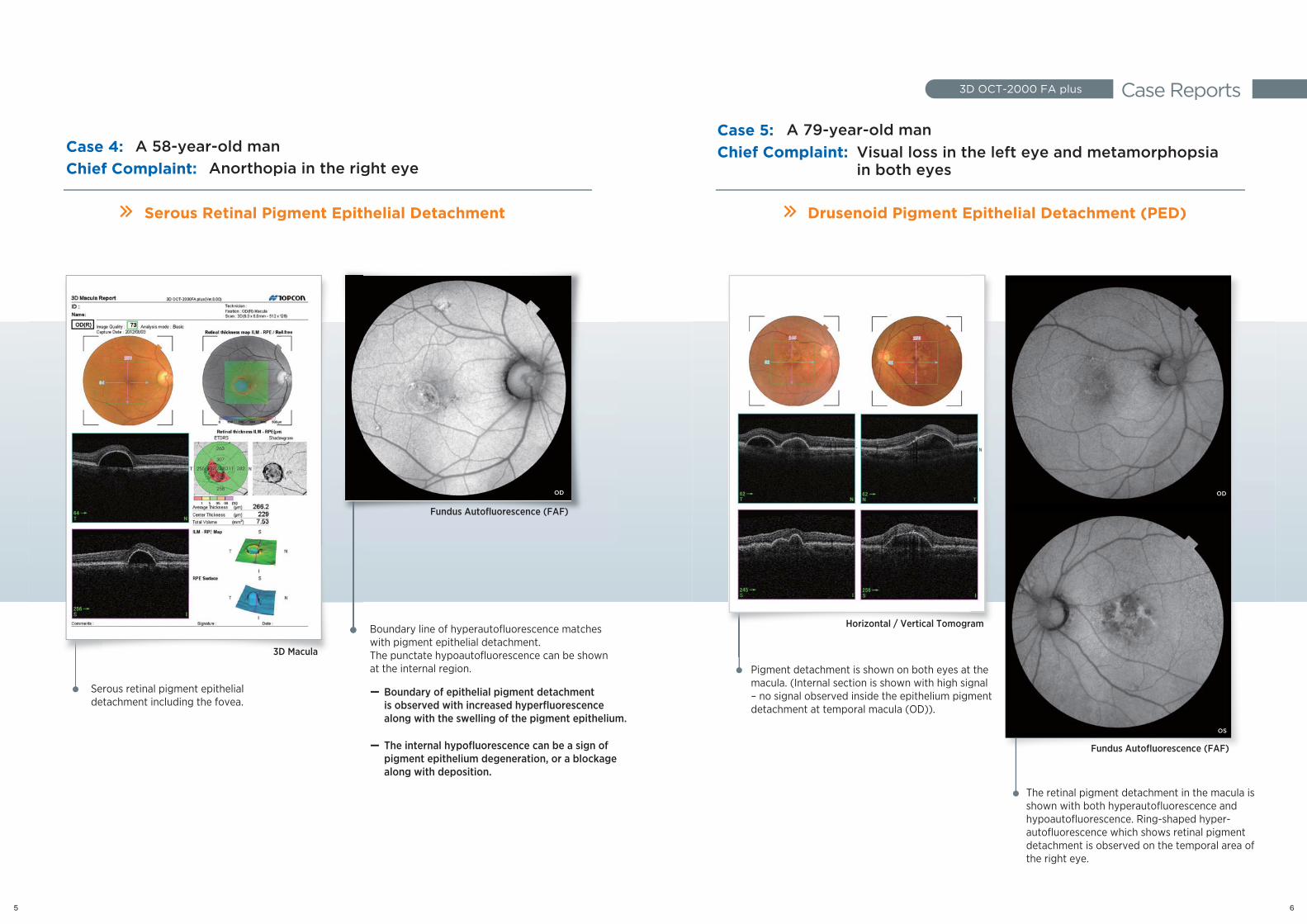
Serous retinal pigment epithelial detachment including the fovea.
Pigment detachment is shown on both eyes at the macula. (Internal section is shown with high signal – no signal observed inside the epithelium pigment detachment at temporal macula (OD)).
The retinal pigment detachment in the macula is shown with both hyperautofluorescence and hypoautofluorescence. Ring-shaped hyper-autofluorescence which shows retinal pigment detachment is observed on the temporal area of the right eye.
Boundary of epithelial pigment detachment is observed with increased hyperfluorescence along with the swelling of the pigment epithelium.
Boundary line of hyperautofluorescence matches with pigment epithelial detachment.The punctate hypoautofluorescence can be shown at the internal region.
OD
右眼
左眼
S ti l i t ith li l
thegnal ment
右眼
左眼
Serous Retinal Pigment Epithelial Detachment Drusenoid Pigment Epithelial Detachment (PED)
Case 4: Chief Complaint:
A 58-year-old manCase 5: Chief Complaint:
A 79-year-old man
Anorthopia in the right eye
3D Macula
Horizontal / Vertical Tomogram
Fundus Autofluorescence (FAF)
Fundus Autofluorescence (FAF)
Visual loss in the left eye and metamorphopsia in both eyes
OD
OS
5 6
The internal hypofluorescence can be a sign of pigment epithelium degeneration, or a blockage along with deposition.
(Reticular Pseudo Drusen)
Case Reports3D OCT-2000 FA plus
Horizontal Line Scan
Fundus Autofluorescence (FAF)
3D Macula5 Line Raster Scan
Fundus Autofluorescence (FAF)Fundus Autofluorescence (FAF)
(Re
Fundus Autofluorescence (FAF)
3D Macula Horizontal Line Scan5 Line Raster Scan
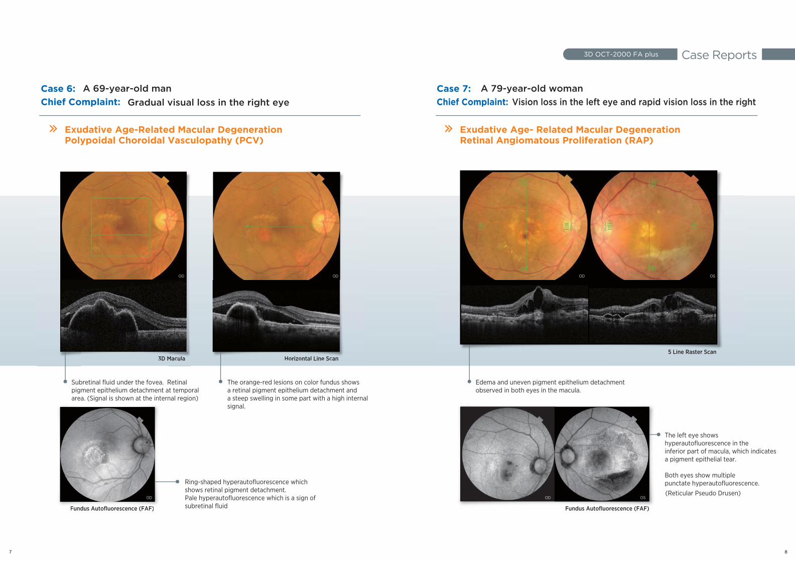
Subretinal fluid under the fovea. Retinal pigment epithelium detachment at temporal area. (Signal is shown at the internal region)
The orange-red lesions on color fundus shows a retinal pigment epithelium detachment and a steep swelling in some part with a high internal signal.
The left eye shows hyperautofluorescence in the inferior part of macula, which indicatesa pigment epithelial tear.
Both eyes show multiple punctate hyperautofluorescence.
Edema and uneven pigment epithelium detachment observed in both eyes in the macula.
Ring-shaped hyperautofluorescence which shows retinal pigment detachment.Pale hyperautofluorescence which is a sign of subretinal fluid
Exudative Age-Related Macular DegenerationPolypoidal Choroidal Vasculopathy (PCV)
Exudative Age- Related Macular DegenerationRetinal Angiomatous Proliferation (RAP)
OD
OD
OD OD
OD OS
OS
Case 6: Chief Complaint:
A 69-year-old manGradual visual loss in the right eye
Case 7: Chief Complaint: Vision loss in the left eye and rapid vision loss in the right
A 79-year-old woman
7 8
Case Reports3D OCT-2000 FA plus
case1
case2
Fundus Autofluorescence (FAF)
Horizontal Line Scan
Fundus Autofluorescence (FAF)
Color
Color
Fundus Autofluorescence (FAF)
Fundus Autofluorescence (FAF)
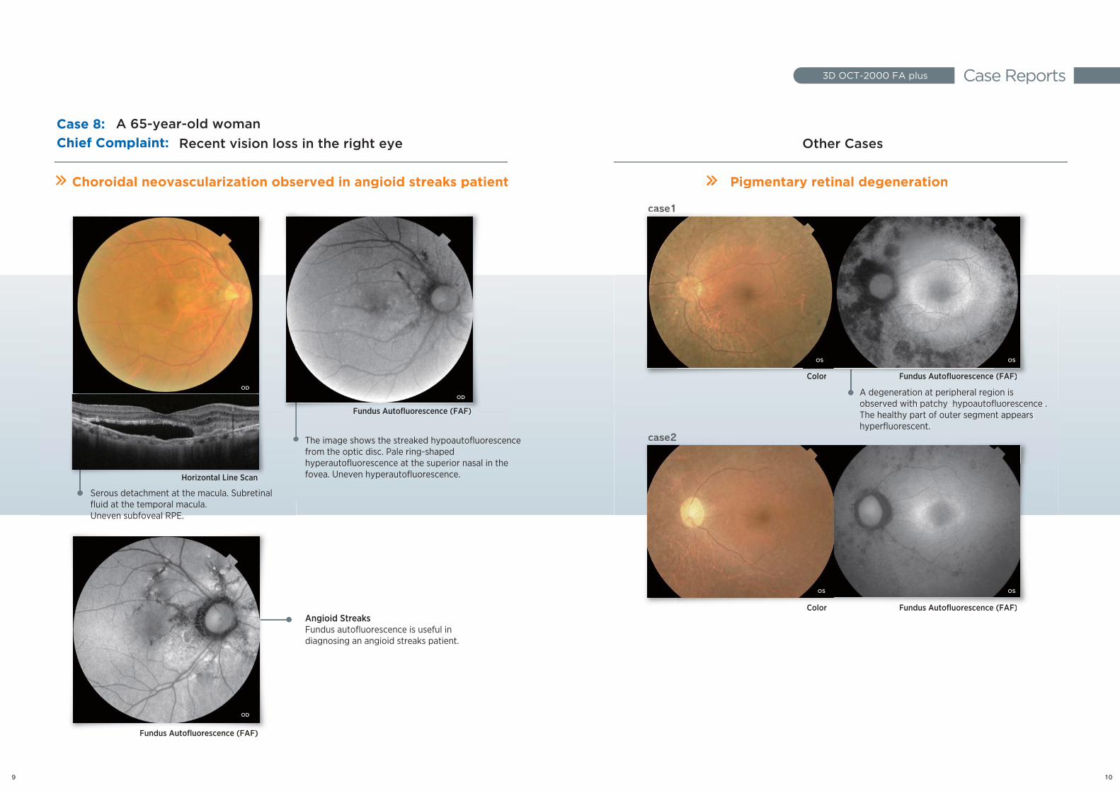
Choroidal neovascularization observed in angioid streaks patient Pigmentary retinal degeneration
Angioid StreaksFundus autofluorescence is useful in diagnosing an angioid streaks patient.
A degeneration at peripheral region is observed with patchy hypoautofluorescence .The healthy part of outer segment appears hyperfluorescent.
Fundus Autofluorescence (FAF)
on observed in angioid streaks patient
Fundus Autofluorescence (FAF)
case1
Color Fundus Autofluorescence (FAF)
Pigmentary retinal degeneration
A degeneration at peripheral region is observed with patchy hypoautofluorescence
Horizontal Line Scan
Choroidal neovascularizatioon obs
case2
Color Fundus Autofluorescence (FAF)
The healthy part of outer segment appears hyperfluorescent.
Case 8: Chief Complaint:
A 65-year-old womanOther CasesRecent vision loss in the right eye
Serous detachment at the macula. Subretinal fluid at the temporal macula. Uneven subfoveal RPE.
The image shows the streaked hypoautofluorescence from the optic disc. Pale ring-shaped hyperautofluorescence at the superior nasal in the fovea. Uneven hyperautofluorescence.
OD
OD
OD
OSOS
OSOS
9 10


![Topcon 3D OCTTopcon 3D OCT-2000 2000 · (Microsoft PowerPoint - fdfsd [modalit\340 compatibilit\340]) Author: ivana Created Date: 1/12/2010 10:06:33 AM](https://img.pdfslide.tips/doc/110x75/60543825ba4f024c1533aa8a/topcon-3d-octtopcon-3d-oct-2000-2000-microsoft-powerpoint-fdfsd-modalit340.jpg)