Embed Size (px)
Citation preview

�4 Integrin and Laminin 5 Are Aberrantly Expressedin Polycystic Kidney Disease
Role in Increased Cell Adhesion and Migration
Dominique Joly,* Viviane Morel,*Aurelie Hummel,* Antonella Ruello,*Patrick Nusbaum,* Natacha Patey,†Laure-Helene Noel,* Patricia Rousselle,‡ andBertrand Knebelmann*§
From INSERM 507,* the Departments of Nephrology § and
Pathology,† Necker Hospital, and Universite Paris V, Paris; and
Institut de Biologie et Chimie des Proteines,‡ CNRS UMR 5086,
Universite Lyon, Lyon, France
Extracellular matrix alterations have been suggestedto be part of the early events occurring in AutosomalDominant Polycystic Kidney Disease (ADPKD), a dis-ease characterized by formation of renal cysts andprogressive renal failure. Here we report that cDNAarray analysis identified �4 integrin aberrant expres-sion in ADPKD cells. Furthermore, laminin 5 (Ln-5),the main �6�4 integrin ligand, was also found to beabnormally expressed in ADPKD. Studies performedwith ADPKD cyst-lining epithelial cells (CC) by com-parison with normal tubular cells indicate that inte-grin �6�4-Ln-5 interactions are involved in cellularevents of potential importance for cystogenesis: 1)laminin 5 is a preferential adhesion substrate for CC,mainly through �6�4 interaction, 2) CC increasedhaptotactic and chemotactic motility depends on thepresence of Ln-5 and requires integrin �3�1 coopera-tion, and 3) CC haptotactic or chemotactic migrationis specifically increased by mAb-mediated �4 integrinligation, through an �3�1 integrin-dependent and in-dependent pathway, respectively. These results high-light the role of Ln-5 and �6�4 integrin in adhesiveand motility properties of cyst-lining epithelial cells,and further suggest that integrins and extracellularmatrix modifications may be of general relevance tokidney epithelial cell cyst formation. (Am J Pathol2003, 163:1791–1800)
Autosomal dominant polycystic kidney disease (ADPKD),the most frequently inherited kidney disease, is due togermline and somatic PKD1 or PKD2 gene mutations.The main clinical features is the progressive appearanceand growth of multiple renal cysts, resulting in frequentend-stage renal failure. The proteins encoded by PKD1
and PKD2, polycystins 1 (Pc-1) and 2 (Pc-2), are longtransmembrane proteins located in the primary cilia thatinteract and function together as a non-selective cationchannel.1 The long extracellular domain of Pc-1 may beinvolved in cell-cell and cell-matrix interactions. Signaltransduction through Pc-1 (Wnt signaling, AP1, and Gproteins activation) and Pc-2 (p38 and AP1 activation)has been reported, and a role for polycystins in celldifferentiation and proliferation has been suggested (re-viewed in2,3).
In ADPKD renal cysts, somatic mutations of the wild-type allele of PKD1 and PKD2 and subsequent loss of thefunctional polycystin complex presumably trigger a cas-cade of signaling and gene expression events.3 To fur-ther understand how the Pc-1/Pc-2 disruption leads tocystogenesis, we performed cDNA array experiments toidentify abnormally expressed genes in ADPKD. Amonggenes differentially expressed between cells derivedfrom control and ADPKD kidneys, we decided to focus onthose involved in cell proliferation, cell adhesion, and cellmigration, because these events presumably play a keyrole in cystogenesis.
We were particularly interested by the detection of astrong overexpression of �4 integrin in ADPKD cyst-de-rived cells. �4 subunit associates with �6 to form �6�4
integrin, which is primarily expressed at the basal surfaceof most epithelia, and may participate in both cell adhe-sion and migration. Integrin �6�4 is a key component ofhemidesmosomes4 that link the keratin cytoskeleton withlaminins in the basement membrane.4 Integrin �4 activa-tion can also trigger intracellular signaling through itsparticularly long cytoplasmic tail. Epidermal growth factor(EGF) stimulation leads to �4 cytoplasmic tail phosphor-ylation, disruption of hemidesmosomes, and mobilizationof �4 to actin protrusions.5 Integrin �4 interaction withlaminin 5 (Ln-5) may also trigger intracellular signal trans-duction.6 Epithelial cells adhere to Ln-5 via two adhesivestructures, focal adhesions and hemidesmosomes,
Supported by Institute National pour la Sante et la Recherche Medicale,Assistance Publique-Hopitaux de Paris, Association pour l’Utilisation duRein Artificiel, Association pour l’Information et la Recherche dans lesmaladies Genetiques renales, and Amgen.
Accepted for publication July 15, 2003.
Address reprint requests to Dominique Joly, 149 rue de Sevres, IN-SERM U 507, hopital Necker 75015 Paris. E-mail: [email protected].
American Journal of Pathology, Vol. 163, No. 5, November 2003
Copyright © American Society for Investigative Pathology
1791

through binding to �3�1 or �6�1 and �6�4 integrins, re-spectively.7 On processing, Ln-5 may also trigger inte-grin-dependent migration and integrin-independent cellscattering.8,9 Ln-5 ligation to �6�4 integrin receptor, forinstance, may activate PI3K signaling and subsequentstimulation of other integrins implicated in cell motility,such as �3�1.
10
In this study, we identify �6�4 integrin and Ln-5 aber-rant expression in ADPKD, and characterize the func-tional consequences of �6�4 integrin-Ln-5 interactions onADPKD cell adhesion and migration. We report that 1)Ln-5 enhances �6�4 integrin-mediated adhesion of CC,2) Ln-5 induces �6�4- and �3�1-dependent haptotacticmigration of CC, and 3) under EGF stimulation, �4 integrinligation promotes migration independently of �3�1 inte-grin. These results point to distinct properties of �6�4 inLn-5 expressing epithelia, that may contribute in vivo torenal cyst enlargement in ADPKD.
Experimental Procedures
Kidney Specimens
We had the unique opportunity to benefit from an ADPKDkidney harvested before the onset of renal failure. This32-year-old woman was pronounced brain-dead after aruptured cerebral aneurysm and underwent organ har-vesting. However, both kidneys appeared enlarged andmulticystic and were refused for cadaveric donation, thusbecoming available for research purposes. The serumcreatinine at the time of nephrectomy was subnormal (15mg/dl). The clinical diagnosis of ADPKD was retrospec-tively confirmed, based on 1) bilateral enlarged polycys-tic kidneys with typical histological features, 2) intracere-bral aneurysm, and 3) a family history of autosomaldominant polycystic kidney disease. We also handlednine other polycystic kidneys, retrieved from ADPKD pa-tients with end-stage renal failure before renal transplan-tation. As control, we handled normal portions of kidneyscontaining localized adenocarcinoma obtained from fourage-matched patients.
Primary Cell Cultures of Cystic and Non-CysticEpithelium
ADPKD and control kidneys were used to rise primarycultures of cystic renal tubular epithelial cells (CC) andnon-cystic renal tubular epithelial cells (NC). The detailedmethods have been published.11 Each primary culturewas derived from a pool of all cysts dissected within akidney. Cells were grown in Dulbecco’s modified Eagle’smedium (DMEM) (Invitrogen, Carlsbad, CA) containing1% fetal bovine serum, 5 �g/ml insulin, 10 �g/ml trans-ferrin, 5 ng/ml sodium selenite, 6.5 ng/ml Triiodothyronin,10 ng/ml EGF, 500 ng/ml hydrocortisone, and 1% HEPES(Invitrogen). Reagents were purchased from Sigma (St.Louis, MO) unless indicated. For functional studies (ad-hesion, migration; see below) cells were cultivated 24hours in DMEM, 1% HEPES (referred as “starvation”),harvested with cell dissociation buffer (Invitrogen),washed and resuspended in DMEM, 1% HEPES, even-
tually supplemented with various antibodies or EGF. Weelected to exclusively use these primary cultures be-tween passages 2 and 4.
cDNA Arrays
RNA was isolated using RNeasy miniKit (QIAGEN,Hilden, Germany) according to manufacturer. Atlas Hu-man cDNA array-1 (BD Biosciences Clontech, Palo Alto,CA) was used for expression analysis per manufacturer’sinstructions. Briefly, 5 �g of DNAse-treated total RNA wasconverted to 32P-labeled first-strand cDNA and purifiedusing column chromatography to remove unincorporatednucleotides. Labeled cDNA was then used to hybridizeAtlas cDNA nylon array in ExpressHyb mix (BD Bio-sciences Clontech) at 68°C overnight. The membranewas then washed twice in 2X SSC at 65°C for 30 minuteseach and exposed to autoradiography for 24 hours. Theorientation grid supplied by the manufacturer was thenused to identify the positions of the hybridization signals.
Antibodies, Extracellular Matrix Proteins, andReagents
Monoclonal antibodies used in this study were: anti-inte-grin �2 (clone P1E6), anti-integrin �3 (clone P1B5), anti-integrin �6 (clone NKI-GoH3), anti-integrin �4 (clone3E1), and anti-integrin �1 (clone 6S6) from Chemicon(Temecula, CA); anti-laminin 5: clone BM-165 against the�3 chain (Dr. Patricia Rousselle, Lyon, France) and cloneD4B5 against the �2 chain (Chemicon); anti-cytokeratin(clone AE1-AE3) from DAKO (Glostrup, Denmark). Poly-clonal antibody anti-�4 (H-101) was from Santa Cruz Bio-technology (Santa Cruz, CA). Human laminin 5 (Ln-5;�3�3�2) was purified from SCC25 cells;12 collagen I andcollagen IV were purchased from Sigma.
Immunohistochemistry
Cryostat sections of tissues were fixed with acetone andstored at �20°C. Before use, specimens were fixed againin chloroform, blocked with 5% goat serum and 1% bo-vine serum albumin (BSA) in PBS, incubated with theprimary antibody for 1 hour, and treated with LSAB2-system horseradish peroxidase (HRP) (DAKO) accordingto the manufacturer’s recommendations. Peroxidasestaining was examined with an Ortho LEICA microscopecoupled to a CDD camera (Olympus). Cells grown oncoverslips (Costar, Cambridge, MA) were fixed with ac-etone, incubated with 3% hydrogen peroxide (DAKO),incubated 1 hour with primary antibody or negative con-trol reagent, and subsequently treated as above.
Immunofluorescence
Monolayers were fixed with 4% paraformaldehyde inphosphate-buffered saline (PBS) for 10 minutes at roomtemperature, treated with 0.25% Triton X-100 for 10 min-utes, and saturated for 30 minutes with 2% BSA in PBS.Cell monolayers were successively (after intermediatewashes in PBS) incubated for 1 hour with a monoclonal
1792 Joly et alAJP November 2003, Vol. 163, No. 5

antibody to Ln-5 �2 chain (D4B5, 1:100), and a fluores-cein isothiocyanate goat anti-mouse antibody (JacksonImmunoresearch Laboratories, West Grove, PA). Thecoverslips were mounted with Aquapolymount antifadingsolution (AGAR, Stansted, Essex, UK) onto glass slidesand the slides were observed under a Leica fluorescencemicroscope.
RT-PCR Analysis
Aliquots of total RNA extracted from cells (1 �g, RNeasymini Kit; QIAGEN) or tissues (20 �g, Trizol reagent; In-vitrogen) were used as a template for cDNA synthesis.Reverse transcription was carried with 200 ng/�l randomhexamers (C1181; Promega, Madison, WI) and M-MLVreverse transcriptase (Invitrogen). Polymerase chain re-action (PCR) was performed using 50 ng (cells) or 250 ng(tissues) of cDNA samples, 2U TaqDNA polymerase(M1665; Promega) in 10 mmol/L (pH 9) TrisHCl, 50mmol/L KCl, 1.5 mmol/L MgCl2, 200 �mol/L of each de-oxynucleotide, and 500 nmol/L of each primer pair. Prim-ers and PCR conditions are listed in Table 1. PCR prod-ucts were analyzed by electrophoresis using 1.5%agarose gels and photographed as ethidium bromidefluorescent bands. GAPDH was used as control, allowingquantitative estimation of gene expression by computeddensitometry (NIH Imager 4.1 software).
Western Blotting
Cells were harvested with cell dissociation buffer (Invitro-gen-BRL), lysed and centrifuged; proteins (20 �g) wereboiled under denaturing condition, separated on 7.5%SDS-PAGE, and blotted to PVDF membranes (MilliporeCorp., Bedford, MA). Blots were saturated 1 hour at 37°Cwith TBST (20 mmol/L/L Tris-HCl, pH 8, 137 mmol/L/LNaCl, 0.1% Tween-20) containing 2% BSA and incubated1 hour at room temperature with 1:200 anti-�4 antibodysc-9090 (Santa Cruz); �4 integrin was visualized using anenhanced chemiluminescence system (ECL plus; Amer-sham Biosciences Inc., Buckinghamshire, UK) and Bi-omax films (Kodak). Membranes were rehybridized withanti-ERK1/2 (Sigma) to ensure equal loading.
Cell Adhesion and Motility
Adhesion
Adhesion assays were performed in 96-well plates.Wells were coated 3 hours at 37°C with 10 �g/ml Ln-5,
collagen I or collagen IV, washed in PBS, and blockedwith 1% BSA (Sigma) at 37° for 2 hours. 1 � 104 cellswere then plated and allowed to attach for 1 hour. Non-adherent cells were washed away. Adherent cells werefixed with absolute ethanol, colored (crystal violet 0.2% in2% ethanol), visualized using a �20 objective, andcounted under a light microscope with an eyepiece grid.At least three optic fields in triplicate wells were countedfor each condition.
Modified Boyden Chamber Motility Assays
Haptotactic migration (ie, migration toward ECM com-ponents) was studied using modified Boyden chambers(Transwell 24-well plates, 8.0-�m pore size; Costar). Thelower chambers of the plate were loaded with DMEM andoverlaid by a 8.0 �m polycarbonate filter eventuallycoated with Ln-5, collagen I, or collagen IV (as above).CC or NC starved cells (2 � 104 in 100 �l of DMEM) werethen added to the upper chamber of the filters and al-lowed to migrate through the filters for 12 hours at 37°C in5% CO2. On completion filters were removed and fixedwith absolute ethanol. Cells that had not migrated wereremoved from the top of the filter with a cotton swab; cellsthat had migrated (remaining on the bottom side of thefilter) were stained (crystal violet 0.2% in 2% ethanol) andnumerated as above (cf adhesion assays).
The same modified Boyden chambers were used toquantify EGF-stimulated migration. For these assays,starved CC or NC cells (4 � 104 in 100 �l) were stimu-lated by EGF at the time of cell plating and allowed tomigrate through uncoated filters for 18 hours. Cells thatmigrated through the filters were identified and countedas outlined above.
Wound Healing
When reaching 90% to 100% confluence, NC or CCcells grown on plastic coverslips (Costar) were starvedand the following day, a straight and uniform scratchwas made on the monolayer with a 200-�l plastic pi-pette tip. Monolayers were washed gently and incu-bated 20 hours at 37°C in DMEM � EGF. For eachexperimental condition, the area of the wound wasassessed at various time points in 10 fields initiallymarked for reference, using a 100-unit eyepiece opticgrid under a light microscope. The difference in num-ber of optic units filled by cells before and after woundrepair is expressed as mean � SD.
Table 1. Primers and PCR Conditions
Gene Sequence of F and R primersPCR product
(bp) Tam (°C)Number of
cycles
�4 Integrin F 5�-CCG TGT GGA TAA GGA CTG CG-3� 764bp 60 35R 5�-TGT AGG TGC CCG TGG TGT CC-3�
Laminin 5 F 5�-AAT GGG AAG TCC AGG CAG TGT ATC-3� 407bp 58 33(�2 chain) R 5�-ACA GCG TTC TCC AGT AAC AGC TG-3�GAPDH F 5�-TCT CTG CCC CCT CTG CTG AT-3� 301bp 58 23
R 5�-ATG ACC TTG CCC ACA GCC TT-3�
F, forward; R, reverse.
�4 Integrin and Laminin 5 in ADPKD 1793AJP November 2003, Vol. 163, No. 5

Inhibition Experiments
For adhesion and Transwell migration inhibition as-says, cells were first pre-incubated for 30� at 4°C withanti-integrin antibodies. For wound healing inhibition as-says, antibodies (and EGF) were added to the mediumlater, at the time of stimulation. The concentrations of theantibodies used for inhibition experiments (10 �g/ml)were equal or up to two times the concentration givingthe maximal inhibition in preliminary dose-responseexperiments.
Statistical Analyses
Experiments were performed in triplicate at least threetimes each, with primary cultures of different origin. Dataare expressed as means � SD. Statistical significancewas determined using Student’s t-test. P � 0.05 wasconsidered to be significant.
Results
Cystic Epithelial Cells of ADPKD PatientsAberrantly Express �4 Integrin and Ln-5
mRNA isolated from a set of ADPKD cyst-derived cells(CC, n � 3) and control kidney tubular cells (NC, n � 3)were used to perform cDNA arrays. Comparison of hy-bridization signals by optic density scanning suggestedthat the gene encoding �4 integrin was overexpressed inADPKD cyst-derived cells (not shown). These resultswere confirmed by semiquantitative RT-PCR: the �4/GAPDH ratio was consistently higher (two to four-fold)when tested in 10 CC when compared to four NC primarycultures (Figure 1A). To confirm that this difference holdstrue at the protein level, we performed Western blot anal-ysis of cell lysates using H-101 polyclonal antibody. In-deed, a much stronger �4 protein expression was de-tected in CC than in NC (Figure 1B). To test whether �4
integrin overexpression was specific, or a more general
Figure 1. �4 integrin overexpression in primary cultures derived from ADPKD kidneys. A: RT-PCR products for �4 and GAPDH genes. Each bar represents the�4/GAPDH ratio for primary tubular epithelial cells derived from normal (white bars, NC 1 to 4) or ADPKD kidneys (black bars, 1 to 10). B: Immunoblot analysisof �4 integrin in control (NC) and cystic cells (CC); anti-ERK1/2 was used as a control for loading. C: Immunocytochemical peroxidase staining on subconfluentCC and NC with the following mAbs: anti-�4 3E-1; anti-�3 P1B5; anti-�6 GoH3; anti-�1 6S6; anti-cytokeratin positivity assesses the epithelial nature of the cells.Negative isotypic controls are not shown.
1794 Joly et alAJP November 2003, Vol. 163, No. 5

feature of integrin expression in ADPKD, we performedimmunocytochemical experiments. �4 integrin was unde-tectable in NC and positive in CC with two different an-ti-�4 antibodies, 3E1 (Figure 1C) and ASC-9 (not shown);by contrast, �3, �6, and �1 integrins were strongly ex-pressed in both types of cells (Figure 1C).
We next wanted to test whether �4 overexpression wasalso a feature of in vivo kidneys. With a set of primersdesigned to amplify a band of 764 bp, which corre-sponds to all four known �4 isoforms, RT-PCR amplifica-tion showed that the �4/GAPDH transcript ratio was muchhigher (three to fourfold) in two ADPKD kidneys com-pared to three control kidneys (Figure 2A). Immunohisto-chemistry with the 3E1 antibody, directed against theextracellular domain of �4 integrin, showed no detectable�4 integrin expression in normal adult kidney [Figure2B(a)]. By contrast, in five different adult ADPKD patients,70% to 80% of the kidney cyst-lining epithelia exhibited astrong �4 basolateral expression [Figure 2B(b)]. Of note,aberrant �4 expression was also observed on undilatedtubules in a patient with APDKD and normal renal func-tion [Figure 2B(c)], suggesting that it is an early event.Since ADPKD epithelial cells have been reported to ex-press fetal markers, we also studied �4 immunostaining
in two fetal kidneys (33 weeks of gestation), which wasfound positive in the medullar collecting ducts [Figure2B(d)]. Additional stainings performed on serial sectionsshowed a perfect match between �4 and �6 integrinstainings, as expected [Figure 2B(e to f)]. The proportionof cysts of proximal origin (stained by lotus tetragonolo-bus lectin), of distal origin (stained by �3 chain of colla-gen IV and epithelial membrane antigen antibodies) andoriginating from the collecting ducts (stained by dolichosbiflorus agglutinin lectin) or showing no nephron segmentspecificity were the same, whether the cysts were posi-tive or negative for �4 staining (not shown). Positive �4
staining was also independent of the size of the cysts.We then asked whether Ln-5, an �6�4 integrin ligand,
was expressed in ADPKD kidneys. RT-PCR was per-formed using primers designed to amplify the LAM�2gene, encoding for the Ln-5-specific �2 chain. LAM�2/GAPDH transcript ratio was two- to 3.5-fold higher inADPKD than in normal renal tissues (Figure 3A). Addi-tionally, immunohistochemical staining for Ln-5-specific�2 chain (D4B5 Ab) and Ln-5 �3 chain (BM 165 Ab)showed strong positivity in BMs adjacent to cystic epi-thelia in ADPKD patients [Figure 3B(a and b)]. By con-trast, no significant Ln-5 staining was observed in control
Figure 2. �4 integrin is overexpressed in ADPKDkidneys. A: Integrin �4 mRNA levels in controland ADPKD kidneys. Bottom, ethidium bromidestained RT-PCR products. Top, graphic represen-tation of the �4/GAPDH ratio in kidneys retrievedfrom three control subjects (open bars, normaltissue, NT 1 to 3) and 2 ADPKD patients (filledbars, cystic tissue, CT 1 and 2). B, a to d: �4
immunohistochemical peroxidase staining on kid-ney sections with 3E1 antibody. a: Normal adultkidney (�10); b, ADPKD patient (�10, * denoteslarge cysts); c, undilated tubules in a ADPKDpatient with normal renal function (�40); d, nor-mal fetal kidney at 33 weeks of gestation (�40). eand f: Serial sections of an ADPKD kidney,stained with anti-�4 3E1(c, �5) and anti-�6 GoH3(d, �5).
�4 Integrin and Laminin 5 in ADPKD 1795AJP November 2003, Vol. 163, No. 5

adult kidneys [Figure 3B(c)]. Overall, Ln-5 positivity wasfound in 70% of ADPKD cysts. Importantly, Ln-5 pericys-tic aberrant expression was already observed in a patientwith preserved renal function [Figure 3B(b)]. Interest-ingly, Ln-5 and �4 integrin aberrant expression corre-lated nicely in individual cysts (not shown). When fetalkidneys were studied, Ln-5 expression was found early indevelopment, at 12 weeks, in the ureteric bud [Figure3B(d)]. As for �4 integrin, this suggested aberrant reex-pression of a fetal marker in ADPKD. To test the speci-ficity of this aberrant laminin expression, we studied theexpression of laminin 10/11 �5 chain; immunohistochem-istry showed a strong positivity in ADPKD but similar tocontrol kidneys (not shown).
�6�4 Integrin Mediates Enhanced Cystic CellsAdhesion to Ln-5
We first assessed the binding of CC and NC on plasticsupport coated with increasing concentrations of Ln-5 (1to 50 �g/ml). As shown on Figure 4A, Ln-5 is a preferen-tial adhesion substrate for CC, in a dose-dependent man-ner, with an optimal differential adhesion seen at 50 �g/ml. The 10 �g/ml Ln-5 concentration was chosen forsubsequent adhesion assays. The Ln-5 specificity of thisadhesion preference was demonstrated by comparingadherence of CC and NC to other ECM components, ie,collagen I and IV. No significant differences were seen onplastic, collagen I- or collagen IV- coated dishes,whereas adhesion to Ln-5 was more than twofold higherfor CC (Figure 4B). A role for �6�4 integrin in preferentialCC adhesion to Ln-5 was studied using monoclonalblocking antibodies (Figure 4C). On a Ln-5 support, anti-
Ln-5 mAb (BM 165) exerted a 70% inhibition of CC at-tachment; mAbs against integrins �6 and �4 similarlydecreased to �40% CC adhesion. By contrast, anti-�2,-�3, and -�1 mAbs had little effect on cell binding, sug-gesting that �6�4 integrin was the major mediator of CCadhesion to Ln-5 (Figure 4C). NC adhesion to Ln-5 wasnot blocked by anti-�4 mAb, and was partially blocked byanti �6, �3, and �1 mAb, suggesting that the two otherintegrin receptors for Ln-5, ie, integrins �3�1 and �6�1,were responsible for NC adherence to Ln-5 (not shown).On a collagen I support, a preferential ligand for �2�1
integrin, CC adhesion was not influenced by anti-Ln-5and anti-integrins �6 or �4 mAbs, showing the specificityof the effects observed in Figure 4C, while anti-�2 andanti-�1 blocking mAbs exerted a strong inhibitory effect,as expected (Figure 4D).
Laminin 5 Is a Strong Haptoattractant for CCand Is Required for EGF-StimulatedChemotactic Migration
To study the role of Ln-5 in CC motility, we first performedhaptotactic migration assays using Ln-5-coated Trans-well filters. Quiescent CC displayed a two-fold higherspontaneous migration toward Ln-5 than NC (Figure 5A).The Ln-5 specificity of this migration preference wasdemonstrated by comparing haptotaxis of CC and NCtoward other ECM components; both cell types showedweak and comparable spontaneous migration towardcollagen I or IV, and virtually no migration through un-coated filters (Figure 5A). Ln-5 displayed a dose-depen-dent haptotactic effect on CC (Figure 5B), and a 10 �g/ml
Figure 3. Ln-5 is overexpressed in ADPKD kid-neys. A: Overexpression of Ln-5 �2 chain(LAM�2) mRNA in ADPKD kidneys. Results arepresented as in Figure 2A. B: Immunohistochem-ical peroxidase staining of ADPKD kidneys usinganti-Ln-5 �2 chain (D4B5) antibody on an end-stage renal disease ADPKA patient (a) and anti-Ln-5 �3 chain (BM 165) antibody on the earlyADPKD kidney with preserved renal function (b).c: Immunohistochemical staining of control kid-ney with D4B5 Ab. d: Fetal kidney at 12 weeks ofgestation, stained with BM 165 Ab. All magnifica-tions, �10.
1796 Joly et alAJP November 2003, Vol. 163, No. 5
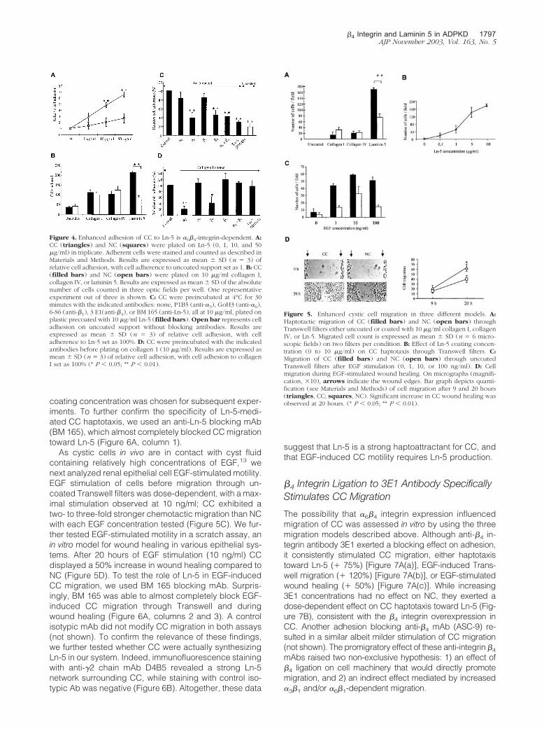
coating concentration was chosen for subsequent exper-iments. To further confirm the specificity of Ln-5-medi-ated CC haptotaxis, we used an anti-Ln-5 blocking mAb(BM 165), which almost completely blocked CC migrationtoward Ln-5 (Figure 6A, column 1).
As cystic cells in vivo are in contact with cyst fluidcontaining relatively high concentrations of EGF,13 wenext analyzed renal epithelial cell EGF-stimulated motility.EGF stimulation of cells before migration through un-coated Transwell filters was dose-dependent, with a max-imal stimulation observed at 10 ng/ml; CC exhibited atwo- to three-fold stronger chemotactic migration than NCwith each EGF concentration tested (Figure 5C). We fur-ther tested EGF-stimulated motility in a scratch assay, anin vitro model for wound healing in various epithelial sys-tems. After 20 hours of EGF stimulation (10 ng/ml) CCdisplayed a 50% increase in wound healing compared toNC (Figure 5D). To test the role of Ln-5 in EGF-inducedCC migration, we used BM 165 blocking mAb. Surpris-ingly, BM 165 was able to almost completely block EGF-induced CC migration through Transwell and duringwound healing (Figure 6A, columns 2 and 3). A controlisotypic mAb did not modify CC migration in both assays(not shown). To confirm the relevance of these findings,we further tested whether CC were actually synthesizingLn-5 in our system. Indeed, immunofluorescence stainingwith anti-�2 chain mAb D4B5 revealed a strong Ln-5network surrounding CC, while staining with control iso-typic Ab was negative (Figure 6B). Altogether, these data
suggest that Ln-5 is a strong haptoattractant for CC, andthat EGF-induced CC motility requires Ln-5 production.
�4 Integrin Ligation to 3E1 Antibody SpecificallyStimulates CC Migration
The possibility that �6�4 integrin expression influencedmigration of CC was assessed in vitro by using the threemigration models described above. Although anti-�4 in-tegrin antibody 3E1 exerted a blocking effect on adhesion,it consistently stimulated CC migration, either haptotaxistoward Ln-5 (� 75%) [Figure 7A(a)], EGF-induced Trans-well migration (� 120%) [Figure 7A(b)], or EGF-stimulatedwound healing (� 50%) [Figure 7A(c)]. While increasing3E1 concentrations had no effect on NC, they exerted adose-dependent effect on CC haptotaxis toward Ln-5 (Fig-ure 7B), consistent with the �4 integrin overexpression inCC. Another adhesion blocking anti-�4 mAb (ASC-9) re-sulted in a similar albeit milder stimulation of CC migration(not shown). The promigratory effect of these anti-integrin �4
mAbs raised two non-exclusive hypothesis: 1) an effect of�4 ligation on cell machinery that would directly promotemigration, and 2) an indirect effect mediated by increased�3�1 and/or �6�1-dependent migration.
Figure 4. Enhanced adhesion of CC to Ln-5 is �6�4-integrin-dependent. A:CC (triangles) and NC (squares) were plated on Ln-5 (0, 1, 10, and 50�g/ml) in triplicate. Adherent cells were stained and counted as described inMaterials and Methods. Results are expressed as mean � SD (n � 3) ofrelative cell adhesion, with cell adherence to uncoated support set as 1. B: CC(filled bars) and NC (open bars) were plated on 10 �g/ml collagen I,collagen IV, or laminin 5. Results are expressed as mean � SD of the absolutenumber of cells counted in three optic fields per well. One representativeexperiment out of three is shown. C: CC were preincubated at 4°C for 30minutes with the indicated antibodies: none, P1B5 (anti-�3), GoH3 (anti-�6),6-S6 (anti-�1), 3 E1(anti-�4), or BM 165 (anti-Ln-5), all at 10 �g/ml, plated onplastic precoated with 10 �g/ml Ln-5 (filled bars). Open bar represents celladhesion on uncoated support without blocking antibodies. Results areexpressed as mean � SD (n � 3) of relative cell adhesion, with celladherence to Ln-5 set as 100%. D: CC were preincubated with the indicatedantibodies before plating on collagen 1 (10 �g/ml). Results are expressed asmean � SD (n � 3) of relative cell adhesion, with cell adhesion to collagenI set as 100% (* P � 0.05; ** P � 0.01).
Figure 5. Enhanced cystic cell migration in three different models. A:Haptotactic migration of CC (filled bars) and NC (open bars) throughTranswell filters either uncoated or coated with 10 �g/ml collagen I, collagenIV, or Ln-5. Migrated cell count is expressed as mean � SD (n � 6 micro-scopic fields) on two filters per condition. B: Effect of Ln-5 coating concen-tration (0 to 10 �g/ml) on CC haptotaxis through Transwell filters. C:Migration of CC (filled bars) and NC (open bars) through uncoatedTranswell filters after EGF stimulation (0, 1, 10, or 100 ng/ml). D: Cellmigration during EGF-stimulated wound healing. On micrographs (magnifi-cation, �10), arrows indicate the wound edges. Bar graph depicts quanti-fication (see Materials and Methods) of cell migration after 9 and 20 hours(triangles, CC; squares, NC). Significant increase in CC wound healing wasobserved at 20 hours. (* P � 0.05; ** P � 0.01).
�4 Integrin and Laminin 5 in ADPKD 1797AJP November 2003, Vol. 163, No. 5

�4 Ligation to 3E1 Stimulates �3�1-DependentHaptotaxis and �3�1-Independent Chemotaxis
To further address these hypotheses, we performed mi-gration assays with anti-integrins blocking antibodies. Inthe Ln-5 haptotaxis model, migration was dramaticallyreduced by preincubating cells with anti-integrin �3
(�80%), anti-�6 (�75%) and anti-�1 (�70%) mAbs, butnot by anti-�2 mAb (Figure 8A). The stimulating effect of3E1 mAb was almost completely blocked by antibodies
directed against �3�1, and partially blocked by antibod-ies directed against �6�1 (Figure 8A, last 3 columns),suggesting that the stimulating effects of �4 ligation op-erate mainly through an �3�1-dependent pathway. In theEGF-stimulated wound repair assay, anti-�3 mAbstrongly inhibited CC migration (�70%), while anti-�2,anti-�6, and anti-�1 mAb exerted a moderate inhibition(�27%, �5% and �33%, respectively; Figure 8B), sug-gesting that in this assay, CC migration is also mainly�3�1-dependent. In this assay however, the promigratoryeffect of 3E1 was not blocked by the combination ofanti-�1 and either anti-�3 or anti-�6, suggesting that �4
integrin ligation is able to promote migration indepen-dently of these integrins.
DiscussionNormal renal tubular epithelia synthesize and interactwith the adjacent basement membrane, a specializedregion of the extracellular matrix (ECM) containing type IVcollagen, laminins, fibronectin, entactin, and proteogly-cans. In ADPKD, it has long been stated that abnormalinteractions between cyst-lining epithelia and surround-ing basement membrane may play an important role incystogenesis.3
Through cDNA arrays screening of differentially ex-pressed genes, we identified a strong overexpression of�4 integrin in ADPKD cystic cells. RT-PCR analysis con-firmed that �4 integrin transcripts were overexpressed inADPKD compared to normal kidneys. Immunohistochem-istry further demonstrated a strong �4 integrin expressionin most ADPKD cyst-lining epithelia, whereas no expres-sion was detected in normal adult tubules. Our observa-tion of �4 integrin overexpression in slightly dilated AD-PKD tubules in a patient with preserved renal function[Figure 2B(c)] suggests it is an early event in the cystformation process and that this finding is not related toend-stage renal disease. Strong �4 integrin expressionduring normal fetal kidney development suggests that �4
integrin may be either persistently expressed after birth inADPKD tubules, or reexpressed later during cystogen-esis. Costaining with specific markers ruled out �4 inte-grin aberrant expression as a nephron segment-specificevent. Expression of other integrins (�2, �3, �6, and �1) byimmunohistochemistry was similar in ADPKD and control
Figure 6. Inhibitory effect of 10 �g/ml BM165 anti-Ln-5 antibody on threeCC migration models. A, panel a: inhibition of CC haptotaxis toward lami-nin-5 coated filters (filled bars,10 �g/ml; first lane, uncoated support).Results are expressed as mean � SD (n � 3) of relative cell migration, withmigration toward Ln-5-coated filters set as 100%. b: Inhibition of CC chemo-taxis. Where indicated, CC were incubated with 10 ng/ml EGF (filled bars)� BM165 before plating. Results are expressed as mean � SD (n � 3) ofrelative cell migration, with EGF-stimulated migration set as 100%. c: Inhibi-tion of wound healing. Serum-starved CC were allowed to migrate from theedges to re-epithelialize the wounded surface for 20 hours in DMEM � EGF(10 ng/ml, filled bars) � BM165. Results are expressed as mean � SD (n �3) of relative cell migration, with EGF-stimulated wound healing set as 100%.B: Endogenous Ln-5 deposition by CC in cultures (a, �40) and negativeisotypic control (b).
Figure 7. Anti-�4 integrin 3E1 mAb enhances CC haptotactic and EGF-induced chemotactic migration. A: Stimulating effect of 3E1 mAb on threecell migration assays (performed as in Figure 6). Anti-�4 integrin mAb 3E1stimulates haptotactic migration toward Ln-5 (a), EGF-stimulated chemotac-tic migration through Transwell filters (b), and EGF-stimulated wound heal-ing (c). B: 3E1 stimulates CC but not NC haptotaxis toward Ln-5. CC (trian-gles) and NC (squares) were preincubated with or without 3E1 (1, 5, or 10�g/ml). Spontaneous haptotactic migration toward Ln-5 was set as 100%.
Figure 8. Haptotactic migration of CC on Ln-5 is �3�1 integrin-dependent,while chemotactic CC migration becomes �3�1 independent after stimulationwith �4 antibody 3E1. A: Haptotactic CC migration toward Ln-5 coated filterswas performed and quantified as described in Figure 6. Cells were preincu-bated with 10 �g/ml P1E6 (anti-�2), P1B5 (anti-�3), GoH3 (anti-�6), 6-S6(anti-�1), or 3E1 (anti-�4). B: EGF-stimulated CC wound repair was per-formed and quantified as described in Figure 6. Cells were incubated duringwound healing with 10 �g/ml P1E6 (anti-�2), P1B5 (anti-�3), GoH3 (anti-�6), 6-S6 (anti-�1), or 3E1 (anti-�4).
1798 Joly et alAJP November 2003, Vol. 163, No. 5

kidneys (not shown), highlighting the specificity of �4
integrin overexpression in ADPKD. The reason why a fewcysts did not express �4 integrin remains obscure.
Ln-5, the main ligand for �6�4, was also aberrantlyexpressed in most of the cyst-surrounding ECM. Otherauthors have reported the strong expression of unspec-ified laminin in the Cy rat model of ADPKD,14 and thepresence of uncharacterized laminin breakdown prod-ucts in the cyst fluid of ADPKD patients.15 As expected,we observed a good correlation between Ln-5 and �4
integrin-positive staining among individual cysts. In ac-cordance with previous data, no expression of Ln-5 wasdetected in normal adult tubules.16 By contrast, we de-tected Ln-5 expression in fetal kidney ureteric bud, asreported by others [Figure 3B(d)].17
To clarify the role of �6�4 integrin-Ln-5 interactions inADPKD, we set up in vitro conditions to analyze cellanchorage and cell motility. As shown in Figure 1, cellsderived from ADPKD cysts in primary culture maintainhigh production of �4 integrin mRNA and protein. Inshort-term adhesion studies, CC and NC adhere similarlyto collagens I and IV, while Ln-5 induce specifically CCbut not NC to adhere more strongly (two-fold) in a dose-dependent manner (Figure 4, A and B). This result con-tradicts the previously reported weaker adhesion of AD-PKD cyst-lining cells to laminin than to other ECMcomponents,18 but the laminin composition and experi-mental design were clearly different in this work. Most ofCC adhesion to Ln-5 was blocked by anti-�6 or �4 mAbs,suggesting that the strong and specific adhesion of CC toLn-5 is mainly �6�4-dependent, with �3�1 and �6�1 play-ing a minor role (Figure 4C).
We then tested in Transwell assays the ability of exog-enous matrix substrates to stimulate haptotactic migra-tion, and found that Ln-5 is a much stronger haptoattrac-tant for CC than collagen I or collagen IV (Figure 5A).Furthermore, complete inhibition of EGF-stimulated mi-gration by anti-Ln5 mAb [Figure 6(b and c)] suggests thatLn-5 endogenous production and extracellular deposi-tion as a substrate is necessary for CC to migrate.19
Blocking experiments further indicated that among Ln-5-binding integrins, �3�1 was the major mediator of CChaptotaxis toward Ln-5 and EGF-stimulated wound repair(Figure 8A), in agreement with findings reported in mi-grating keratinocytes.20–22 These studies failed to dem-onstrate a role for �6�4 in migration, but mAbs used toblock integrin �6�4 function were generally raised againstthe �6 subunit. In this study we show that the anti-�4
subunit mAb 3E1 stimulates CC migration in the threedifferent models tested (Figure 7). Integrin �4 ligation to3E1 could indirectly enhance migration by preventingadhesion. However, a similar observation was made on acolon cancer cell line in which 3E1 had no effect onadhesion.23 Indeed, our blocking experiments suggestthat that 3E1 ligation to �4 may transactivate �3�1 integrinin the Ln-5-driven haptotaxis assays (Figure 8A), andeven directly stimulate the promigratory cell machinery inthe EGF-stimulated CC motility assays (Figure 8B). Inter-estingly, it was reported that both 3E1 mAb ligation to �4
and EGF receptor binding to �6�4 could both trigger �4
cytoplasmic domain phosphorylation, downstream signal
transduction, and subsequent epithelial cell migrationand tumor invasion.5,24,25
To date, �6�4 integrin and Ln-5 overexpression havemostly been reported in neoplastic diseases and weredescribed separately. Expression of �6�4 integrin waspositively correlated to the progression of various carci-nomas26 and facilitated cell motility, tumor invasion, andmetastatic potential.27 Classically, �6�4 signaling is trig-gered by its ligation to the tumor-produced laminins,26
but it has also been described in laminin-free ECM envi-ronments.28 Overexpression of Ln-5 was described invarious tumors29 and was shown to modulate cell adhe-sion,30 migration, and proliferation.10,31 Furthermore, dis-ruption of �6�4-Ln-5 interaction was shown to inhibit tu-mor growth.32
A role for �6�4 and Ln-5 in kidney tubulogenesis wasrecently suggested by Zent et al,33 who reported thatblocking antibodies to �6 or Ln-5 could inhibit uretericbud branching morphogenesis in whole embryonic kid-ney organ culture as well as in isolated ureteric budculture. Of note, a role for Ln-5 acting not only through�6�4 but also through �3�1 was reported in this model, inaccordance with the involvement of �3�1 integrin in CCmigration.
In ADPKD, the mechanism of �4 integrin and Ln-5cystic aberrant expression remains unknown. However,we found that �4 integrin mRNA expression by CC wasdose-dependently increased by EGF (data not shown),as described in other cell types.34 The positive regulationof �4 integrin expression by EGF may be biologicallyrelevant in ADPKD because cysts contain high levels ofEGF, which is able to interact with apical EGF receptorsin vivo.35 Altogether, our in vitro findings suggest a role for�6�4 and Ln-5 in kidney cystogenesis. Ln-5-�6�4 com-plex mediates both adhesion and migration of cyst-de-rived cells. Several factors may account for this apparentduality, including the proteolytic cleavage of Ln-5 �2chain8 and the activation of EGF receptor pathway.5,25
One can speculate that stable anchorage of the epithe-lium is necessary to maintain the cystic architecture,while cyst growth requires cell migration. Further work iscurrently under way to clarify the role of Ln-5-�6�4 inter-action in dysregulated proliferation, another cardinal fea-ture of ADPKD cystic epithelia.
AcknowledgementsWe thank Drs. Eric Thervet, Yves Chretien, and ArnaudMejean for providing us with kidney specimens; Dr. LiseHalbwachs-Mecarelli for critical reading of the manu-script; Jean-Pierre Grunfeld and Philippe Lesavre forstimulating discussions; and Youcef Meftali for technicalassistance.
References
1. Nauli SM, Alenghat FJ, Luo Y, Williams E, Vassilev P, Li X, Elia AE, LuW, Brown EM, Quinn SJ, Ingber DE, Zhou J: Polycystins 1 and 2mediate mechanosensation in the primary cilium of kidney cells. NatGenet 2003, 33:129–137
�4 Integrin and Laminin 5 in ADPKD 1799AJP November 2003, Vol. 163, No. 5

2. Arnaout MA: Molecular genetics and pathogenesis of autosomaldominant polycystic kidney disease. Annu Rev Med 2001, 52:93–123
3. Wilson PD: Polycystin: new aspects of structure, function, and regu-lation. J Am Soc Nephrol 2001, 12:834–845
4. Sonnenberg A, Calafat J, Janssen H, Daams H, van der Raaij-HelmerLM, Falcioni R, Kennel SJ, Aplin JD, Baker J, Loizidou M, et al: Integrin�6/�4 complex is located in hemidesmosomes, suggesting a majorrole in epidermal cell-basement membrane adhesion. J Cell Biol1991, 113:907–917
5. Mainiero F, Pepe A, Yeon M, Ren Y, Giancotti FG: The intracellularfunctions of �6�4 integrin are regulated by EGF. J Cell Biol 1996,134:241–253
6. Giancotti FG, Ruoslahti E: Integrin signaling. Science 1999, 285:1028–1032
7. Dogic D, Rousselle P, Aumailley M: Cell adhesion to laminin 1 or 5induces isoform-specific clustering of integrins and other focal adhe-sion components. J Cell Sci 1998, 111:793–802
8. Giannelli G, Falk-Marzillier J, Schiraldi O, Stetler-Stevenson WG,Quaranta V: Induction of cell migration by matrix metalloprotease-2cleavage of laminin-5. Science 1997, 277:225–228
9. Grassi M, Moens G, Rousselle P, Thiery JP, Jouanneau J: The SFLactivity secreted by metastatic carcinoma cells is related to laminin 5and mediates cell scattering in an integrin-independent manner.J Cell Sci 1999, 112:2511–2520
10. Nguyen BP, Gil SG, Carter WG: Deposition of laminin 5 by keratino-cytes regulates integrin adhesion and signaling. J Biol Chem 2000,275:31896–31907
11. Wallace DP, Grantham JJ, Sullivan LP: Chloride and fluid secretion bycultured human polycystic kidney cells. Kidney Int 1996, 50:1327–1336
12. Rousselle P, Aumailley M: Kalinin is more efficient than laminin inpromoting adhesion of primary keratinocytes and some other epithe-lial cells and has a different requirement for integrin receptors. J CellBiol 1994, 125:205–214
13. Munemura C, Uemasu J, Kawasaki H: Epidermal growth factor andendothelin in cyst fluid from autosomal dominant polycystic kidneydisease cases: possible evidence of heterogeneity in cystogenesis.Am J Kidney Dis 1994, 24:561–568
14. Cowley BD Jr, Gudapaty S, Kraybill AL, Barash BD, Harding MA,Calvet JP, Gattone VH II: Autosomal-dominant polycystic kidney dis-ease in the rat. Kidney Int 1993, 43:522–534
15. Slade MJ, Kirby RB, Pocsi I, Jones JK, Price RG: Presence of lamininfragments in cyst fluid from patients with autosomal dominant poly-cystic kidney disease (ADPKD): role in proliferation of tubular epithe-lial cells. Biochim Biophys Acta 1998, 1401:203–210
16. Mizushima H, Miyagi Y, Kikkawa Y, Yamanaka N, Yasumitsu H,Misugi K, Miyazaki K: Differential expression of laminin-5/ladsin sub-units in human tissues and cancer cell lines and their induction bytumor promoter and growth factors. J Biochem (Tokyo) 1996, 120:1196–1202
17. Mizushima H, Koshikawa N, Moriyama K, Takamura H, Nagashima Y,Hirahara F, Miyazaki K: Wide distribution of laminin-5 �2 chain inbasement membranes of various human tissues. Horm Res 1998,50:7–14
18. Wilson PD, Geng L, Li X, Burrow CR: The PKD1 gene product,“polycystin-1,” is a tyrosine-phosphorylated protein that colocalizeswith �2�1-integrin in focal clusters in adherent renal epithelia. LabInvest 1999, 79:1311–1323
19. Larjava H, Salo T, Haapasalmi K, Kramer RH, Heino J: Expression of
integrins and basement membrane components by wound keratino-cytes. J Clin Invest 1993, 92:1425–1435
20. Carter WG, Ryan MC, Gahr PJ: Epiligrin, a new cell adhesion ligandfor integrin �3 �1 in epithelial basement membranes. Cell 1991,65:599–610
21. Goldfinger LE, Stack MS, Jones JC: Processing of laminin-5 and itsfunctional consequences: role of plasmin and tissue-type plasmino-gen activator. J Cell Biol 1998, 141:255–265
22. Zhang K, Kramer RH: Laminin 5 deposition promotes keratinocytemotility. Exp Cell Res 1996, 227:309–322
23. Daemi N, Thomasset N, Lissitzky JC, Dumortier J, Jacquier MF,Pourreyron C, Rousselle P, Chayvialle JA, Remy L: Anti-�4 integrinantibodies enhance migratory and invasive abilities of human colonadenocarcinoma cells and their MMP-2 expression. Int J Cancer2000, 85:850–856
24. Mainiero F, Pepe A, Wary KK, Spinardi L, Mohammadi M, Schless-inger J, Giancotti FG: Signal transduction by the �6 �4 integrin:distinct �4 subunit sites mediate recruitment of Shc/Grb2 and asso-ciation with the cytoskeleton of hemidesmosomes. EMBO J 1995,14:4470–4481
25. Mariotti A, Kedeshian PA, Dans M, Curatola AM, Gagnoux-Palacios L,Giancotti FG: EGF-R signaling through Fyn kinase disrupts the func-tion of integrin �6�4 at hemidesmosomes: role in epithelial cell mi-gration and carcinoma invasion. J Cell Biol 2001, 155:447–458
26. Mercurio AM, Rabinovitz I, Shaw LM: The �6�4 integrin and epithelialcell migration. Curr Opin Cell Biol 2001, 13:541–545
27. Shaw LM, Rabinovitz I, Wang HH, Toker A, Mercurio AM: Activation ofphosphoinositide 3-OH kinase by the �6�4 integrin promotes carci-noma invasion. Cell 1997, 91:949–960
28. O’Connor KL, Shaw LM, Mercurio AM: Release of cAMP gating by the�6�4 integrin stimulates lamellae formation and the chemotactic mi-gration of invasive carcinoma cells. J Cell Biol 1998, 143:1749–1760
29. Lohi J: Laminin-5 in the progression of carcinomas. Int J Cancer2001, 94:763–767
30. Orian-Rousseau V, Aberdam D, Rousselle P, Messent A, Gavrilovic J,Meneguzzi G, Kedinger M, Simon-Assmann P: Human colonic cancercells synthesize and adhere to laminin-5: their adhesion to laminin-5involves multiple receptors among which is integrin �2�1. J Cell Sci1998, 111:1993–2004
31. Gonzales M, Haan K, Baker SE, Fitchmun M, Todorov I, Weitzman S,Jones JC: A cell signal pathway involving laminin-5, �3�1 integrin,and mitogen-activated protein kinase can regulate epithelial cell pro-liferation. Mol Biol Cell 1999, 10:259–270
32. Dajee M, Lazarov M, Zhang JY, Cai T, Green CL, Russell AJ, Marink-ovich MP, Tao S, Lin Q, Kubo Y, Khavari PA: NF-�B blockade andoncogenic Ras trigger invasive human epidermal neoplasia. Nature2003, 421:639–643
33. Zent R, Bush KT, Pohl ML, Quaranta V, Koshikawa N, Wang Z,Kreidberg JA, Sakurai H, Stuart RO, Nigam SK: Involvement of lami-nin binding integrins and laminin-5 in branching morphogenesis ofthe ureteric bud during kidney development. Dev Biol 2001, 238:289–302
34. Song QH, Singh RP, Trinkaus-Randall V: Injury and EGF mediate theexpression of �6�4 integrin subunits in corneal epithelium. J CellBiochem 2001, 80:397–414
35. Wilson PD, Du J, Norman JT: Autocrine, endocrine and paracrineregulation of growth abnormalities in autosomal dominant polycystickidney disease. Eur J Cell Biol 1993, 61:131–138
1800 Joly et alAJP November 2003, Vol. 163, No. 5