Embed Size (px)
Citation preview
Aalborg Universitet
The Potential of Nano-Porous Surface Structure for Pain Therapeutic Applications
Surface Properties and Evaluation of Pain Perception
Wu, Ching-Zong; Hsu, Ling-Chuan; Chou, Hsin-Hua; Barnkob, Sanne; Eggert, Tobias;Nielsen, Pernille Lind; Young, Roger; Vase, Lene; Wang, Kelun; Svensson, Peter; Ou, Keng-Liang; Baad-Hansen, LenePublished in:Applied Sciences
DOI (link to publication from Publisher):10.3390/app10134578
Creative Commons LicenseCC BY 4.0
Publication date:2020
Document VersionPublisher's PDF, also known as Version of record
Link to publication from Aalborg University
Citation for published version (APA):Wu, C-Z., Hsu, L-C., Chou, H-H., Barnkob, S., Eggert, T., Nielsen, P. L., Young, R., Vase, L., Wang, K.,Svensson, P., Ou, K-L., & Baad-Hansen, L. (2020). The Potential of Nano-Porous Surface Structure for PainTherapeutic Applications: Surface Properties and Evaluation of Pain Perception. Applied Sciences, 10(13),[4578]. https://doi.org/10.3390/app10134578
General rightsCopyright and moral rights for the publications made accessible in the public portal are retained by the authors and/or other copyright ownersand it is a condition of accessing publications that users recognise and abide by the legal requirements associated with these rights.
- Users may download and print one copy of any publication from the public portal for the purpose of private study or research. - You may not further distribute the material or use it for any profit-making activity or commercial gain - You may freely distribute the URL identifying the publication in the public portal -
Take down policyIf you believe that this document breaches copyright please contact us at [email protected] providing details, and we will remove access tothe work immediately and investigate your claim.

applied sciences
Article
The Potential of Nano-Porous Surface Structure forPain Therapeutic Applications: Surface Propertiesand Evaluation of Pain Perception
Ching-Zong Wu 1,2,3,†, Ling-Chuan Hsu 1,†, Hsin-Hua Chou 1,4, Sanne Barnkob 5, Tobias Eggert 5,Pernille Lind Nielsen 5, Roger Young 1, Lene Vase 6, Kelun Wang 7, Peter Svensson 5,8,Keng-Liang Ou 9,10,11,12,* and Lene Baad-Hansen 5
1 School of Dentistry, College of Oral Medicine, Taipei Medical University, Taipei 110, Taiwan;[email protected] (C.-Z.W.); [email protected] (L.-C.H.); [email protected] (H.-H.C.);[email protected] (R.Y.)
2 Department of Dentistry, Taipei Medical University Hospital, Taipei 110, Taiwan3 Department of Dentistry, Lotung Poh-Ai Hospital, Yilan 265, Taiwan4 Dental Department of Wan-Fang Hospital, Taipei Medical University, Taipei 116, Taiwan5 Section of Orofacial Pain and Jaw Function, Department of Dentistry and Oral Health, Aarhus University,
8000 Aarhus, Denmark; [email protected] (S.B.); [email protected] (T.E.);[email protected] (P.L.N.); [email protected] (P.S.); [email protected] (L.B.-H.)
6 Department of Psychology and Behavioral Sciences, Aarhus University, 8000 Aarhus, Denmark;[email protected]
7 The Faculty of Medicine, Department of Health Science and Technology, Center for Sensory-MotorInteraction, Aalborg University Hospital, 9100 Aalborg, Denmark; [email protected]
8 Department of Dental Medicine, Karolinska Institute, 14157 Huddinge, Sweden9 Department of Dentistry, Taipei Medical University-Shuang Ho Hospital, New Taipei City 235, Taiwan10 Department of Oral Hygiene Care, Ching Kuo Institute of Management and Health, Keelung 203, Taiwan11 Biomedical Technology R & D Center, China Medical University Hospital, Taichung 404, Taiwan12 3D Global Biotech Inc. (Spin-off Company from Taipei Medical University), New Taipei City 221, Taiwan* Correspondence: [email protected]† Authors contribute equally.
Received: 18 May 2020; Accepted: 26 June 2020; Published: 1 July 2020�����������������
Featured Application: The potential application of the work is in the area of acupuncture therapy.The study focuses on the nanostructured surfaces to stimulate connective tissue interaction at theneurological sites for pain therapy.
Abstract: The objective of this study was to evaluate the biomaterial properties of nano-modifiedsurface acupuncture needles and the effect of such needles on human pain perception by usingpressure pain threshold (PPT) measurements. It is known that changing a material’s surfacenano-topography or nanostructure has strong effects on its physical, chemical, and biological surfaceproperties. However, there is no information in the literature about the stimulation characteristics ofacupuncture needles with nano-topography or nanostructured surfaces. Based on the knowledge onnanostructured surfaces, it may be possible to potentiate the effects of acupuncture needle stimulation.The pressure pain sensitivity of the masseter muscle in the orofacial region was studied in 21 healthyvolunteers in two randomized, double-blinded sessions: an active session of manual acupuncturemanipulation with nano-modified surface needles, and an inactive session of sham acupuncturestimulation to control for possible placebo effects. Three acupuncture points were selected fromclassical Chinese medicine literature: LI4 (Hegu) on the hand, ST6 (Jiache) on the lower masseterregion, and ST7 (Xiaguan) on the upper masseter region. PPT measurements, perceived sensations,and pain from the acupuncture were recorded. The results showed discrete yet significant differencesin PPT values between the active and inactive acupuncture treatments and significantly higher pain
Appl. Sci. 2020, 10, 4578; doi:10.3390/app10134578 www.mdpi.com/journal/applsci

Appl. Sci. 2020, 10, 4578 2 of 14
scores from active acupuncture stimulation than from sham acupuncture. These results indicate subtlebut significant effects of acupuncture stimulation with nano-modified surface needles, compared tosham acupuncture in healthy participants.
Keywords: acupuncture; nano-modified surface; pressure pain threshold; orofacial pain
1. Introduction
Acupuncture is a complementary therapy applied widely in pain management. The numberof reports on its therapeutic effects in various orofacial pain conditions has been on the rise [1].However, a major obstacle in conducting these studies is to adequately control for placebo effects [2].The present study was designed to investigate the effects of acupuncture on orofacial musculoskeletalpain sensitivity, and was specifically targeted towards the masseter muscle, which is often involvedin temporomandibular disorders (TMDs) [3–6]. The results of our previous study indicated thatthe condyle was asymmetrical during jaw closing, and that both the condyle and disc were slightlyasymmetrical during jaw opening in patients with TMD [7]. One-third of adults are reported to havesymptoms of TMDs, which include headaches, joint pain, and clicking or muscle tenderness [8,9].Many therapies have been applied for TMD management, such as self-care after mirror experiences,intra-oral devices, night-time biofeedback, and complementary acupuncture therapy [10,11].
According to the classical Chinese medicine literature, a physiological phenomenon called “Deqi”is required to achieve good therapeutic outcomes from acupuncture treatment [12,13]. The sensationsdescribed by the patients can involve dullness, heaviness, distension, numbness, and aches during theDeqi phase of the acupuncture [14,15]. The classical Chinese medicine literature, Huangdi Neijing(Huangdi’s canon of medicine), states that Deqi should be felt by the acupuncturists who also need toconcentrate in order to hold it. Thus, the process of Deqi with needles is experienced by both patient andacupuncturist. The acupuncturist feels an increase in resistance while manipulating the needles (needlegrasping), and the sensation is described as tense, tight, or heavy [15,16]. Needle grasping (Deqi) isthought to be important for clinical efficacy, according to the traditional Chinese medicine theory,and there is some scientific support for this claim. Langevin’s team investigated the physiologicalchanges during the sensations of Deqi. They observed connective tissue grasping and winding aroundthe inserted needle during the rotational manipulation of acupuncture needles [12]. Their work alsoshowed that the needle grasping phenomenon necessitated more pull-out force to withdraw the needle,and increased the tissue volume around the needle insertion site [17,18]. The needle grasping or tissuewinding induced by needle movements stimulates the local tissues, such as the connective tissue,thereby delivering a mechanical signal that leads to downstream effects [19,20]. This includes a changein the gene expression or alteration of the composition of the extracellular matrix. Connective tissue isthe main tissue type affected by needle manipulation. The winding of the connective tissue is believedto be the reason for local, remote, and long-term therapeutic effects of acupuncture [21]. To summarizeLangevin’s research, acupuncture creates changes in the connective tissue, which impact the body atthe cellular level via mechano-transduction [20]. These mechanical signals may also stimulate theendocrine system to release endogenous opioids, which act as analgesics of specific neurological sites.This may be the biochemical mechanism by which acupuncture blocks the nociceptive inputs andtransmission, thereby eliciting an analgesic response [16,22,23].
It is well known that changing a material’s surface nano-topography or nanostructure has strongeffects on its physical, chemical, and biological properties [24–27]. The nano-topography of thesurfaces has also been demonstrated to be able to alter gene expression, facilitate or direct stem celldifferentiation, induce changes in the cell morphology, and stimulate the proliferation and spreadingof different cell types [28–32]. Such changes in surface properties have been extensively used in dental,orthopedic, and other biomedical applications. However, there is no information in the literature
Appl. Sci. 2020, 10, 4578 3 of 14
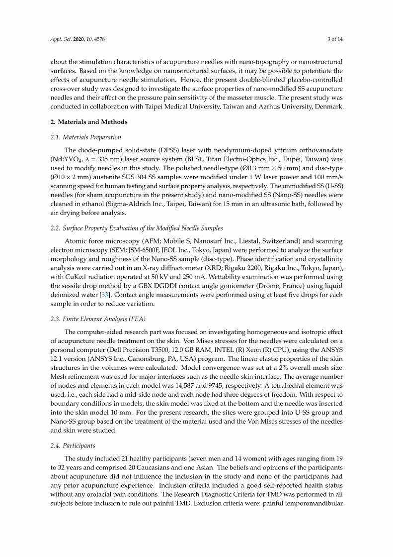
about the stimulation characteristics of acupuncture needles with nano-topography or nanostructuredsurfaces. Based on the knowledge on nanostructured surfaces, it may be possible to potentiate theeffects of acupuncture needle stimulation. Hence, the present double-blinded placebo-controlledcross-over study was designed to investigate the surface properties of nano-modified SS acupunctureneedles and their effect on the pressure pain sensitivity of the masseter muscle. The present study wasconducted in collaboration with Taipei Medical University, Taiwan and Aarhus University, Denmark.
2. Materials and Methods
2.1. Materials Preparation
The diode-pumped solid-state (DPSS) laser with neodymium-doped yttrium orthovanadate(Nd:YVO4, λ = 335 nm) laser source system (BLS1, Titan Electro-Optics Inc., Taipei, Taiwan) wasused to modify needles in this study. The polished needle-type (Ø0.3 mm × 50 mm) and disc-type(Ø10 × 2 mm) austenite SUS 304 SS samples were modified under 1 W laser power and 100 mm/sscanning speed for human testing and surface property analysis, respectively. The unmodified SS (U-SS)needles (for sham acupuncture in the present study) and nano-modified SS (Nano-SS) needles werecleaned in ethanol (Sigma-Aldrich Inc., Taipei, Taiwan) for 15 min in an ultrasonic bath, followed byair drying before analysis.
2.2. Surface Property Evaluation of the Modified Needle Samples
Atomic force microscopy (AFM; Mobile S, Nanosurf Inc., Liestal, Switzerland) and scanningelectron microscopy (SEM; JSM-6500F, JEOL Inc., Tokyo, Japan) were performed to analyze the surfacemorphology and roughness of the Nano-SS sample (disc-type). Phase identification and crystallinityanalysis were carried out in an X-ray diffractometer (XRD; Rigaku 2200, Rigaku Inc., Tokyo, Japan),with CuKα1 radiation operated at 50 kV and 250 mA. Wettability examination was performed usingthe sessile drop method by a GBX DGDDI contact angle goniometer (Drôme, France) using liquiddeionized water [33]. Contact angle measurements were performed using at least five drops for eachsample in order to reduce variation.
2.3. Finite Element Analysis (FEA)
The computer-aided research part was focused on investigating homogeneous and isotropic effectof acupuncture needle treatment on the skin. Von Mises stresses for the needles were calculated on apersonal computer (Dell Precision T3500, 12.0 GB RAM, INTEL (R) Xeon (R) CPU), using the ANSYS12.1 version (ANSYS Inc., Canonsburg, PA, USA) program. The linear elastic properties of the skinstructures in the volumes were calculated. Model convergence was set at a 2% overall mesh size.Mesh refinement was used for major interfaces such as the needle-skin interface. The average numberof nodes and elements in each model was 14,587 and 9745, respectively. A tetrahedral element wasused, i.e., each side had a mid-side node and each node had three degrees of freedom. With respect toboundary conditions in models, the skin model was fixed at the bottom and the needle was insertedinto the skin model 10 mm. For the present research, the sites were grouped into U-SS group andNano-SS group based on the treatment of the material used and the Von Mises stresses of the needlesand skin were studied.
2.4. Participants
The study included 21 healthy participants (seven men and 14 women) with ages ranging from 19to 32 years and comprised 20 Caucasians and one Asian. The beliefs and opinions of the participantsabout acupuncture did not influence the inclusion in the study and none of the participants hadany prior acupuncture experience. Inclusion criteria included a good self-reported health statuswithout any orofacial pain conditions. The Research Diagnostic Criteria for TMD was performed in allsubjects before inclusion to rule out painful TMD. Exclusion criteria were: painful temporomandibular
Appl. Sci. 2020, 10, 4578 4 of 14
disorders (temporomandibular joint click and crepitation without pain was allowed), any criticalphysical or mental disorders, intake of any medicine that affects the central nervous system, and anyprior nerve damage affecting the orofacial area. All participants signed a statement of informed consentand were paid a compensation of 28 Euros (200 DKK) each for participating in the study.
2.5. Study Design
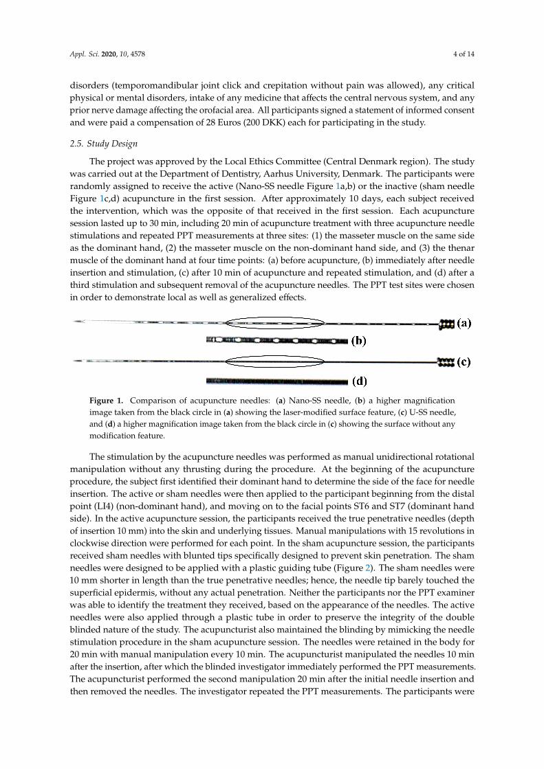
The project was approved by the Local Ethics Committee (Central Denmark region). The studywas carried out at the Department of Dentistry, Aarhus University, Denmark. The participants wererandomly assigned to receive the active (Nano-SS needle Figure 1a,b) or the inactive (sham needleFigure 1c,d) acupuncture in the first session. After approximately 10 days, each subject receivedthe intervention, which was the opposite of that received in the first session. Each acupuncturesession lasted up to 30 min, including 20 min of acupuncture treatment with three acupuncture needlestimulations and repeated PPT measurements at three sites: (1) the masseter muscle on the same sideas the dominant hand, (2) the masseter muscle on the non-dominant hand side, and (3) the thenarmuscle of the dominant hand at four time points: (a) before acupuncture, (b) immediately after needleinsertion and stimulation, (c) after 10 min of acupuncture and repeated stimulation, and (d) after athird stimulation and subsequent removal of the acupuncture needles. The PPT test sites were chosenin order to demonstrate local as well as generalized effects.
Figure 1. Comparison of acupuncture needles: (a) Nano-SS needle, (b) a higher magnificationimage taken from the black circle in (a) showing the laser-modified surface feature, (c) U-SS needle,and (d) a higher magnification image taken from the black circle in (c) showing the surface without anymodification feature.
The stimulation by the acupuncture needles was performed as manual unidirectional rotationalmanipulation without any thrusting during the procedure. At the beginning of the acupunctureprocedure, the subject first identified their dominant hand to determine the side of the face for needleinsertion. The active or sham needles were then applied to the participant beginning from the distalpoint (LI4) (non-dominant hand), and moving on to the facial points ST6 and ST7 (dominant handside). In the active acupuncture session, the participants received the true penetrative needles (depthof insertion 10 mm) into the skin and underlying tissues. Manual manipulations with 15 revolutions inclockwise direction were performed for each point. In the sham acupuncture session, the participantsreceived sham needles with blunted tips specifically designed to prevent skin penetration. The shamneedles were designed to be applied with a plastic guiding tube (Figure 2). The sham needles were10 mm shorter in length than the true penetrative needles; hence, the needle tip barely touched thesuperficial epidermis, without any actual penetration. Neither the participants nor the PPT examinerwas able to identify the treatment they received, based on the appearance of the needles. The activeneedles were also applied through a plastic tube in order to preserve the integrity of the doubleblinded nature of the study. The acupuncturist also maintained the blinding by mimicking the needlestimulation procedure in the sham acupuncture session. The needles were retained in the body for20 min with manual manipulation every 10 min. The acupuncturist manipulated the needles 10 minafter the insertion, after which the blinded investigator immediately performed the PPT measurements.The acupuncturist performed the second manipulation 20 min after the initial needle insertion andthen removed the needles. The investigator repeated the PPT measurements. The participants were

Appl. Sci. 2020, 10, 4578 5 of 14
subjected to the same experimental procedure and investigation by the same study personnel duringthe second visit.
Figure 2. Inactive acupuncture guiding tube. Concaved needle handle anchor on the top with twoinsertion site markers on the side and adhesive button part.
Prior to the beginning of the first session, participants were requested to complete a form withpersonal information, health status, and a series of questions regarding their knowledge of acupuncturetherapy. The subjects were asked about their belief of the beneficial effects of acupuncture and anyexpectation they had from the treatment. Immediately after each needle insertion, the investigatorasked the participants whether he/she felt anything (yes/no). The participants were then asked to ratethe sensations evoked by the acupuncture needle on a numerical rating scale (NRS) of 0–10 (0 being nopain and 10 being the highest imaginable pain). Furthermore, the participants were asked to specifythe sensation as “sharp pain”, “pricking”, “tingling”, or “other sensation”. The participant´s and theinvestigator´s thoughts, as to which needle (active or sham) was used, were recorded after needleinsertion and at the end of the session. Participants were advised to take a light meal before theycame to the session to minimize the possibility of side effects such as fainting, dizziness or discomfort.During the entire experiment, the investigator carefully monitored the participant’s condition to ensuresafety and efficiency of the experiment.
2.6. Acupuncture
Based on the Chinese medicine literature on facial pain (targeted on trigeminal neuralgiadistribution), ST6 Jiache and ST7 Xiaguan points were selected for the activation of the masseter muscleon the side of the dominant hand. These two acupuncture points coincide with the distribution of themaxillary nerve branches. The distal point LI4 Hegu is located contralateral to the dominant side andsubjected to the same acupuncture manipulations as those of the points on the face. Both the active andinactive needles were concealed inside plastic guiding tubes before the treatment began. The guidingtube was made opaque and there were two markers at the base of the tube to ensure that the methodof insertion was accurate.
2.7. PPT
A standardized PPT measurement using an electronic pressure algometer (Somedic Inc., Sösdala,Sweden) was performed at four different time points: (1) at baseline, (2) immediately after the firstinsertion and manipulation of the acupuncture needle, (3) 10 min after needle insertion (after thesecond manipulation), and (4) immediately after the third-needle stimulation and subsequent needlewithdrawal. All PPT measurements were performed according to the standard research protocolestablished by the German Research Network on Neuropathic Pain (DFNS) [34–36]. The PPT wasmeasured in triplicate on both masseter muscles (between ST6 and ST7) and on the thenar on thedominant hand (the side opposite to that of the LI4 acupuncture point). The pressure algometer had arubber tip with a contact area of 1 cm2, which was pressed against the skin on the test region with a

Appl. Sci. 2020, 10, 4578 6 of 14
gradual increase of the pressure of 30 kPa/s. The test subject notified the investigator when the pressurebecame just slightly painful by pressing a stop button.
2.8. Statistics
Data are presented as mean ± SD or as percentages (%). Three (one for each PPT test site) two-wayAnalyses of Variance (ANOVAs) with acupuncture session type (active vs. sham) and PPT measurementtime as factors were performed. Where appropriate, Tukey´s Honestly Significant Difference (HSD)tests with corrections for multiple comparisons were performed for post hoc analyses. McNemar´s testswere performed to compare frequencies of sensation of needle insertion between sessions. Effect ofthe blinding procedure was evaluated by quantification of the proportion (%) of participants andexaminers, who were able to correctly identify active or placebo needles. Differences were regarded assignificant when p < 0.05. Statistical analyses were performed by using the STATISTICA statisticalsoftware (StatSoft 13 version, Hugecon Inc., Kaohsiung, Taiwan).
3. Results
3.1. Nano-Modified Surface Properties
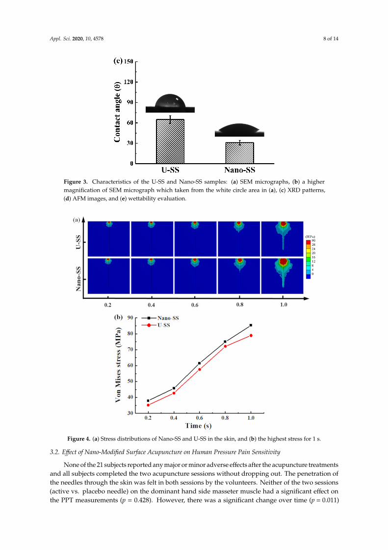
Figure 3a shows the SEM image of a Nano-SS needle sample. Laser modification resulted ina wave like structure on the surface. A higher magnification SEM image of the white circle area inthe Nano-SS sample shows the presence of nano-porous structures on the surface of the Nano-SSsample (Figure 3b). Figure 3c shows the XRD patterns from the U-SS and Nano-SS samples. All thepeaks were analyzed using the database from the Joint Committee on Powder Diffraction Standards.Only typical austenite phase (γ) peaks were detected in the U-SS and Nano-SS samples. The crystallinestructure of the γ phase belongs to face centered cubic structure. No other precipitates or intermetalliccompounds could be found on the surface of the Nano-SS sample. According to the AFM observation,the surface roughness (represented as root mean square) of the nano-porous structure was about18.6 nm (Figure 3d). The contact angle of the U-SS and the Nano-SS samples is presented in Figure 3e.The Nano-SS needle sample exhibited a more hydrophilic surface (~31◦) than the U-SS sample(~65◦) [37]. Thus, a hydrophilic wave like surface layer with nano-porous structure was formed on thesurface of the Nano-SS sample.
Figure 4a shows the stress distribution for the Nano-SS and U-SS models on the skin at 1 s.The maximum stress varied from 35.2 MPa to 85.3 MPa in all models, as shown in Figure 4b. In theNano-SS group, the stress (85.3 MPa) was larger than the maximum stress of 78.9 MPa in the U-SSgroup at 1 s. The stress on the skin while using treated needles was larger than that exerted by theneedles without surface treatment. However, the stress patterns in both models were similar to eachother. Analysis of the present study indicated that stresses were induced by the needle.
Figure 3. Cont.

Appl. Sci. 2020, 10, 4578 7 of 14
Figure 3. Cont.
Appl. Sci. 2020, 10, 4578 8 of 14
Figure 3. Characteristics of the U-SS and Nano-SS samples: (a) SEM micrographs, (b) a highermagnification of SEM micrograph which taken from the white circle area in (a), (c) XRD patterns,(d) AFM images, and (e) wettability evaluation.
Figure 4. (a) Stress distributions of Nano-SS and U-SS in the skin, and (b) the highest stress for 1 s.
3.2. Effect of Nano-Modified Surface Acupuncture on Human Pressure Pain Sensitivity
None of the 21 subjects reported any major or minor adverse effects after the acupuncture treatmentsand all subjects completed the two acupuncture sessions without dropping out. The penetration ofthe needles through the skin was felt in both sessions by the volunteers. Neither of the two sessions(active vs. placebo needle) on the dominant hand side masseter muscle had a significant effect onthe PPT measurements (p = 0.428). However, there was a significant change over time (p = 0.011)

Appl. Sci. 2020, 10, 4578 9 of 14
and a tendency towards a significant interaction between session and time (p = 0.081). A focusedpost hoc analysis of the tendency showed that 10 min after needle insertion, PPT in the active sessionwas significantly increased (decreased pressure pain sensitivity) compared with the placebo session(p = 0.006). There was no statistically significant change from baseline in either session (p > 0.239).
No significant main effects of session or time were detected in PPTs from the masseter musclecontralateral to the dominant hand (Session: p = 0.413; Time: p = 0.969) or from the thenar muscle(Session: p = 0.407; Time: p = 0.846). No significant interactions between factors were found at thesetwo test sites (p > 0.227).
The thenar muscle showed higher PPTs than the facial regions (p < 0.001). There were nodifferences in PPT values of masseter muscles between sides (p = 0.940) (Table 1).
Table 1. Presentation of means (SD) of pressure pain thresholds (PPT in kPa) of the three testsites at different time points. The values in bold indicates a significant difference from placebo.Dom = Dominant hand side masseter muscle, Non = Non-dominant hand side masseter muscle.
Inactive Active
Site Baseline Immediately 10 min 20 min Baseline Immediately 10 min 20 min
Dom 195.1(74.7)
175.9(62.9)
190.3(70.4)
200.7(72.7)
203.7(71.3)
195.1(76.8)
220.9(88.4)
204.1(76.1)
Non 191.4(77.7)
179.6(69.6)
188.7(73.0)
184.3(73.3)
196.7(77.8)
203.2(78.3)
198.7(81.2)
204.5(84.5)
Thenar 294.0(132.0)
312.5(131.4)
285.5(136.6)
286.2(174.6)
321.5(166.8)
319.9(160.6)
320.5(169.3)
326.1(194.7)
Total 226.8(108.0)
222.7(111.9)
221.5(106.8)
223.7(123.7)
240.6(126.1)
239.4(124.5)
246.7(129.4)
244.9(140.5)
The analyses of the sensations evoked by acupuncture needles showed that the sham acupunctureneedle was felt by 57.1% of the participants on the hand site, 66.7% on the lower masseter site and76.2% on the upper masseter site. The active needles were felt by 87.7% on the hand site, 85.7% on thelower masseter site and 81.0% on the upper masseter site. From the McNemar´s tests, it was found thatthe nano-modified acupuncture needles were felt by more participants than the sham needles for allsites (p < 0.039) (Figure 5).
The mean perceived intensity of the acupuncture evoked pain on the 0–10 NRS in the shamacupuncture session was 0.8 (±1.1) in the distal insertion site (hand), 1.1 (±1.0) in the lower masseterregion and 1.2 (±1.0) in the upper masseter region. In the active acupuncture session, the meanperceived intensity of the acupuncture evoked pain was 2.7 (±2.0) on the distal insertion site (hand),3.4 (±2.0) in the lower masseter region and 3.1 (±2.5) in the upper masseter region. Hence, the activeacupuncture in the three insertion sites (Hand, Lower masseter and Upper masseter) evoked statisticallyhigher levels of pain than the sham acupuncture (p < 0.05).
In the first session, 28.6% of the participants correctly identified the treatment they received while23.8% were wrong about the treatment they received and 47.6% indicated that they did not know. In thesecond session, more participants were able to identify the treatment they received (42.9%), but 38.1%still did not know. When the data from both sessions were combined, 33.3% of the subjects were trulyblinded and only 14.3% were able to identify correctly, the treatment they received in both sessions.In the first session, the blinded investigators guessed the treatment correctly for 52.4%, wrongly for28.6% and could not make a guess for 19.1% of the participants. In the second session, the investigatorsguessed correctly for only 9.5% of participants and could not make a guess for the remaining 90.5%of participants.

Appl. Sci. 2020, 10, 4578 10 of 14
Figure 5. The nano-modified acupuncture needle has the ability to increase tissue winding effect due tothe increased surface area on the needle shaft: (a) tissue winding effect of the U-SS needle and (b) tissuewinding effect of the Nano-SS needle.
4. Discussion
The main finding of this study was that no overall significant differences between active andsham acupuncture, as measured by the PPTs, could be detected in healthy participants. However,there was a modest tendency towards a small increase in PPT (reduced pressure pain intensity) for themasseter muscle of the acupuncture needle side in the active session as compared with that in the shamsession. As shown in Table 1, the minor difference between active and sham needle sessions of thePPT measurements may be seen as a validation of the earlier reported placebo-effect of acupuncturetherapy [38,39]. The sham needle may have stimulated the superficial epidermis. Since the shamneedle did not penetrate the subject’s skin and into the acupoint, it did not reach our stimulationgoal, Deqi, in the study. Therefore, a possible explanation could be that the participants in the shamsession might have experienced peripheral sensory stimulation and in some cases even a sharp painperception, even though the needles did not exactly penetrate the skin [40,41]. It is certainly difficult toinsert acupuncture needles without evincing any sensory stimulation. Although the sham needle isshort and blunt, a very gentle touch from the needle tip may still exert an effect [42,43].
The active needles induced significantly more frequent and intense sensations than the shamneedles. This can probably be attributed to the depth of needle insertion, but also partly to the increasedsurface area of the Nano-SS needle, which provides more space for tissue attachments leading to greaterstimulation. As compared to smooth surfaces (U-SS needle), the textured needle shaft surfaces of theNano-SS needle present more surface area for interaction with and stimulation of the tissues. It can beexpected that the textured surface has a positive influence on connective tissue grasping and winding,

Appl. Sci. 2020, 10, 4578 11 of 14
which may result in a better stimulation result (Figure 5) [16,17,44–46]. The contact area of the needlehas a lot to do with the stimulation effect of the acupuncture manipulation techniques. The techniqueof rotational manipulation increases needle pull out forces, when acupuncture needles are insertedinto the connective tissue layer (unidirectional and bidirectional). The winding of connective tissuebecause of the acupuncture manipulation may transduce a series of mechanical signals and stimulateendocrine physiological events [20,47,48]. Finite element method detected hard stimulation in thegroup receiving acupuncture with surface treated needles. The discontinuous structure induced lots ofstress confusing to stimulate the neural pain sensation to promote the clinical treatment effect [49,50].
FEA has been used for decades in mechanical engineering and design. In the dental field,models have been used to describe the stresses and strains in different structures, such as the TMJ,dentition, and facial skeleton. Biomechanical models of the human masticatory system using the FEAprogram were not perfect but are based on various assumptions and simplifications. Moreover, it wasdifficult to compare the earlier studies clearly because of the differences in muscle force, materialsetting, mesh structure, and other constraints. The result of FEA was also very difficult to compare toa real model because of the mechanical and physical properties of the model in the biological field.The human organs have a very complex structure. For example, the condyle cartilage layer is composedmainly of large proteoglycans such as versican and aggrecan [51]. In general, large proteoglycansare more suited to resisting compressive force than articular disc which is composed mainly ofcollagen fibers [52]. An accurate representation of the condyle cartilage layer in TMJ finite elementmodels was of vital importance, but it was very difficult to achieve in the computer-aided program.Therefore, simplification was a common way to study. Mechanical forces, temperature and electricityhave been applied to a variety of nerve stimulation including different tissues [53–55]. These studieshave led to new insights into how tissue to transmit signals by mechanical stimulation. Mechanicalstimulation is also a kind of substrate for cell adhesion, growth, and differentiation [56]. During boneremodeling or wound healing, a stress simulation mechanism must start to enhance the response ofgene expression. These advances have significantly increased our understanding of the process ofmechanical transductions inducing stimulation using the acupuncture.
The nano-porous structures of the Nano-SS needle probably plays an important role in increasingthe surface area, which leads to more space for connective tissue interaction at the neurologicalsites during the acupuncture. However, more clinical research on the influence of nano surface andnanostructures of acupuncture needles is clearly needed to better understand their potential advantagescompared with conventional needles. It should also be mentioned that acupuncture with surfacemodified needles in this study did not result in any significant complications. None of the subjectsreported any adverse effects or major concerns during and after the sessions. Owing to the smallsample size, the study findings must be interpreted cautiously and the possible effects on clinical painshould be investigated further.
5. Conclusions
A hydrophilic wave-like surface layer with nano-porous structures was formed on the surface ofthe modified acupuncture needles used in the present study. The results showed subtle yet significantdifferences in PPT measurements between the active and sham acupuncture groups, and significantlyhigher pain scores from active acupuncture stimulation than from sham acupuncture. These resultsindicate subtle effects of acupuncture with nano-modified needles compared to effects of shameacupuncture in healthy participants. Further studies in patients with clinical pain are needed.
Author Contributions: For this research article, C.-Z.W. and L.-C.H. were the main authors responsible for theoriginal draft. L.-C.H. was also responsible for the software analysis along with H.-H.C., R.Y. contributed to theinvestigation. S.B., T.E. and P.L.N. were responsible for the data collection. L.V. and K.W. contributed to thevalidation. P.S., L.B.-H. and K.-L.O. were responsible for the study design, supervision, and manuscript review &editing. All authors have read and agreed to the published version of the manuscript.

Appl. Sci. 2020, 10, 4578 12 of 14
Funding: The authors are grateful to the Biomate Medical Devices Technology co., ltd and ANMC Healthcare,Inc. for their support with experimental devices and testing instruments. This study is also supported in part byLo-Hsu Foundation Lotung Poh-Ai Hospital (104-E126) and the Danish Dental Association. Special thanks areextended to the Department of Dentistry and Oral Health, Aarhus University, Denmark and 3D Global BiotechInc., Taiwan, for their skillful technical assistance.
Conflicts of Interest: The authors declare no conflict of interest in this work.
References
1. List, T.; Axelsson, S. Management of TMD: Evidence from systematic reviews and meta-analyses. J. OralRehabil. 2010, 37, 430–451. [CrossRef] [PubMed]
2. Vase, L.; Baram, S.; Takakura, N.; Yajima, H.; Takayama, M.; Kaptchuk, T.J.; Schou, S.; Jensen, T.S.; Zachariae, R.;Svensson, P. Specifying the nonspecific components of acupuncture analgesia. Pain 2013, 154, 1659–1667.[CrossRef] [PubMed]
3. Welsh, D.; Hengerer, A.S. The diagnosis and treatment of intramuscular hemangiomas of the massetermuscle. Am. J. Otolaryngol. 1980, 1, 186–190. [CrossRef]
4. Lao, L. Acupuncture techniques and devices. J. Altern. Complement. Med. 1996, 2, 23–25. [CrossRef]5. Mupparapu, M. Evidence Based Approach for the Diagnosis of Temporomandibular Joint Disorders (TMD).
J. Indian Prosthodont. Soc. 2013, 13, 387–388. [CrossRef]6. Ott, K.H. Diagnosis and therapy of masseter hypertrophy. HNO 1983, 31, 207–211. [PubMed]7. Cheng, H.Y.; Peng, P.W.; Lin, Y.J.; Chang, S.T.; Pan, Y.N.; Lee, S.C.; Ou, K.L.; Hsu, W.C. Stress analysis
during jaw movement based on vivo computed tomography images from patients with temporomandibulardisorders. Int. J. Oral Maxillofac. Surg. 2013, 42, 386–392. [CrossRef] [PubMed]
8. Kurita, K.; Westesson, P.-L.; Yuasa, H.; Toyama, M.; Machida, J.; Ogi, N. Natural Course of UntreatedSymptomatic Temporomandibular Joint Disc Displacement without Reduction. J. Dent. Res. 1998, 77,361–365. [CrossRef] [PubMed]
9. Kalpakci, K.N.; Willard, V.P.; Wong, M.E.; Athanasiou, K.A. An Interspecies Comparison of theTemporomandibular Joint Disc. J. Dent. Res. 2011, 90, 193–198. [CrossRef] [PubMed]
10. Pereira, L.J.; Steenks, M.H.; de Wijer, A.; Speksnijder, C.M.; van der Bilt, A. Masticatory function in subacuteTMD patients before and after treatment. J. Oral Rehabil. 2009, 36, 391–402. [CrossRef]
11. Durham, J.; Steele, J.; Moufti, M.A.; Wassell, R.; Robinson, P.; Exley, C. Temporomandibular disorder patients’journey through care. Community Dent. Oral 2011, 39, 532–541. [CrossRef] [PubMed]
12. Kou, W.; Gareus, I.; Bell, J.D.; Goebel, M.U.; Spahn, G.; Pacheco-Lopez, G.; Backer, M.; Schedlowski, M.;Dobos, G.J. Quantification of DeQi sensation by visual analog scales in healthy humans afterimmunostimulating acupuncture treatment. Am. J. Chin. Med. 2007, 35, 753–765. [CrossRef] [PubMed]
13. Liang, Z.H.; Xie, C.C.; Li, Z.P.; Zhu, X.P.; Lu, A.P.; Fu, W.B. Deqi sensation in placebo acupuncture: A crossoverstudy on Chinese medicine students. Evid. Based Complement. Altern. Med. 2013, 2013, 620671. [CrossRef][PubMed]
14. Zhang, S.; Mu, W.; Xiao, L.; Zheng, W.K.; Liu, C.X.; Zhang, L.; Shang, H.C. Is deqi an indicator of clinicalefficacy of acupuncture? A systematic review. Evid. Based Complement. Altern. Med. 2013, 2013, 750140.[CrossRef]
15. Park, J.; Park, H.; Lee, H.; Lim, S.; Ahn, K.; Lee, H. Deqi sensation between the acupuncture-experienced andthe naive: A Korean study II. Am. J. Chin. Med. 2005, 33, 329–337. [CrossRef]
16. Yang, X.Y.; Shi, G.X.; Li, Q.Q.; Zhang, Z.H.; Xu, Q.; Liu, C.Z. Characterization of deqi sensation andacupuncture effect. Evid. Based Complement. Altern. Med. 2013, 2013, 319734. [CrossRef] [PubMed]
17. Langevin, H.M.; Churchill, D.L.; Wu, J.; Badger, G.J.; Yandow, J.A.; Fox, J.R.; Krag, M.H. Evidence ofconnective tissue involvement in acupuncture. FASEB J. 2002, 16, 872–874. [CrossRef]
18. Langevin, H.M.; Storch, K.N.; Snapp, R.R.; Bouffard, N.A.; Badger, G.J.; Howe, A.K.; Taatjes, D.J. Tissuestretch induces nuclear remodeling in connective tissue fibroblasts. Histochem. Cell Biol. 2010, 133, 405–415.[CrossRef]
19. Langevin, H.M. Connective tissue: A body-wide signaling network? Med. Hypotheses 2006, 66, 1074–1077.[CrossRef]
20. Langevin, H.M.; Churchill, D.L.; Cipolla, M.J. Mechanical signaling through connective tissue: A mechanismfor the therapeutic effect of acupuncture. FASEB J. 2001, 15, 2275–2282. [CrossRef]

Appl. Sci. 2020, 10, 4578 13 of 14
21. Langevin, H.M.; Bouffard, N.A.; Fox, J.R.; Palmer, B.M.; Wu, J.; Iatridis, J.C.; Barnes, W.D.; Badger, G.J.;Howe, A.K. Fibroblast cytoskeletal remodeling contributes to connective tissue tension. J. Cell Physiol. 2011,226, 1166–1175. [CrossRef] [PubMed]
22. Joshi, N.; Araque, H. Neurophysiologic basis for the relief of human pain by acupuncture. Acupunct. Electrother.Res. 2009, 34, 165–174. [CrossRef] [PubMed]
23. Hui, K.K.; Nixon, E.E.; Vangel, M.G.; Liu, J.; Marina, O.; Napadow, V.; Hodge, S.M.; Rosen, B.R.; Makris, N.;Kennedy, D.N. Characterization of the “deqi” response in acupuncture. BMC Complement. Altern Med. 2007,7, 33. [CrossRef] [PubMed]
24. Yim, E.K.; Darling, E.M.; Kulangara, K.; Guilak, F.; Leong, K.W. Nanotopography-induced changes infocal adhesions, cytoskeletal organization, and mechanical properties of human mesenchymal stem cells.Biomaterials 2010, 31, 1299–1306. [CrossRef]
25. Liu, X. Correlation analysis of surface topography and its mechanical properties at micro and nanometrescales. Wear 2013, 305, 305–311. [CrossRef]
26. Arsiwala, A.; Desai, P.; Patravale, V. Recent advances in micro/nanoscale biomedical implants. J. Controll.Release 2014, 189, 25–45. [CrossRef]
27. Saber, O.; Hefny, N.; al Jaafari, A.A. Improvement of physical characteristics of petroleum waxes by usingnano-structured materials. Fuel Process. Technol. 2011, 92, 946–951. [CrossRef]
28. Martinez, E.; Engel, E.; Planell, J.A.; Samitier, J. Effects of artificial micro- and nano-structured surfaces oncell behavior. Ann. Anat. Anat. Anz. 2009, 191, 126–135. [CrossRef] [PubMed]
29. Andersson, A.S.; Backhed, F.; von Euler, A.; Richter-Dahlfors, A.; Sutherland, D.; Kasemo, B. Nanoscalefeatures influence epithelial cell morphology and cytokine production. Biomaterials 2003, 24, 3427–3436.[CrossRef]
30. Dalby, M.J.; McCloy, D.; Robertson, M.; Agheli, H.; Sutherland, D.; Affrossman, S.; Oreffo, R.O.C.Osteoprogenitor response to semi-ordered and random nanotopographies. Biomaterials 2006, 27, 2980–2987.[CrossRef] [PubMed]
31. Dalby, M.J.; Gadegaard, N.; Tare, R.; Andar, A.; Riehle, M.O.; Herzyk, P.; Wilkinson, C.D.; Oreffo, R.O.The control of human mesenchymal cell differentiation using nanoscale symmetry and disorder. Nat. Mater.2007, 6, 997–1003. [CrossRef] [PubMed]
32. Dalby, M.J.; Gadegaard, N.; Oreffo, R.O. Harnessing nanotopography and integrin-matrix interactions toinfluence stem cell fate. Nat. Mater. 2014, 13, 558–569. [CrossRef] [PubMed]
33. Bachmann, J.; Ellies, A.; Hartge, K.H. Development and application of a new sessile drop contact anglemethod to assess soil water repellency. J. Hydrol. 2000, 231, 66–75. [CrossRef]
34. Maier, C.; Baron, R.; Tolle, T.R.; Binder, A.; Birbaumer, N.; Birklein, F.; Gierthmuhlen, J.; Flor, H.; Geber, C.;Huge, V.; et al. Quantitative sensory testing in the German Research Network on Neuropathic Pain (DFNS):Somatosensory abnormalities in 1236 patients with different neuropathic pain syndromes. Pain 2010, 150,439–450. [CrossRef]
35. Rolke, R.; Baron, R.; Maier, C.; Tolle, T.R.; Treede, R.D.; Beyer, A.; Binder, A.; Birbaumer, N.; Birklein, F.;Botefur, I.C.; et al. Quantitative sensory testing in the German Research Network on Neuropathic Pain(DFNS): Standardized protocol and reference values. Pain 2006, 123, 231–243. [CrossRef]
36. Pigg, M.; Baad-Hansen, L.; Svensson, P.; Drangsholt, M.; List, T. Reliability of intraoral quantitative sensorytesting (QST). Pain 2010, 148, 220–226. [CrossRef]
37. Drelich, J.; Chibowski, E.; Meng, D.D.; Terpilowski, K. Hydrophilic and superhydrophilic surfaces andmaterials. Soft Matter 2011, 7, 9804–9828. [CrossRef]
38. Takeshige, C.; Sato, M. Comparisons of pain relief mechanisms between needling to the muscle, static magneticfield, external qigong and needling to the acupuncture point. Acupunct. Electrother. Res. 1996, 21, 119–131.[CrossRef]
39. Karst, M.; Rollnik, J.D.; Fink, M.; Reinhard, M.; Piepenbrock, S. Pressure pain threshold and needleacupuncture in chronic tension-type headache–a double-blind placebo-controlled study. Pain 2000, 88,199–203. [CrossRef]
40. Orhan, E.K.; Deymeer, F.; Oflazer, P.; Parman, Y.; Baslo, M.B. Jitter analysis with concentric needle electrodein the masseter muscle for the diagnosis of generalised myasthenia gravis. Clin. Neurophysiol. 2013, 124,2277–2282. [CrossRef]

Appl. Sci. 2020, 10, 4578 14 of 14
41. Itoh, K.; Minakawa, Y.; Kitakoji, H. Effect of acupuncture depth on muscle pain. Chin. Med. 2011, 6, 24.[CrossRef] [PubMed]
42. Ulett, G.A. Acupuncture treatments for pain relief. JAMA 1981, 245, 768–769. [CrossRef] [PubMed]43. Asghar, A.U.; Green, G.; Lythgoe, M.F.; Lewith, G.; MacPherson, H. Acupuncture needling sensation:
The neural correlates of deqi using fMRI. Brain Res. 2010, 1315, 111–118. [CrossRef] [PubMed]44. Langevin, H.M.; Yandow, J.A. Relationship of acupuncture points and meridians to connective tissue planes.
Anat. Rec. 2002, 269, 257–265. [CrossRef] [PubMed]45. Langevin, H.M.; Storch, K.N.; Cipolla, M.J.; White, S.L.; Buttolph, T.R.; Taatjes, D.J. Fibroblast spreading
induced by connective tissue stretch involves intracellular redistribution of alpha- and beta-actin. Histochem.Cell Biol. 2006, 125, 487–495. [CrossRef] [PubMed]
46. Eadie, M.J. Acupuncture and the relief of pain. Med. J. Aust. 1990, 153, 180–181. [CrossRef]47. Seminowicz, D.A. Acupuncture and the CNS: What can the brain at rest suggest? Pain 2008, 136, 230–231.
[CrossRef]48. Rong, P.J.; Zhu, B.; Huang, Q.F.; Gao, X.Y.; Ben, H.; Li, Y.H. Acupuncture inhibition on neuronal activity of
spinal dorsal horn induced by noxious colorectal distention in rat. World J. Gastroenterol. 2005, 11, 1011–1017.[CrossRef]
49. Woolf, C.J.; Salter, M.W. Neuronal Plasticity: Increasing the Gain in Pain. Science 2000, 288, 1765–1768.[CrossRef] [PubMed]
50. Melzack, R. Evolution of the Neuromatrix Theory of Pain. The Prithvi Raj Lecture: Presented at the ThirdWorld Congress of World Institute of Pain, Barcelona 2004. Pain Pract. 2005, 5, 85–94. [CrossRef]
51. Mao, J.J.; Rahemtulla, F.; Scott, P.G. Proteoglycan Expression in the Rat Temporomandibular Joint in Responseto Unilateral Bite Raise. J. Dent. Res. 1998, 77, 1520–1528. [CrossRef] [PubMed]
52. Berkovitz, B.K.B.; Robertshaw, H. Ultrastructural quantification of collagen in the articular disc of thetemporomandibular joint of the rabbit. Arch. Oral Biol. 1993, 38, 91–95. [CrossRef]
53. Liu, M.; Tanswell, A.K.; Post, M. Mechanical force-induced signal transduction in lung cells. Am. J. Physiol.Lung Cell. Mol. Physiol. 1999, 277, L667–L683. [CrossRef] [PubMed]
54. Cope, F.W. Piezoelectricity and pyroelectricity as a basis for force and temeprature detection by nervereceptors. Bull. Math. Biol. 1973, 35, 31–41. [CrossRef]
55. Yamanishi, T.; Yasuda, K. Electrical stimulation for stress incontinence. Int. Urogynecol. J. 1998, 9, 281–290.[CrossRef] [PubMed]
56. Chiquet, M. Regulation of extracellular matrix gene expression by mechanical stress. Matrix Biol. 1999, 18,417–426. [CrossRef]
© 2020 by the authors. Licensee MDPI, Basel, Switzerland. This article is an open accessarticle distributed under the terms and conditions of the Creative Commons Attribution(CC BY) license (http://creativecommons.org/licenses/by/4.0/).