Embed Size (px)
Citation preview

Additions to Phaeosphaeriaceae (Pleosporales) 59
Additions to Phaeosphaeriaceae (Pleosporales): Elongaticollum gen. nov., Ophiosphaerella taiwanensis sp. nov., Phaeosphaeriopsis beaucarneae sp. nov. and a new
host record of Neosetophoma poaceicola from Musaceae
Danushka S. Tennakoon1,2,3, Kasun M. Thambugala4, Dhanushka N. Wanasinghe5, Eleni Gentekaki2,3,
Itthayakorn Promputtha6,7, Chang-Hsin Kuo1, Kevin D. Hyde2,3,5,6,8
1 Department of Plant Medicine, National Chiayi University, 300 Syuefu Road, Chiayi City 60004, Taiwan 2 School of Science, Mae Fah Luang University, Chiang Rai 57100, Thailand 3 Center of Excellence in Fungal Research, Mae Fah Luang University, Chiang Rai, 57100, Thailand 4 Genetics and Molecular Biology Unit, Faculty of Applied Sciences, University of Sri Jayewardenepura, Gangodawila, Nugegoda, Sri Lanka 5 CAS Key Laboratory for Plant Biodiversity and Biogeography of East Asia (KLPB), Kunming Institute of Botany, Chinese Academy of Science, Kunming 650201, Yunnan, China 6 Department of Biology, Faculty of Science, Chiang Mai University, Chiang Mai 50200, Thailand 7 Environmental Science Research Center, Faculty of Science, Chiang Mai University, Chiang Mai, 50200, Thailand 8 Institute of Plant Health, Zhongkai University of Agriculture and Engineering, Haizhu District, Guangzhou 510225, China
Corresponding author: Chang-Hsin Kuo ([email protected])
Academic editor: Huzefa Raja | Received 27 April 2020 | Accepted 3 June 2020 | Published 27 July 2020
Citation: Tennakoon DS, Thambugala KM, Wanasinghe DN, Gentekaki E, Promputtha I, Kuo CH, Hyde KD (2020) Additions to Phaeosphaeriaceae (Pleosporales): Elongaticollum gen. nov., Ophiosphaerella taiwanensis sp. nov., Phaeosphaeriopsis beaucarneae sp. nov. and a new host record of Neosetophoma poaceicola from Musaceae. MycoKeys 70: 59–88. https://doi.org/10.3897/mycokeys.70.53674
AbstractA novel ascomycetous genus, Elongaticollum, occurring on leaf litter of Hedychium coronarium (Zingib-eraceae) in Taiwan, is described and illustrated. Elongaticollum is characterized by dark brown to black, superficial, obpyriform, pycnidial conidiomata with a distinct elongate neck, and oval to oblong, hyaline, aseptate conidia. Phylogenetic analyses (maximum likelihood, maximum parsimony and Bayesian) of combined ITS, LSU, SSU and tef1-α sequence data revealed Elongaticollum as a distinct genus within the family Phaeosphaeriaceae with high statistical support. In addition, Ophiosphaerella taiwanensis and Phae-osphaeriopsis beaucarneae are described as new species from dead leaves of Agave tequilana and Beaucarnea recurvata (Asparagaceae), respectively. Neosetophoma poaceicola is reported as a new host record from dead leaves of Musa acuminata (Musaceae). Newly described taxa are compared with other similar species and comprehensive descriptions and micrographs are provided.
Copyright Danushka S. Tennakoon et al. This is an open access article distributed under the terms of the Creative Commons Attribution License (CC BY 4.0), which permits unrestricted use, distribution, and reproduction in any medium, provided the original author and source are credited.
MycoKeys 70: 59–88 (2020)
doi: 10.3897/mycokeys.70.53674
http://mycokeys.pensoft.net
A peer-reviewed open-access journal
MycoKeysLaunched to accelerate biodiversity research
ReseARCh ARtiCle

Danushka S. Tennakoon et al. / MycoKeys 70: 59–88 (2020)60
KeywordsAsparagaceae, Dothideomycetes, leaf litter, new taxa, Zingiberaceae
introduction
Plant litter is considered as one of the main contributors to net above-ground primary productivity of terrestrial ecosystems (Swift et al. 1979; Berg and McClaugherty 2008; Krishna and Mohan 2017). Since plant litter is returned back to the soil, it represents a major source of organic carbon in forest soils (Berg 2003). Plant litter can be defined as a collection of fallen leaves, twigs, seeds and other woody debris that accumulate on the ground as a natural part of the forest ecosystem (Johnson and Catley 2002; Berg and McClaugherty 2008). In particular, leaf litter is the main source of organic matter and nutrients of the soil, compared to other litter types (Robertson and Paul 1999; Berg and McClaugherty 2008; Krishna and Mohan 2017). Leaf litter decomposition is a key process contributing to biogeochemical cycles in any forest ecosystem. Microorganisms are the primary agents in this process (Purahong et al. 2016; Mlambo et al. 2019). Fungi are considered as the “key players” in leaf litter decomposition, because of their ability to produce a wide range of extracellular enzymes (Pointing et al. 2005; Berg and McClaugherty 2008; Bani et al. 2018). Many researchers have been carrying out stud-ies of fungal species inhabiting leaf litter and have described numerous new species in Dothideomycetes (Hyde et al. 2019; Phookamsak et al. 2019; Tennakoon et al. 2019).
The family Phaeosphaeriaceae is considered to be one of the most species-rich fam-ilies in Dothideomycetes and includes species that inhabit a wide range of ecosystems (i. e., marine, terrestrial, and mangroves) (Phookamsak et al. 2014, 2017; Bakhshi et al. 2019; Jones et al. 2019; Luo et al. 2019; Tennakoon et al. 2019). Phaeosphaeriaceae was established by Barr (1979), who designated Phaeosphaeria I. Miyake as the generic type of the family. Phaeosphaeriaceae species have immersed to superficial, globose to subglobose ascomata, short papilla, bitunicate asci and hyaline to pigmented, fusiform to ellipsoidal, filiform, or muriform ascospores (Bakhshi et al. 2019; Chaiwan et al. 2019; Maharachchikumbura et al. 2019; Yang et al. 2019). Members of Phaeospha-eriaceae are cosmopolitan, since they exhibit diverse lifestyles as saprobes, endophytes and pathogens of economically important plants (Barr 1992; Phookamsak et al. 2014, 2017; Yang et al. 2016; Hyde et al. 2020; Mapook et al. 2020). Apart from being cosmopolitan in nature, it appears that this family is phylogenetically highly diverse. Thus, recent studies have revealed a large number of new genera in this family. For instance, in the space of two years, eleven genera have been introduced, viz. Bhagirathi-myces S.M. Singh & S.K. Singh (Hyde et al. 2020), Hydeomyces Maharachchikumbura et al. (Maharachchikumbura et al. 2019), Hydeopsis J.F. Zhang et al. (Zhang et al. 2019), Neostagonosporella C.L. Yang, et al. (Yang et al. 2019), Parastagonosporella M. Bakhshi, Arzanlou & Crous (Bakhshi et al. 2019), Pseudoophiosphaerella J.F. Zhang

Additions to Phaeosphaeriaceae (Pleosporales) 61
et al. (Zhang et al. 2019), Murichromolaenicola Mapook & K.D. Hyde (Mapook et al. 2020), Neoophiobolus Mapook & K.D. Hyde (Mapook et al. 2020), Paraleptospora Mapook & K.D. Hyde (Mapook et al. 2020), Pseudostaurosphaeria Mapook & K.D. Hyde (Mapook et al. 2020) and Vittaliana Devadatha et al. (Devadatha et al. 2019). Currently, more than 70 genera are accommodated in this family (Wanasinghe et al. 2018; Bakhshi et al. 2019; Maharachchikumbura et al. 2019; Phookamsak et al. 2019; Hongsanan et al. 2020; Hyde et al. 2020).
We are investigating the diversity of microfungi on leaf litter in the tropics with the aim of clarifying their taxonomy based on morphology coupled with multi-gene phylogeny. As a part of this study, we have collected and isolated four taxa from Tai-wan, which belong to the family Phaeosphaeriaceae. We present herein comprehensive morphological descriptions and an in-depth phylogenetic investigation of the newly introduced species.
Materials and methods
Sample collection, morphological studies and isolation
Decaying leaf litter samples of Agave tequilana F.A.C. Weber (Asparagaceae), Beau-carnea recurvata Lem. (Asparagaceae), Hedychium coronarium J.Koenig (Zingiber-aceae), and Musa acuminata Colla (Musaceae) were collected from Dahu Forest Area in Chiayi, Taiwan and taken to the laboratory in Zip lock plastic bags. Specimens were examined with a LEICA EZ4 stereomicroscope. Micro-morphological charac-ters were determined using AXIOSKOP 2 PLUS compound microscope and images were captured with a Zeiss AXIOCAM 506 COLOR digital camera. Observations and photomicrographs were made from materials mounted in water. Permanent slides were preserved in lactoglycerol, sealed by applying nail-polish around the margins of cover slip. All measurements were made with ZEN2 (blue edition) and images used for figures were processed with Adobe Photoshop CS3 Extended version 10.0 software (Adobe Systems, USA).
Single ascospore and conidial isolation was carried out following the method de-scribed in Phookamsak et al. (2014). The single germinated spore was picked up and transferred to potato dextrose agar (PDA) and incubated at 25 °C in natural light. Sub-sequent sub-culturing was done carefully to obtain pure culture and ensure absence of contaminants. Culture characteristics were observed after three weeks. Colonies were photographed and colonial characters were noted and described. Type specimens of new taxa were deposited at the herbarium of Mae Fah Luang University (MFLU) and National Chiayi University Herbarium (NCYU). Living cultures were deposited in Mae Fah Luang University Culture Collection (MFLUCC) and National Chiayi Uni-versity Culture Collection (NCYUCC). Faces of Fungi and Index Fungorum numbers were provided as in Jayasiri et al. (2015) and Index Fungorum (2020).

Danushka S. Tennakoon et al. / MycoKeys 70: 59–88 (2020)62
DNA extraction and PCR amplification
Total genomic DNA was extracted from scraped fresh fungal mycelium using the DNA extraction kit E.Z.N.A Fungal DNA Mini Kit (D3390-02, Omega Bio-Tek) following the manufacturer’s protocol. The DNA product was kept at 4 °C for DNA amplification and maintained at -20 °C for long term storage. DNA was amplified by polymerase chain reaction (PCR) for four genes, the large subunit (28S, LSU), small subunit (18S, SSU), internal transcribed spacers including the 5.8s rDNA (ITS1-5.8S-ITS2) and translation elongation factor 1 alpha (tef1-α). The partial LSU gene was amplified by using the primer combination LR0R and LR5 (Vilgalys and Hester 1990; Rehner and Samuels 1994); partial SSU was amplified with NS1 and NS4 (White et al. 1990), nuclear ITS was amplified with primers ITS5 and ITS4 (White et al. 1990), and tef1-α gene was amplified using the primers EF1-983F and EF1-2218R (Rehner et al. 2001). Amplification reactions were performed in 25 µl of total reaction that contained 9.5 µl of sterilized water, 12.5 µl of 2×Power Taq PCR MasterMix (Tri-I Biotech, Taipei, Taiwan), 1 µl of each forward and reverse primers and 1 µl of DNA template. The PCR thermal cycle program of ITS, LSU, SSU and tef1-α gene was processed initially at 94 °C for 3 minutes, followed by 35 cycles of denaturation at 94 °C for 30 seconds, annealing at 55 °C for 50 seconds, elongation at 72 °C for 1 minute and a final extension at 72 °C for 10 minutes and a holding temperature of 4 °C. The PCR products were analyzed by 1.5% agarose gels containing the Safeview DNA stain (GeneMark, Taipei, Taiwan) to confirm their expected molecular weight. PCR products were purified and sequenced with prim-ers mentioned above by Tri-I Biotech, Taipei, Taiwan. Nucleotide sequences were deposited in GenBank (Table 1).
Phylogenetic analysis
Phylogenetic analyses were performed using a combined LSU, SSU, ITS and tef1-α se-quence dataset. Newly generated sequence data were initially subjected to blast search in NCBI to obtain the closest matches in GenBank. Sequences generated from this study were analyzed with related taxa in the family Phaeosphaeriaceae, which were obtained from GenBank and from recently published data (Bakhshi et al. 2019; Hyde et al. 2019; Maharachchikumbura et al. 2019; Yang et al. 2019; Mapook et al. 2020) (Table 1). The combined dataset consisted of 168 sequences including our newly gen-erated sequences. Multiple alignments were automatically made with MAFFT v. 7 at the web server (http://mafft.cbrc.jp/alignment/server), using default settings (Katoh and Standley 2013). The alignment was refined manually with BioEdit v. 7.0.5.2 (Hall 1999), where necessary.
Evolutionary models for phylogenetic analyses were selected independently for each locus using MrModeltest v. 3.7 (Posada and Crandall 1998) under the Akaike Information Criterion (AIC). Phylogenetic trees were obtained from Randomized Ac-celerated Maximum Likelihood (RAxML), maximum parsimony analysis (MP) and
Additions to Phaeosphaeriaceae (Pleosporales) 63
table 1. GenBank and culture collection accession numbers of species included in the present phyloge-netic study. Newly generated sequences are shown in bold.
Species Strain/Voucher no. GenBank accession no.LSU SSU ITS tef1–α
Acericola italica MFLUCC 13-0609 MF167429 MF167430 MF167428 –Allophaeosphaeria muriformia MFLUCC 13-0277 KX910089 KX950400 KX926415 –Alloneottiosporina thailandica MFLUCC 15-0576 – – – –Amarenographium ammophilicola MFLU 17-2571 MN017847 MN017913 MN047087 MN077065Amarenomyces dactylidis KUMCC 18-0154 MK356345 MK356359 MK356371 –Arezzomyces cytisi MFLUCC 15-0649 KT306950 KT306954 KT306947 –Banksiophoma australiensis CBS 142163 KY979794 – KY979739 KY979889Bhagirathimyc es himalayensis AMH 10127 MK836020 MN121697 MK836021 –Bhatiellae rosae MFLUCC 17-0664 MG828989 MG829101 MG828873 –Brunneomurispora lonicerae KUMCC 18-0157 MK356346 MK356360 MK356373 MK359065Camarosporioides phragmitis MFLUCC 13-0365 KX572345 KX572350 KX572340 KX572354Chaetosphaeronema achilleae MFLUCC 16-0476 KX765266 – KX765265 –C. hispidulum CBS 216.75 KF251652 EU754045 KF251148 KF253108Dactylidina shoemaker MFLUCC 14-0963 MG829003 MG829114 MG828887 MG829200Dematiopleospora cirsii MFLUCC 13-0615 KX274250 – KX274243 KX284708D. mariae MFLUCC 15-0612 KJ749653 KJ749652 KX274244 KJ749655Didymocyrtis xanthomendozae CBS 129666 – – KP170651 KP170677Diederichomyces ficuzzae CBS 128019 JQ238616 – KP170647 KP170673Dlhawksworthia clematidicola MFLUCC 17-0693 MG829038 MG829144 MG828929 –D. lonicera MFLUCC 14-0955 MG829012 MG829121 MG828902 MG829203Edenia gomezpompae JLCC 34533 – – KC193601 –
LVPEI 3225 – – KU578033 –Elongaticollum hedychii MFLUCC 18-1638 MT321810 MT321803 MT321796 MT328753E. hedychii MFLUCC 17-2653 MT321811 MT321804 MT321797 MT328754
NCYUCC 19-0286 MT321812 MT321805 MT321798 MT328755Embarria clematidis MFLUCC 14-0652 KT306953 KT306956 KT306949 –
MFLUCC 14-0976 MG828987 MG829099 MG828871 MG829194Equiseticola fusispora MFLUCC 14-0522 KU987669 KU987670 KU987668 MG520895Galiicola baoshanensis HKAS 102234 MK356348 MK356362 MK356374 MK359066G. pseudophaeosphaeria MFLU 14-0524 – – – MG520896Hydeomyces desertipleosporoides SQUCC 15259 MK290839 MK290843 MK290841 MK290848
SQUCC 15260 MK290840 MK290844 MK290842 MK290849Hydeopsis verrucispora SD 2016-5 MK522498 MK522504 MK522508 MK523388Italica achilleae MFLUCC 14-0955 MG829012 MG829121 MG828902 MG829203I. luzulae MFLUCC 14-0932 KT306951 – – –Jeremyomyces labinae CBS 144617 MK442529 – MK442589 MK442695Juncaceicola italica MFLUCC 13-0750 – – KX500110 MG520897J. luzulae MFLUCC 13-0780 KX449530 KX449531 KX449529 –Kwanghwaensis miscanthi FU31017 MK503823 MK503829 MK503817 MT009126Leptosphaeria doliolum CBS 505.75 GU301827 GU296159 JF740205 GU349069Leptospora rubella CPC 11006 DQ195792 DQ195803 DQ195780 –L. thailandica MFLUCC 16-0385 KX655549 KX655554 KX655559 KX655564Longispora clematidis MFLU 15–1277Loratospora aestuarii CBS 117592 – – MH863024 –Mauginiella scaettae CBS 239.58 MH869303 – MH857770 –Melnikia anthoxanthii MFLUCC 14-1011 KU848204 KU848205 – –Murichromolaenicola chiangraiensis MFLUCC 17-1488 MN994559 MN994605 MN994582 MN998163M. chromolaenae MFLUCC 17-1489 MN994560 MN994606 MN994583 MN998164Muriphaeosphaeria galatellae MFLUCC 14-0614 KT438329 KT438331 KT438333 MG520900
MFLUCC 15-0769 KT438330 KT438332 – –Neoophiobolus chromolaenae MFLUCC 17-1467 MN994562 MN994606 MN994583 MN998164

Danushka S. Tennakoon et al. / MycoKeys 70: 59–88 (2020)64
Species Strain/Voucher no. GenBank accession no.LSU SSU ITS tef1–α
N. chromolaenae MFLUCC 17-1449 MN994561 MN994607 MN994584 MN998165Neosetophoma sp. MFLUCC 17-0844 MG829035 MG829141 MG828926 MG829219N. aseptata CBS 145363 MK540024 – MK539953 –N. camporesii MFLUCC 15-0682 KU302778 – KU302779 –N. clematidis MFLUCC 13-0734 KP684153 KP684154 KP744450 –N. garethjonesii MFLUCC 14-0528 – KY501126 – KY514402N. guiyangensis GZ13 MH018132 MH018136 MH018134 MH051889N. italica MFLU 14-0809 KP711361 KP711366 KP711356 –N. lonicerae KUMCC 18-0155 MK356349 MK356363 MK356375 MK359067N. lunariae CPC 26671 KX306789 – KX306763 –N. miscanthi MFLU 18-2675 MK503826 MK503832 MK503820 –N. phragmitis CBS 145364 MK540025 – MK539954 MK540148N. poaceicola MFLUCC 16-0886 KY550382 KY550383 KY568986 –
MFLUCC 18-1632 MT321809 MT321802 MT321795 –N. rosae MFLUCC 17-0844 MG829035 MG829141 MG828926 MG829219N. rosaena MFLUCC 17-0768 MG829037 MG829143 MG828928 –N. rosarum MFLU 17-0308 MG829036 MG829142 MG828927 –N. salicis MFLU 17-0118 MK608026 – MK608025 –N. samarorum CBS 138.96 KF251664 GQ387517 MH862569 KF253119N. sambuci CBS 145365 MK540026 – MK539955 MK540149N. shoemakeri MFLU 16-1606 MG602199 MG602201 MG602203 MG844352
MFLUCC 17-0780 MG844348 MG844350 MG844346 MG844352N. tienshanensis MFLUCC 17-0844 MG829035 MG829141 MG828926 MG829219N. xingrensis GZAAS18 0100 MH018133 – MH018135 –Neosphaerellopsis thailandica CPC 21659 KP170721 – KP170652 KP170678Neostagonospora caricis CBS 135092 KF251667 – KF251163 –N. phragmitis MFLUCC 16-0493 KX910090 KX950401 KX926416 MG520902Neostagonosporella sichuanensis MFLUCC 18-1228 – – – MK313854
MFLUCC 18-1231 – – – MK313851Neosulcatispora agaves CPC 26407 KT950867 – KT950853 –Nodulosphaeria multiseptata MFLUCC 15-0078 KY496728 – KY496748 –N. scabiosae MFLUCC 14-1111 KU708846 KU708842 KU708850 KU708854Ophiobolopsis italica MFLUCC 17-1791 MG520959 MG520977 MG520939 MG520903Ophiobolus disseminans MFLUCC 17-1787 MG520961 MG520980 MG520941 MG520906O. rossicus MFLU 17-1639 MG520964 MG520983 MG520944 MG520909Ophiosimulans tanaceti MFLUCC 14-0525 KU738891 KU738892 KU738890 MG520910Ophiosphaerella agrostidis MFLUCC 11-0152 KM434281 KM434290 KM434271 KM434299
MFLUCC 12-0007 KM434282 KM434291 KM434272 KM434300MFLUCC 16-0895 MF197563 MF351604 MF351996 –
IGM35 MF197563 MF351604 – –MFLUCC 11-0152 KM434281 KM434290 KM434271 KM434299
O. aquatica MFLUCC 14-0033 KX767089 KX767090 KX767088 MG520911MFLUCC 14-0033 KX767089 KX767090 KX767088 MG520911
O. herpotricha k28 – – KP690992 KP691016KS29 – – KP690986 KP691015
O. korrae ATCC 56289 – – KC848509 KC848515O. narmari ATCC 64688 – – KC848510 KC848516
ATCC 201719 – – KC848508 KC848514O. taiwanensis MFLU 18-2534 MT321815 MT321808 MT321801 MT328758O. taiwanica NTUCC 17-024 MN082419 – MN082417 –
NTUCC 17-025 MN082420 – MN082418 –Paraleptosphaeria dryadis CBS 643.86 GU301828 KC584632 JF740213 GU349009Paraleptospora chromolaenae MFLUCC 17-1481 MN994563 MN994609 MN994587 MN998167

Additions to Phaeosphaeriaceae (Pleosporales) 65
Species Strain/Voucher no. GenBank accession no.LSU SSU ITS tef1–α
P. chromolaenicola MFLUCC 17-1450 MN994564 MN994610 MN994588 MN998168Paraophiobolus arundinis MFLUCC 17-1789 MG520965 MG520984 MG520945 MG520912P. plantaginis MFLUCC 17-0245 KY815010 KY815012 KY797641 MG520913Paraloratospora camporesii MFLU 18-0915 MN756637 MN756635 MN756639 –Paraphoma chrysanthemicola CBS 522.66 KF251670 GQ387521 KF251166 KF253124P. radicina CBS 111.79 KF251676 EU754092 KF251172 KF253130Parastagonospora dactylidis MFLUCC 13-0375 KU058722 – KU058712 –Parastagonosporella fallopiae CBS 135981 MH460545 – MH460543 MH460549P. fallopiae CCTU 1151-1 MH460546 – MH460544 MH460550Phaeopoacea muriformis MFLUCC 17-0372 MF611638 MF611639 MF611637 –P. festucae MFLUCC 17-0056 KY824767 KY824769 KY824766 –Phaeoseptoriella zeae CBS 144614 MK442547 – MK442611 MK442702Phaeosphaeria musae MFLUCC 11-0133 KM434277 KM434287 KM434267 KM434296P. oryzae CBS 110110 KF251689 GQ387530 KF251186 –P. papayae CBS 135416 – – MH866082 –Phaeosphaeriopsis agapanthi CPC 26303 KX228311 – KX228260 –P. agavacearum CPC 29122 KY173520 – KY173430 –P. agavensis CBS 102206 KY090669 KY090693 KY090635 –P. aloes CBS 145367 MK540030 – MK539959 MK540153P. aloicola CBS 145368 MK540031 – MK539960 MK540154P. amblyospora CBS 110131 – – MH862851 –P. beaucarneae MFLU 18-2586 MT321813 MT321806 MT321799 MT328756
MFLU 18-2587 MT321814 MT321807 MT321800 MT328757P. dracaenicola MFLUCC 11-0157 KM434283 KM434292 KM434273 KM434301P. glaucopunctata MFLUCC 13-0265 KJ522477 KJ522481 KJ522473 MG520918P. grevilleae CBS 145369 MK540032 – MK539961 MK540155P. nolinae CBS 102205 KY090667 KY090694 KY090637 –P. obtusispora CBS 246.64 JX681119 – KY090644 –P. omaniana SQUCC:14333 MT075849 – MT075840 –P. phacidiomorpha CBS 198.35 AF275496 AF275515 FJ462742 –P. pseudoagavacearum CBS 145370 MK540033 – MK539962 –
MFLU 17-1800A MN750592 MN750607 MN750613 MN756837P. triseptata MFLUCC 13-0271 KJ522479 KJ522484 KJ522475 MG520919P. yuccae MFLUCC 16-0558 KY554481 KY554480 KY554482 MG520920Piniphoma wesendahlina CBS 145032 MK442551 – MK442615 MK442706Populocrescentia ammophilae MFLUCC 17-0665 MG829059 MG829164 MG828949 MG829231P. rosacea MFLU 17-0128 MG829060 MG829165 – MG829232Pseudoophiobolus achilleae MFLU 17-0925 MG520966 – MG520946 –P. galii MFLUCC 17-2257 MG520967 MG520989 MG520947 MG520926Pseudoophiosphaerella huishuiensis HS13 MK522499 MK522505 MK522509 MK523389Pseudophaeosphaeria rubi MFLUCC 14-0259 KX765299 KX765300 KX765298 MG520934Pseudostaurosphaeria chromolaena MFLUCC 17-1490 MN994570 MN994616 MN994593 MN998174P. chromolaenicola MFLUCC 17-1491 MN994571 MN994617 MN994594 MN998175Poaceicola arundinis MFLU 16-0158 MG829057 MG829162 MG828947 MG829229P. bromi MFLUCC 13-0739 KU058727 – KU058717 –Sclerostagonospora rosicola MFLUCC 15-0129 MG829068 MG829172 MG828957 MG829237Scolicosporium minkeviciusii MFLUCC 12-0089 KF366382 KF366383 – –Septoriella phragmitis CPC 24118 KR873279 – KR873251 –S. pseudophragmitis CBS 145417 – – MK560161 MK559452Setomelanomma holmii CBS 110217 GU301871 GU296196 KT389542 GU349028Setophoma antiqua LC6594 MK511947 – MK511909 MK525070S. chromolaenae CBS 135105 KF251747 – KF251244 KF253195S. endophytica LC3163 MK511956 – MK511931 MK525092

Danushka S. Tennakoon et al. / MycoKeys 70: 59–88 (2020)66
Species Strain/Voucher no. GenBank accession no.LSU SSU ITS tef1–α
S. longinqua LC6593 MK511946 – MK511908 MK525069S. pseudosacchari CBS 145373 MK540039 – MK539969S. sacchari MFLUCC 11-0154 KJ476146 KJ476148 KJ476144 KJ461319
MFLUCC 12-0241 KJ476147 KJ476149 KJ476145 KJ461318S. terrestris CBS 335.29 KF251749 GQ387526 KF251246 KF253196S. vernoniae CBS 137988 KJ869198 – KJ869141 MK540162S. yingyisheniae LC12696 MK511950 – MK511914 MK525075S. yunnanensis LC6532 MK511945 – MK511907 MK525068Stagonospora foliicola CBS 110111 KF251759 EU754118 KF251256 KF253206Sulcispora sp. MFLUCC 14-0995 KP271444 KP271445 KP271443 MH665366Sulcispora pleurospora CBS 460.84 – – AF439498 –Tintelnotia destructans CBS 127737 KY090664 KY090698 KY090652 –T. opuntiae CBS 376.91 GU238123 GU238226 KY090651 –Vagicola vagans CBS 604.86 KU058727 – KF251193 KF253149Vittaliana mangrovei NFCCI 4251 MG767312 MG767313 MG767311 MG767314Vrystaatia aloeicola CBS 135107 KF251781 – KF251278 –Wingfieldomyces cyperi CBS 141450 KX228337 – KX228286 MK540163Wojnowiciella eucalypti CPC 25024 KR476774 – KR476741 LT990617W. kunmingensis KUMCC 18-0159 MK356354 MK356368 MK356380 MK359071Xenophoma puncteliae CBS 128022 JQ238619 – – KP170686Xenoseptoria neosaccardoi CBS 120.43 KF251783 – KF251280 KF253227
CBS 128665 KF251784 – KF251281 KF253228Yunnanensis chromolaenae MFLUCC 17-1486 MN994573 MN994619 MN994596 MN998177
MFLUCC 17-1487 MN994574 MN994620 MN994597 MN998178Yunnanensis phragmitis MFLUCC 17-0315 MF684863 MF684867 MF684862 MF683624
MFLUCC 17-1361 MF684865 MF684864 MF684869 –
Bayesian inference analyses (BI). ML trees were generated using the RAxML-HPC2 on XSEDE (8.2.8) (Stamatakis et al. 2008; Stamatakis 2014) in the CIPRES Science Gateway platform (Miller et al. 2010) using GTR+I+G model of evolution. The MP analysis was performed using PAUP (Phylogenetic Analysis Using Parsimony) version 4.0b10 (Swofford 2002), with parameters as described in Tennakoon et al. (2019). Descriptive tree statistics for parsimony, such as Tree Length (TL), Consistency Index (CI), Retention Index (RI), Relative Consistency Index (RC) and Homoplasy Index (HI) were calculated.
The BI analysis was conducted with MrBayes v. 3.1.2 (Huelsenbeck and Ronquist 2001) to evaluate posterior probabilities (PP) (Rannala and Yang 1996; Zhaxybayeva and Gogarten 2002) by Markov Chain Monte Carlo sampling (MCMC). Six MCMC chains were run simultaneously, starting from random trees for 3,000,000 genera-tions. Trees were sampled every 100th generation for a total of 30,000 trees. The first 6,000 trees were discarded as the burn-in phase of each analysis. Posterior probabilities (Rannala and Yang 1996) were determined from a majority-rule consensus tree gener-ated with the remaining 24,000 trees. Phylograms were visualized with FigTree v1.4.0 (Rambaut 2012) and annotated in Microsoft Power Point (2010). Sequences of the new strains generated in this study are deposited in GenBank. The final alignment and trees were deposited in TreeBASE, submission ID: 26088.

Additions to Phaeosphaeriaceae (Pleosporales) 67
Results
Phylogenetic analysis
The combined dataset of ITS, LSU, SSU and tef1-α sequences comprised 3423 characters, of which 2418 characters are constant, 697 characters are parsimony-in-
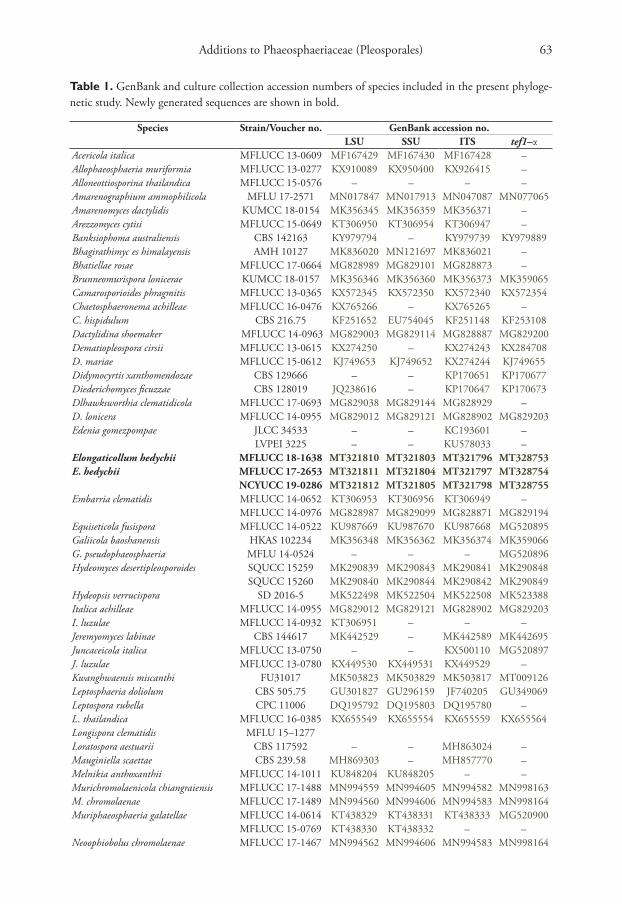
Figure 1. RAxML tree inferred from combined dataset of ITS, LSU, SSU and tef1-α partial sequences of 168 strains of Phaeosphaeriaceae. Bootstrap support values for maximum likelihood (ML), maximum par-simony (MP) values ≥70%, and Bayesian posterior probabilities (BYPP) ≥0.95 are given above each branch respectively. The new species are highlighted in red, and the new record in green. Ex-type strains are in bold. The tree is rooted by Leptosphaeria doliolum (CBS 505.75) and Paraleptosphaeria dryadis (CBS 643.86).
Danushka S. Tennakoon et al. / MycoKeys 70: 59–88 (2020)68
Figure 1. Continued.
formative, while 308 variable characters are parsimony-uninformative in the maxi-mum parsimony (MP) analysis (TL = 6364, CI = 0.250, RI = 0.657, RC = 0.164, HI = 0.750). The RAxML analysis of the combined dataset yielded a best scoring tree (Figure 1) with a final ML optimization likelihood value of – 34492.801018. The matrix had 1331 distinct alignment patterns, with 37.25% of undetermined characters or gaps. Estimated base frequencies are; A = 0.247120, C = 0.228182, G = 0.268238, T = 0.256459; substitution rates AC = 1.250439, AG = 3.526348, AT = 2.517351, CG = 0.798250, CT = 6.907432, GT = 1.000; proportion of in-

Additions to Phaeosphaeriaceae (Pleosporales) 69
Figure 1. Continued.
variable sites I = 0.596400; gamma distribution shape parameter α = 0.492378. All analyses (ML, MP and BI) gave similar results and are in agreement with previous studies based on multi-gene analyses (Hyde et al. 2019, 2020; Phookamsak et al. 2019). Phylogenetic analyses of the combined data matrix resulted in well-resolved clades, many of which had considerably high statistical support (Figure 1). Boot-strap support values for maximum likelihood, maximum parsimony ≥70%, and Bayesian posterior probabilities (BYPP) ≥0.95 are given above each branch in that order (Figure 1). Phylogenetic position and statistical support are noted in the tax-onomy section.

Danushka S. Tennakoon et al. / MycoKeys 70: 59–88 (2020)70
Taxonomy
Elongaticollum Tennakoon, C.H. Kuo & K.D. Hyde, gen. nov.Index Fungorum number: IF 557486Facesoffungi number: FoF07849
Etymology. Refers to the fact that the pycnidia have elongated necks.Diagnosis. Saprobic on dead leaves of Hedychium coronarium J. Koenig. Sexual
morph: Undetermined. Asexual morph: Coelomycetous. Conidiomata pycnidial, solitary, superficial, dark brown to black, obpyriform, papillate. Neck elongate, dark brown, usually straight, but sometimes slightly curved. Conidiomatal wall composed of 4–5 layers of light brown cells, arranged in textura angularis. Conidiophores reduced to conidiogenous cells. Conidiogenous cells hyaline, aseptate, smooth, ampulliform, arising from the inner cell wall of the apex. Conidia oval to oblong, smooth and thin-walled, hyaline, aseptate, with 1–2-minute guttules.
Type species. Elongaticollum hedychii Tennakoon, C.H. Kuo & K.D. Hyde.
Elongaticollum hedychii Tennakoon, C.H. Kuo & K.D. Hyde, sp. nov.Index Fungorum number: IF 557487Facesoffungi number: FoF07850Figure 2
Etymology. Name reflects the host Hedychium coronarium J. Koenig, from which the holotype was collected.
Figure 1. Continued.

Additions to Phaeosphaeriaceae (Pleosporales) 71
Holotype. MFLU 18-2542.Diagnosis. Saprobic on dead leaves of Hedychium coronarium J. Koenig. Sexual
morph: Undetermined. Asexual morph: Coelomycetous. Conidiomata 120–140 µm high, 60–70 µm diam., pycnidial, solitary, scattered, superficial, visible as small black spots on host surface, dark brown to black, obpyriform, papillate. Neck up to 80–100 µm long, 20–30 µm diam., elongated, dark brown, usually straight, but some-times slightly curved. Conidiomatal wall 10–20 µm wide, composed of 4–5 layers of light brown, thick-walled cells, arranged in textura angularis. Conidiophores reduced to conidiogenous cells. Conidiogenous cells 3–4 × 3–3.5 µm (x̄ = 3.6 × 3.2 µm, n = 10), arising from the inner cell wall of the apex, hyaline, aseptate, smooth, ampulliform. Conidia 4–5 × 1.8–2.2 µm (x̄ = 4.6 × 2.1 µm, n = 30), oval to oblong, smooth, thin-walled, hyaline, aseptate, with 1–2-minute guttules.
Culture characteristics. Colonies on PDA reaching 30 mm diameter after 3 weeks at 20–25 °C, colonies medium sparse, circular, raised, surface slightly rough with entire edge, margin entire, colony from above: light brown to grey at the margin, dark brown at middle, dark brown to black at the center; reverse, light brown to yellowish at the margin, brown at middle, dark brown to black at the center; mycelium light brown to grey with tufts; not producing pigments in PDA.
Material examined. Taiwan, Chiayi, Fanlu Township area, Dahu Forest, dead leaves of Hedychium coronarium J. Koenig (Zingiberaceae), 15 August 2018 (23°27.514'N, 120°36.302'E), D.S. Tennakoon, TLF031-A (MFLU 18-2542, holotype), ex-type living culture (MFLUCC 18-1638 = NCYUCC 19-0163); ibid. 20 August 2018 (23°27.530'N, 120°36.314'E), TLF031-B (NCYU19-0139, paratype), living culture (NCYUCC19-0286); ibid. 25 August 2018 (23°27.512'N, 120°36.301'E), TLF031-C (NCYU19-0140, paratype), living culture (NCYUCC 19-0287).
Notes. The genus Elongaticollum differs from other asexual morphs in Phaeospha-eriaceae in dark brown to black, superficial, obpyriform, pycnidial conidiomata with distinct elongate necks (80–100 µm) and a globose base and oval to oblong, hyaline, aseptate conidia (Figure 2). Multi-gene phylogenetic analyses (LSU, SSU, ITS, tef1-α), show Elongaticollum strains constitute a highly supported independent lineage nested between Setophoma sensu lato and Neostagonosporella (97% ML, 80% MP, 1.00 BYPP, Figure 1). However, the asexual morph of Setophoma can be distinguished from Elonga-ticollum in having setose conidiomata without elongate necks and oblong to ellipsoidal conidia, whereas, Elongaticollum have conidiomata with distinct elongate necks and lacking setae and oval to oblong conidia (De Gruyter et al. 2010; Phookamsak et al. 2014). Despite some Setophoma species not having setae (i.e. S. antiqua, S. endophytica, and S. yunnanensis) (Liu et al. 2019), Elongaticollum species can be distinguished by its superficial conidiomata with elongate necks.
The asexual morph of Neostagonosporella differs from Elongaticollum in having multiloculate conidiomata without distinct elongate necks and two types of conidia (macroconidia: subcylindrical to cylindrical, transversely multi-septate, hyaline and microconidia oval, ellipsoidal or long ellipsoidal, aseptate, hyaline), whereas Elongati-collum has uni-loculate conidiomata with distinct elongate necks and oval to oblong conidia (Figure 2, Yang et al. 2019).

Danushka S. Tennakoon et al. / MycoKeys 70: 59–88 (2020)72
Figure 2. Elongaticollum hedychii (MFLU 18-2542, holotype) a specimen b appearance of conidiomata on host c close up of conidiomata on host d vertical section through conidioma e, f squash mount of conidioma g conidioma wall h, i elongated conidiomatal necks j conidiogenous cells k conidia l, m ger-minated conidia n colony from below o colony from above p, q pycnidia formed on PDA. Scale bars: 100 µm (c), 50 µm (d–h), 10 µm (g), 30 µm (i), 3 µm (j–m), 100 µm (p, q).
Phylogenetic investigations herein provide insights into the taxonomy of Setopho-ma as well (Figure 1). Two major clades of Setophoma are recovered (Setophoma sensu stricto and Setophoma sensu lato). The Setophoma sensu stricto clade includes S. brachy-podii, S. poaceicola and S. terrestris (type species). Setophoma sensu lato comprises S. antiqua, S. chromolaenae, S. endophytica, S. pseudosacchari, S. sacchari, S. vernoniae, S. yingyisheniae and S. yunnanensis (Figure 1). Elongaticollum, differs from Setophoma sen-su lato in having distinct superficial, obpyriform, pycnidial conidiomata with a globose base and distinct elongated necks (Figure 2, Liu et al. 2019). Further work is needed to resolve relationships between Setophoma sensu stricto and Setophoma sensu lato.
Ophiosphaerella Speg., Anal. Mus. nac. B. Aires, Ser. 3 12: 401 (1909)
Notes. Ophiosphaerella was introduced by Spegazzini (1909) to accommodate O. graminicola Speg. as the type species. The species of this genus are characterized by papillate ascomata bearing fissitunicate, cylindrical asci frequently narrower near the

Additions to Phaeosphaeriaceae (Pleosporales) 73
base, with a short furcate pedicel and filamentous, pale brown, multi-septate ascospores without swollen cells or separating into part spores. Barr (1987) placed Ophiosphaerella in Phaeosphaeriaceae and this was confirmed by Zhang et al. (2009, 2012) and Hyde et al. (2013) based on molecular phylogeny. Most Ophiosphaerella species are often found as pathogens or saprobes worldwide on Poaceae and Cyperaceae (Câmara et al. 2000). Currently, twelve Ophiosphaerella species are listed in Index Fungorum (2020). In this study, we introduce Ophiosphaerella taiwanensis from Agave tequilana F.A.C. Weber (Asparagaceae) as a new species.
Ophiosphaerella taiwanensis Tennakoon, C.H. Kuo & K.D. Hyde, sp. nov.Index Fungorum number: IF 557488Facesoffungi number: FoF07851Figure 3
Etymology. Named after Taiwan, where this fungus was collected.Holotype. MFLU 18-2534.Diagnosis. Saprobic on dead leaf of Agave tequilana F.A.C. Weber (Asparagaceae).
Sexual morph: Ascomata 270–310 µm high, 220–260 µm diam., solitary, scattered, immersed to slightly erumpent through host tissue with papilla, visible as raised, small black dots in host surface, globose to subglobose, uniloculate, glabrous, dark brown to black, ostiole central, periphysate. Peridium 20–25 µm wide, thick-walled, of equal thickness, composed of 6–7 layers of small, flattened, brown to dark brown pseudo-parenchymatous cells, hyaline towards the inside, arranged in a textura angularis, fusing and indistinguishable from the host tissues. Hamathecium of 1.5–2.5 µm wide, cellular, septate, rarely branching, pseudoparaphyses, anastomosing mostly above the asci and embedded in a mucilaginous matrix. Asci 115–140 × 8.5–10 µm (x̄ = 121.6 × 9.2 µm, n = 20), 8-spored, bitunicate, fissitunicate, cylindrical to cylindric-clavate, short pedi-cellate, apically rounded, with a well-developed ocular chamber. Ascospores 110–132 × 2.2–2.7 µm (x̄ = 117.2 × 2.4 µm, n = 20), fasciculate, parallel, scolecosporous, fili-form, 12–13-septate, narrowing towards ends, pale brown to brown, smooth-walled. Asexual morph: Undetermined.
Culture characteristics. Colonies on PDA reaching 25 mm diameter after 3 weeks at 20–25 °C, colonies medium sparse, circular, raised, surface slightly rough with entire edge, margin well-defined, colony from above: gray to light brown at the margin, gray to cream at the center; reverse, gray to light brown at the margin, dark brown to black at the center; mycelium whitish gray with tufting; not producing pigments in PDA.
Material examined. Taiwan, Chiayi, Fanlu Township area, Dahu Forest, dead leaf of Agave tequilana F.A.C. Weber (Asparagaceae), 15 August 2018 (23°27.520'N, 120°36.310'E), D.S. Tennakoon, TLF016 (MFLU 18-2534, holotype); ibid. (NCYU19-0131, isotype), ex-type living culture, NCYUCC 19-0152.
Notes. The scolecosporous specimen was collected from dead leaves of Agave te-quilana (Asparagaceae) in Taiwan. The multi-gene phylogenetic analysis (Figure 1)

Danushka S. Tennakoon et al. / MycoKeys 70: 59–88 (2020)74
Figure 3. Ophiosphaerella taiwanensis (MFLU 18-2534, holotype) a, b appearance of ascomata on host c close-up of ascomata d vertical section through ascoma e apex of ascoma f peridium g pseudoparaphyses h–j asci k, l ascospores m germinated ascospore in PDA n colony from above o colony from below. Scale bars: 100 µm (d, e), 15 µm (f), 50 µm (g–m).
shows our strain (Ophiosphaerella taiwanensis, NCYUCC 19-0152), cluster with other Ophiosphaerella species, in particular with close affinity to Ophiosphaerella agrostidis with high bootstrap support (88% ML, 70% MP, 0.99 BYPP, Figure 1). Morphologi-cal characters of our collection (NCYUCC 19-0152) differ from Ophiosphaerella agros-tidis in having periphyses in the ostiole, 12–13 septate ascospores and host occurrence (Asparagaceae). Ophiosphaerella agrostidis was introduced by Câmara et al. (2000) on Agrostis palustris (Poaceae), and is lacking periphyses, comprises 15-septate ascospores (Phookamsak et al. 2014). A comparison of the 619 nucleotides across the tef1-α gene region of Ophiosphaerella taiwanensis and O. agrostidis (MFLUCC 11-0152) reveals 17 base pair differences (2.74%).
Phaeosphaeriopsis M.P.S. Câmara, M.E. Palm & A.W. Ramaley, Mycol. Res. 107(5): 519 (2003)
Notes. The genus Phaeosphaeriopsis was introduced by Câmara et al. (2003) to ac-commodate Paraphaeosphaeria-like taxa, viz. P. agavensis A.W. Ramaley, M.E. Palm &

Additions to Phaeosphaeriaceae (Pleosporales) 75
M.E. Barr, P. glaucopunctata (Grev.) Shoemaker & C.E. Babc., P. nolinae A.W. Rama-ley, P. obtusispora (Speg.) O.E. Erikss, Phaeosphaeriopsis amblyspora A. W. Ramaley and Phaeosphaeriopsis amblyspora A. W. Ramaley. The genus is typified by P. glaucopunctata and characterized by having immersed, sub-epidermal, globose to subglobose to pyri-form ascomata, cylindric asci and septate, punctate or verrucose ascospores (Câmara et al. 2003; Phookamsak et al. 2014; Thambugala et al. 2014; Tibpromma et al. 2017). Currently, 17 Phaeosphaeriopsis species are accepted in Index Fungorum (2020). In this paper, Phaeosphaeriopsis beaucarneae is introduced from Beaucarnea recurvata (As-paragaceae) as a new species and the sexual/asexual morph connection between strains isolated from the natural habitat was established based on molecular sequence data.
Phaeosphaeriopsis beaucarneae Tennakoon, C.H. Kuo & K.D. Hyde, sp. nov.Index Fungorum number: IF 557489Facesoffungi number: FoF07852Figures 4, 5
Etymology. Name reflects the host Beaucarnea recurvata Lem., from which the holo-type was collected.
Holotype. MFLU 18-2586.Diagnosis. Saprobic on dead leaf of Beaucarnea recurvata Lem. (Asparagaceae).
Sexual morph: Ascomata 160–200 µm high, 220–250 µm diam., scattered, solitary, gregarious, coriaceous, immersed to semi-immersed, slightly raised, erumpent, visible as black spots on host surface, uniloculate, dark brown to black, globose to subglobose, ostiolate. Ostiole central, papillate. Peridium 20–30 µm wide, thick-walled, of equal thickness, composed of 4–5 layers of dark brown to brown, thick-walled, pseudo-parenchymatous cells of textura angularis. Hamathecium of 1.5–2.5 µm wide, cellular, septate, rarely branching, pseudoparaphyses, anastomosing mostly above the asci and embedded in a mucilaginous matrix. Asci 80–90 × 9–10 µm (x̄ = 86.5 × 9.6 µm, n = 25), 8-spored, bitunicate, fissitunicate, cylindrical to cylindric-clavate, short pedi-cellate, apically rounded, with a well-developed ocular chamber. Ascospores 20–25 × 5.5–7 µm (x̄ = 22.6 × 6.2 µm, n = 20), overlapping 1–2-seriate, oblong to cylindrical, yellowish to light brown, slightly narrowing towards the end cells, mostly 5-septate, constricted at the septa, enlarged at the 4th cell from above, verruculose, straight to curved, lacking a mucilaginous sheath. Asexual morph: Conidiomata 180–200 µm high, 140–160 µm diam., pycnidial, solitary, immersed to erumpent, small black spots on host surface, globose to subglobose with centrally placed ostiole. Conidiomatal wall 28–34 µm wide, composed of 6–7 layers of dark brown cells, arranged in textu-ra angularis. Conidiophores reduced to conidiogenous cells. Conidiogenous cells 3–4 × 2.6–3.1 µm, holoblastic, phialidic, single, discrete, sometimes integrated, ampulliform or cylindric-clavate, hyaline, arising from basal stratum. Conidia 6.8–7.4 × 3–4 µm (x̄ = 7.1 × 3.4 µm, n = 30), 1-celled, globose to subglobose, initially hyaline, becoming brown to dark brown, aseptate, rough-walled.

Danushka S. Tennakoon et al. / MycoKeys 70: 59–88 (2020)76
Figure 4. Phaeosphaeriopsis beaucarneae (MFLU 18-2586, holotype) a appearance of ascomata on host b close up of ascoma c vertical section through ascoma d peridium e pseudoparaphyses f–i asci j–n as-cospores o germinated ascospore in PDA p colony from above q colony from below. Scale bars: 100 µm (c), 15 µm (d), 50 µm (e–i), 10 µm (j–o).
Culture characteristics. Colonies on PDA reaching 27 mm diameter after 3 weeks at 20–25 °C, colonies medium sparse, circular, raised, surface slightly rough with entire edge, margin irregular, colony from above: light brown at the margin, white to cream at the center; reverse, yellow to light brown at the margin, light brown to brown at the center; mycelium white to cream with tufting; not producing pigments in PDA.
Material examined. Taiwan, Chiayi, Fanlu Township area, Dahu Forest, dead leaf of Beaucarnea recurvata Lem. (Asparagaceae), 21 July 2018 (23°27.514'N, 120°36.302'E), D.S. Tennakoon, SV027 (MFLU 18-2586, holotype); ibid. (NCYU19-0184, isotype), ex-type living culture, NCYUCC 19-0106; ibid., Dahu forest, dead leaf of Beaucarnea recurvata Lem. (Asparagaceae), 25 July 2018 (23°26.534'N, 120°36.220'E), D.S. Ten-nakoon, SV028 (MFLU 18-2587, paratype); living culture, NCYUCC 19-0107.
Notes. Phaeosphaeriopsis beaucarneae is similar to other Phaeosphaeriopsis spe-cies in having scattered, semi-immersed to erumpent, globose to subglobose, ostio-late ascomata and cylindrical to clavate asci and light brown, verrucose ascospores (Phookamsak et al. 2014; Thambugala et al. 2014; Hyde et al. 2020). According to

Additions to Phaeosphaeriaceae (Pleosporales) 77
Figure 5. Phaeosphaeriopsis beaucarneae (MFLU 18-2586, paratype) a appearance of conidiomata on host b close up of conidiomata c vertical section through conidioma d conidiomatal wall e, f conid-iogenous cells and developing conidia g–i conidia j germinated conidium in PDA k colony from above l colony from below. Scale bars: 100 µm (c), 20 µm (d), 3 µm (e, f), 5 µm (g–j).
the present multi-gene phylogenetic analyses (Figure 1), Phaeosphaeriopsis beaucarneae is grouped with other Phaeosphaeriopsis species, in particularly closely to P. grevilleae (CBS 145369) with high statistical support (70% ML, 75% MP, 0.99 BYPP, Figure 1). The asexual morph of P. grevilleae was isolated from leaves of Grevillea sp. (Proteaceae) and introduced by Marin-Felix et al. (2019). Phaeosphaeriopsis beaucarneae differs from P. grevilleae in having larger conidia (6.8–7.4 × 3–4 µm), whereas P. grevilleae has com-paratively smaller conidia (5 × 3.5 µm). A comparison of the 516 nucleotides across the ITS (+5.8S rDNA) gene region of Phaeosphaeriopsis beaucarneae and P. grevilleae (CBS 145369) revealed 16 base pair differences (3.10%). In addition, we compared our new taxon with P. grevilleae based on base pair differences in the tef1-α gene region. We found a total of 19 base pair differences (3.06%) across 619 nucleotides.
Recent studies have revealed that Phaeosphaeriopsis is a species rich genus and numerous Phaeosphaeriopsis species have been described during the last few years (Thambugala et al. 2014; Tibpromma et al. 2017; Marin-Felix et al. 2019; Al-Jaradi et al. 2020; Hyde et al. 2020). With this study, the number of Phaeosphaeriopsis spe-cies increases to 18.

Danushka S. Tennakoon et al. / MycoKeys 70: 59–88 (2020)78
Neosetophoma Gruyter, Aveskamp & Verkley, Mycologia 102(5): 1075 (2010)
Notes. Neosetophoma was introduced by de Gruyter et al. (2010), typified by N. sama-rarum (Desm.) Gruyter, Aveskamp. & Verkley. Species of Neosetophoma are character-ized by globose to irregular conidiomata, with papillate ostioles, and yellowish conidia that are attenuate at one end (De Gruyter et al. 2010; Liu et al. 2015). Tibpromma et al. (2017) introduced Neosetophoma garethjonesii Tibpromma, E.B.G. Jones & K.D. Hyde as the first report of the sexual morph of Neosetophoma. Neosetophoma species have a diverse distribution as saprobes, endophytes, plant pathogens and soil fungi (Phookamsak et al. 2014; Hernandez-Restrepo et al. 2016; Karunarathna et al. 2017; Tibpromma et al. 2017; Wanasinghe et al. 2018). Currently, 19 Neosetophoma species are accepted in Index Fungorum (2020). In this study, we found Neosetophoma poacei-cola Goonas., Thambug. & K.D. Hyde from dead leaves of Musa acuminata Colla in Taiwan. This is the first Neosetophoma species recorded from the plant family Musaceae.
Neosetophoma poaceicola Goonas., Thambug. & K.D. Hyde. Mycosphere 8: 742 (2017)Index Fungorum number: IF552974Facesoffungi number: FoF00262Figure 6
Diagnosis. Saprobic on dead leaf petioles of Musa acuminata Colla (Musaceae). Sex-ual morph: Ascomata 70–100 µm high, 90–130 µm diam., solitary, gregarious, co-riaceous, immersed to semi-immersed, slightly raised, visible as black spots on host surface, uni-loculate, dark brown to black, globose to ovoid. Peridium 15–20 µm wide, thick-walled, of equal thickness, composed of several layers of dark brown to brown, pseudoparenchymatous cells of textura angularis. Hamathecium of 1–2 µm wide, cel-lular, rarely branching, pseudoparaphyses, anastomosing mostly above the asci and embedded in a mucilaginous matrix. Asci 60–80 × 7–8 µm (x̄ = 70.6 × 7.6 µm, n = 30), 8-spored, bitunicate, fissitunicate, cylindric-clavate with a short, rounded pedicel, api-cally rounded. Ascospores 20–30 × 3–4 µm (x̄ = 25.5 × 3.7 µm, n = 40), overlapping 1–2-seriate, hyaline, fusiform, with acute ends, 1-septate, 3–4 eu-septate, cell near the septum slightly larger, slightly constricted at the septum, straight to curved, smooth-walled, guttulate. Asexual morph: Undetermined.
Culture characteristics. Colonies on PDA reaching 30 mm diameter after 3 weeks at 20–25 °C, colonies medium sparse, circular, flat, surface slightly rough with entire edge, margin well-defined, colony from above: yellow to light brown at the margin, brown at the center; reverse, yellow to light brown at the margin, dark brown at the cent-er; mycelium light brown to whitish grey with tufting; not producing pigments in PDA.
Material examined. Taiwan, Chiayi, Fanlu Township area, Dahu Forest, dead leaf petiole of Musa acuminata Colla (Musaceae), 21 July 2018 (23°27.530'N, 120°36.340'E), D.S. Tennakoon, SV049 (MFLU 18-2597, new host record), living culture, MFLUCC 18-1632, NCYUCC 19-0119.
Additions to Phaeosphaeriaceae (Pleosporales) 79
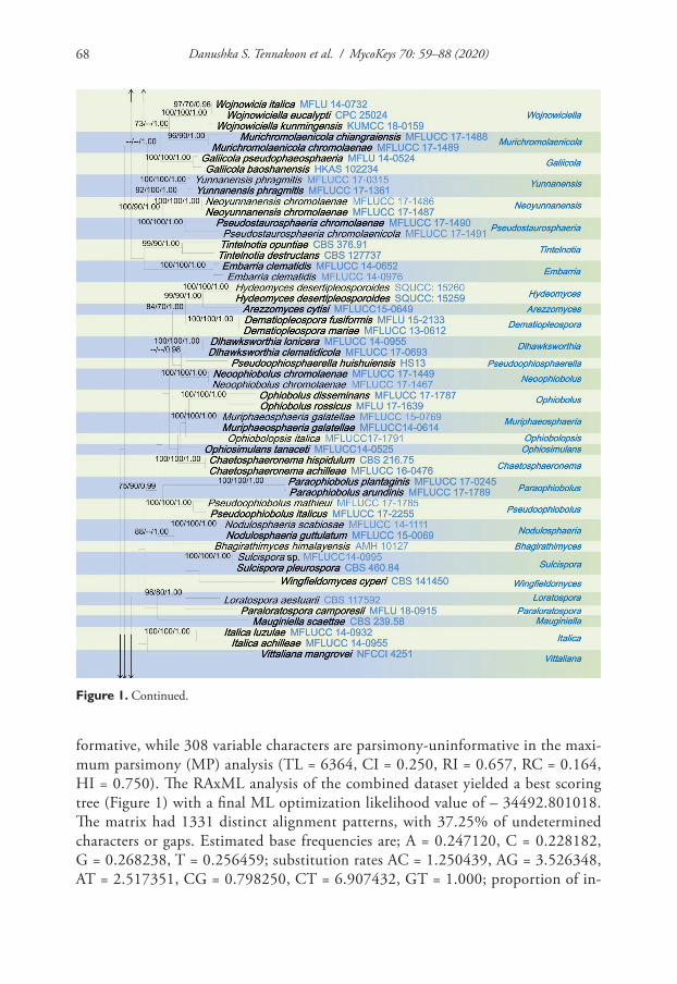
Notes. As morphological characters (immersed to semi-immersed ascomata, cylin-dric-clavate, apically rounded asci with short rounded pedicel and hyaline, fusiform, 1-septate ascospores) largely overlap with those of Neosetophoma poaceicola (MFLUCC 16–0886), we report our collection (MFLUCC 18-1632) as a new host record of N. poa-ceicola from dead leaves of Musa acuminata (Musaceae) in Taiwan. Combined multi-gene (LSU, SSU, ITS and tef1-α) based phylogenies also showed that our collection clustered with Neosetophoma poaceicola (MFLUCC 16-0886), with high bootstrap support (100% ML, 100% MP, 1.00 BYPP, Figure 1). Neosetophoma poaceicola was introduced by Tham-bugala et al. (2017) from dead leaves of grass species in Thailand. However, our collection slightly differs from Neosetophoma poaceicola (MFLUCC 16-0886) in having compara-tively slightly larger ascospores (20–30 × 3–4 µm, versus 18.5–22.5 × 3.5–5 µm).
Neosetophoma species have been recorded from various host families, viz. Brassi-caceae, Caprifoliaceae, Iridaceae, Malvaceae, Ranunculaceae, Salicaceae, but most are reported from Poaceae (Phookamsak et al. 2014; Karunarathna et al. 2017; Tibpromma et al. 2017, Wanasinghe et al. 2018; Marin-Felix et al. 2019). Interestingly, this is the first Neosetophoma species record (MFLU 18-2597) from the plant family Musaceae.
Figure 6. Neosetophoma poaceicola (MFLU 18–2597, new host record) a appearance of ascomata on host b close up of ascomata c vertical section through ascoma d peridium e pseudoparaphyses f–h asci i–k as-cospores l germinated ascospore in PDA m colony from above n colony from below. Scale bars: 50 µm (c), 20 µm (d), 30 µm (e–h), 15 µm (i–l).

Danushka S. Tennakoon et al. / MycoKeys 70: 59–88 (2020)80
Discussion
The taxonomy of Phaeosphaeriaceae has been subjected to several changes in recent years. Traditionally, morphology-based identification was the main means for identi-fying Phaeosphaeriaceae species (Barr 1979, 1992; Tomilin 1993). However, species identification has been revolutionized by the application of molecular based approach-es incorporating DNA sequence data in Phaeosphaeriaceae (Phookamsak et al. 2014, 2017; Tennakoon et al. 2016; Wanasinghe et al. 2018; Bakhshi et al. 2019; Chethana et al. 2020; Hyde et al. 2020). Phaeosphaeriaceae species are adapted to a wide range of ecological environments and are present in soils, fresh and marine habitats and cause infections in humans (Yuan 1994; Phookamsak et al. 2014, 2017; Ahmed et al. 2017; Maharachchikumbura et al. 2019; Valenzuela-Lopez et al. 2019). Members of the Phaeosphaeriaceae have also been recorded from both temperate and tropical countries (i.e. Austria, Belgium, Bulgaria, Canada, China, Germany, Italy, Japan, Nor-way, Poland, Thailand, Sweden, Switzerland) and from different host families (i. e. Ac-oraceae, Arecaceae, Cyperaceae, Asparagaceae, Brassicaceae, Fabaceae, Poaceae, Maran-taceae) (Shoemaker and Babcock 1989; Phookamsak et al. 2014, 2019; Wanasinghe et al. 2018; Maharachchikumbura et al. 2019; Farr and Rossman 2020). Due to their cosmopolitan distribution, in the last few years, many researchers have paid significant attention to the Phaeosphaeriaceae (Phookamsak et al. 2014, 2019; Tennakoon et al. 2016; Wanasinghe et al. 2018; Bakhshi et al. 2019; Hyde et al. 2020).
The fungi that decay leaf litter are highly diverse and may be host-specific (Pa-rungao et al. 2002). Several studies have examined the succession of leaf degrading communities and found unique sets of species on different types of litter (Promput-tha et al. 2002, 2017; Duong et al. 2008). Additional ecological studies are therefore needed to establish whether these fungi are generalists or specialists. This study pro-vides evidence to indicate the fungal diversity in leaf litter, even within a single family, Phaeosphaeriaceae. Additional work is necessary to identify if the newly described species are host specific.
Acknowledgments
The authors would like to thank T.K. Goh for his valuable suggestions and help. Shaun Pennycook is thanked for checking species names. This research work was partially supported by Chiang Mai University and K.D. Hyde thanks Chiang Mai University for the award of Visiting Professorship. He also thanks the Thailand Research Fund for the Grant No. RDG613001, entitled “Impact of Climate Change on Fungal Diversity and Biogeography in the Greater Mekong Subregion”. D.N. Wanasinghe would like to thank the CAS President’s International Fellowship Initiative (PIFI) for funding his postdoctoral research (number 2019PC0008), the National Science Foundation of China and the Chinese Academy of Sciences for financial support under the following grants: 41761144055, 41771063 and Y4ZK111B01. Wanasinghe also thanks the 64th batch of China Postdoctoral Science Foundation (grant no: Y913083271).

Additions to Phaeosphaeriaceae (Pleosporales) 81
References
Al-Jaradi AJ, Maharachchikumbura SS, Al-Sadi AM (2020) Phaeosphaeriopsis omaniana (Phaeosphaeriaceae, Pleosporales), a novel fungus from Oman. Phytotaxa 436: 187–192. https://doi.org/10.11646/phytotaxa.436.2.8
Ahmed SA, Hofmueller W, Seibold M, de Hoog GS, Harak H, Tammer I, Van Diepeningen AD, Behrens‐Baumann W (2017) Tintelnotia, a new genus in Phaeosphaeriaceae harbour-ing agents of cornea and nail infections in humans. Mycoses 60: 244–253. https://doi.org/10.1111/myc.12588
Bakhshi M, Arzanlou M, Groenewald JZ, Quaedvlieg W, Crous PW (2019) Parastagonosporella fallopiae gen. et sp. nov. (Phaeosphaeriaceae) on Fallopia convolvulus from Iran. Mycologi-cal Progress 18: 203–214. https://doi.org/10.1007/s11557-018-1428-z
Bani A, Pioli S, Ventura M, Panzacchi P, Borruso L, Tognetti R, Tonon G, Brusetti L (2018) The role of microbial community in the decomposition of leaf litter and deadwood. Ap-plied soil ecology 126: 75–84. https://doi.org/10.1016/j.apsoil.2018.02.017
Barr ME (1979) A classification of Loculoascomycetes. Mycologia 71: 935–957. https://doi.org/10.1080/00275514.1979.12021099
Barr ME (1987) New taxa and combinations in the Loculoascomycetes. Mycotaxon 29: 501–505.Barr ME (1992) Additions to and notes on the Phaeosphaeriaceae (Pleosporales, Loculoasco-
mycetes). Mycotaxon 43: 371–400.Berg B, McClaugherty C (2003) Plant Litter. Decomposition, Humus Formation, Carbon
Sequestration. Springer-Verlag, Berlin Heidelberg, New York.Berg B, McClaugherty C (2008) Plant Litter. Decomposition, Humus Formation, Carbon
Sequestration (2nd ed.). Springer. https://doi.org/10.1007/978-3-540-74923-3Câmara MP, O’Neill NR, Berkum PV, Dernoeden PH, Palm ME (2000) Ophiosphaerella agros-
tis sp. nov. and its relationship to other species of Ophiosphaerella. Mycologia 92: 317–325. https://doi.org/10.1080/00275514.2000.12061162
Câmara MPS, Ramaley AW, Castlebury LA, Palm ME (2003) Neophaeosphaeria and Phaeospha-eriopsis, segregates of Paraphaeosphaeria. Mycological Research 107: 516–522. https://doi.org/10.1017/S0953756203007731
Chaiwan N, Wanasinghe DN, Camporesi E, Tibpromma S, Boonmee S, Lumyong S, Hyde KD (2019) Molecular taxonomy reveals the sexual morph of Nodulosphaeria digitalis in Phaeosphaeriaceae from Campanula trachelium in Italy. Phytotaxa 400: 1–13. https://doi.org/10.11646/phytotaxa.400.1.1
Chethana KWT, Jayawardena RS, Hyde KD (2020) Hurdles in fungal taxonomy: Effectiveness of recent methods in discriminating taxa. Megataxa 1: 114–122. https://doi.org/10.11646/megataxa.1.2.2
De Gruyter J, Woudenberg JH, Aveskamp MM, Verkley GJ, Groenewald JZ, Crous PW (2010) Systematic reappraisal of species in Phoma section Paraphoma, Pyrenochaeta and Pleurophoma. Mycologia 102: 1066–1081. https://doi.org/10.3852/09-240
Devadatha B, Mehta N, Wanasinghe DN, Baghela A, Sarma VV (2019) Vittaliana mangrovei gen. nov, sp. nov. (Phaeosphaeriaceae), from mangroves near Pondicherry (India), based on morphology and multigene phylogeny. Cryptogamie Mycologie 40: 117–132. https://doi.org/10.5252/cryptogamiemycologie2019v40a7
Danushka S. Tennakoon et al. / MycoKeys 70: 59–88 (2020)82
Duong LM, McKenzie EHC, Lumyong S, Hyde KD (2008) Fungal succession on senescent leaves of Castanopsis diversifolia in Doi Suthep-Pui National Park, Thailand. Fungal Diver-sity 30: 23–36. http://cmuir.cmu.ac.th/jspui/handle/6653943832/60073
Farr DF, Rossman AY (2020) Fungal databases, Systematic mycology and microbiology labora-tory, ARS, USDA. [Retrieved April 10, 2020] http://nt.ars-grin.gov/fungaldatabases
Hall TA (1999) BioEdit: a user-friendly biological sequence alignment editor and analysis pro-gram for Windows 95/98/NT. Nucleic Acids Symposium Series 41: 95–98.
Hernandez-Restrepo M, Schumacher RK, Wingfield MJ, Ishtiaq A, Cai L, Duong TA, Edwards J, Gene J, Groenewald JZ, Sana J, Khalid AN (2016) Fungal systematics and evolution: FUSE 2. Sydowia 68: 193–230. https://doi.org/10.12905/0380.sydowia68-2016-0193
Hongsanan S, Hyde KD, Phookamsak R, Wanasinghe DN, McKenzie HCE, Sarma VV, Boon-mee S, Lücking R, Pem D, Bhat JD, Liu N, Tennakoon DS, Karunarathna A, Jiang SH, Jones EBG, Phillips AJL, Manawasinghe I, Tibpromma S, Jayasiri SC, Sandamali D, Jaya-wardena RS, Wijayawardene NN, Ekanayaka AH, Jeewon R, Lu YZ, Dissanayake AJ, Zeng XY, Luo Z, Tian Q, Phukhamsakda C, Thambugala KM, Dai D, Chethana TKW, Ertz D, Doilom M, Liu JK, Pérez-Ortega S, Suija A, Senwanna C, Wijesinghe SN, Konta S, Niranjan M, Zhang SN, Ariyawansa HA, Jiang HB, Zhang JF, de Silva NI, Thiyagaraja V, Zhang H, Bezerra JDP, Miranda-Gonzáles R, Aptroot A, Kashiwadani H, Harishchan-dra D, Aluthmuhandiram JVS, Abeywickrama PD, Bao DF, Devadatha B, Wu HX, Moon KH, Gueidan C, Schumm F, Bundhun D, Mapook A, Monkai J, Chomnunti P, Samara-koon MC, Suetrong S, Chaiwan N, Dayarathne MC, Jing Y, Rathnayaka AR, Bhunjun CS, Xu J, Zheng J, Liu G, Feng Y, Xie N (2020) Refined families of Dothideomycetes. Fungal diversity. [In press]
Huelsenbeck JP, Ronqvist F (2001) MRBAYES: Bayesian inference of phylogenetic trees. Bio-informatics 17: 754–755. https://doi.org/10.1093/bioinformatics/17.8.754
Hyde KD, Jones EBG, Liu JK, Ariyawansa H, Boehm E, Boonmee S, Braun U, Chomnunti P, Crous PW, Dai DQ, Diederich P, Dissanayake A, Doilom M, Doveri F, Hongsanan S, Jayawardena R, Lawrey JD, Li YM, Liu YX, Lücking R, Monka J, Muggia L, Nelsen MP, Pang KL, Phookamsak R, Senanayake IC, Shearer CA, Suetrong S, Tanaka K, Thambugala KM, Wijayawardene NN, Wikee S, Wu HX, Zhang Y, Begoña AH, Alias SA, Aptroot A, Bahkali AH, Bezerra JL, Bhat DJ, Camporesi E, Chukea E, Gueidan C, Hawksworth DL, Hirayama K, Hoog SD, Kang JK, Knudsen K, Li WJ, Li XH, Liu ZY, Mapook A, Mckenzie EHC, Miller AN, Mortimer PE, Phillips AJL, Raja HA, Scheuer C, Schumm F, Taylor JE, Tian Q, Tibpromma S, Wanasinghe DN, Wang Y, Xu JC, Yacharoen S, Yan JY, Zang M (2013) Families of Dothideomycetes. Fungal Diversity 63: 1–313. https://doi.org/10.1007/s13225-013-0263-4
Hyde KD, Tennakoon DS, Jeewon R, Bhat DJ, Maharachchikumbura SSN, Rossi W, Leonardi M, Lee HB, Mun HY, Houbraken J, Nguyen TTT, Jeon SJ, Frisvad JC, Dhanushka N, Wanasinghe DN, Luücking R, Aptroot A, Cáceres MES, Karunarathna SC, Hongsanan S, Phookamsak R, de Silva NI, Thambugala KM, Jayawardena RS, Senanayake IC, Boonmee S, Chen J, Luo ZL, Phukhamsakda C, Pereira OL, Abreu VP, Rosado AWC, Bart B, Ran-drianjohany E, Hofstetter V, Gibertoni TB, da Silva Soares AM, Plautz Jr HL, Sotão HMP, Xavier WKS, Bezerra JDP, de Oliveira TGL, de Souza-Motta CM, Magalhães OMC, Bun-dhun D, Harishchandra D, Manawasinghe IS, Dong W, Zhang SN, Bao DF, Samarakoon
Additions to Phaeosphaeriaceae (Pleosporales) 83
MC, Pem D, Karunarathna A, Lin CG, Yang J, Perera RH, Kumar V, Huang SK, Dayar-athne MC, Ekanayaka AH, Jayasiri SC, Xiao YP, Konta S, Niskanen T, Liimatainen K, Dai YC, Ji XH, Tian XM, Mešić A, Singh SK, Phutthacharoen K, Cai L, Sorvongxay T, Thiyagaraja V, Norphanphoun C, Chaiwan N, Lu YZ, Jiang HB, Zhang JF, Abeywickrama PD, Aluthmuhandiram JVS, Brahmanage RS, Zeng M, Chethana T, Wei DP, Réblová M, Fournier J, Nekvindová J, do Nascimento Barbosa R, dos Santos JEF, de Oliveira NT, Li GJ, Ertz D, Shang QJ, Phillips AJL, Kuo CH, Camporesi E, Bulgakov TS, Lumyong S, Jones EBG, Chomnunti P, Gentekaki E, Bungartz F, Zeng XY, Fryar S, Tkalčec Z, Liang J, Li GS, Wen TC, Singh PN, Gafforov Y, Promputtha I, Yasanthika E, Goonasekara ID, Zhao RL, Zhao Q, Kirk PM, Liu JK, Yan JY, Mortimer PE, Xu JC (2019) Fungal diversity notes 1036–1150: taxonomic and phylogenetic contributions on genera and species of fungal taxa. Fungal Diversity 96: 1–242. https://doi.org/10.1007/s13225-019-00429-2
Hyde KD, Dong Y, Phookamsak R, Jeewon R, Bhat DJ, Jones EBG, Liu NG, Abeywickrama PD, Mapook A, Wei DP, Perera RH, Manawasinghe IS, Pem D, Bundhun D, Karunar-athna A, Ekanayaka AH, Bao DF, Li JF, Samarakoon MC, Chaiwan N, Lin CG, Phut-thacharoen K, Zhang SN, Senanayake IC, Goonasekara ID, Thambugala KM, Phukham-sakda C, Tennakoon DS, Jiang HB, Yang J, Zeng M, Huanraluek N, Liu JK, Wijesinghe SN, Tian Q, Tibpromma S, Brahmanage RS, Boonmee S, Huang SK, Thiyagaraja V, Lu YZ, Jayawardena LS, Dong W, Yang EF, Singh SK, Singh SM, Rana S, Lad SS, Anand G, Devadatha B, Niranjan M, Sarma VV, Liimatainen K, Aguirre-Hudson B, Niskanen T, Overall A, Alvarenga RLM, Gibertoni TB, Pliegler WP, Horváth E, Imre A, Alves AL, San-tos ACDS, Tiago RV, Bulgakov TS, Wanasinghe DN, Bahkali AH, Doilom M, Elgorban AM, Maharachchikumbura SSN, Rajeshkumar KC, Haelewaters D, Mortimer PE, Zhao Q, Lumyong S, Xu JC, Sheng J (2020) Fungal diversity notes 1151–1276: taxonomic and phylogenetic contributions on genera and species of fungal taxa. Fungal Diversity 100: 1–273. https://doi.org/10.1007/s13225-020-00439-5
Index Fungorum (2020) Index Fungorum. http://www.indexfungorum.org/names/Names.asp [accessed 6 April 2020]
Jayasiri SC, Hyde KD, Ariyawansa HA, Bhat DJ, Buyck B, Cai L, Dai YC, Abd-Elsalam KA, Ertz D, Hidayat I, Jeewon R, Jones EBG, Bahkali AH, Karunarathna SC, Liu JK, Luangsa-ard JJ, Lumbsch HT, Maharachchikumbura SSN, McKenzie EHC, Moncalvo JM, Ghobad-Ne-jhad M, Nilsson H, Pang KA, Pereira OL, Phillips AJL, Raspé O, Rollins AW, Romero AI, Etayo J, Selçuk F, Stephenson SL, Suetrong S, Taylor JE, Tsui CKM, Vizzini A, Abdel-Wa-hab MA, Wen TC, Boonmee S, Dai DQ, Daranagama DA, Dissanayake AJ, Ekanayaka AH, Fryar SC, Hongsanan S, Jayawardena RS, Li WJ, Perera RH, Phookamsak R, de Silva NI, Thambugala KM, Tian Q, Wijayawardene NN, Zhao RL, Zhao Q, Kang JC, Promputtha I (2015) The faces of fungi database: fungal names linked with morphology, phylogeny and human impacts. Fungal Diversity 74: 3–18. https://doi.org/10.1007/s13225-015-0351-8
Johnson EA, Catley KM (2002) Life in the Leaf Litter. American Museum of Natural His-tory, New York.
Jones EBG, Pang KL, Abdel-Wahab MA, Scholz B, Hyde KD, Boekhout T, Ebel R, Rateb ME, Henderson L, Sakayaroj J, Suetrong S, Dayarathne MC, Kumar V, Raghukumar S, Sridhar KR, Bahkali AH, Gleason FH, Norphanphoun C (2019) An online resource for marine fungi. Fungal Diversity 96: 347–433. https://doi.org/10.1007/s13225-019-00426-5

Danushka S. Tennakoon et al. / MycoKeys 70: 59–88 (2020)84
Karunarathna A, Papizadeh, M, Senanayake IC, Jeewon R, Phookamsak R, Goonasekara ID, Wanasinghe DN, Wijayawardene NN, Amoozegar MA, Shahzadeh Fazeli SA, Camporesi E (2017) Novel fungal species of Phaeosphaeriaceae with an asexual/sexual morph connec-tion. Mycosphere 8: 1818–1834. https://doi.org/10.5943/mycosphere/8/10/8
Katoh K, Standley DM (2013) MAFFT multiple sequence alignment software version 7: im-provements in performance and usability. Molecular Biology and Evolution 30: 772–780. https://doi.org/10.1093/molbev/mst010
Krishna MP, Mohan M (2017) Litter decomposition in forest ecosystems: a review. Energy Ecology and Environment 2: 236–249. https://doi.org/10.1007/s40974-017-0064-9
Liu F, Wang J, Li H, Wang W, Cai L (2019) Setophoma spp. on Camellia sinensis. Fungal Sys-tematics and Evolution 4: 43–57. https://doi.org/10.3114/fuse.2019.04.05
Liu JK, Hyde KD, Jones EBG, Ariyawansa HA, Bhat DJ, Boonmee S, Maharachchikumbura SSN, McKenzie EHC, Phookamsak R, Phukhamsakda C, Shenoy BD, Abdel-Wahab MA, Buyck B, Chen J, Chethana KWT, Singtripop C, Dai DQ, Dai YC, Daranagama DA, Dis-sanayake AJ, Doilom M, D’souza MJ, Fan XL, Goonasekara ID, Hirayama K, Hongsanan S, Jayasiri SC, Jayawardena RS, Karunarathna SC, Li WJ, Mapook A, Norphanphoun C, Pang KL, Perera RH, Peršoh D, Pinruan U, Senanayake IC, Somrithipol S, Suetrong S, Tanaka K, Thambugala KM, Tian Q, Tibpromma S, Udayanga D, Wijayawardene NN, Wanasinghe DN, Wisitrassameewong K, Zeng XY, Abdel-Aziz FA, Adamčík S, Bahkali AH, Boonyuen N, Bulgakov T, Callac P, Chomnunti P, Greiner K, Hashimoto A, Hofstet-ter V, Kang JC, Lewis D, Li XH, Liu XZ, Liu ZY, Matsumura M, Mortimer PE, Rambold G, Randrianjohany E, Sato G, Sri-Indrasutdhi V, Tian CM, Verbeken A, von Brackel W, Wang Y, Wen TC, Xu JC, Yan JY, Zhao RL, Camporesi E (2015) Fungal diversity notes 1–110: taxonomic and phylogenetic contributions to fungal species. Fungal Diversity 72: 1–197. https://doi.org/10.1007/s13225-015-0324-y
Luo ZL, Hyde KD, Liu JK, Maharachchikumbura SSN, Jeewon R, Bao DF, Bhat DJ, Lin CG, Li WL, Yang J, Liu NG, Lu YZ, Jayawardena RS, Li JF, Su HY (2019) Freshwater Sordari-omycetes. Fungal Diversity 99: 451–660. https://doi.org/10.1007/s13225-019-00438-1
Maharachchikumbura SSN, Ariyawansa HA, Wanasinghe DN, Dayarathne MC, Al-Saady NA, Al-Sadi AM (2019) Phylogenetic classification and generic delineation of Hydeomyces desertipleosporoides gen. et sp. nov., (Phaeosphaeriaceae) from Jebel Akhdar Mountain in Oman. Phytotaxa 391: 28–38. https://doi.org/10.11646/phytotaxa.391.1.2
Mapook A, Hyde KD, McKenzie EHC, Jones EBG, Bhat DJ, Jeewon R, Stadler M, Samara-koon MC, Malaithong M, Tanunchai B (2020) Taxonomic and phylogenetic contribu-tions to fungi associated with the invasive weed Chromolaena odorata (Siam weed). Fungal Diversity. [In press] https://doi.org/10.1007/s13225-020-00444-8
Marin-Felix Y, Hernández-Restrepo M, Iturrieta-González I, García D, Carnegie AJ, Chee-wangkoon R, Gramaje D, Groenewald JZ, Guarnaccia V, Halleen F, Lombard L (2019) Genera of phytopathogenic fungi: GOPHY 3. Studies in Mycology 94: 1–124. https://doi.org/10.1016/j.simyco.2018.04.002
Miller MA, Pfeiffer W, Schwartz T (2010) Creating the CIPRES Science Gateway for Infer-ence of Large Phylogenetic Trees. SC10 Workshop on Gateway Computing Environments (GCE10). https://doi.org/10.1109/GCE.2010.5676129
Additions to Phaeosphaeriaceae (Pleosporales) 85
Mlambo MC, Paavola R, Fritze H, Louhi P, Muotka T (2019) Leaf litter decomposition and decomposer communities in streams affected by intensive forest biomass removal. Ecologi-cal indicators 101: 364–372. https://doi.org/10.1016/j.ecolind.2019.01.035
Parungao MM, Fryar SC, Hyde KD (2002) Diversity of fungi on rainforest litter in North Queensland, Australia. Biodiversity & Conservation 11: 1185–1194. https://doi.org/10.1023/A:1016089220042
Phookamsak R, Liu JK, McKenzie EHC, Manamgoda DS, Ariyawansa HA, Thambugala KM, Dai DQ, Camporesi E, Chukeatirote E, Wijayawardene NN, Bahkali AH, Mortimer PE, Xu JC, Hyde KD (2014) Revision of Phaeosphaeriaceae. Fungal Diversity 68: 159–238. https://doi.org/10.1007/s13225-014-0308-3
Phookamsak R, Wanasinghe DN, Hongsanan S, Phukhamsakda C, Huang SK, Tennakoon DS, Norphanphoun C, Camporesi E, Bulgakov TS, Promputtha I, Mortimer PE (2017) Towards a natural classification of ophiobolus and ophiobolus-like taxa; introducing three novel genera Ophiobolopsis, Paraophiobolus and Pseudoophiobolus in Phaeoshaeriaceae (Ple-osporales). Fungal Diversity 87: 299–339. https://doi.org/10.1007/s13225-017-0393-1
Phookamsak R, Hyde KD, Jeewon R, Bhat DJ, Jones EBJ, Maharachchikumbura SSN, Ras-pé O, Karunarathna SC, Wanasinghe DN, Hongsanan S, Doilom M, Tennakoon DS, Machado AR, Firmino AL,Ghosh A, Karunarathna A, Mešić A, Dutta AK, Thongbai B, Devadatha B, Norphanphoun C, Senwanna C, Wei D, Pem D, Ackah FK, Wang GN, Jiang HB, Madrid H, Lee HB, Goonasekara ID, Manawasinghe IS, Kušan Cano J, Gené J, Li J, Das K, Acharya K, Raj KNA, Latha KPD, Chethana KWT, He MQ, Dueñas M, Jadan M, Martín MP, Samarakoon MC, Dayarathne MC, Raza M, Park MS, Telleria MT, Chaiwan N, Matočec N, de Silva NI, Pereira OL, Singh PN, Manimohan P, Uniyal P, Shang QJ, Bhatt RP, Perera RH, Alvarenga RLM, Nogal-Prata S, Singh SK,Vadthanarat S, Oh SY, Huang SK, Rana S, Konta S, Paloi S, Jayasiri SC, Jeon SJ, Mehmood T, Gibertoni TB, Nguyen TTT, Singh U, Thiyagaraja V, Sarma VV, Dong W, Yu XD, Lu YZ, Lim YW, Chen Y, Tkalčec Z, Zhang ZF, Luo ZL, Daranagama DA, Thambugala KM, Tibpromma S, Camporesi E, Bulgakov T, Dissanayake AJ, Senanayake IC, Dai DQ, Tang LZ, Khan S, Zhang H, Promputtha I, Cai L, Chomnunti P, Zhao RL, Lumyong S, Boonmee S, Wen TC, Mortimer PE, Xu J (2019) Fungal diversity notes 929–1036: taxonomic and phylo-genetic contributions on genera and species of fungal taxa. Fungal Diversity 95: 1–273. https://doi.org/10.1007/s13225-019-00421-w
Pointing SB, Pelling AL, Smith GJD, Hyde KD (2005) Screening of basidiomycetes and xy-lariaceous fungi for lignin peroxidase and laccase gene-specific sequences. Mycological Re-search 109: 115–124. https://doi.org/10.1017/S0953756204001376
Posada D, Crandall KA (1998) Modeltest: testing the model of DNA substitution. Bioinfor-matics 14: 817–818. https://doi.org/10.1093/bioinformatics/14.9.817
Promputtha I, Lumyong S, Lumyong P, McKenzie EC, Hyde KD (2002) Fungal succession on senescent leaves of Manglietia garrettii in Doi Suthep-Pui National Park, northern Thai-land. Fungal Diversity 10: 89–100.
Promputtha I, Mckenzie EH, Tennakoon DS, Lumyong S, Hyde KD (2017) Succession and natural occurrence of saprobic fungi on leaves of Magnolia liliifera in a tropical forest. Cryptogamie Mycologie 38: 213–225. https://doi.org/10.7872/crym/v38.iss2.2017.213

Danushka S. Tennakoon et al. / MycoKeys 70: 59–88 (2020)86
Purahong W, Wubet T, Lentendu G, Schloter M, Pecyna MJ, Kapturska D, Hofrichter M, Krüger D, Buscot F (2016) Life in leaf litter: novel insights into community dynamics of bacteria and fungi during litter decomposition. Molecular Ecology 25: 4059–4074. https://doi.org/10.1111/mec.13739
Rambaut A (2012) FigTree version 1.4.0. http://tree.bio.ed.ac.uk/software/figtree/ [accessed 10 March 2020]
Rannala B, Yang Z (1996) Probability distribution of molecular evolutionary trees: a new meth-od of phylogenetic inference. Journal of Molecular Evolution 43: 304–311. https://doi.org/10.1007/BF02338839
Rehner SA, Samuels GJ (1994) Taxonomy and phylogeny of Gliocladium analysed from nuclear large subunit ribosomal DNA sequences. Mycological Research 98: 625–634. https://doi.org/10.1016/S0953-7562(09)80409-7
Rehner S (2001) Primers for Elongation Factor 1-α (EF1-α). http://ocid.NACSE.ORG/re-search/deephyphae/EF1primer.pdf
Robertson GP, Paul EA (1999) Decomposition and soil organic matter dynamics. In: Sala OE, Jackson RB, Mooney HA, Howarth RW (Eds) Methods of Ecosystem Science. Springer, New York, 104–116. https://doi.org/10.1007/978-1-4612-1224-9_8
Shoemaker RA, Babcock CE (1989) Phaeosphaeria. Canadian Journal of Botany 67: 1500–1599. https://doi.org/10.1139/b89-199
Spegazzini C (1909) Mycetes Argentinenses. Series IV. Anales del Museo Nacional de Historia Natural Buenos Aires. Ser. 3, 12: 257–458.
Stamatakis A, Hoover P, Rougemont J (2008) A rapid bootstrap algorithm for the RAxML web servers. Systematic biology 57: 758–771. https://doi.org/10.1080/10635150802429642
Stamatakis A (2014) RAxML version 8: a tool for phylogenetic analysis and post-analysis of large phylogenies. Bioinformatics 30: 1312–1313. https://doi.org/10.1093/bioinformat-ics/btu033
Swift MJ, Heal OW, Anderson MM (1979) Decomposition in Terrestrial Ecosystems. Black-well Scientific Publications, Oxford.
Swofford DL (2002) PAUP: phylogenetic analysis using parsimony, version 4.0 b10. Sinauer Associates, Sunderland.
Tennakoon DS, Hyde KD, Phookamsak R, Wanasinghe DN, Camporesi E, Promputtha I (2016) Taxonomy and phylogeny of Juncaceicola gen. nov.(Phaeosphaeriaceae, Pleospori-nae, Pleosporales). Cryptogamie Mycologie 37: 135–156. https://doi.org/10.7872/crym/v37.iss2.2016.135
Tennakoon DS, Jeewon R, Gentekaki E, Kuo CH. Hyde KD (2019) Multi-gene phylogeny and morphotaxonomy of Phaeosphaeria ampeli sp. nov. from Ficus ampelas and a new re-cord of P. musae from Roystonea regia. Phytotaxa 406: 111–128. https://doi.org/10.11646/phytotaxa.406.2.3
Tibpromma S, Hyde KD, Jeewon R, Maharachchikumbura SSN, Liu JK, Bhat DJ, Jones EBG, McKenzie E, Camporesi E, Bulgakov TS, Doilom M, Santiago AM, Das K, Manimo-han P, Gibertoni TB, Lim YW, Ekanayaka AH, Thongbai B, Lee HB, Yang J, Kirk PM, Sysouphanthong P, Singh SK, Boonmee S, Dong W, Raj KN, Latha KP, Phookamsak R, Phukhamsakda C, Konta S, Jayasiri SC, Norphanphoun C, Tennakoon D, Li J, Da-

Additions to Phaeosphaeriaceae (Pleosporales) 87
yarathne MC, Perera RH, Xiao Y, Wanasinghe DN, Senanayake IC, Goonasekara ID, Silva NI, Mapook A, Jayawardena RS, Dissanayake AJ, Manawasinghe IS, Chethana KW, Luo Z, Hapuarachchi KK, Baghela A, Soares AM, Vizzini A, Meiras-Ottoni A, Mešić A, Dutta AK, Souza CA, Richter C, Lin C, Chakrabarty D, Daranagama DA, Lima DX, Chakraborty D, Ercole E, Wu F, Simonini G, Vasquez G, Silva GA, Plautz HL, Ariyawansa HA, Lee HS, Kušan I, Song J, Sun J, Karmakar J, Hu K, Semwal KC, Thambugala KM, Voigt K, Acharya K, Rajeshkumar KC, Ryvarden L, Jadan M, Hosen MI, Mikšík M, Sa-marakoon MA, Wijayawardene NN, Kim NK, Matočec N, Singh PN, Tian Q, Bhatt RP, Oliveira RJ, Tulloss RE, Aamir S, Kaewchai S, Marathe SD, Khan S, Hongsanan S, Adhikari S, Mehmood T, Bandyopadhyay TK, Svetasheva TY, Nguyen TT, Antonín V, Li W, Wang Y, Indoliya Y, Tkalčec Z, Elgorban AM, Bahkali AH, Tang A, Su H, Zhang H, Promputtha I, Luangsa-Ard J, Xu J, Yan J, Kang JC, Stadler M, Mortimer PE, Chomnunti P, Zhao Q, Phillips AJ, Nontachaiyapoom S, Wen T, Karunarathna SC (2017) Fungal diversity notes 491–602: taxonomic and phylogenetic contributions to fungal taxa. Fungal Diversity 83: 1–261. https://doi.org/10.1007/s13225-017-0378-0
Thambugala KM, Camporesi E, Ariyawansa HA, Phookamsak R, Liu ZY, Hyde KD (2014) Phylogeny and morphology of Phaeosphaeriopsis triseptata sp. nov., and Phaeosphaeriopsis glaucopunctata. Phytotaxa 176: 238–250. https://doi.org/10.11646/phytotaxa.176.1.23
Thambugala KM, Wanasinghe DN, Phillips AJL, Camporesi E, Bulgakov TS, Phukhamsakda C, Ariyawansa HA, Goonasekara ID, Phookamsak R, Dissanayake A, Tennakoon DS, Tibprom-ma S, Chen YY, Liu ZY, Hyde KD (2017) Mycosphere notes 1–50: Grass (Poaceae) inhabit-ing Dothideomycetes. Mycosphere 8: 697–796. https://doi.org/10.5943/mycosphere/8/4/13
Tomilin BA (1993) New species of Loculoascomycetes (fam. Phaeosphaeriaceae Barr.). Novosti Sistematiki Nizshikh Rasteniĭ 29: 69–73.
Valenzuela-Lopez N, Sutton DA, Cano-Lira JF, Paredes K, Wiederhold N, Guarro J, Stchigel AM (2017) Coelomycetous fungi in the clinical setting: morphological convergence and cryptic diversity. Journal of clinical microbiology 55: 552–567. https://doi.org/10.1128/JCM.02221-16
Vilgalys R, Hester M (1990) Rapid genetic identification and mapping of enzymatically ampli-fied ribosomal DNA from several Cryptococcus species. Journal of Bacteriology 172: 4238–4246. https://doi.org/10.1128/JB.172.8.4238-4246.1990
Wanasinghe DN, Phukhamsakda C, Hyde KD, Jeewon R, Lee HB, Jones EBG, Tibpromma S, Tennakoon DS, Dissanayake AJ, Jayasiri SC, Gafforov Y, Camporesi E, Bulgakov TS, Ekanayake AH, Perera RH, Samarakoon MC, Goonasekara ID, Mapook A, Li WJ, Sena-nayake IC, Li JF, Norphanphoun C, Doilom M, Bahkali AH, Xu JC, Mortimer PE, Ti-bell L, Tibell S, Karunarathna SC (2018) Fungal diversity notes 709–839: taxonomic and phylogenetic contributions to fungal taxa with an emphasis on fungi on Rosaceae. Fungal Diversity 89: 1–236. https://doi.org/10.1007/s13225-018-0395-7
White TJ, Bruns T, Lee SJWT, Taylor JW (1990) Amplification and direct sequencing of fungal ribosomal RNA genes for phylogenetics. PCR protocols: a guide to methods and applica-tions 18: 315–322. https://doi.org/10.1016/B978-0-12-372180-8.50042-1
Yang CL, Xu XL, Wanasinghe DN, Jeewon R, Phookamsak R, Ying-Gao L, Li-Juan L, Hyde KD (2019) Neostagonosporella sichuanensis gen. et sp. nov. (Phaeosphaeriaceae, Pleospo-

Danushka S. Tennakoon et al. / MycoKeys 70: 59–88 (2020)88
rales) on Phyllostachys heteroclada (Poaceae) from Sichuan Province, China. MycoKeys 46: 119–150. https://doi.org/10.3897/mycokeys.46.32458
Yang JW, Yeh YH, Kirschner R (2016) A new endophytic species of Neostagonospora (Pleospo-rales) from the coastal grass Spinifex littoreus in Taiwan. Botany 94: 593–598. https://doi.org/10.1139/cjb-2015-0246
Yuan ZQ (1994) Barria, a new ascomycetous genus in the Phaeosphaeriaceae. Mycotaxon 51: 313–316.
Zhang Y, Schoch CL, Fournier J, Crous PW, De Gruyter J, Woudenberg JHC, Hirayama K, Tanaka K, Pointing SB, Spatafora JW, Hyde KD (2009) Multi-locus phylogeny of Ple-osporales: a taxonomic, ecological and evolutionary re-evaluation. Studies in Mycology 64: 85–102. https://doi.org/10.3114/sim.2009.64.04
Zhang Y, Crous PW, Schoch CL, Hyde KD (2012) Pleosporales. Fungal Diversity 52: 1–225. https://doi.org/10.1007/s13225-011-0117-x
Zhang JF, Liu JK, Jeewon R, Wanasinghe DN, Liu ZY (2019) Fungi from Asian Karst forma-tions III. Molecular and morphological characterization reveal new taxa in Phaeospha-eriaceae. Mycosphere 10: 202–220. https://doi.org/10.5943/mycosphere/10/1/3
Zhaxybayeva O, Gogarten JP (2002) Bootstrap, Bayesian probability and maximum likelihood mapping: exploring new tools for comparative genome analyses. MBC genomics 3: 1–4. https://doi.org/10.1186/1471-2164-3-4