Embed Size (px)
Citation preview

1
AGATHISFLAVONE, A FLAVONOID DERIVED FROM POINCIANELLA PYRAMIDALIS (Tul.), ENHANCES NEURONAL POPULATION AND PROTECTS AGAINST GLUTAMATE EXCITOTOXICITY
Cleide dos Santos Souza1, Maria Socorro grangeiro1, Erica Patricia Lima Pereira1,
Cleonice Creusa dos Santos1, Alessandra Bispo da Silva1, Geraldo Pedral
Sampaio1, Daiana Dias Ribeiro Figueiredo3, Jorge Mauricio David2, Juceni Pereira
David3, Victor Diogenes Amaral da Silva1, Arthur Morgan Butt4, Silvia Lima Costa1*,
1Department of Biochemistry and biophysics, Institute of Health Sciences,
Universidade Federal da Bahia, Brazil; 2Department of General and Inorganic
Chemistry, Institute of Chemistry, Universidade Federal da Bahia, Brazil; 3Departament of Medication, Faculty of Pharmacy, Universidade Federal da Bahia,
Brazil; 4School of Pharmacy and Biomedical Science, University of Portsmouth,
United Kingdom
*Corresponding author: Instituto de Ciências da Saúde Av. Reitor Miguel Calmon
s/nº, Vale do Canela, Salvador-Bahia, 40110-902, Brazil. Tel.: 55 71 3283 8919; fax:
+55 71 3283 8927. E-mail address: [email protected] [email protected]

2
Abstract Flavonoids are bioactive compounds that are known to be neuroprotective against glutamate-mediated excitotoxicity, one of the major causes of neurodegeneration. The mechanisms underlying these effects are unresolved, but recent evidence indicates flavonoids may modulate estrogen signaling, which can delay the onset and ameliorate the severity of neurodegenerative disorders. Furthermore, the roles played by glial cells in the neuroprotective effects of flavonoids are poorly understood. The aim of this study was to investigate the effects of the flavonoid agathisflavone (FAB) in primary neuron-glial co-cultures from postnatal rat cerebral cortex. Compared to controls, treatment with FAB significantly increased the number of neuronal progenitors and mature neurons, without increasing astrocytes or microglia. These pro-neuronal effects of FAB were suppressed by antagonists of estrogen receptors (ERα and ERβ). In addition, treatment with FAB significantly reduced cell death induced by glutamate and this was associated with reduced expression levels of pro-inflammatory (M1) microglial cytokines, including TNFα, IL1β and IL6, which are associated with neurotoxicity, and increased expression of IL10 and Arginase 1, which are associated with anti-inflammatory (M2) neuroprotective microglia. We also observed that FAB increased neuroprotective trophic factors, such as BDNF, NGF, NT4 and GDNF. The neuroprotective effects of FAB were also associated with increased expression of glutamate regulatory proteins in astrocytes, namely glutamine synthetase (GS) and Excitatory Amino Acid Transporter 1 (EAAT1). These findings indicate that FAB acting via estrogen signaling stimulates production of neurons in vitro and enhances the neuroprotective properties of microglia and astrocytes to significantly ameliorate glutamate-mediated neurotoxicity.
Key words: Neuroprotection, flavonoid, anti-inflammatory, phytoestrogen

3
Introduction
With the increase in life expectancy over the last century, the prevalence of
age-related disorders, such as neurodegenerative diseases continues to rise. This is
the case of Alzheimer's disease (AD), Parkinson's disease (PD), Huntington's
disease (HD) and other neurodegenerative diseases (Procaccini et al., 2016). In
addition, ischemic stroke is one of the main causes of death in the world and occurs
due to the significant decrease or occlusion of blood flow in a particular brain area,
which may be temporary or permanent (Jean et al., 2012). A key cause of neuronal
loss is glutamate-mediated excitotoxicity, which is known to be involved in the
pathogenesis of AD, HD, PD and ischemic stroke (Xu et al., 2016, Pallo et al., 2016,
Douaud et al., 2009, Xing et al., 2012). Glutamate is the main excitatory
neurotransmitter in the central nervous system (CNS), but elevated levels of
glutamate result in neuronal excitotoxicity (Olney, 1969). Astrocytes are responsible
for the removal and recycling of extracellular glutamate via the glutamate transporter
EAAT1 and glutamine synthetase (GS) (Hertz, 2014). This is essential to maintain
spatial and temporal resolution of synaptic signaling and to prevent excitotoxicity
(Danbolt, 2001, Meeker et al., 2015). Hence, astroglial glutamate regulation is a
potential therapeutic target in multiple neuropathologies. Furthermore, oxidative
stress and neuroinflammation have been implicated in the progression of AD, HD,
PD and ischemic stroke (Agostinho et al., 2010, Amor et al., 2010, Uttara et al.,
2009, Chen et al., 2016, Xing et al., 2012). Neuroinflammation constitutes a
beneficial process involved in the maintenance of organ homeostasis and the brain
response to infection or injury (Glass et al., 2010). However, sustained
neuroinflammatory processes contribute to the cascade of events leading to the
progressive neuronal damage observed in ageing (Barrientos et al., 2015). In this
context, as the resident immune cells of the brain, microglia is central to
neuroinflammation and controlling their responses is relevant to most
neuropathologies (Salter and Stevens, 2017).
Flavonoids are polyphenolic compounds that are ubiquitously present in plants and
have biological effects on animal cells (Pandey and Rizvi, 2009). Plants containing
these bioactive compounds have been used for centuries because of their beneficial

4
effects on human health, reducing inflammation, promoting cognition and preventing
cancer (Scalbert et al., 2005, Spencer et al., 2008, Williams and Spencer, 2012,
Spencer et al., 2012, Sokolov et al., 2013, Busch et al., 2015). Moreover, dietary
flavonoids can be neuroprotective and anti-inflammatory and successfully reduce the
risk or delay the onset or progression of AD (Dai et al., 2006). The effects of
flavonoids are generally related to their antioxidant properties and modulation of
intracellular signaling pathways, such as ERK1–ERK2 and PI3K neuroprotective
signalling. In addition, flavonoids may act via estrogen signaling (Lehart et al., 2005),
although at present this is poorly defined in neuropathology. Nonetheless, estrogen
has been shown to affect neural development, maturation, function, and plasticity, in
particular influencing synaptogenesis, being anti- apoptotic (Brinton, 2013, Sehara et
al., 2013) and stimulating neurite outgrowth (Rozovsky et al., 2002, Islamov et al.,
2002). Estrogens induce their effects through estrogen receptors (ER), which exist
mainly as ERα and ERβ forms. In particular, it has been discovered that ERs
coordinate multiple neuroprotective signaling cascades, either directly or through
interactions of ERs with the receptors for other neuroprotective factors (Arevalo et
al., 2015). For example, studies on primary cortical neurons have shown that the
synthetic estrogen estradiol activates ERK1–ERK2 and PI3K neuroprotective signal-
ling in parallel in the same neurons (Mannella and Brinton, 2006).
Notably, estradiol is used therapeutically in humans, but its therapeutic use in
controlling neurodegeneration is limited because of the increased risk of some
estrogen-dependent cancers. Therefore, the role of ERs in the activation of
neuroprotective mechanisms has led researchers to assess the neuroprotective
potency of different ER ligands, such as selective ER modulators (SERMs) (Arevalo
et al., 2015). The possibility that flavonoids can act as SERMs represents a potential
alternative to estrogen that avoids its side-effects for the treatment or the prevention
of neurodegeneration, since these compounds may activate multiple neuroprotective
mechanisms of action. Here, we have investigated the neuroprotective and anti-
inflammatory effect of agathisflavone a biflavonoid extracted from Poincianella
pyramidalis (Tul.), an abundant plant at the northeast of Brazil, in primary cultures of
neurons and glial cells. The results indicate agathisflavone acts as an SERM to
promote generation of neurons in vitro and is a potent neuroprotective agent against
glutamate-mediated excitoxicity, acting at least in part by polarizing microglia

5
towards an anti-inflammatory and neuroprotective M2 phenotype and enhancing
glutamate regulation in astrocytes.
Methods Neuron/Glial cell primary co-cultures
Cell cultures were prepared from cerebral hemispheres from Wistar rats,
obtained from the Department of Physiology of the Institute of Health Sciences of the
Federal University of Bahia (Salvador, BA, Brazil). All experiments were performed in
accordance with the local Ethical Committee for Animal Experimentation of the
Health Sciences Institute (protocol nº027/2012).
Glial cell primary cultures were obtained from cerebral hemispheres of Wistar
rats as previously described (Silva et al., 2013). Cerebral hemispheres from one-
day-old postnatal Wistar rat pups were isolated aseptically, and the meninges were
mechanically removed. The cerebral cortex was dissected out and then gently forced
through a sterile 70-µm Falcon™ Cell Strainer. Cells were suspended in DMEM
HAM F12 medium (Gibco®), supplemented with 2 mM L-glutamine, 0.011 g/l
pyruvate, 10% FBS, 3.6 g/L Hepes, 33 mM glucose (Cultilab, SP, Brazil), 100 IU/mL
penicillin G and100 µg/ml streptomycin, and cultured in 100mm Ø plates in a
humidified atmosphere with 5% CO2 at 37°C. Culture medium was changed every
two days, and cells were cultured for 15 days (Fig. 1A). Cells were then washed 3X
with PBS, detached with trypsin (Trypsin EDTA) and plated at a density of 1x10⁵
cell/cm2 and maintained in culture for 48 h. After incubation, neurons obtained from
cerebral hemispheres of 15 day-old Wistar rat embryos, using the same method
described above for glial isolation, were suspended in supplemented DMEM/HAM
F12 and seeded at half the amount of glial cells (5x10⁴ cells/cm2) onto the astroglial
monolayer. Cells were incubated in a humidified atmosphere with 5% CO2 at 37°C
for 8 days, when treatments were performed.
Microglial Cultures
Microglial cultures were obtained from the cerebral cortex of new born Wistar
rats as described previously (Mecha et al., 2011). The brain meninges were carefully
stripped off, and cerebral tissues were washed in PBS with 0.6 % glucose (Sigma
6
Aldrich, St. Louis, MO, USA), dissociated mechanically and resuspended in DMEM
(Cultilab, SP, Brazil), supplemented with 10% FBS (Gibco®), 10% HS, 4 mM L-
glutamine, 100 U/mL penicillin, and 100 µg/mL streptomycin. The cells were
cultured on poly-D-lysine (25 µg/mL)-coated flasks. The cultures were incubated at
37°C in humidified 5% CO2upon reaching confluence (7-10 days). The microglia
were isolated from astrocytes at 13 DIV by shaking for 3 h at 37°C at 165 rpm.
Isolated microglia were seeded at a density of 3x10⁴/cm2, and experiments
performed after 24 h. For LPS and IL4 treatment, microglial cells were incubated for
an additional 24 h with DMEM medium without FBS (control), with LPS (1 μg/mL,
Sigma Aldrich, St. Louis, MO, USA) and IL4 (20ng/mL, Sigma Aldrich, St. Louis, MO,
USA).
Treatments
Agathisflavone was extracted from Poincianella pyramidalis (Tul.) leaves as
previously described (Mendes et al., 2000), stored at 100 mM in dimethyl sulfoxide
(DMSO; Sigma Chemical Co), and kept out of light at -20oC until use. 17β-estradiol
(EST, the primary estrogen hormone) was obtained from Tocris (2824) stored at 100
mM in dimethyl sulfoxide (DMSO; Sigma Chemical Co), and kept out of light at -20oC
until use.
To determine the effects of agathisflavone on neuronal and neuronal
progenitor number, neuron-glial co-cultures were treated for 72 h with10 µM of
agathisflavone, diluted in culture medium; control cultures were treated with DMSO
the vehicle of dilution of FAB (Fig. 1A).
To induce excitotoxicity, co-cultures were treated with 1 mM glutamate (Glut;
Sigma Chemical Co) for 4 h, then the medium was removed and replaced with
medium containing agathisflavone (0.1-10 µM), 17β-estradiol (100 nM) or vehicle.
The experiments were performed after 24 h and 72 h after treatment (Fig. 1B).

7
Fig.1. Experimental design. (A) Co-cultures of neuron and glial cells were obtained from cerebral hemispheres from Wistar rats. Co-cultures were treated with agathisflavone (FAB, 10 µM) for 72 h when the analyses for neurogenesis were performed. (B) Excitotoxicity: Co-cultures of neuron and glial cells were pretreated with 1mM of glutamate for 4 h, after glutamate treatment cells were treated with FAB (0.1-10 µM) or 17β Estradiol (100 nM) for 24 and 72 h.
ER Antagonists Treatments
To establish if the effects of agathisflavone were mediated through estrogen
receptors (ER), neuron-glial co-cultures were treated with specific ER antagonists
starting 2 h before agathisflavone treatment through 72 h (during FAB treatment). In
this study, we used selective antagonists for ER-α 1,3-Bis(4-hydroxyphenyl)-4-
methyl-5-[4-(2-piperidinylethoxy)phenol]-1H-pyrazole dihydrochloride (MPP
dihydrochloride; 10 nM, from Sigma), or for ER-β 4-[2-Phenyl-5,7-bis(trifluoromethyl)
pyrazolo[1,5-a]pyrimidin-3-yl]phenol (PHTPP) at 1 μM (Tocris). Control cells were
treated with the vehicle of dilution of agathisflavone (DMSO).
Immunostaining
For immunostaining, cells were washed with phosphate-buffered saline (PBS)
three times and fixed with 4% paraformaldehyde for 15 min at room temperature
(RT). Cultures were washed three times with PBS, incubated with 0.3% Triton X-100
in PBS (Sigma) for 5 min and blocked by incubation with PBS containing 5% bovine
serum albumin (BSA) (Sigma) for 1 h. After blocking, samples were incubated with
primary antibodies diluted in PBS containing 1% of BSA overnight. Cells were
washed with PBS three times. Then, secondary antibodies were added to cells and
incubated for 2 h. The cells were washed with PBS three more times and incubated

8
with 1.0 μg/mL 4,6-diamidino-2-phenylindole (DAPI) for nuclear staining. Staining
was visualized on a confocal microscope (Leica, TCS-SP5). Images were captured
with either a 40×objective or a 63×oil immersion objective. The following primary
antibodies were used at the indicated dilutions: anti-Tubulin β3 (mouse, 1:1000;
BioLegend, 801201), anti-doublecortin (rabbit, 1:1000; Abcam, ab18723), anti-MAP2
(Mouse, 1:500; Sigma, M1406), anti-neurofilament (mouse, 1:400; Abcam,
AB24574), anti-vGlut2 (mouse, 1:500; Abcam, AB79157), anti-GFAP (rabbit, 1:300;
DAKO, Z0334), anti-Glutamine Synthetase (GS, rabbit, 1:500; Abcam, ab49873),
anti-EAAT1(rabbit, 1:200; Abcam, ab416), anti-Iba1 (rabbit, 1:200; Wako, 019-
19741), anti-CD11b/c [OX42] (mouse, 1:200; Abcam, ab1211), anti-CD68 (rat,1:100;
Abcam, ab53444), anti-CD206 (mouse, 1:100; BioRad, MCA2235GA), anti-active
caspase-3 (rabbit, 1:300; Chemicon, ab3623).The following secondary antibodies
were used at the indicated dilutions: Alexa Fluor 488-conjugated goat anti-mouse
IgG conjugated (1:400; Molecular Probes, A11001), Alexa Fluor 594-conjugated goat
anti-rabbit IgG(1:400; Molecular Probes, A11037). Alexa Fluor 555-conjugated goat
anti-rat IgG (1:400; Molecular Probes, A21434), Alexa Fluor 488-conjugated goat
anti-rabbit IgG conjugated (1:400; Molecular Probes, A11008). All experiments
included cultures where the primary antibodies were not added, unspecific stained
was not observed in such negative controls.
Fluoro-Jade B staining
Fluoro-Jade B (FJ-B) stain (Schmued and Hopkins, 2000) was used to
investigate neuronal loss. Neuron-glial co-cultures were cultivated in 96 well black
plates, 1.5x10⁴ cells/cm2, and were treated with 1 mM glutamate (Glut) for 4 h, then
the medium was removed and replaced with medium contain agathisflavone or 17β-
estradiol, and kept for 72 h. After treatment, the cultures were fixed in ethanol at 4°C
for 10 min. Cultures were washed three times with PBS then incubated with 0.3%
Triton X-100 in PBS (Sigma) for 10 min. After washes in PBS three times, cells were
incubated with 0.001% Fluoro-jade B in PBS for 30 min at RT, under agitation and
protected from the light. After incubation, the cells were washed three times with
PBS and incubated for 5 min at RT in the dark with 1.0 μg/mL 4,6-diamidino-2-
phenylindole (DAPI) for nuclear staining, and then washed 3 times with PBS.
Analyses were performed in a spectrophotometer (Varioskan™ Flash Multimode
Reader, Thermo Plate), and the fluorescence intensity of each sample was

9
measured at480 nm for fluoro-Jade B and 350 nm for DAPI. The values of
absorbance of fluoro Jade B of each well were normalized to the DAPI absorbance
in the same well.
RNA extraction and qPCR
After 24 h of treatment, cells were processed for qPCR. Total RNA was
isolated using Trizol reagent (Ambiom™ 15596018) and subjected to DNase
treatment using TURBO DNA-free™ Kit (Invitrogen, AM1906), following
manufacturer’s instructions. The concentration of the RNA samples was determined
spectrophotometrically. Complementary DNA was generated from 2.5 μg total RNA
using the SuperScript® VILO™ Master Mix according to manufacturer
recommendations. Expression of mRNA of target genes and the endogenous
controls genes Actin B and HPRT1 were assessed by real-time PCR (with TaqMan
Gene Expression Assay products, Applied Biosystems), according to the
manufacturer’s recommendations. Expression levels for each gene of interest were
calculated by normalizing the quantified mRNA amount to Actin b and HPRT1.
Relative gene expression was determined and used to test significance between
different groups. Real-time PCR was performed in QuantStudio™ 7 Flex Real-
Time PCR System (AppliedBiosystems, CA, USA) using TaqMan Universal PCR
Master Mix II (Applied Biosystems™ 4440044), TaqMan probes and primers
provided by Applied Biosystems. The assay ID provided by the manufacturer are the
following: IL-1b (Rn00580432_m1), IL-6 (Rn01410330_m1), TNF (Rn01525859_g1),
IL-10 (Rn00563409_m1), TGFB1 (Rn00572010_m1), Arg1 (Rn00691090_m1),
BDNF (Rn02531967_s1), NGF (Rn01533872_m1), NTF4 (Rn00566076_s1), GDNF
(Rn00569510_m1), CDNF (Rn01765001_m1), HPRT1 (Rn01527840_m1), Actin B
(ACTB; Rn00667869_m1).
Flow Cytometry Analysis
After treatments, the cells were analyzed by flow cytometry. Cells were
dissociated with trypsin, washed with PBS, and fixed with 4% paraformaldehyde for
15 min. Cell permeabilization was achieved with PBS containing 0.3% Triton X-100
for 5 min and blocking was performed in PBS with 5% BSA (Sigma). Following 3
washes with PBS, cells were incubated overnight with anti-β-tubulin III (mouse,
1:2000, BioLegend 801202), anti-doublecortin (rabbit, 1:2000; Abcam, ab18723),

10
anti-MAP2 (Mouse, 1:500; Sigma, M1406), anti-vGlut2 (Mouse, 1:1000; Abcam,
AB79157),anti-GFAP (rabbit, 1:500; DAKO, Z0334), anti-Iba1(rabbit, 1:500; Wako,
019-19741) antibodies in PBS with 1% BSA. Cells were washed with PBS three
times and hybridized with Alexa 488- or Alexa 633-conjugated secondary antibodies
for 2 h (1:1000; Invitrogen), after which three more PBS washes were performed.
Flow cytometry was performed using a FACSCalibur cytometer (Beckman Coulter,
Brea, CA). Data analyses were performed using FlowJo or WinMDI software (v.2.9).
Fluorescence thresholds and immune positive cell rates were calculated using the
same program. For all experiments, the isotype antibodies were used as controls.
Western Blot
After treatments, neuron-glial co-cultures were collected and lysis buffer was
added, total protein was extracted with buffer (4M urea, 2% SDS, 2 mM EGTA,
62,5mM Tris – HCl pH 6,8, 2 mM EDTA, 0,5% Triton X-100) supplemented with
protease inhibitor cocktail (Sigma-Aldrich, P8340). Thirty micrograms of protein per
lane were electrophoretically separated on a 10% polyacrylamide gel. After
separation, proteins were transferred into a polyvinylidenedifluoride (PVDF)
membrane (Hybond, Amersham, Piscataway, USA) in a semi-dry system (Bio-Rad,
Hercules, USA) for 120 min at a constant current of 0.15 mA. Membranes were
blocked with 5% nonfat milk in tris-buffered saline containing 0.05% Tween-20 (TBS-
T) for 1 h at RT under agitation. After blocking, membranes were incubated with
primary antibodies for either Anti-Glutamine Synthetase (GS, rabbit, 1:10000;
Abcam, ab49873) or Anti-Cyclophilin B (Rabbit 1:5000 Abcam, ab178397) overnight
at 4°C. Afterward, membranes were washed with TBS-T three times and incubated
for 1 h at RT under agitation with goat anti-rabbit peroxidase-conjugated (1:10000;
Molecular probes, G21234) secondary antibody diluted in 5% nonfat milk in TBS-T.
Membranes were then washed three times under agitation in TBS-T. Blots were
developed using Amersham ECL Prime (GE). After protein detection, densitometric
analyses were performed using ImageJ software. The values of each protein were
normalized to the Cyclophilin B amount in the same lane.
Statistical analyses
Statistical analyses were performed using GraphPad Prism 5 and validated
with Student's t-tests or one-way analysis of variance (ANOVA) when more than 2

11
groups were compared. Confidence intervals were defined at 95% confidence level
(p< 0.05 was considered to be statistically significant). Fold change was calculated
by dividing the average (mean) value of the experimental group to that of the control
group. In all figures, error bars represent SEM, of at least 3 independent
experiments.
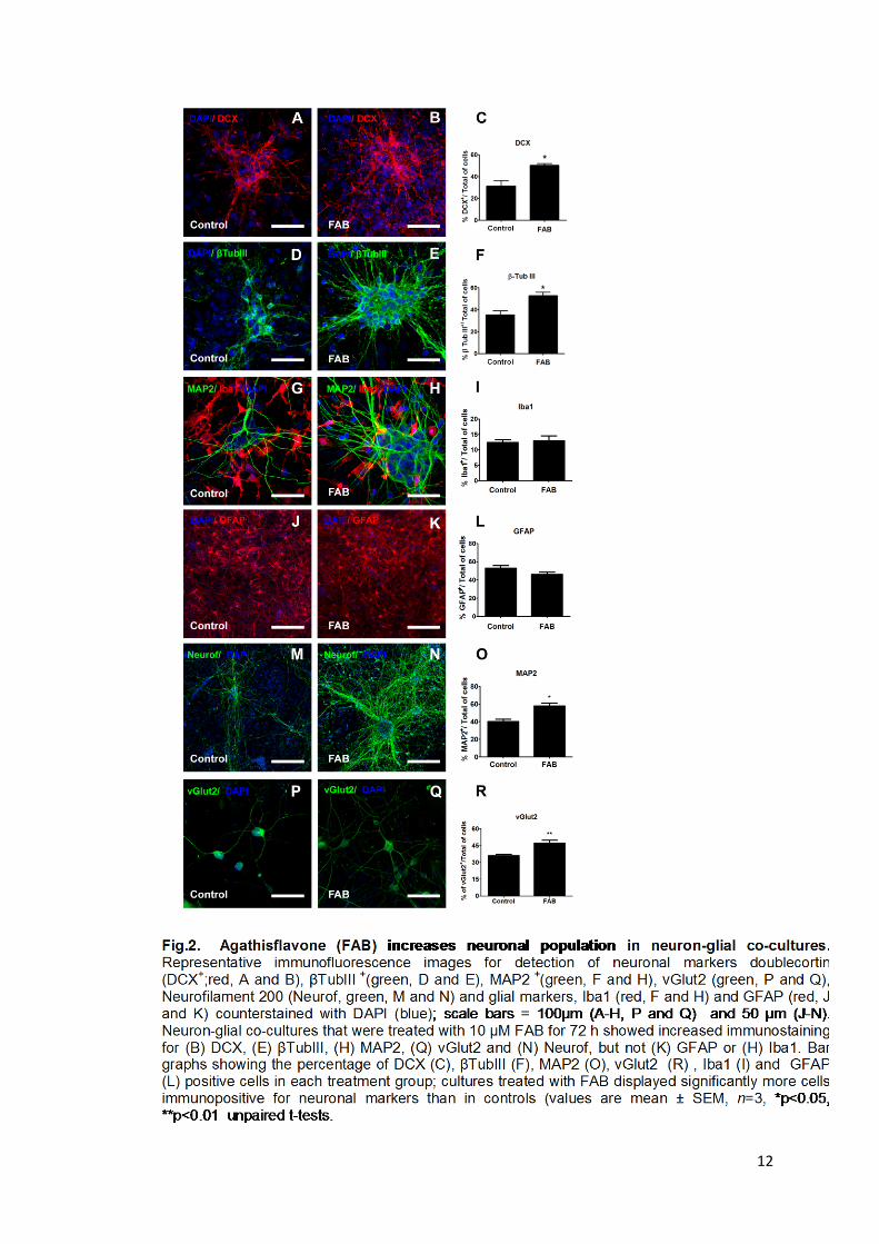
Results The flavonoid agathisflavone enhances generation of neurons
The effects of agathisflavone (FAB) on the neuronal population in primary
cortical co-cultures of neurons and glial cells were investigated by quantification of
the number of neuronal cells after 72 h of treatment. Compared to controls, cultures
treated with agathisflavone showed significant increases in the proportion of cells
expressing the neuronal markers DCX (from 33% in controls to 52% in
agathisflavone, p<0.05; Fig. 2A- C), βTubulin III (37% in controls to 58%
agathisflavone, p<0.05; Fig. 2D- F), MAP2(40% in controls to 58% in agathisflavone,
p<0.05; Fig 2G, H and I) and vGlut2 (36% in controls to 47% in agathisflavone,
p<0.05; Fig 2P-R). We also observed an apparent increase in the expression of
Neurofilament 200, although this was not quantified (Fig. 2M, N). On the other hand,
agathisflavone had no significant effect on the percentage of Iba+ microglia (12.5%
in control and 13% in agathisflavone, p>0.05; Fig 2F-O) or GFAP+ astrocytes (53 %
in control and 46% in agathisflavone, p>0.05; Fig 2J-L).
12

13
The pro-neuronal effects of agathisflavone involve estrogen receptors
To investigate the involvement of Estrogen receptor (ER) signaling on the
effects of agathisflavone on neuronal and neuronal progenitor number, we carried
out experiments with antagonists to ERα and ERβ subtypes. Antagonists were
added to cultures 2h before and concomitant with agathisflavone treatment and the
number of neurons was measured after 72 h. Pharmacological antagonism of ERα
with methyl-piperidinopyrazole (MPP), a selective and widely used antagonist of this
receptor, markedly reduced the number of βTubIII+ cells compared with
agathisflavone-treated cultures, from 50% in agathisflavone-treated cultures to 21%
in MPP + agathisflavone (Fig. 3B, C and E; p<0.05, unpaired t-test). Similarly,
blocking ERβ with pyrazolo[1,5-a]pyrimidine (PHTPP) resulted in a significant
decrease in the number of βTubIII+ cells, from 50% in agathisflavone to 32% in
PHTPP + agathisflavone (Fig. 3B, D and E; p<0.05, unpaired t-test).

14
Agathisflavone protects against glutamate-mediated excitotoxicity
Excitotoxicity is a pathological event characterized by neuronal excitation
through overstimulation of neurons by excitatory glutamate receptors contributing to
neuronal degeneration in many acute and chronic CNS diseases. To investigate the
neuroprotective potential of agathisflavone against glutamate-mediated
excitotoxicity, neuron-glial co-cultures were exposed to glutamate (1mM) for 4 h and
then treated with agathisflavone for 24h or 72h; 17β estradiol (EST) has known
neuroprotective effects (Arevalo et al., 2015) and was used as a positive control.
First, we used Fluoro Jade B (FJ-B) to stain neurons undergoing degeneration
and compared the effects of different concentrations of agathisflavone (0.1-10µM) on
glutamate-mediated excitotoxicity (Fig.4B). As expected, we observed a significant
increase in FJ-B Fluorescence Intensity after treatment with 1mM of glutamate (0.60,
Fig. 4B), when compared with control treated with DMSO vehicle (0.31, Fig. 4B). In
contrast, treatment with agathisflavone after exposure to glutamate significantly
reduced FJ-B fluorescence intensity at all concentrations of agathisflavone tested
(0.33 agathisflavone 0.1 µM, 0.35 agathisflavone 1 µM, 0.37 agathisflavone 10 µM,
Fig. 4B), significantly less than glutamate treated cells (0.60, Fig. 4B); similar results
were observed in cells treated with EST after exposure to glutamate (0.42; Fig. 4B).
The FJ-B Fluorescence Intensity was similar in the experimental groups treated with
agathisflavone or EST alone when compared with control groups (Fig. 5B),
suggesting that agathisflavone and EST have no effect on cell death in cortical co-
cultures in the absence of a cytotoxic insult (0.33 agathisflavone 0.1 µM, 0.27
agathisflavone 1 µM, 0.39 agathisflavone 10 µM, 0.39 EST 0.1 µM, Fig. 4B). In
addition, we examined whether agathisflavone (10 µM) affected glutamate-mediated
apoptosis, using TUNEL staining, and the results indicated that agathisflavone
decreased significantly the amount of TUNEL-positive cells (Fig. 4A).
To evaluate whether agathisflavone was protecting specifically by reducing
caspase 3, we also evaluated by immunofluorescence the amount of cleaved
caspase-3 in co-culture (Fig. 4C). Cultures exposed to glutamate (Glut), showed a
significant increase in the percentage of caspase-3+ cells (14.5% Fig. 4C and D),
compared with controls (4.4% Fig. 4C and D). Compared to the glutamate treated
group (14.5%), agathisflavone treatment significantly reduced the percentage of
Caspase 3 positive cells (5.7% Fig. 4C and D), and similar results were observed in
cells treated with EST (5.4% Fig. 4C and D); agathisflavone or EST treatment did not

15
significantly alter the proportion of Casp3+ cells in normal medium (Fig. 4C and D).
These data show that agathisflavone protects against glutamate-mediated neuronal
excitotoxicity.

16
Agathisflavone reduces neuroinflammation induced by glutamate Immunofluorescence analyses of microglia was performed in neuron-glial co-
cultures 72 h after treatment to investigate the activation of microglia after glutamate-
mediated excitotoxicity (Fig. 5A). A significant increase in the percentage of Ox42+
microglial cells was observed after glutamate treatment (10.1%) compared with
controls (2.0%). In contrast, agathisflavone significantly reduced microglial activation
induced by glutamate, as observed by the reduction in the percentage of Ox42+cells
(3.6%), and similar results were observed following treatment with 17β Estradiol
(4.8%). We did not observe differences in the percentage of Ox42+ cells in groups
treated only with agathisflavone or EST (Fig. 5B).
In order to evaluate if glutamate-mediated excitotoxicity induces distinct
cytokine expression, qPCR was performed in neuron-glial co-cultures 24 h after
treatments. Glutamate (Glut) induced an increase of mRNA expression of TNF (1.4),
IL1β (2.0), IL6 (2.4), arginase1 (1.46) and IL10 (1.35), compared with control (1.0);
however, the expression levels of TGF-β was not altered. In contrast, agathisflavone
reduces the expression of the inflammatory cytokines TNF (1.2), IL1β (1.5), IL6 (1.4),
induced by glutamate, whereas expression of anti-inflammatory markers arginase1
(2.26) and IL10 (4.31) were increased by agathisflavone (Fig. 5C). EST also reduced
the levels of TNF (1.2), IL1β (1.7), IL6 (1.3) and increased the expression of
arginase1 (2.2) and IL10 (5.3) when compared with glutamate. Treatment with
agathisflavone or EST alone showed increased expression of IL6 (1.2 and 1.4
respectively) and IL10 (1.2, 1.4) but not other targets (IL1β, TNF, IL1β, arginase and
TGF β), when compared with control group.
To investigate the molecular mechanisms involved in the neuroprotection
induced by agathisflavone, qPCR of neurotrophins was performed in neuron-glial co-
cultures 24 h after treatments. Glutamate induced an increase in mRNA expression
of GDNF (1.3), but reduced the levels of BDNF (0.7), when compared with control
(1.0); no differences were observed in the expression of NGF, NT4 and CDNF.
Treatment with glutamate plus agathisflavone increased the levels of BDNF (2.2),
NGF (1.7), NT4 (2.4) and GDNF (2.2), but not CDNF (1.05), when compared with
glutamate treated cells. Similarly, treatment with EST plus glutamate elevated the
levels of BDNF (1.5), NGF (1.4), NT4 (4.0) and GDNF (1.9), but not CDNF (1.12),
compared to glutamate treated cells. Treatment with agathisflavone alone increased
the expression of BDNF (1.2), NT4 (1.2, 1.4) and CDNF when compared with control

17
medium group; EST alone elevated the levels of BDNF (1.5) and GDNF (1.3) (Fig.
5D).
Fig.5. Agathisflavone reduced microglia activation induced by glutamate. (A) Representative immunofluorescence images staining for GFAP (green) and OX42 (red) counterstained with DAPI (blue) in co-cultures. (B) Bar graph showing the percentage of Ox42+cells in each treatment group. (C) FAB and EST modulate cytokine expression after glutamate treatment. qPCR analysis showing that cells treated with FAB or EST reduced expression of the pro-inflammatory cytokines TNFα, IL1β, and IL6, whereas there was increased expression of the anti-inflammatory markers IL10 and ARG1, but not TGFβ. (D) FAB modulates the expression of neurotrophins: qPCR analysis showing that cells treated with FAB or EST increased expression the neurotrophins BDNF, NGF, NT4 and GDNF, but not CDNF after glutamate induced excitotoxicity. (E) Confocal photomicrographs of isolated microglia cultures immunostained for the microglial marker Iba1 (green) and the M1 microglial marker CD68 (red); nuclei were counterstained with DAPI (blue). Cells were incubated with 0.01% of vehicle (DMSO) or treated with 1mM Glutamate (Glu),1 µg/ mL of LPS, 20ng/mL of IL4, 10 µM of agathisflavone (FAB), 100nM of 17βEstradiol (EST) or the combination of 1 mM Glu plus 10µM FAB (Glu + FAB) or the combination of 1 mM Glu plus 100nM EST (Glu + EST). (F) Confocal photomicrographs of isolated microglia cultures, immunostained for the microglial marker Iba1 (red) and the M2 microglial marker CD206 (green); nuclei were counterstained with DAPI (blue).Values in B-D are expressed as the mean±SEM, n=3; significant differences are indicated with *P≤0.05 when compared with control, #P<0.05 when compared with glutamate treatment and &p<0.05 when compared with Glut +FAB treatment, one way ANOVA. Scale bar: 50 mm (A) and 100mm (E and F).

18
To confirm the effects of agathisflavone on the apparent change in the M1/M2
polarization of microglial cells after glutamate-induced excitotoxicity, isolated
microglial cultures were pre-treated with glutamate and then treated with
agathisflavone and EST and processed for immunofluorescence for Iba1, a general
marker for microglia, plus CD68 and CD206 for the M1 and M2 phenotypes,
respectively. Cultures treated with 1 µg/mL of LPS were used as a positive control of
the M1 phenotype, and cultures treated with 20 ng/mL IL4 were used as a control of
the M2 phenotype. We observed an increase in the levels of the M1 marker CD68 in
cells treated with LPS and EST, when compared with control, whereas
agathisflavone and glutamate did not change the levels of CD68 in these conditions
(Fig. 5E). In comparison, the M2 marker CD206 was increased in IL4 treated groups
and reduced in cells treated with LPS or glutamate, when compared with control
(Fig. 5F). Notably, agathisflavone and EST treatment alone and after pre-treatment
with glutamate resulted in an increase in CD206 expression. Together, these results
show that glutamate-mediated excitotoxicity induces microglial activation and that
agathisflavone reduces inflammation induced by glutamate and polarizes microglia
towards an M2 phenotype.
Agathisflavone increases the levels of EAAT1 and GS in astrocytes
Since one of the most important functions of astrocytes in the brain is their
control of glutamate clearance, whose accumulation in the extracellular space can
trigger excessive activation of glutamatergic receptors and lead to excitotoxicity, a
characteristic of many neurodegenerative diseases (Murphy-Royal et al., 2017).
Based on these observations, we postulated that astrocytes could have a role in the
neuroprotective effects of agathisflavone. To investigate this, we performed
immunofluorescence and western blot analysis of EAAT1 and GS expression on co-
cultures 72 h after treatments. Cultures exposed to glutamate (Glut), showed a
decrease in the intensity EAAT1 fluorescence (7.61) compared with control groups
(9.45, Fig. 6A and B). agathisflavone and EST treatment increased the levels of
EAAT1 (13.52 and 13.56 respectively) when compared with control treated with the
vehicle and when compared with glutamate treated group . Treatment with
agathisflavone after glutamate induced excitotoxicity markedly increased the levels
of EAAT1 (21.85), compared with control (9.45, a) and glutamate (7.61); EST also

19
elevated the levels of EAAT1 after glutamate induced excitotoxicity (17.99).
Furthermore, EAAT1 expression was increased to a larger extent in agathisflavone
than EST after glutamate induced excitotoxicity (Fig. 6A and B).
Fig.6. Agathisflavone increases the levels of Excitatory Amino Acid Transporter 1 (EAAT1) and Glutamine synthetase (GS) in astrocytes. Cells were incubated with 0.01% of vehicle (DMSO) or treated with 1mM Glutamate (Glut), 10 µM of agathisflavone (FAB), 0.1 µM of 17βEstradiol (EST) or the combination of 1 mM Glut plus 10µM FAB (Glut + FAB) or the combination of 1 mM Glu plus 0.1 µM EST (Glut + EST). (A) Representative immunofluorescence images stained for β Tubulin III (green) and EAAT1 (red), counterstained with DAPI (blue) in neuron-glial co-cultures. (B) Bar graph of the fluorescence intensity as a percentage of the total number of cells showing significantly increased EAAT1 staining in Glut + FAB and Glut + EST-treated cells compared to glutamate. (C) Representative immunofluorescence images stained for β Tubulin III (green) and GS (red), counterstained with DAPI (blue), in neuron-glial co-cultures. (D) Bar graph of the fluorescence intensity as a percentage of the total number of cells showing significantly increased GS staining in Glut + FAB and Glut + EST-treated cells compared to glutamate. (E) Western blot showed increased expression of GS in treated cells, the results were normalized relative to control group considered as 100% and at least three independent experiments. The values are expressed as the mean±SEM; n=3; significant differences are indicated with *P≤0.05, ***p<0.001 when compared with control, #P<0.05, ###p<0.001 when compared with glutamate treatment and &p<0.05 when compared with Glut +FAB treatment, one way ANOVA. Scale bar: 50 µm.

20
Glutamine synthetase (GS) is an ATP-dependent enzyme found in most
species that synthesizes glutamine from glutamate and ammonia. In the CNS, GS is
exclusively located in astrocytes where it serves to maintain the glutamate-glutamine
cycle (Jayakumar and Norenberg, 2016). We evaluated the levels of GS in co-
cultures by immunofluorescence and western blot. The immunofluorescence showed
a tendency of reduction in the levels of GS in cultures exposed to glutamate (Glut,
Fig. 6C and D), which was confirmed by western blot that showed a significantly
decreased level of GS (23.99%), when compared with control groups (100%, Fig.
6E). The treatment with agathisflavone alone did not change significantly the levels
of GS (84%, Fig. 6E), whereas agathisflavone after glutamate induced excitotoxicity
resulted in an increase of the levels of GS showed by immunofluorescence and
confirmed by western blot (87% Fig. 6E) when compared with glutamate (23.99%);
the results of agathisflavone after glutamate induced excitotoxicity was similar to
control levels. Western blot showed that treatment with EST alone reduced the levels
of GS (65.9%, Fig. 6E), when compared with the control (100%, Fig. 6E), but
increased the level of EAAT1 after glutamate induced excitotoxicity (70.5%), when
compared with glutamate (23.99%). These data show that agathisflavone modulates
the astrocyte response against glutamate-mediated excitotoxicity.
Discussion Glutamate-mediated excitotoxicity is a major factor in neuronal loss in
neurodegenerative diseases. Notably, there is evidence that estrogens are
neuroprotective, but their therapeutic use in humans is limited by the increased risk
of cancer. Here, we provide evidence that the flavonoid agathisflavone acts as a
modulator of estrogen receptors (ER) to promote the generation of neurons in vitro
and protects against glutamate-mediated neurotoxicity as effectively as the synthetic
estrogen estradiol. In neuron-glial co-cultures, we show that the protective effects of
agathisflavone again glutamate-mediated neurotoxicity are mediated at least in part
by polarizing microglia towards an anti-inflammatory M2 phenotype and increased
expression of neuroprotective trophic factors. In addition, agathisflavone treatment
maintained the expression of the glutamate regulatory proteins EAAT1 and GS in
astrocytes, which is likely to be important in protecting neurons against elevated
levels of glutamate. The neuroprotective and anti-inflammatory potency of

21
agathisflavone suggest it may provide a potential alternative to estrogens for the
treatment of neurodegeneration.
The increased neuronal population in cortical cultures after agathisflavone
treatment gave rise to the population of doublecortin-(DCX)-expressing cells that
comprises the late intermediate progenitor cells as well as immature neurons.
Agathisflavone also increased the population of neurons (βTubIII + and MAP2+ Cells).
This effect was dependent, at least partly, on estrogen receptor (ER) activation,
since ER antagonists were capable of reducing the generation of these cells. Natural
compounds and their potential effect on brain plasticity have become particularly
interesting for their beneficial effects to both the general public and scientists (Ortiz-
Lopez et al., 2016). Preclinical studies, in vitro and in vivo suggest that natural
compounds such as polyphenols modulate neuroplasticity (Dias et al., 2012).
Several natural compounds reportedly favored the generation of new neurons (Dias
et al., 2012). For example, epigallo-catechin-3-gallate (EGCG), a polyphenol
compound mainly found in green tea leaves (Camellia sinensis) increased the
population of doublecortin-(DCX)-expressing cells and also significantly increased
net neurogenesis in the adult hippocampus (Ortiz-Lopez et al., 2016). On the other
hand, some flavonoids, in particular apigenin, have been shown to activate estrogen
receptors (Mak et al., 2006), which affect the development, maturation, function, and
plasticity of the nervous system. Furthermore, it has been previously shown that the
flavonoid apigenin induces neural differentiation of human pluripotent stem cells and
this neural conversion was dependent, at least partly, on ER activation (Souza et al.,
2015). We previously demonstrated that agathisflavone, a product of the oxidative
coupling of an apigenin dimer, enhances neural differentiation induced by retinoic
acid (RA) in murine pluripotent stem cells by increasing the expression of all trans
retinoic acid (RA) receptors (RAR) (Paulsen et al., 2011). The results of the present
study show that agathisflavone also modulates ER signaling to promote the
generation of neurons in vitro.
The influence of the agathisflavone on cell death induced by glutamate has
not been explored in co-culture previously. Glutamate is one of the most abundant
excitatory neurotransmitters in the CNS and is particularly involved in the cortical and
hippocampal regions, which deal especially with synaptic plasticity, memory and
learning among other functions (Collingridge and Lester, 1989, Esposito et al.,

22
2013). Excess levels of glutamate and other excitatory molecules results in over
excitation of the ionotropic glutamatergic receptors N-methyl-d-aspartate (NMDA)
and 2-amino-3- (3-hydroxy-5-methylisoxazol-4-yl) propionate (AMPA), and the
release of other excitotoxins followed by excessive calcium levels in cytosol (Essa et
al., 2013). Excitotoxicity describes the pathological event characterized by neuronal
excitation through overstimulation of neurons by excitatory amino acids receptors of
glutamate and aspartate (Olney, 1969). Glutamate induced excitotoxicity is
considered a direct cause of cell degeneration in several neurological disorders,
such as HD (Douaud et al., 2009, Malkki, 2016), PD (Rodriguez et al., 1998, Marti et
al., 2000) and AD (Bezprozvanny and Mattson, 2008, Zhang et al., 2016). Here, we
demonstrate that the flavonoid agathisflavone reduced neuronal cell death induced
by glutamate excitotoxicity in neuron-glial co-cultures. The reduction of
neurodegeneration by agathisflavone is consistent with previous results that show
that flavonoids can reduce cell death (Zong et al., 2016, Cong et al., 2016). Our
results show that agathisflavone reduced the percentage of cells expressing caspase
3, a member of the caspase family of proteases that play a pivotal role in apoptosis.
In healthy cells, caspase-3 is present in the cytoplasm as an inactive proenzyme,
pro-caspase-3. During apoptosis, pro-caspase-3 is hydrolyzed to the active-caspase-
3 (D'Amelio et al., 2011).
Flavonoids are widely known as antioxidants and the mechanisms by which
they protect against cell death induced by glutamate are likely to be multifarious.
Recently, much interest has focused on the suggested anti-inflammatory and
neuroprotective effects of dietary derived polyphenols (Vauzour et al., 2015). We
demonstrated that the flavonoid agathisflavone (bis-apigenin) reduces the
neuroinflammation induced by glutamate, by modulating microglial activation and
reducing the expression of pro-inflammatory cytokines TNF, IL6 and IL1β, and
increasing the expression of anti-inflammatory markers. The anti-inflammatory
effects of flavonoids are well established (Rahimifard et al., 2017). For example, in
an in vivo model, apigenin showed a significant reduction in severity of experimental
autoimmune encephalomyelitis (EAE) progression and in relapses observed in
C57BL/6 (progressive) and SJL/J (relapse-remitting) mouse models of MS, where
apigenin modulated microglial activation via inhibition of STAT1-induced CD40
expression (Rezai-Zadeh et al., 2008). In addition, a recent study using the human
induced pluripotent stem cell (hiPSC) model of familial and sporadic AD to assess

23
the neuroprotective activity of apigenin, demonstrated that hiPSC-derived AD
neurons exhibited a hyper-excitable calcium signalling phenotype, elevated levels of
nitrite, increased apoptosis, reduced neurite length and increased susceptibility to
inflammatory stress challenge from activated murine microglia, in comparison to
neurons derived from healthy controls. In this study, they identified that the flavonoid
apigenin had potent anti-inflammatory effects with the ability to protect neurites and
cell viability by promoting a down-regulation of cytokine and nitric oxide (NO) release
in inflammatory cells (Balez et al., 2016).
Neurotrophins (NTFs) are endogenous peptides secreted from neurons and
glial cells, and are associated with regulating brain function, survival, and
development of individual cells and neuronal networks across the entire brain.
Specifically, NTFs regulate synaptic plasticity, protect neurons from apoptosis, and
stimulate neurogenesis (Skaper, 2012, Leal et al., 2015, Kuipers et al., 2016,
Wurzelmann et al., 2017). The neuroprotective effect of agathisflavone was
associated with the increase of the NTFs BDNF, NGF, CDNG, NT4 and GDNF.
Survival signaling is important to suppress apoptosis and counterbalance death
signaling in the nervous system. Several studies have shown that polyphenolic
compounds enhance neuronal survival and increase the expression levels of NTFs
(Moosavi et al., 2016). For example, apigenin reverses depression-like behavior
induced by chronic corticosterone treatment in mice. In addition, the administration of
apigenin ameliorated the levels of BDNF in corticosterone-treated mice. The
antidepressant-like effects of apigenin have been suggested to be mediated, at least
partly, by up-regulation of BDNF levels in the hippocampus (Weng et al., 2016).
BDNF, the main neurotrophin expressing in the central nervous system, has an
important role in promoting survive, growth and synaptic plasticity in the CNS (Huang
and Reichardt, 2001).
Astrocytes, earlier presumed to serve as supportive roles for the neuronal
network, have recently been shown to play an active role in the synaptic dysfunction,
impairment of homeostasis, inflammation, as well as excitotoxicity in relation to
several neurological disorders (Dezsi et al., 2015). The role of astrocytes in the
neuroprotective effects of agathisflavone against glutamate-induced excitotoxicity
was evaluated. In physiological conditions to prevent over-stimulation, glutamate is
removed from the synaptic cleft by astrocytes trough EAAT located in the plasma
membrane and converted through the action of GS to L-glutamine, which is released

24
to the extracellular fluid and taken up by neurons (Danbolt, 2001, Hazell et al., 2001,
Walton and Dodd, 2007). We demonstrated that treatment with agathisflavone after
excitotoxicity induced by glutamate increased the levels of EAAT1 and GS in
astrocytes, suggesting that an important neuroprotective effect of agathisflavone is
via glutamate uptake and recycling in astrocytes.
In conclusion, we showed that treatment of co-cultures of neurons and glial
cells with agathisflavone increases the neuronal population at least in part via ER.
Furthermore, we demonstrate that agathisflavone has broad neuroprotective effects
against glutamate induced excitotoxicity, associated with anti-inflammatory effects on
microglia and increased expression of neuroprotective cytokines and trophic factors.
Taken together, these data suggest that agathisflavone could be a potential agent for
treatment of excitotoxicity-related diseases.
Conflict of interest and funding The authors report no conflicts of interest, including personal or financial.
Acknowledgements This work was supported by the UK/ Brazil interchange cooperation (Edital RCUK-
CONFAP/FAPESB- Research Partnerships Call 2014) grant, by the Bahia State
Research Foundation (FAPESB – Project Nº 2957/2013, RED0016/2013), and the
National Council for Scientific and Technological Development (CNPq, EU Edital
MCTI/CNPq/Universal 14/2014 Processo 443723/2014-1). The authors thank the
Program for Technological Development in Tools for Health-PDTIS-FIOCRUZ for
use of Confocal Microscopy Service-CPqGM facilities.

25
References Agostinho, P., Cunha, R. A. and Oliveira, C. (2010) 'Neuroinflammation, oxidative stress and the
pathogenesis of Alzheimer's disease', Curr Pharm Des, 16(25), pp. 2766-78. Amor, S., Puentes, F., Baker, D. and van der Valk, P. (2010) 'Inflammation in neurodegenerative
diseases', Immunology, 129(2), pp. 154-69. Arevalo, M. A., Azcoitia, I. and Garcia-Segura, L. M. (2015) 'The neuroprotective actions of oestradiol
and oestrogen receptors', Nat Rev Neurosci, 16(1), pp. 17-29. Balez, R., Steiner, N., Engel, M., Munoz, S. S., Lum, J. S., Wu, Y., Wang, D., Vallotton, P., Sachdev, P.,
O'Connor, M., Sidhu, K., Munch, G. and Ooi, L. (2016) 'Neuroprotective effects of apigenin against inflammation, neuronal excitability and apoptosis in an induced pluripotent stem cell model of Alzheimer's disease', Sci Rep, 6, pp. 31450.
Barrientos, T., Laothamatas, I., Koves, T. R., Soderblom, E. J., Bryan, M., Moseley, M. A., Muoio, D. M. and Andrews, N. C. (2015) 'Metabolic Catastrophe in Mice Lacking Transferrin Receptor in Muscle', EBioMedicine, 2(11), pp. 1705-17.
Bezprozvanny, I. and Mattson, M. P. (2008) 'Neuronal calcium mishandling and the pathogenesis of Alzheimer's disease', Trends Neurosci, 31(9), pp. 454-63.
Brinton, R. D. (2013) 'Neurosteroids as regenerative agents in the brain: therapeutic implications', Nat Rev Endocrinol, 9(4), pp. 241-50.
Busch, C., Burkard, M., Leischner, C., Lauer, U. M., Frank, J. and Venturelli, S. (2015) 'Epigenetic activities of flavonoids in the prevention and treatment of cancer', Clin Epigenetics, 7, pp. 64.
Chen, W. W., Zhang, X. and Huang, W. J. (2016) 'Role of neuroinflammation in neurodegenerative diseases (Review)', Mol Med Rep, 13(4), pp. 3391-6.
Collingridge, G. L. and Lester, R. A. (1989) 'Excitatory amino acid receptors in the vertebrate central nervous system', Pharmacol Rev, 41(2), pp. 143-210.
Cong, L., Cao, C., Cheng, Y. and Qin, X. Y. (2016) 'Green Tea Polyphenols Attenuated Glutamate Excitotoxicity via Antioxidative and Antiapoptotic Pathway in the Primary Cultured Cortical Neurons', Oxid Med Cell Longev, 2016, pp. 2050435.
D'Amelio, M., Cavallucci, V., Middei, S., Marchetti, C., Pacioni, S., Ferri, A., Diamantini, A., De Zio, D., Carrara, P., Battistini, L., Moreno, S., Bacci, A., Ammassari-Teule, M., Marie, H. and Cecconi, F. (2011) 'Caspase-3 triggers early synaptic dysfunction in a mouse model of Alzheimer's disease', Nat Neurosci, 14(1), pp. 69-76.
Dai, Q., Borenstein, A. R., Wu, Y., Jackson, J. C. and Larson, E. B. (2006) 'Fruit and vegetable juices and Alzheimer's disease: the Kame Project', Am J Med, 119(9), pp. 751-9.
Danbolt, N. C. (2001) 'Glutamate uptake', Prog Neurobiol, 65(1), pp. 1-105. Dezsi, L., Tuka, B., Martos, D. and Vecsei, L. (2015) 'Alzheimer's disease, astrocytes and kynurenines',
Curr Alzheimer Res, 12(5), pp. 462-80. Dias, G. P., Cavegn, N., Nix, A., do Nascimento Bevilaqua, M. C., Stangl, D., Zainuddin, M. S., Nardi, A.
E., Gardino, P. F. and Thuret, S. (2012) 'The role of dietary polyphenols on adult hippocampal neurogenesis: molecular mechanisms and behavioural effects on depression and anxiety', Oxid Med Cell Longev, 2012, pp. 541971.
Douaud, G., Behrens, T. E., Poupon, C., Cointepas, Y., Jbabdi, S., Gaura, V., Golestani, N., Krystkowiak, P., Verny, C., Damier, P., Bachoud-Levi, A. C., Hantraye, P. and Remy, P. (2009) 'In vivo evidence for the selective subcortical degeneration in Huntington's disease', Neuroimage, 46(4), pp. 958-66.
Esposito, Z., Belli, L., Toniolo, S., Sancesario, G., Bianconi, C. and Martorana, A. (2013) 'Amyloid beta, glutamate, excitotoxicity in Alzheimer's disease: are we on the right track?', CNS Neurosci Ther, 19(8), pp. 549-55.
Essa, M. M., Braidy, N., Vijayan, K. R., Subash, S. and Guillemin, G. J. (2013) 'Excitotoxicity in the pathogenesis of autism', Neurotox Res, 23(4), pp. 393-400.
Glass, C. K., Saijo, K., Winner, B., Marchetto, M. C. and Gage, F. H. (2010) 'Mechanisms underlying inflammation in neurodegeneration', Cell, 140(6), pp. 918-34.

26
Hazell, A. S., Rao, K. V., Danbolt, N. C., Pow, D. V. and Butterworth, R. F. (2001) 'Selective down-regulation of the astrocyte glutamate transporters GLT-1 and GLAST within the medial thalamus in experimental Wernicke's encephalopathy', J Neurochem, 78(3), pp. 560-8.
Hertz, L. (2014) 'Book review: “Glial physiology and pathophysiology” by Alexei Verkhratsky and Arthur Butt, Wiley-Blackwell, 2013', Frontiers in Systems Neuroscience, 8, pp. 17.
Huang, E. J. and Reichardt, L. F. (2001) 'Neurotrophins: roles in neuronal development and function', Annu Rev Neurosci, 24, pp. 677-736.
Islamov, R. R., Hendricks, W. A., Jones, R. J., Lyall, G. J., Spanier, N. S. and Murashov, A. K. (2002) '17Beta-estradiol stimulates regeneration of sciatic nerve in female mice', Brain Res, 943(2), pp. 283-6.
Jayakumar, A. R. and Norenberg, M. D. (2016) 'Glutamine Synthetase: Role in Neurological Disorders', Adv Neurobiol, 13, pp. 327-350.
Jean, S. M., Preuss, T. M., Sharma, P., Anderson, D. C., Provenzale, J. M., Strobert, E., Ross, S. R. and Stroud, F. C. (2012) 'Cerebrovascular accident (stroke) in captive, group-housed, female chimpanzees', Comp Med, 62(4), pp. 322-9.
Kuipers, S. D., Trentani, A., Tiron, A., Mao, X., Kuhl, D. and Bramham, C. R. (2016) 'BDNF-induced LTP is associated with rapid Arc/Arg3.1-dependent enhancement in adult hippocampal neurogenesis', Sci Rep, 6, pp. 21222.
Leal, G., Afonso, P. M., Salazar, I. L. and Duarte, C. B. (2015) 'Regulation of hippocampal synaptic plasticity by BDNF', Brain Res, 1621, pp. 82-101.
Mak, P., Leung, Y. K., Tang, W. Y., Harwood, C. and Ho, S. M. (2006) 'Apigenin suppresses cancer cell growth through ERbeta', Neoplasia, 8(11), pp. 896-904.
Malkki, H. (2016) 'Huntington disease: New study challenges the hypothesis of glutamate transporter dysfunction in Huntington disease', Nat Rev Neurol, 12(5), pp. 251.
Mannella, P. and Brinton, R. D. (2006) 'Estrogen receptor protein interaction with phosphatidylinositol 3-kinase leads to activation of phosphorylated Akt and extracellular signal-regulated kinase 1/2 in the same population of cortical neurons: a unified mechanism of estrogen action', J Neurosci, 26(37), pp. 9439-47.
Marti, M., Sbrenna, S., Fuxe, K., Bianchi, C., Beani, L. and Morari, M. (2000) 'Increased responsivity of glutamate release from the substantia nigra pars reticulata to striatal NMDA receptor blockade in a model of Parkinson's disease. A dual probe microdialysis study in hemiparkinsonian rats', Eur J Neurosci, 12(5), pp. 1848-50.
Mecha, M., Iñigo, P. M., Mestre, L., Hernangómez, M., Borrell, J. and Guaza, C. (2011) 'An easy and fast way to obtain a high number of glial cells from rat cerebral tissue: A beginners approach'.
Meeker, K. D., Meabon, J. S. and Cook, D. G. (2015) 'Partial Loss of the Glutamate Transporter GLT-1 Alters Brain Akt and Insulin Signaling in a Mouse Model of Alzheimer's Disease', J Alzheimers Dis, 45(2), pp. 509-20.
Mendes, C. C., Bahia, M. V., David, J. M. and David, J. P. (2000) 'Constituents of Caesalpinia pyramidalis', Fitoterapia, 71(2), pp. 205-7.
Moosavi, F., Hosseini, R., Saso, L. and Firuzi, O. (2016) 'Modulation of neurotrophic signaling pathways by polyphenols', Drug Des Devel Ther, 10, pp. 23-42.
Murphy-Royal, C., Dupuis, J., Groc, L. and Oliet, S. H. (2017) 'Astroglial glutamate transporters in the brain: Regulating neurotransmitter homeostasis and synaptic transmission', J Neurosci Res.
Olney, J. W. (1969) 'Brain lesions, obesity, and other disturbances in mice treated with monosodium glutamate', Science, 164(3880), pp. 719-21.
Ortiz-Lopez, L., Marquez-Valadez, B., Gomez-Sanchez, A., Silva-Lucero, M. D., Torres-Perez, M., Tellez-Ballesteros, R. I., Ichwan, M., Meraz-Rios, M. A., Kempermann, G. and Ramirez-Rodriguez, G. B. (2016) 'Green tea compound epigallo-catechin-3-gallate (EGCG) increases neuronal survival in adult hippocampal neurogenesis in vivo and in vitro', Neuroscience, 322, pp. 208-20.

27
Pallo, S. P., DiMaio, J., Cook, A., Nilsson, B. and Johnson, G. V. (2016) 'Mechanisms of tau and Abeta-induced excitotoxicity', Brain Res, 1634, pp. 119-31.
Pandey, K. B. and Rizvi, S. I. (2009) 'Plant polyphenols as dietary antioxidants in human health and disease', Oxid Med Cell Longev, 2(5), pp. 270-8.
Paulsen, B. S., Souza, C. S., Chicaybam, L., Bonamino, M. H., Bahia, M., Costa, S. L., Borges, H. L. and Rehen, S. K. (2011) 'Agathisflavone enhances retinoic acid-induced neurogenesis and its receptors alpha and beta in pluripotent stem cells', Stem Cells Dev, 20(10), pp. 1711-21.
Procaccini, C., Santopaolo, M., Faicchia, D., Colamatteo, A., Formisano, L., de Candia, P., Galgani, M., De Rosa, V. and Matarese, G. (2016) 'Role of metabolism in neurodegenerative disorders', Metabolism, 65(9), pp. 1376-90.
Rahimifard, M., Maqbool, F., Moeini-Nodeh, S., Niaz, K., Abdollahi, M., Braidy, N., Nabavi, S. M. and Nabavi, S. F. (2017) 'Targeting the TLR4 signaling pathway by polyphenols: A novel therapeutic strategy for neuroinflammation', Ageing Res Rev, 36, pp. 11-19.
Rezai-Zadeh, K., Ehrhart, J., Bai, Y., Sanberg, P. R., Bickford, P., Tan, J. and Shytle, R. D. (2008) 'Apigenin and luteolin modulate microglial activation via inhibition of STAT1-induced CD40 expression', J Neuroinflammation, 5, pp. 41.
Rodriguez, M. C., Obeso, J. A. and Olanow, C. W. (1998) 'Subthalamic nucleus-mediated excitotoxicity in Parkinson's disease: a target for neuroprotection', Ann Neurol, 44(3 Suppl 1), pp. S175-88.
Rozovsky, I., Wei, M., Stone, D. J., Zanjani, H., Anderson, C. P., Morgan, T. E. and Finch, C. E. (2002) 'Estradiol (E2) enhances neurite outgrowth by repressing glial fibrillary acidic protein expression and reorganizing laminin', Endocrinology, 143(2), pp. 636-46.
Salter, M. W. and Stevens, B. (2017) 'Microglia emerge as central players in brain disease', Nat Med, 23(9), pp. 1018-1027.
Scalbert, A., Manach, C., Morand, C., Remesy, C. and Jimenez, L. (2005) 'Dietary polyphenols and the prevention of diseases', Crit Rev Food Sci Nutr, 45(4), pp. 287-306.
Schmued, L. C. and Hopkins, K. J. (2000) 'Fluoro-Jade B: a high affinity fluorescent marker for the localization of neuronal degeneration', Brain Res, 874(2), pp. 123-30.
Sehara, Y., Sawicka, K., Hwang, J. Y., Latuszek-Barrantes, A., Etgen, A. M. and Zukin, R. S. (2013) 'Survivin Is a transcriptional target of STAT3 critical to estradiol neuroprotection in global ischemia', J Neurosci, 33(30), pp. 12364-74.
Silva, V. D., Pitanga, B. P., Nascimento, R. P., Souza, C. S., Coelho, P. L., Menezes-Filho, N., Silva, A. M., Costa Mde, F., El-Bacha, R. S., Velozo, E. S. and Costa, S. L. (2013) 'Juliprosopine and juliprosine from prosopis juliflora leaves induce mitochondrial damage and cytoplasmic vacuolation on cocultured glial cells and neurons', Chem Res Toxicol, 26(12), pp. 1810-20.
Skaper, S. D. (2012) 'The neurotrophin family of neurotrophic factors: an overview', Methods Mol Biol, 846, pp. 1-12.
Sokolov, A. N., Pavlova, M. A., Klosterhalfen, S. and Enck, P. (2013) 'Chocolate and the brain: neurobiological impact of cocoa flavanols on cognition and behavior', Neurosci Biobehav Rev, 37(10 Pt 2), pp. 2445-53.
Souza, C. S., Paulsen, B. S., Devalle, S., Lima Costa, S., Borges, H. L. and Rehen, S. K. (2015) 'Commitment of human pluripotent stem cells to a neural lineage is induced by the pro-estrogenic flavonoid apigenin', Advances in Regenerative Biology, 2(1), pp. 29244.
Spencer, J. P., Abd El Mohsen, M. M., Minihane, A. M. and Mathers, J. C. (2008) 'Biomarkers of the intake of dietary polyphenols: strengths, limitations and application in nutrition research', Br J Nutr, 99(1), pp. 12-22.
Spencer, J. P., Vafeiadou, K., Williams, R. J. and Vauzour, D. (2012) 'Neuroinflammation: modulation by flavonoids and mechanisms of action', Mol Aspects Med, 33(1), pp. 83-97.
Uttara, B., Singh, A. V., Zamboni, P. and Mahajan, R. T. (2009) 'Oxidative stress and neurodegenerative diseases: a review of upstream and downstream antioxidant therapeutic options', Curr Neuropharmacol, 7(1), pp. 65-74.

28
Vauzour, D., Martinsen, A. and Laye, S. (2015) 'Neuroinflammatory processes in cognitive disorders: Is there a role for flavonoids and n-3 polyunsaturated fatty acids in counteracting their detrimental effects?', Neurochem Int, 89, pp. 63-74.
Walton, H. S. and Dodd, P. R. (2007) 'Glutamate-glutamine cycling in Alzheimer's disease', Neurochem Int, 50(7-8), pp. 1052-66.
Weng, L., Guo, X., Li, Y., Yang, X. and Han, Y. (2016) 'Apigenin reverses depression-like behavior induced by chronic corticosterone treatment in mice', Eur J Pharmacol, 774, pp. 50-4.
Williams, R. J. and Spencer, J. P. (2012) 'Flavonoids, cognition, and dementia: actions, mechanisms, and potential therapeutic utility for Alzheimer disease', Free Radic Biol Med, 52(1), pp. 35-45.
Wurzelmann, M., Romeika, J. and Sun, D. (2017) 'Therapeutic potential of brain-derived neurotrophic factor (BDNF) and a small molecular mimics of BDNF for traumatic brain injury', Neural Regen Res, 12(1), pp. 7-12.
Xing, C., Arai, K., Lo, E. H. and Hommel, M. (2012) 'Pathophysiologic cascades in ischemic stroke', Int J Stroke, 7(5), pp. 378-85.
Xu, D., Chen, H., Mak, S., Hu, S., Tsim, K. W., Hu, Y., Sun, Y., Zhang, G., Wang, Y., Zhang, Z. and Han, Y. (2016) 'Neuroprotection against glutamate-induced excitotoxicity and induction of neurite outgrowth by T-006, a novel multifunctional derivative of tetramethylpyrazine in neuronal cell models', Neurochem Int, 99, pp. 194-205.
Zhang, Y., Li, P., Feng, J. and Wu, M. (2016) 'Dysfunction of NMDA receptors in Alzheimer's disease', Neurol Sci, 37(7), pp. 1039-47.
Zong, N., Li, F., Deng, Y., Shi, J., Jin, F. and Gong, Q. (2016) 'Icariin, a major constituent from Epimedium brevicornum, attenuates ibotenic acid-induced excitotoxicity in rat hippocampus', Behav Brain Res, 313, pp. 111-9.

![Residuated implications derived from quasi-overlap functions ...arXiv:2002.12267v1 [cs.LO] 27 Feb 2020 Residuated implications derived from quasi-overlap functions on lattices Rui](https://img.pdfslide.tips/doc/110x75/6065d0f065c50f701a4e3e26/residuated-implications-derived-from-quasi-overlap-functions-arxiv200212267v1.jpg)