Embed Size (px)
Citation preview
Altered expression of the a2 laminin chain in psoriatic skin:the effect of treatment with cyclosporin
P.TOTI, M.PELLEGRINO,* M.VILLANOVA,† M.L.FLORI,* C.MIRACCO,S.BARTOLOMMEI AND L.ANDREASSI*Institutes of Pathology, *Dermatology and †Neurological Sciences, University of Siena, Via delle Scotte, 53100 Siena, Italy
Accepted for publication 1 April 1998
Summary The histopathological pattern of psoriasis is characterized by dermal inflammatory reaction andhyperproliferation of the epidermis. The mechanism of the epidermal hyperproliferation is notcompletely understood, but it is probably modulated by the basal lamina (BL), the alterations ofwhich have not been described. We performed the present study to evaluate the expression of the a1,a2, b1 and g1 laminin chains and collagen IV in the BL of active psoriasis vulgaris before and aftercyclosporin treatment administered until the psoriasis was in remission. The results showed that thea2 chain is weak and irregular in the lesions, while the a1, b1 and g1 chains and collagen IV arenormal, with intense and continuous reaction. In the same subjects, this alteration was absent inskin that was clinically unaffected. After treatment with cyclosporin, the altered expression of the a2chain returned to normal in the healing lesions.
Psoriasis vulgaris is a chronic, recurring inflammatoryskin disorder characterized by hyperproliferation of theepidermis with parakeratosis, granulocytic infiltratesand oedema in the epidermis, and lymphocytic infil-trates and formation of new vessels in the dermis.1 Suchhistological alterations have been correlated with theepidermal infiltration of T lymphocytes capable of trig-gering the hyperproliferation of basal keratinocytes2
and with molecular alterations, such as an increasedexpression of cytokines in psoriatic lesions3 or theanomalous expression of integrins in newly formedvessels of the dermis.4 However, changes in the epider-mal basal lamina, which determines keratinocyte polar-ity and therefore allows for regulation of theproliferation of basal cells5,6 and controls inflammatorycell migration inside the tissues, have not been describedin the literature.
Basal laminae (BLs) are composed of several largeglycoproteins. Collagen IV forms a structural frameworkfor the BL.7 Laminin, the primary non-collagenousprotein found in BLs, is a large glycoprotein composedof three subunits designated a, b and g.8 Even if mostBLs share several molecular features, it has generallybeen acknowledged that they are somewhat heteroge-neous. The most common form of laminin throughoutthe body is laminin 1, composed of the a1, b1 and g1chains.9 Recently, homologues of each laminin chain
have been identified with restricted tissue distribution.10
For example, laminin 2 (formerly indicated as merosinor laminin M) contains the a2 chain isoform instead ofthe more common a1 chain, together with the classicb1 and g1 chains. Laminin 2 was first identified in thetrophoblast BL of human placenta and in BLs ofstriated muscle and Schwann cells.11 The presence oflaminin 2 has also been described in the BL of thenormal human brain, surrounding meninges andintraparenchymal vessels12 and, more recently, alsoin the normal epidermis.13,14
In the present immunohistochemical study, we exam-ined the expression of the a1, a2, b1 and g1 lamininchains in skin biopsies from healthy patients and frompatients with psoriasis vulgaris, before and after cyclo-sporin treatment administered until the lesions were inremission.
Materials and methods
Patients
Ten patients (seven men and three women), between theages of 30 and 55 years, were prospectively enrolled inthe study before any topical or systemic treatment. Thepatients were affected by severe psoriasis vulgaris,according to the Psoriasis Area Severity Index (with avalue equal to or greater than 18).15 Skin samples were
British Journal of Dermatology 1998; 139: 375–379.
375q 1998 British Association of Dermatologists
Correspondence: Dr Maria Laura Flori.
taken after local anaesthesia from the border of the plaquesvia Stiefel punch biopsy (4 mm in diameter). Controls wererepresented by analogous skin samples of areas unaffectedby psoriasis from the same subjects. Furthermore, sectionsof normal skin were examined from five healthy patients,comparable in age and sex, who had undergone surgeryfor the excision of naevocytic lesions.
The patients were treated with 5 mg/kg per day cyclo-sporin (Sandimmun Neoral; Novartis) for 2 months, bywhich time the skin lesions were in almost completeclinical remission in all subjects. A skin biopsy wasrepeated at the site of the previous psoriatic lesions2 months after the beginning of treatment.
Immunohistochemistry
Skin fragments were immersed in OCT and immediatelyfrozen in liquid nitrogen. Mounted 5-mm-thick cryostatsections were fixed in acetone and stained with haema-toxylin and eosin to verify the presence of lesions. Unfixedsections were incubated overnight at 4 8C with thefollowing primary monoclonal antibodies to lamininchains: a1 chain (Chemicon, Temecula, CA, U.S.A.,mAb 1924, diluted 1 : 100); a2 chain (Chemicon, mAb
1922, diluted 1 : 1000); b1 chain (Chemicon, mAb1921, diluted 1 : 200) and g1 chain (Chemicon, mAb1920, diluted 1 : 2000). A monoclonal antibody againstcollagen IV (Dakopatts, Glostrup, Denmark, diluted1 : 100) was also used to check the amount and locationof BLs. Tissues were immunostained using the avidin–biotin complex (ABC) technique for monoclonal antibodiesusing Dakopatts (Glostrup, Denmark) and Amersham(Amersham Life Science, Amersham, U.K.) reagents. Col-ours were developed with 3,30-diaminobenzidine (DAB)tetrahydrochloride. Sections were dehydrated, counter-stained with Harris’ haematoxylin, coverslipped withEukitt (O.Kindler, Germany) and observed under lightmicroscopy. Preincubation with phosphate-bufferedsaline and 0·1% bovine serum albumin, incubation withnon-immune mouse IgG or the omission of the primaryantibody were used as negative controls.
Results
Controls
Light microscopy examinations of haematoxylin and
376 P.TOTI et al.
q 1998 British Association of Dermatologists, British Journal of Dermatology, 139, 375–379
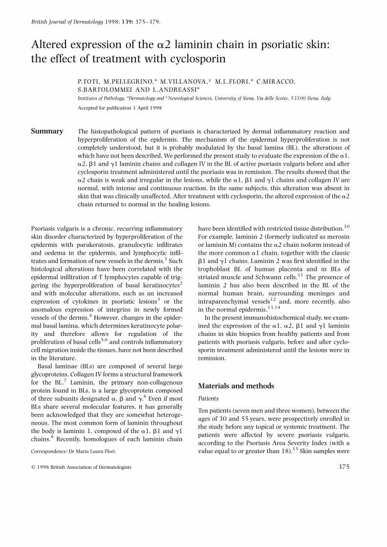
Figure 1. a2 laminin, healthy skin. The immunohistochemical reac-tion at the dermoepidermal junction is intense and continuous. Nerveendings are very evident in the dermis. Original magnification, × 60.
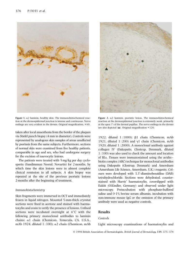
Figure 2. a2 laminin, psoriatic lesion. The immunohistochemicalreaction at the dermoepidermal junction is extremely weak, primarilyat the apex (*) of the dermal papillae. The nerve endings in the dermisare also depicted (O). Original magnification × 120.
eosin-stained cryostat sections appeared to be comple-tely normal in healthy skin from patients with psor-iasis as well as in normal skin. All the laminin chainsexamined (a1, a2, b1, g1), in addition to collagen IV,presented a uniform immunohistochemical reactionrunning along the basal layer of the epidermis and theepidermal BL, which were therefore impossible todistinguish under light microscopy (Fig. 1). Further-more, the a1, b1 and g1 laminin chains and collagenIV were expressed in the BL of haematic and lymphaticdermal vessels, while the a2, b1 and g1 chains andcollagen IV were expressed in the terminal branches ofthe peripheral nerves. As such, our results completelycoincide with the data presented in the literature todate.
Psoriatic lesions
Examination of haematoxylin and eosin-stained sec-tions confirmed the clinical diagnosis of psoriasis inour patients, with hyperkeratosis, parakeratosis andinflammatory granulocytic infiltration in the epidermis,
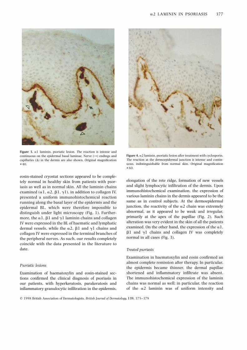
elongation of the rete ridge, formation of new vesselsand slight lymphocytic infiltration of the dermis. Uponimmunohistochemical examination, the expression ofvarious laminin chains in the dermis appeared to be thesame as in control subjects. At the dermoepidermaljunction, the reactivity of the a2 chain was extremelyabnormal, as it appeared to be weak and irregular,primarily at the apex of the papillae (Fig. 2). Suchalteration was very evident in the skin of all the patientsexamined. On the other hand, the expression of the a1,b1 and g1 chains and collagen IV was completelynormal in all cases (Fig. 3).
Treated psoriasis
Examination in haematoxylin and eosin confirmed analmost complete remission after therapy. In particular,the epidermis became thinner, the dermal papillaeshortened and inflammatory infiltrate was absent.The immunohistochemical expression of the lamininchains was normal as well; in particular, the reactionof the a2 laminin was of uniform intensity and
a2 LAMININ IN PSORIASIS 377
q 1998 British Association of Dermatologists, British Journal of Dermatology, 139, 375–379
Figure 3. a1 laminin, psoriatic lesion. The reaction is intense andcontinuous on the epidermal basal laminae. Nerve (→) endings andcapillaries (D) in the dermis are also shown. Original magnification× 40.
Figure 4. a2 laminin, psoriatic lesion after treatment with cyclosporin.The reaction at the dermoepidermal junction is intense and contin-uous, indistinguishable from normal skin. Original magnification× 60.
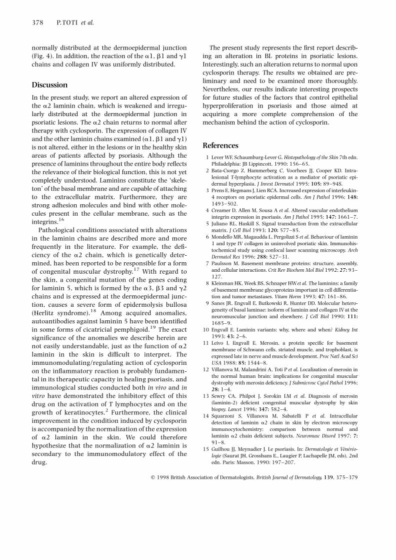
normally distributed at the dermoepidermal junction(Fig. 4). In addition, the reaction of the a1, b1 and g1chains and collagen IV was uniformly distributed.
Discussion
In the present study, we report an altered expression ofthe a2 laminin chain, which is weakened and irregu-larly distributed at the dermoepidermal junction inpsoriatic lesions. The a2 chain returns to normal aftertherapy with cyclosporin. The expression of collagen IVand the other laminin chains examined (a1, b1 and g1)is not altered, either in the lesions or in the healthy skinareas of patients affected by psoriasis. Although thepresence of laminins throughout the entire body reflectsthe relevance of their biological function, this is not yetcompletely understood. Laminins constitute the ‘skele-ton’ of the basal membrane and are capable of attachingto the extracellular matrix. Furthermore, they arestrong adhesion molecules and bind with other mole-cules present in the cellular membrane, such as theintegrins.16
Pathological conditions associated with alterationsin the laminin chains are described more and morefrequently in the literature. For example, the defi-ciency of the a2 chain, which is genetically deter-mined, has been reported to be responsible for a formof congenital muscular dystrophy.17 With regard tothe skin, a congenital mutation of the genes codingfor laminin 5, which is formed by the a3, b3 and g2chains and is expressed at the dermoepidermal junc-tion, causes a severe form of epidermolysis bullosa(Herlitz syndrome).18 Among acquired anomalies,autoantibodies against laminin 5 have been identifiedin some forms of cicatricial pemphigoid.19 The exactsignificance of the anomalies we describe herein arenot easily understandable, just as the function of a2laminin in the skin is difficult to interpret. Theimmunomodulating/regulating action of cyclosporinon the inflammatory reaction is probably fundamen-tal in its therapeutic capacity in healing psoriasis, andimmunological studies conducted both in vivo and invitro have demonstrated the inhibitory effect of thisdrug on the activation of T lymphocytes and on thegrowth of keratinocytes.2 Furthermore, the clinicalimprovement in the condition induced by cyclosporinis accompanied by the normalization of the expressionof a2 laminin in the skin. We could thereforehypothesize that the normalization of a2 laminin issecondary to the immunomodulatory effect of thedrug.
The present study represents the first report describ-ing an alteration in BL proteins in psoriatic lesions.Interestingly, such an alteration returns to normal uponcyclosporin therapy. The results we obtained are pre-liminary and need to be examined more thoroughly.Nevertheless, our results indicate interesting prospectsfor future studies of the factors that control epithelialhyperproliferation in psoriasis and those aimed atacquiring a more complete comprehension of themechanism behind the action of cyclosporin.
References1 Lever WF, Schaumburg-Lever G. Histopathology of the Skin 7th edn.
Philadelphia: JB Lippincott, 1990: 156–65.2 Bata-Csorgo Z, Hammerberg C, Voorhees JJ, Cooper KD. Intra-
lesional T-lymphocyte activation as a mediator of psoriatic epi-dermal hyperplasia. J Invest Dermatol 1995; 105: 89–94S.
3 Prens E, Hegmans J, Lien RCA. Increased expression of interleukin-4 receptors on psoriatic epidermal cells. Am J Pathol 1996; 148:1493–502.
4 Creamer D, Allen M, Sousa A et al. Altered vascular endotheliumintegrin expression in psoriasis. Am J Pathol 1995; 147: 1661–7.
5 Juliano RL, Haskill S. Signal transduction from the extracellularmatrix. J Cell Biol 1993; 120: 577–85.
6 Mondello MR, Magaudda L, Pergolizzi S et al. Behaviour of laminin1 and type IV collagen in uninvolved psoriatic skin. Immunohis-tochemical study using confocal laser scanning microscopy. ArchDermatol Res 1996; 288: 527–31.
7 Paulsson M. Basement membrane proteins: structure, assembly,and cellular interactions. Crit Rev Biochem Mol Biol 1992; 27: 93–127.
8 Kleinman HK, Week BS, Schnaper HWet al. The laminins: a familyof basement membrane glycoproteins important in cell differentia-tion and tumor metastases. Vitam Horm 1993; 47: 161–86.
9 Sanes JR, Engvall E, Butkowski R, Hunter DD. Molecular hetero-geneity of basal laminae: isoform of laminin and collagen IV at theneuromuscular junction and elsewhere. J Cell Biol 1990; 111:1685–9.
10 Engvall E. Laminin variants: why, where and when? Kidney Int1993; 43: 2–6.
11 Leivo I, Engvall E. Merosin, a protein specific for basementmembrane of Schwann cells, striated muscle, and trophoblast, isexpressed late in nerve and muscle development. Proc Natl Acad SciUSA 1988; 85: 1544–8.
12 Villanova M, Malandrini A, Toti P et al. Localization of merosin inthe normal human brain: implications for congenital musculardystrophy with merosin deficiency. J Submicrosc Cytol Pathol 1996;28: 1–4.
13 Sewry CA, Philpot J, Sorokin LM et al. Diagnosis of merosin(laminin-2) deficient congenital muscular dystrophy by skinbiopsy. Lancet 1996; 347: 582–4.
14 Squarzoni S, Villanova M, Sabatelli P et al. Intracellulardetection of laminin a2 chain in skin by electron microscopyimmunocytochemistry: comparison between normal andlaminin a2 chain deficient subjects. Neuromusc Disord 1997; 7:91–8.
15 Guilhou JJ, Meynadier J. Le psoriasis. In: Dermatologie et Venereo-logie (Saurat JH, Grosshans E., Laugier P, Lachapelle JM, eds), 2ndedn. Paris: Masson, 1990: 197–207.
378 P.TOTI et al.
q 1998 British Association of Dermatologists, British Journal of Dermatology, 139, 375–379
16 Aumalley M, Krieg T. Laminins: a family of diverse multifunctionalmolecules of basement membranes. J Invest Dermatol 1996; 106:209–14.
17 Tome F, Evangelista T, Leclerc A et al. Congenital musculardystrophy with merosin deficiency. CR Acad Sci Paris 1994; 317:351–7.
18 Vidal F, Baudoin C, Miquel C et al. Cloning of the laminin alpha 3
gene (LAMA3) and identifications of a homozygous deletion in apatient with Herlitz junctional epidermolysis bullosa. Genomics1995; 30: 273–80.
19 Shimuzu H, Masunaga T, Ishiko A et al. Autoantibodies frompatients with cicatricial pemphigoid target different sites in epi-dermal basement membrane. J Invest Dermatol 1995; 104: 370–3.
a2 LAMININ IN PSORIASIS 379
q 1998 British Association of Dermatologists, British Journal of Dermatology, 139, 375–379