Embed Size (px)
Citation preview
RSC Advances
PAPER
Publ
ishe
d on
08
Apr
il 20
14. D
ownl
oade
d by
Dal
hous
ie U
nive
rsity
on
29/0
6/20
14 0
9:19
:05.
View Article OnlineView Journal | View Issue
Department of Materials Engineering, India
India. E-mail: [email protected]
Cite this: RSC Adv., 2014, 4, 19086
Received 30th January 2014Accepted 7th April 2014
DOI: 10.1039/c4ra00875h
www.rsc.org/advances
19086 | RSC Adv., 2014, 4, 19086–190
Amine-functionalized multiwall carbon nanotubesimpart osteoinductive and bactericidal propertiesin poly(3-caprolactone) composites
Sachin Kumar, Suryasarathi Bose and Kaushik Chatterjee*
Poly(3-caprolactone) (PCL) is an aliphatic polyester widely used for biomedical applications but lacks the
mechanical properties desired for many load-bearing orthopedic applications. The objective of this study
was to prepare and characterize PCL composites incorporating multiwall carbon nanotubes (MWNTs)
with different surface functional groups. PCL composites were prepared by melt-mixing with three
different types of MWNTs: pristine (pMWNT), amine functionalized (aMWNT), and carboxyl functionalized
(cMWNT). Melt rheology and scanning electron microscopy indicated good dispersion of the nanotubes
in the matrix. Tensile strength and elastic modulus of the polymer was significantly increased by the
incorporation of MWNTs and further enhanced by favorable interactions between PCL and aMWNTs.
Thermal analysis revealed that MWNTs act as heterogeneous nucleation sites for crystallization of PCL
and increase polymer crystallinity. Incorporation of functionalized MWNTs increased the surface water
wettability of PCL. Osteoblast proliferation and differentiation was significantly enhanced on
functionalized composites. aMWNT composites also exhibited the best bactericidal response. This study
demonstrates that surface functionalization of MWNTs profoundly influences the properties of PCL and
amine-functionalization offers the optimal combination of mechanical properties, osteogenesis and
antimicrobial response. These results have important implications for designing nanocomposites for use
in orthopedics.
1. Introduction
There are growing incidences of chronic, degenerative diseasesglobally largely driven by sedentary, stressful modern lifestylesand age-related injuries with increasing life spans.1 As a result,there is an ever-increasing demand for therapies that canfacilitate regeneration and replacement of damaged tissues.2
Globally there is huge demand for bioactive materials for bonetissue engineering (BTE). Ideally, for BTE application a bonesubstitute material minimally should be biocompatible andosteoconductive to enhance new bone growth.3,4 In recent years,several types of bioactive materials such as bioactive glass,hydroxyapatite, b-tricalcium phosphate, glass ceramics, etc.,have been used for BTE, but these materials tend to be brittleand many are non-biodegradable.5
With progressive advancement in the eld of tissue engi-neering, interest has shied to the use of synthetic biodegrad-able polymers as scaffold materials.5 These synthetic polymersare gaining attention because they can offer structural integrityand mechanical strength, and eventually slowly degrade leavingbehind newly formed tissue which is expected to take over themechanical loads.5 Many synthetic biodegradable polymers
n Institute of Science, Bangalore 560012,
net.in; Tel: +91-80-22933408
98
have been used as scaffolds for tissue engineering, amongwhich poly(3-caprolactone) (PCL) has been extensively studied.6
PCL is a semicrystalline, hydrophobic polymer with a slowdegradation rate. PCL also exhibits excellent bioresorbabilityand biocompatibility. However, PCL lacks mechanical proper-ties desired for use in load-bearing applications limiting its usein orthopaedics.6
There are various routes available to improve the mechanicalproperties of a polymer but one the most effective ways is byreinforcing the so matrix with hard nanoparticles.7–10 Amongthe many possible nanollers, multiwall carbon nanotube(MWNT) reinforcement has been shown to effectivelystrengthen polymers due to its goodmechanical properties.7,10,11
In a recent review various aspects of polymer/CNT compositeshave been addressed.12 MWNT–polymer composites are beingincreasingly studied for potential use in biomedical applica-tions.13 Zhang et al. showed that the addition of MWNTs inpolylactic acid (PLA) improved mechanical properties butinhibited growth of broblast on the composite surface.14 Panet al. reported that bone marrow derived stem cell osteogenesiswas enhanced in PCL scaffolds with the incorporationMWNTs.7
Studies have also shown that the use of surface functionalizedMWNTs, surfactant-coated MWNTs and MWNTs decoratedwith block co-polymers help in better dispersion and polymer–nanotube interfacial interactions. This results in more effective
This journal is © The Royal Society of Chemistry 2014
Paper RSC Advances
Publ
ishe
d on
08
Apr
il 20
14. D
ownl
oade
d by
Dal
hous
ie U
nive
rsity
on
29/0
6/20
14 0
9:19
:05.
View Article Online
stress transfer to MWNTs resulting in improved mechanicalproperties of the composites.15–17 Lin et al. showed thatcarboxylated MWNTs were better dispersed than non-func-tionalized MWNTs in poly(lactic-co-glycolic acid) (PLGA).18
Better nanoparticle dispersion resulted in further improvementin mechanical properties of the polymer. Other studies havereported that functionalization of MWNTs leads to reducedcytotoxicity in contrast to non-functionalized and surfactant-coated nanotubes.19,20
However, the role, if any, of surface functional groups ofMWNT llers on the physical properties and biologicalresponse to biomedical polyesters has not been studied. Thus,the aim of this study was to systematically evaluate the effect ofusing functionalized MWNTs in PCL and compare with non-functionalized MWNT-reinforced PCL and neat polymer. Wehypothesized that the presence of functional groups on thenanoparticles will lead to differences in physical properties andbiological responses. PCL/MWNT composites containing 1%MWNTs were prepared by melt mixing and their thermal,rheological and mechanical properties were characterized tostudy the effect of MWNT functionalization. Osteoblast attach-ment, proliferation and mineralization along with bactericidalproperty were studied to characterize potential differences inbiological response.
2. Materials and methods2.1. Materials
Chemical vapor deposition-synthesized, thin puried MWNTs:pristine (pMWNT), –COOH functionalized (cMWNT) and –NH2
functionalized (aMWNT) were obtained commercially (NanocylCA). Table 1 presents average diameter, length, aspect ratio andpercentage functionalization of the different MWNTs. pMWNTswere processed to enhance their dispersion by dissolving indimethylformamide (DMF, Merck) followed by probe sonication(Hielsher, Ultrasound technology) and vacuum drying overnightat 80 �C. cMWNTs and aMWNTs were used as receivedwithout further treatment. PCL (average molecular weightMn ¼45 000 g mol�1, Sigma-Aldrich) obtained in pellet form wasdried overnight in a vacuum oven at 30 �C prior to preparingcomposites.
2.2. Preparation of PCL/MWNT nanocomposites
Nanocomposites of PCL/MWNT were prepared by melt mixing.1 wt% (MWNT to PCL ratio) of the respective MWNTs was mixedwith PCL using a conical twin screw microcompounder (HaakeMini lab II) at 80 �C with rotating speed of 60 rpm undernitrogen supply for 20 min to form nanocomposites of
Table 1 Average diameter, length and percentage functionalization of t
MWNT Code no. Average diam
Pristine (pMWNT) NC-7000 9.5–COOH functionalized (cMWNT) NC-3151 9.5–NH2 functionalized (aMWNT) NC-3152 9.5
This journal is © The Royal Society of Chemistry 2014
PCL/pMWNT, PCL/cMWNT and PCL/aMWNT. Melt extrudedstrands were compress-molded using a laboratory scale hotpress (Boolani Engineering) at a pressure of 30 kgf m�2, meltingtemperature 80 �C and holding time of 2 min into circular discs(5 mm diameter � 0.5 mm height).
2.3. Physical and chemical characterization
Surface morphology of neat PCL and its PCL/MWNT nano-composites was analyzed using a scanning electron microscope(SEM). The extruded strands were cryo-fractured in liquidnitrogen. Cryo-fractured samples were mounted on analuminium stub and sputter-coated with gold prior to imaging(FEI Sirion XL30 FEG).
Thermal properties were characterized by differential scan-ning calorimetry (DSC, Mettler-Toledo DSC). The samples wereheated from 25 �C to 100 �C, held for 3 min, cooled to 25 �C, andreheated to 100 �C to eliminate prior thermal history, at 10 �Cmin�1 under argon atmosphere. The degree of crystallization(Xc) of samples were calculated as follows: Xc ¼ (DHc/DH0) �100, where DHc is the heat of crystallization of the sample andDH0 is heat of fusion of a fully crystalline material (DH0 for PCLwas taken as 139.5 J g�1).21 X-ray diffraction (XPERTPro, PAN-alytical) patterns were recorded at ambient temperature using aCu Ka radiation source (40 kV and 20 mA) in the 2q range of 10�
to 40�.The viscoelastic properties of neat PCL and its nano-
composite were measured by a stress controlled rheometer(Discovery Hybrid Rheometer, DHR-3, TA Instruments) usingparallel plate geometry (25 mm diameter). The samples of 1 mmthickness were melted at 70 �C for 5 min in the xture to removeresidual thermal history, and subsequently small amplitudeoscillatory shear (SAOS) were carried out. The dynamic strainsweep measurements were carried out apriori to ensure that thestrain amplitude was within the linear viscoelastic region.Isothermal dynamic frequency sweep measurements were per-formed on all samples from 0.01 rad s�1 to 100 rad s�1 at 70 �Cunder inert N2 atmosphere to prevent thermal degradation.
Uniaxial tensile tests was performed in accordance withASTM standard D368 type V. Tensile tests (MicroUTM-Mecme-sin) were performed at room temperature using 100 N load cellat a cross head speed of 5 mm min�1. Data are presented asmean � S.D. from ve independent measurements. The elasticmodulus of the sample was calculated using the initial linearpart of the stress–strain curve and tensile strength was taken asthe stress at which facture occurred.
Sessile drop water contact angle goniometry (OCA 15EC,Dataphysics) was used to assess surface wettability. Ultrapurewater (Sartorius Arium) was used as the probe liquid. The
he MWNTs (as provided by the supplier)
eter (nm) (d) Average length (mm) (L) Aspect ratio (L/d)
1.5 1581.0 1050.7 73
RSC Adv., 2014, 4, 19086–19098 | 19087
RSC Advances Paper
Publ
ishe
d on
08
Apr
il 20
14. D
ownl
oade
d by
Dal
hous
ie U
nive
rsity
on
29/0
6/20
14 0
9:19
:05.
View Article Online
contact angles reported represent the mean for three indepen-dent measurements.
Surface topography of neat PCL and its nanocomposite discsused for cell studies were measured using an atomic forcemicroscope (AFM, Bruker Dimension Icon) in tapping mode atresonance frequency of about 300–410 kHz. Surface informationwas expressed as height images from 1.5 � 1.5 mm2 AFM scan.
2.4. Biological studies
MC3T3-E1 subclone 4 mouse osteoblasts (ATCC, USA) werecultured in alpha-minimum essential medium (a-MEM, Gibco,Life Technologies) with 10% (v/v) fetal bovine serum (FBS,Gibco, Life Technologies) and 1% (v/v) penicillin–streptomycin(Sigma) in 5% CO2 atmosphere at 37 �C. Medium was changedevery 3 days until cells were 80% to 90% conuent. The cellswere passaged using 0.25% trypsin (Gibco) and serially sub-cultured. Passage 3 cells were used for all studies reported here.
Compression molded discs (5 mm diameter � 0.5 mmheight) were placed in 96 well polypropylene plates (Sigma, non-surface treated) to minimize cell attachment to the bottom ofthe wells. The well-plates containing the discs were sterilizedusing ethylene oxide (Anprolene gas sterilizer, Andersen) anddegassed for 4 days under vacuum prior to cell seeding. 0.2 mlmedium containing 5 � 103 cells were added to each well andcultured as described above with media changes every 3 days.Three plates were used for this study for measuring cellresponse at 1 day, 6 days, and 10 days aer seeding. Each platecontained at least ve samples each of PCL, PCL/pMWNT, PCL/aMWNT, and PCL/cMWNT wherein at least three samples wereused for DNA quantication and two samples were used fornuclear staining. This experiment was repeated thriceindependently.
Picogreen dsDNA quantication kit (Invitrogen) was used toquantify DNA content on the sample.22,23 To collect the DNA,0.2 ml lysate solution (0.2 mg ml�1 proteinase K and 0.02%sodium dodecyl sulfate) was added for 24 h at 37 �C. 0.1 ml ofthe lysate was transferred to a fresh 96 well-plate and mixedwith equal volume of the picogreen dye solution. Fluorescenceintensity was measured using 485 nm excitation and 520 nmemission in a uorescence spectrophotometer (Synergy HT,BioTek). DNA content was calculated from a standard plotgenerated by serial dilutions of a DNA solution of knownconcentration. A total of fourteen samples (n $ 3 for threeindependent experiments) were used to measure DNA contentfor each composite.
Cells were xed with 3.7% (mass/volume) formaldehyde inPBS for 15 min, permeabilized with 0.2% TritonX-100 for 5 minand stained with 6.6 mM Alexa Fluro 546 (Invitrogen) for 1 h and14.3 mM DAPI for 15 min for actin laments and nuclei,respectively. Stained cells were imaged in the blue and redchannel using an inverted uorescence microscope (OlympusIX-71). ImageJ soware was used to calculate cell area andaspect ratio. An outline of the cell was created to measure itsarea and an ellipse was tted to calculate aspect ratio.
To study mineralization, MC3T3 cells (5 � 103 per well) wereadded in a 96 well plate containing nanocomposites discs as
19088 | RSC Adv., 2014, 4, 19086–19098
above and incubated in growth media for 1 day. Growth mediawas replaced with osteogenic differentiation media (growthmedia supplemented with 50 mg ml�1 ascorbic acid and 10 mMb-glycerophosphate24). Medium was refreshed every 3 days.Cells were xed with 3.7% formaldehyde solution at 14 days and21 days. Calcium deposition was analysed by staining with 2%Alizarin Red S (ARS) (Sigma). Fourier transform infrared (FTIR)spectroscopy was measured from 650 cm�1 to 4000 cm�1 toanalyse the presence of calcium phosphate. For quantication,the stain was desorbed with 0.5 ml of 5% SDS in 0.5 N HCL for30 min at room temperature.25 150 ml of solubilised stain wascollected in a 96 well plate, and absorbance was measured at405 nm using a spectrophotometer.
2.5. Antibacterial test
E. coli (MG 1655) were cultured in sterile Luria Broth (LB) mediaovernight at 200 rpm and 37 �C. The E. coli was harvested bycentrifugation at 3000 rpm for 10 min. The collected pellet wassuspended in phosphate buffer saline (PBS) and the opticaldensity (OD) of the suspension was adjusted to 0.5 at 600 nm.This suspension culture was used as inoculums for furtherwork.
The direct contact test was used to assess the antibacterialproperties of PCL and the composites. 50 ml of the bacterialsuspension was added to a 96 well polystyrene plate with ster-ilized discs as described above for osteoblast studies. Emptywells without discs were used as controls. 200 ml of LB mediawas added to each well and incubated at 37 �C with constantshaking at 50 rpm. The growth of suspended bacteria in contactwith the different surfaces was evaluated at 0 h, 1 h, 3 h, 5 h, 8 h,12 h, 18 h and 24 h by collecting 200 ml of the suspension andmeasuring the OD at 600 nm using a spectrophotometer. Allmeasurements are reported as mean � S.D. for n$ 3. To imageadherent cells, samples were xed with 2.5% glutaraldehyde for1 h, dried and sputter-coated with gold prior to SEM imaging.
2.6. Statistical analysis
For statistical analyses, 1-way ANOVA (analysis of variance) withTukey's test for multiple comparisons was used. Differenceswere considered statistically signicant if p < 0.05 and areindicated by symbols in the gures.
3. Results3.1. State of MWNT dispersion
SEM micrographs (Fig. 1) of cryo-fractured surfaces of neat PCLand the different PCL/MWNT nanocomposites show the state ofdispersionMWNTs in the PCLmatrix. MWNTs are seen as whitespecks indicated by the arrows. Higher magnication imagesare presented as insets in Fig. 1(b)–(d). Fig 1(b)–(d) showuniform dispersion of the different types of MWNTs in PCL.
3.2. Thermal and XRD analyses
DSC was used to analyze melting and crystallization behavior ofneat PCL and its different nanocomposites. Melting endothermfor PCL shown in Fig. 2(a) indicates that the melting
This journal is © The Royal Society of Chemistry 2014
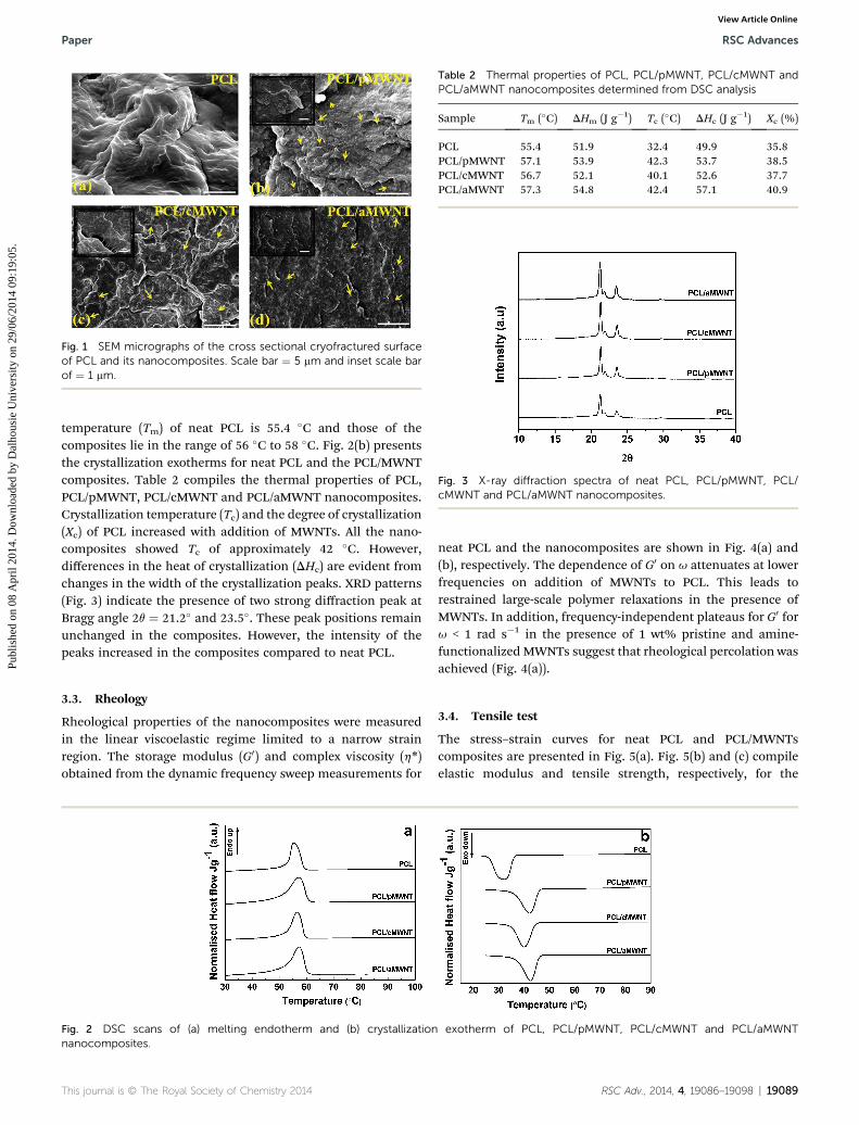
Fig. 1 SEM micrographs of the cross sectional cryofractured surfaceof PCL and its nanocomposites. Scale bar ¼ 5 mm and inset scale barof ¼ 1 mm.
Table 2 Thermal properties of PCL, PCL/pMWNT, PCL/cMWNT andPCL/aMWNT nanocomposites determined from DSC analysis
Sample Tm (�C) DHm (J g�1) Tc (�C) DHc (J g�1) Xc (%)
PCL 55.4 51.9 32.4 49.9 35.8PCL/pMWNT 57.1 53.9 42.3 53.7 38.5PCL/cMWNT 56.7 52.1 40.1 52.6 37.7PCL/aMWNT 57.3 54.8 42.4 57.1 40.9
Fig. 3 X-ray diffraction spectra of neat PCL, PCL/pMWNT, PCL/cMWNT and PCL/aMWNT nanocomposites.
Paper RSC Advances
Publ
ishe
d on
08
Apr
il 20
14. D
ownl
oade
d by
Dal
hous
ie U
nive
rsity
on
29/0
6/20
14 0
9:19
:05.
View Article Online
temperature (Tm) of neat PCL is 55.4 �C and those of thecomposites lie in the range of 56 �C to 58 �C. Fig. 2(b) presentsthe crystallization exotherms for neat PCL and the PCL/MWNTcomposites. Table 2 compiles the thermal properties of PCL,PCL/pMWNT, PCL/cMWNT and PCL/aMWNT nanocomposites.Crystallization temperature (Tc) and the degree of crystallization(Xc) of PCL increased with addition of MWNTs. All the nano-composites showed Tc of approximately 42 �C. However,differences in the heat of crystallization (DHc) are evident fromchanges in the width of the crystallization peaks. XRD patterns(Fig. 3) indicate the presence of two strong diffraction peak atBragg angle 2q ¼ 21.2� and 23.5�. These peak positions remainunchanged in the composites. However, the intensity of thepeaks increased in the composites compared to neat PCL.
3.3. Rheology
Rheological properties of the nanocomposites were measuredin the linear viscoelastic regime limited to a narrow strainregion. The storage modulus (G0) and complex viscosity (h*)obtained from the dynamic frequency sweep measurements for
Fig. 2 DSC scans of (a) melting endotherm and (b) crystallizationnanocomposites.
This journal is © The Royal Society of Chemistry 2014
neat PCL and the nanocomposites are shown in Fig. 4(a) and(b), respectively. The dependence of G0 on u attenuates at lowerfrequencies on addition of MWNTs to PCL. This leads torestrained large-scale polymer relaxations in the presence ofMWNTs. In addition, frequency-independent plateaus for G0 foru < 1 rad s�1 in the presence of 1 wt% pristine and amine-functionalized MWNTs suggest that rheological percolation wasachieved (Fig. 4(a)).
3.4. Tensile test
The stress–strain curves for neat PCL and PCL/MWNTscomposites are presented in Fig. 5(a). Fig. 5(b) and (c) compileelastic modulus and tensile strength, respectively, for the
exotherm of PCL, PCL/pMWNT, PCL/cMWNT and PCL/aMWNT
RSC Adv., 2014, 4, 19086–19098 | 19089
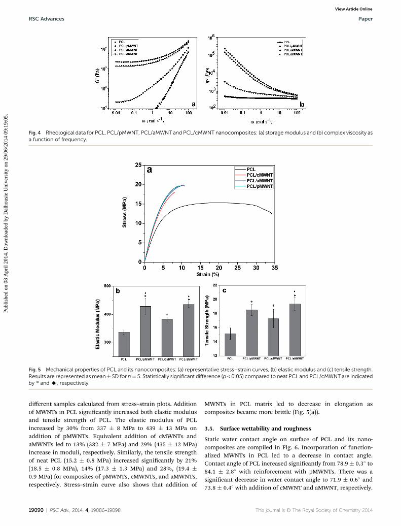
Fig. 4 Rheological data for PCL, PCL/pMWNT, PCL/aMWNT and PCL/cMWNT nanocomposites: (a) storagemodulus and (b) complex viscosity asa function of frequency.
Fig. 5 Mechanical properties of PCL and its nanocomposites: (a) representative stress–strain curves, (b) elastic modulus and (c) tensile strength.Results are represented asmean� SD for n¼ 5. Statistically significant difference (p < 0.05) compared to neat PCL and PCL/cMWNT are indicatedby * and A, respectively.
RSC Advances Paper
Publ
ishe
d on
08
Apr
il 20
14. D
ownl
oade
d by
Dal
hous
ie U
nive
rsity
on
29/0
6/20
14 0
9:19
:05.
View Article Online
different samples calculated from stress–strain plots. Additionof MWNTs in PCL signicantly increased both elastic modulusand tensile strength of PCL. The elastic modulus of PCLincreased by 30% from 337 � 8 MPa to 439 � 13 MPa onaddition of pMWNTs. Equivalent addition of cMWNTs andaMWNTs led to 13% (382 � 7 MPa) and 29% (435 � 12 MPa)increase in moduli, respectively. Similarly, the tensile strengthof neat PCL (15.2 � 0.8 MPa) increased signicantly by 21%(18.5 � 0.8 MPa), 14% (17.3 � 1.3 MPa) and 28%, (19.4 �0.9 MPa) for composites of pMWNTs, cMWNTs, and aMWNTs,respectively. Stress–strain curve also shows that addition of
19090 | RSC Adv., 2014, 4, 19086–19098
MWNTs in PCL matrix led to decrease in elongation ascomposites became more brittle (Fig. 5(a)).
3.5. Surface wettability and roughness
Static water contact angle on surface of PCL and its nano-composites are compiled in Fig. 6. Incorporation of function-alized MWNTs in PCL led to a decrease in contact angle.Contact angle of PCL increased signicantly from 78.9 � 0.3� to84.1 � 2.8� with reinforcement with pMWNTs. There was asignicant decrease in water contact angle to 71.9 � 0.6� and73.8 � 0.4� with addition of cMWNT and aMWNT, respectively.
This journal is © The Royal Society of Chemistry 2014
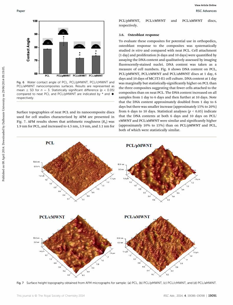
Fig. 6 Water contact angle of PCL, PCL/pMWNT, PCL/cMWNT andPCL/aMWNT nanocomposites surfaces. Results are represented asmean � SD for n ¼ 3. Statistically significant difference (p < 0.05)compared to neat PCL and PCL/pMWNT are indicated by * and Arespectively.
Paper RSC Advances
Publ
ishe
d on
08
Apr
il 20
14. D
ownl
oade
d by
Dal
hous
ie U
nive
rsity
on
29/0
6/20
14 0
9:19
:05.
View Article Online
Surface topographies of neat PCL and its nanocomposite discsused for cell studies characterized by AFM are presented inFig. 7. AFM results shows that arithmetic roughness (Ra) was1.9 nm for PCL, and increased to 4.5 nm, 3.9 nm, and 3.1 nm for
Fig. 7 Surface height topography obtained from AFM micrographs for sa
This journal is © The Royal Society of Chemistry 2014
PCL/pMWNT, PCL/cMWNT and PCL/aMWNT discs,respectively.
3.6. Osteoblast response
To evaluate these composites for potential use in orthopedics,osteoblast response to the composites was systematicallystudied in vitro and compared with neat PCL. Cell attachment(1 day) and proliferation (6 days and 10 days) were quantied byassaying the DNA content and qualitatively assessed by imaginguorescently-stained nuclei. DNA content was taken as ameasure of cell numbers. Fig. 8 shows DNA content on PCL,PCL/pMWNT, PCL/cMWNT and PCL/aMWNT discs at 1 day, 6days and 10 days of MC3T3-E1 cell culture. DNA content at 1 daywas marginally but statistically-signicantly higher on PCL thanthe three composites suggesting that fewer cells attached to thecomposites than on neat PCL. The DNA content increased on allsamples from 1 day to 6 days and then further at 10 days. Notethat the DNA content approximately doubled from 1 day to 6days but there was smaller increase (approximately 15% to 20%)from 6 days to 10 days. Statistical analyses (p < 0.05) indicatethat the DNA contents at both 6 days and 10 days on PCL/cMWNT and PCL/aMWNT were similar and signicantly higher(approximately 10% to 15%) than on PCL/pMWNT and PCL,both of which were statistically similar.
mple: (a) PCL, (b) PCL/pMWNT, (c) PCL/cMWNT, and (d) PCL/aMWNT.
RSC Adv., 2014, 4, 19086–19098 | 19091
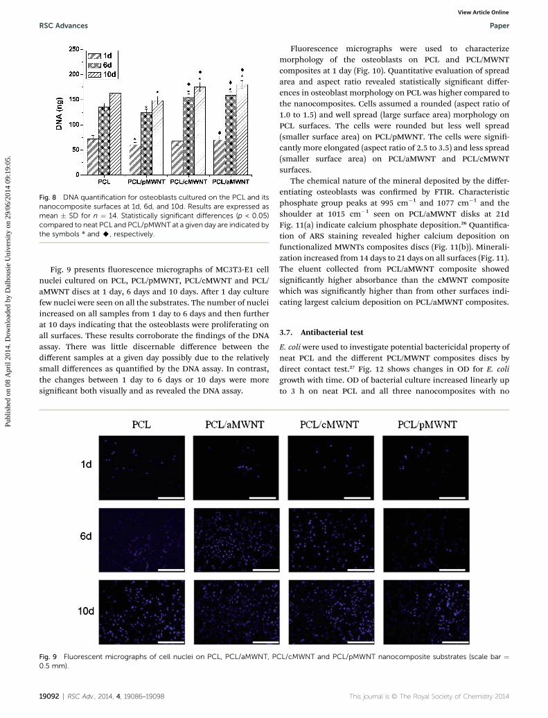
Fig. 8 DNA quantification for osteoblasts cultured on the PCL and itsnanocomposite surfaces at 1d, 6d, and 10d. Results are expressed asmean � SD for n ¼ 14. Statistically significant differences (p < 0.05)compared to neat PCL and PCL/pMWNT at a given day are indicated bythe symbols * and A, respectively.
RSC Advances Paper
Publ
ishe
d on
08
Apr
il 20
14. D
ownl
oade
d by
Dal
hous
ie U
nive
rsity
on
29/0
6/20
14 0
9:19
:05.
View Article Online
Fig. 9 presents uorescence micrographs of MC3T3-E1 cellnuclei cultured on PCL, PCL/pMWNT, PCL/cMWNT and PCL/aMWNT discs at 1 day, 6 days and 10 days. Aer 1 day culturefew nuclei were seen on all the substrates. The number of nucleiincreased on all samples from 1 day to 6 days and then furtherat 10 days indicating that the osteoblasts were proliferating onall surfaces. These results corroborate the ndings of the DNAassay. There was little discernable difference between thedifferent samples at a given day possibly due to the relativelysmall differences as quantied by the DNA assay. In contrast,the changes between 1 day to 6 days or 10 days were moresignicant both visually and as revealed the DNA assay.
Fig. 9 Fluorescent micrographs of cell nuclei on PCL, PCL/aMWNT, PC0.5 mm).
19092 | RSC Adv., 2014, 4, 19086–19098
Fluorescence micrographs were used to characterizemorphology of the osteoblasts on PCL and PCL/MWNTcomposites at 1 day (Fig. 10). Quantitative evaluation of spreadarea and aspect ratio revealed statistically signicant differ-ences in osteoblast morphology on PCL was higher compared tothe nanocomposites. Cells assumed a rounded (aspect ratio of1.0 to 1.5) and well spread (large surface area) morphology onPCL surfaces. The cells were rounded but less well spread(smaller surface area) on PCL/pMWNT. The cells were signi-cantly more elongated (aspect ratio of 2.5 to 3.5) and less spread(smaller surface area) on PCL/aMWNT and PCL/cMWNTsurfaces.
The chemical nature of the mineral deposited by the differ-entiating osteoblasts was conrmed by FTIR. Characteristicphosphate group peaks at 995 cm�1 and 1077 cm�1 and theshoulder at 1015 cm�1 seen on PCL/aMWNT disks at 21dFig. 11(a) indicate calcium phosphate deposition.26 Quantica-tion of ARS staining revealed higher calcium deposition onfunctionalized MWNTs composites discs (Fig. 11(b)). Minerali-zation increased from 14 days to 21 days on all surfaces (Fig. 11).The eluent collected from PCL/aMWNT composite showedsignicantly higher absorbance than the cMWNT compositewhich was signicantly higher than from other surfaces indi-cating largest calcium deposition on PCL/aMWNT composites.
3.7. Antibacterial test
E. coli were used to investigate potential bactericidal property ofneat PCL and the different PCL/MWNT composites discs bydirect contact test.27 Fig. 12 shows changes in OD for E. coligrowth with time. OD of bacterial culture increased linearly upto 3 h on neat PCL and all three nanocomposites with no
L/cMWNT and PCL/pMWNT nanocomposite substrates (scale bar ¼
This journal is © The Royal Society of Chemistry 2014
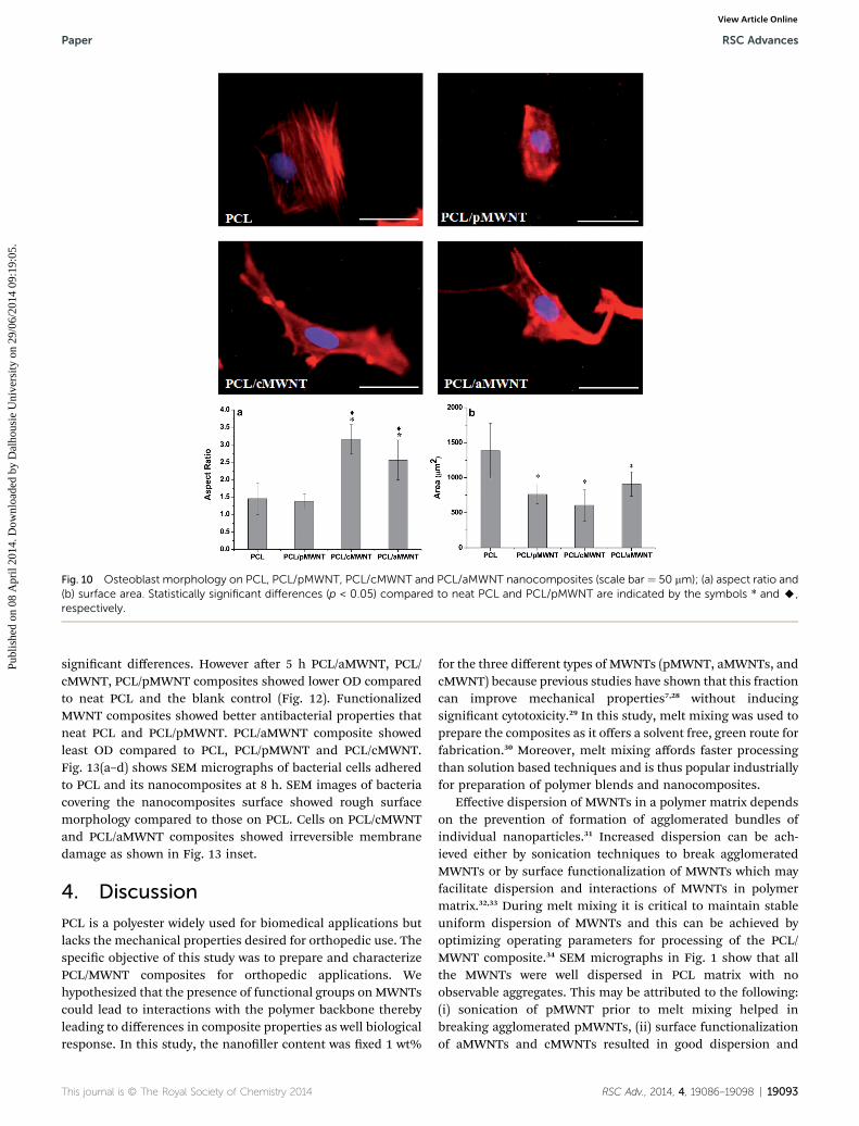
Fig. 10 Osteoblast morphology on PCL, PCL/pMWNT, PCL/cMWNT and PCL/aMWNT nanocomposites (scale bar ¼ 50 mm); (a) aspect ratio and(b) surface area. Statistically significant differences (p < 0.05) compared to neat PCL and PCL/pMWNT are indicated by the symbols * and A,respectively.
Paper RSC Advances
Publ
ishe
d on
08
Apr
il 20
14. D
ownl
oade
d by
Dal
hous
ie U
nive
rsity
on
29/0
6/20
14 0
9:19
:05.
View Article Online
signicant differences. However aer 5 h PCL/aMWNT, PCL/cMWNT, PCL/pMWNT composites showed lower OD comparedto neat PCL and the blank control (Fig. 12). FunctionalizedMWNT composites showed better antibacterial properties thatneat PCL and PCL/pMWNT. PCL/aMWNT composite showedleast OD compared to PCL, PCL/pMWNT and PCL/cMWNT.Fig. 13(a–d) shows SEM micrographs of bacterial cells adheredto PCL and its nanocomposites at 8 h. SEM images of bacteriacovering the nanocomposites surface showed rough surfacemorphology compared to those on PCL. Cells on PCL/cMWNTand PCL/aMWNT composites showed irreversible membranedamage as shown in Fig. 13 inset.
4. Discussion
PCL is a polyester widely used for biomedical applications butlacks the mechanical properties desired for orthopedic use. Thespecic objective of this study was to prepare and characterizePCL/MWNT composites for orthopedic applications. Wehypothesized that the presence of functional groups onMWNTscould lead to interactions with the polymer backbone therebyleading to differences in composite properties as well biologicalresponse. In this study, the nanoller content was xed 1 wt%
This journal is © The Royal Society of Chemistry 2014
for the three different types of MWNTs (pMWNT, aMWNTs, andcMWNT) because previous studies have shown that this fractioncan improve mechanical properties7,28 without inducingsignicant cytotoxicity.29 In this study, melt mixing was used toprepare the composites as it offers a solvent free, green route forfabrication.30 Moreover, melt mixing affords faster processingthan solution based techniques and is thus popular industriallyfor preparation of polymer blends and nanocomposites.
Effective dispersion of MWNTs in a polymer matrix dependson the prevention of formation of agglomerated bundles ofindividual nanoparticles.31 Increased dispersion can be ach-ieved either by sonication techniques to break agglomeratedMWNTs or by surface functionalization of MWNTs which mayfacilitate dispersion and interactions of MWNTs in polymermatrix.32,33 During melt mixing it is critical to maintain stableuniform dispersion of MWNTs and this can be achieved byoptimizing operating parameters for processing of the PCL/MWNT composite.34 SEM micrographs in Fig. 1 show that allthe MWNTs were well dispersed in PCL matrix with noobservable aggregates. This may be attributed to the following:(i) sonication of pMWNT prior to melt mixing helped inbreaking agglomerated pMWNTs, (ii) surface functionalizationof aMWNTs and cMWNTs resulted in good dispersion and
RSC Adv., 2014, 4, 19086–19098 | 19093
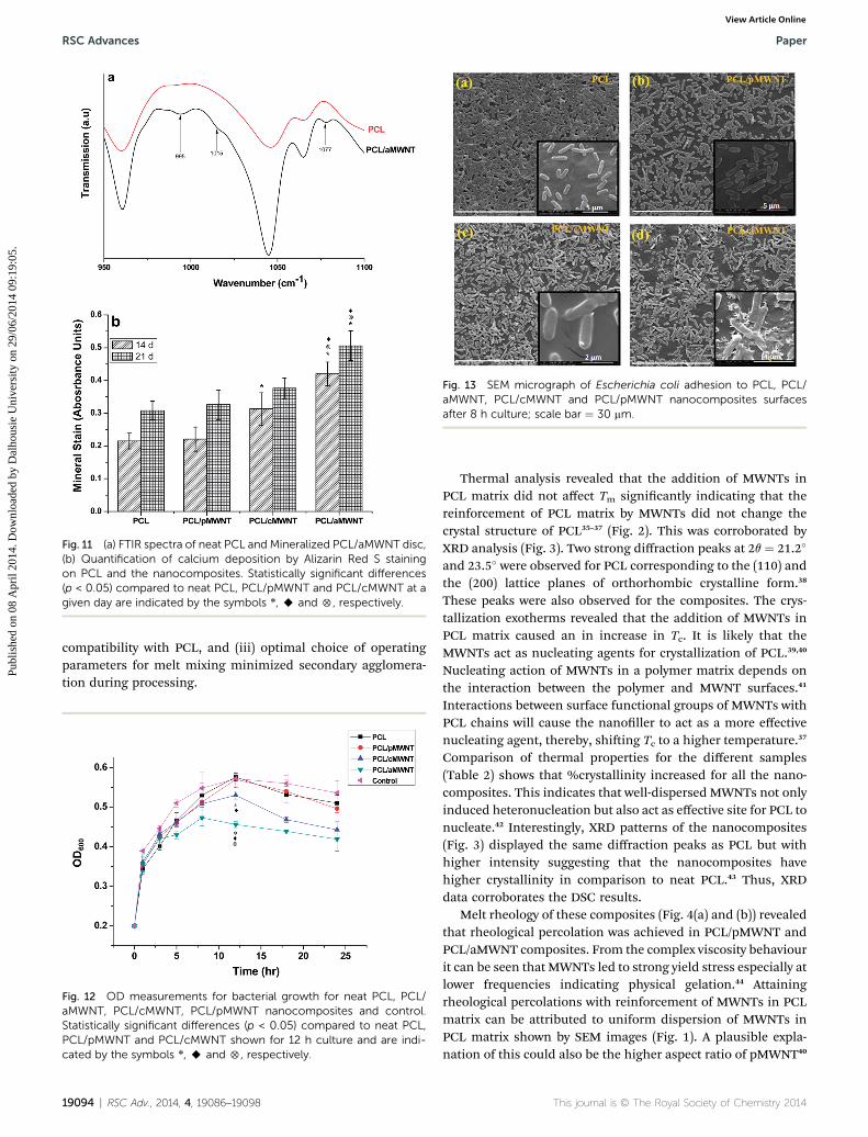
Fig. 11 (a) FTIR spectra of neat PCL and Mineralized PCL/aMWNT disc,(b) Quantification of calcium deposition by Alizarin Red S stainingon PCL and the nanocomposites. Statistically significant differences(p < 0.05) compared to neat PCL, PCL/pMWNT and PCL/cMWNT at agiven day are indicated by the symbols *, A and 5, respectively.
Fig. 13 SEM micrograph of Escherichia coli adhesion to PCL, PCL/aMWNT, PCL/cMWNT and PCL/pMWNT nanocomposites surfacesafter 8 h culture; scale bar ¼ 30 mm.
RSC Advances Paper
Publ
ishe
d on
08
Apr
il 20
14. D
ownl
oade
d by
Dal
hous
ie U
nive
rsity
on
29/0
6/20
14 0
9:19
:05.
View Article Online
compatibility with PCL, and (iii) optimal choice of operatingparameters for melt mixing minimized secondary agglomera-tion during processing.
Fig. 12 OD measurements for bacterial growth for neat PCL, PCL/aMWNT, PCL/cMWNT, PCL/pMWNT nanocomposites and control.Statistically significant differences (p < 0.05) compared to neat PCL,PCL/pMWNT and PCL/cMWNT shown for 12 h culture and are indi-cated by the symbols *, A and 5, respectively.
19094 | RSC Adv., 2014, 4, 19086–19098
Thermal analysis revealed that the addition of MWNTs inPCL matrix did not affect Tm signicantly indicating that thereinforcement of PCL matrix by MWNTs did not change thecrystal structure of PCL35–37 (Fig. 2). This was corroborated byXRD analysis (Fig. 3). Two strong diffraction peaks at 2q ¼ 21.2�
and 23.5� were observed for PCL corresponding to the (110) andthe (200) lattice planes of orthorhombic crystalline form.38
These peaks were also observed for the composites. The crys-tallization exotherms revealed that the addition of MWNTs inPCL matrix caused an in increase in Tc. It is likely that theMWNTs act as nucleating agents for crystallization of PCL.39,40
Nucleating action of MWNTs in a polymer matrix depends onthe interaction between the polymer and MWNT surfaces.41
Interactions between surface functional groups of MWNTs withPCL chains will cause the nanoller to act as a more effectivenucleating agent, thereby, shiing Tc to a higher temperature.37
Comparison of thermal properties for the different samples(Table 2) shows that %crystallinity increased for all the nano-composites. This indicates that well-dispersed MWNTs not onlyinduced heteronucleation but also act as effective site for PCL tonucleate.42 Interestingly, XRD patterns of the nanocomposites(Fig. 3) displayed the same diffraction peaks as PCL but withhigher intensity suggesting that the nanocomposites havehigher crystallinity in comparison to neat PCL.43 Thus, XRDdata corroborates the DSC results.
Melt rheology of these composites (Fig. 4(a) and (b)) revealedthat rheological percolation was achieved in PCL/pMWNT andPCL/aMWNT composites. From the complex viscosity behaviourit can be seen that MWNTs led to strong yield stress especially atlower frequencies indicating physical gelation.44 Attainingrheological percolations with reinforcement of MWNTs in PCLmatrix can be attributed to uniform dispersion of MWNTs inPCL matrix shown by SEM images (Fig. 1). A plausible expla-nation of this could also be the higher aspect ratio of pMWNT40
This journal is © The Royal Society of Chemistry 2014
Paper RSC Advances
Publ
ishe
d on
08
Apr
il 20
14. D
ownl
oade
d by
Dal
hous
ie U
nive
rsity
on
29/0
6/20
14 0
9:19
:05.
View Article Online
which results in the restriction of movement of PCL chains.Thus, PCL/pMWNT composite showed high storage modulusthan neat PCL (Fig. 4(a)). cMWNTs with lower aspect ratio(Table 1) exhibited lower modulus. In contrast, PCL/aMWNTshowed rheological properties similar to those of PCL/pMWNTcomposites although the aspect ratio aMWNTs was the lowestamong all the three MWNTs used. This is indicative of thepresence of strong interfacial interactions between PCL andaMWNTs in the melt putatively between the amine groups ofaMWNTs and the ester groups of the polymer chains. Thesendings can also be corroborated from the low frequencycomplex viscosity (h*) plot (Fig. 4(b)). On addition of pMWNTsand aMWNTs, h* considerably increased at low frequencies.Moreover, the Newtonian plateau of viscosity plots disappeared,suggesting strong shear thinning behaviour.44 This indicatesthe formation of percolation network, also evident fromfrequency sweep for G0.
Tensile tests revealed that incorporation of MWNTs in PCLsignicantly improved elastic modulus and strength of all of thecomposites (Fig. 5(b) and (c)). However, pMWNTs and aMWNTswere most efficient in improving the mechanical properties ofPCL. Improvement in mechanical property of nanocompositesdepends primarily on the dispersion of the nanoparticle, itsaspect ratio and strong ller–matrix interactions.31,45 Increase inthe elastic modulus and tensile strength of PCL with addition ofMWNTs are attributed to (i) uniform dispersion of stifferMWNTs in PCL matrix28 as shown by SEM images of PCL/MWNT (Fig. 1(b)–(d)), and (ii) increase in crystallinity of PCLwith addition of MWNTs as shown by DSC (Table 2), therebystrengthening the mechanical property of the matrix.46
Enhancement in mechanical strength by mechanism ofstress transfer between MWNTs and PCL is inuenced byeffective interaction between ller and matrix, and the length ofnanotube.47–49 Such interactions were evidenced by rheologicalpercolation for PCL/pMWNT composite having longer pMWNTin PCL matrix (Fig. 4(a)), and favourable interaction betweenaMWNTs and PCL. Filler particles with a larger aspect ratio arebelieved to be better suited for effective load transfer in polymercomposites.50 In order to maximize mechanical properties of acomposite, long nanotubes with higher aspect ratio are desiredbecause composites mechanical properties directly depend ontotal surface area, which in turn depends on aspect ratio.45 As aresult, PCL/pMWNT composites incorporating longer pMWNTs(higher aspect ratio, Table 1) showed higher modulus andstrength. Interestingly, PCL/aMWNT showed mechanical prop-erties similar to those of PCL/pMWNT composites althoughaMWNTs had shorter length and lowest aspect ratio among allthe three MWNTs due to the presence of strong interfacialinteractions between PCL and aMWNT as observed inrheology (Fig. 4). A lower elastic modulus and tensile strength ofPCL/cMWNT compared to PCL/pMWNT and PCL/aMWNTnanocomposites is due to the lack of strong interfacial inter-actions between the hydrophilic cMWNTs and the hydrophobicPCL matrix.46 Taken together, uniform dispersion and favour-able interaction of aMWNTs with PCL resulted ineffective improvement in mechanical property PCL/aMWNTcomposite.
This journal is © The Royal Society of Chemistry 2014
Water-wettability of its surface is believed to play a criticalrole in determining biological response to a material. Hydro-phobicity of PCL is one of the major limitations of its use as atissue scaffold. Incorporation of MWNTs to PCL can inuencewater-wettability through potential changes in surface rough-ness and through the availability of functional groups on thesurface. Surface functionalization of MWNTs results in increasein the surface energy of MWNTs due to the presence of the polargroups on MWNT surface.51 Presence of polar components onfunctionalized MWNTs induces dipole–dipole and dipole-induced dipole interactions.31 This may lead to increasedwettability or a decrease in water contact angle with addition offunctionalized MWNTs to PCL matrix. Thus, water contactangles on PCL/aMWNT and PCL/cMWNT were found to belower than PCL or PCL/pMWNT (Fig. 6).
The interactions of cells to designed scaffolds are sensitive tosurface topography.52,53 AFM analysis revealed that PCL/MWNTnanocomposites had higher surface roughness compared toneat PCL (Fig. 7). Change in surface roughness can be attributedto uniform dispersion of MWNTs in polymer matrix and theirspecic interaction with polymer.28 Increase in polymer crys-tallinity can also result in increase of surface roughness.53 As aresult, all nanocomposite surfaces were rougher than PCL dueto good dispersion of MWNTs in PCL matrix (Fig. 1) and theresultant increase in crystallinity (Table 2). Results also indicatethat the increase in surface roughness scaled with the length ofMWNTs (Table 1). pMWNTs being the longest resulted in thelargest roughness in the PCL composites followed by cMWNTand aMWNT nanocomposites.
Biomaterial surface properties play a key role in cell attach-ment, proliferation, morphology and differentiation by modu-lating intracellular signalling events.54–57 In this study,osteoblast attachment and proliferation to the differentsubstrates were compared (Fig. 8 and 9). Cells attached andproliferated on all the four substrates. Although cell attachmentto the composites was marginally lower in contrast to the neatpolymer, signicantly more osteoblasts were observed at 6 daysand 10 days on the functionalized-MWNT composites due todifferences in cell proliferation. Thus, the addition of func-tionalized MWNTs to PCL resulted in enhanced cytocompati-bility of the polymer.7,58 Cell response to material substrates areinuenced by many properties including its chemical proper-ties and physical cues from the surface to the cells. The pres-ence of MWNTs and their functionalization lead to differencesin surface chemistry, nanoscale roughness, etc. potentiallyinducing changes in protein adsorption.59 Here increasedaspect ratio and cell proliferation on the functionalized-MWNTcomposites may be attributed to increased surface waterwettability (Fig. 6) and surface roughness (Fig. 7). Wettabilityand rough surface profoundly inuence protein adsorption tothe surface,60,61 an important event in the biological response tomaterials. Osteoblasts have been observed to attach more tohydrophilic surfaces than to hydrophobic surfaces.61 Differ-ences in surface topography are also known to inuence cellularresponse by changes in focal adhesions and cell signallingpathways involved inmechanotransduction.62 These differencesare oen associated with changes in cell morphology. Cell
RSC Adv., 2014, 4, 19086–19098 | 19095
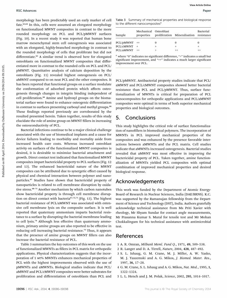
Table 3 Summary of mechanical properties and biological responseto the different nanocompositesa
SampleMechanicalproperties
Osteoblastproliferation Mineralisation
Bacterialresistance
PCL/pMWNT ++ 0 0 0PCL/cMWNT + + + +PCL/aMWNT ++ + ++ ++
a where “0” indicates no signicant difference, “+” indicates a small butsignicant improvement, and “++” indicates a much larger signicantimprovement over PCL.
RSC Advances Paper
Publ
ishe
d on
08
Apr
il 20
14. D
ownl
oade
d by
Dal
hous
ie U
nive
rsity
on
29/0
6/20
14 0
9:19
:05.
View Article Online
morphology has been predictably used an early marker of cellfate.63,64 In this, cells were assumed an elongated morphologyon functionalized MWNT composites in contrast to the morerounded morphology on PCL and PCL/pMWNT surfaces(Fig. 10). In a recent study it was reported that human bonemarrow mesenchymal stem cell osteogenesis was associatedwith an elongated, highly-branched morphology in contrast tothe rounded morphology of cells that proliferate but did notdifferentiate.65 A similar trend is observed here for elongatedosteoblasts on functionalized MWNT composites that differ-entiated more in contrast to the rounded cells on PCL and PCL/pMWNT. Quantitative analysis of calcium deposition by theosteoblasts (Fig. 11) revealed highest osteogenesis on PCL/aMWNT compared to on neat PCL and the other composites. Ithas been reported that functional groups on a surface modulatethe conformation of adsorbed protein which affects osteo-genesis through changes in integrin binding independent ofcell proliferation.66 Amine and hydroxyl groups on the bioma-terial surface were found to enhance osteogenic differentiationin contrast to surfaces presenting carboxyl and methyl groups.66
These ndings reported previously are corroborated by theresulted presented herein. Taken together, results of this studyelucidate the role of amine group onMWNT llers in increasingthe osteoconductivity of PCL.
Bacterial infections continue to be a major clinical challengeassociated with the use of biomedical implants and a cause fordevice failures leading to morbidity and mortality aside fromincreased health care costs. Whereas increased osteoblastactivity on surfaces of the functionalized MWNT composites isdesired, it is desirable to minimize bacterial attachment andgrowth. Direct contact test indicated that functionalized MWNTcomposites impart bactericidal property to PCL surfaces (Fig. 12and 13). The enhanced bactericidal nature of the MWNTcomposites can be attributed due to synergetic effect caused byphysical and chemical interaction between polymer and nano-particles.67 Studies have shown that bactericidal property ofnanoparticles is related to cell membrane disruption by oxida-tive stress.68–71 Another mechanism by which carbon nanotubesshow bactericidal property is through cell membrane disrup-tion on direct contact with bacteria67,72,73 (Fig. 13). The highestbacterial resistance of PCL/aMWNT was associated with exten-sive cell membrane lysis on the composite surface. It is wellreported that quaternary ammonium imparts bacterial resis-tance to a surface by disrupting the bacterial membrane leadingto cell lysis.74 Although less effective than quaternary ammo-nium, primary amine groups are also reported to be effective inreducing cell increasing bacterial resistance.75 Thus, it appearsthat the presence of amine groups on MWNT llers can alsoincrease the bacterial resistance of PCL.
Table 3 summarizes the key outcomes of this work on the useof functionalized MWNTs as llers in PCLmatrix for orthopedicapplications. Physical characterization suggests that the incor-poration of 1 wt% MWNTs enhances mechanical properties ofPCL with the highest improvements observed with the use ofpMWNTs and aMWNTs. Biological studies indicate that PCL/aMWNT and PCL/cMWNT composites were better substrates forproliferation and differentiation of osteoblasts than PCL and
19096 | RSC Adv., 2014, 4, 19086–19098
PCL/pMWNT. Antibacterial property studies indicate that PCL/aMWNT and PCL/cMWNT composites showed better bacterialresistance than PCL and PCL/pMWNT. Thus, surface func-tionalization of MWNTs is critical for preparation of PCLnanocomposites for orthopedic applications and PCL/aMWNTcomposites were optimal in terms of both superior mechanicalproperties and biological outcomes.
5. Conclusions
This study highlights the critical role of surface functionaliza-tion of nanollers in biomedical polymers. The incorporation ofMWNTs in PCL improved mechanical properties of thecomposites and was enhanced by the presence favorable inter-actions between aMWNTs and the PCL matrix. Cell studiesindicate that aMWNTs increased osteogenesis. Bacterial studiesrevealed that aMWNT was most effective in improving thebactericidal property of PCL. Taken together, amine function-alization of MWNTs yielded PCL composites with optimalcombination of improved mechanical properties and desiredbiological response.
Acknowledgements
This work was funded by the Department of Atomic Energy-Board of Research in Nuclear Sciences, India (DAE/BRNS). K.C.was supported by the Ramanujan fellowship from the Depart-ment of Science and Technology (DST), India. Authors gratefullyacknowledge technical assistance from Ms Priti Xavier withrheology, Mr Shyam Sundar for contact angle measurements,Mr Prasanna Kumar S. Mural for tensile test and Mr MohanChokkalingam for his technical assistance with antimicrobialtest.
References
1 A. R. Omran, Milbank Meml. Fund Q., 1971, 49, 509–538.2 R. Langer and D. A. Tirrell, Nature, 2004, 428, 487–492.3 S. L. Ishaug, G. M. Crane, M. J. Miller, A. W. Yasko,M. J. Yaszemski and A. G. Mikos, J. Biomed. Mater. Res.,1997, 36, 17–28.
4 G. M. Crane, S. L. Ishaug and A. G. Mikos, Nat. Med., 1995, 1,1322–1324.
5 L. L. Hench and J. M. Polak, Science, 2002, 295, 1014–1017.
This journal is © The Royal Society of Chemistry 2014
Paper RSC Advances
Publ
ishe
d on
08
Apr
il 20
14. D
ownl
oade
d by
Dal
hous
ie U
nive
rsity
on
29/0
6/20
14 0
9:19
:05.
View Article Online
6 M. A. Woodruff and D. W. Hutmacher, Prog. Polym. Sci.,2010, 35, 1217–1256.
7 L. Pan, X. Pei, R. He, Q. Wan and J. Wang, Colloids Surf., B,2012, 93, 226–234.
8 E. Ural, K. Kesenci, L. Fambri, C. Migliaresi and E. Piskin,Biomaterials, 2000, 21, 2147–2154.
9 C. Wan and B. Chen, Biomed. Mater., 2011, 6, 055010.10 S. D. McCullen, K. L. Stano, D. R. Stevens, W. A. Roberts,
N. A. Monteiro-Riviere, L. I. Clarke and R. E. Gorga, J. Appl.Polym. Sci., 2007, 105, 1668–1678.
11 S. Singh, Y. Pei, R. Miller and P. R. Sundararajan, Adv. Funct.Mater., 2003, 13, 868–872.
12 S. Bose, R. A. Khare and P. Moldenaers, Polymer, 2010, 51,975–993.
13 J. V. Veetil and K. Ye, Biotechnol. Prog., 2009, 25, 709–721.14 D. Zhang, M. A. Kandadai, J. Cech, S. Roth and S. A. Curran,
J. Phys. Chem. B, 2006, 110, 12910–12915.15 F. H. Gojny, J. Nastalczyk, Z. Roslaniec and K. Schulte, Chem.
Phys. Lett., 2003, 370, 820–824.16 L. Vaisman, H. D. Wagner and G. Marom, Adv. Colloid
Interface Sci., 2006, 128, 37–46.17 R. Shvartzman-Cohen, Y. Levi-Kalisman, E. Nativ-Roth and
R. Yerushalmi-Rozen, Langmuir, 2004, 20, 6085–6088.18 C. Lin, Y. Wang, Y. Lai, W. Yang, F. Jiao, H. Zhang, S. Ye and
Q. Zhang, Colloids Surf., B, 2011, 83, 367–375.19 M. Bottini, S. Bruckner, K. Nika, N. Bottini, S. Bellucci,
A. Magrini, A. Bergamaschi and T. Mustelin, Toxicol. Lett.,2006, 160, 121–126.
20 C. M. Sayes, F. Liang, J. L. Hudson, J. Mendez, W. Guo,J. M. Beach, V. C. Moore, C. D. Doyle, J. L. West andW. E. Billups, Toxicol. Lett., 2006, 161, 135–142.
21 V. Crescenzi, G. Manzini, G. Calzolari and C. Borri, Eur.Polym. J., 1972, 8, 449–463.
22 K. Chatterjee, L. Sun, L. C. Chow, M. F. Young andC. G. Simon Jr, Biomaterials, 2011, 32, 1361–1369.
23 K. Chatterjee, S. Hung, G. Kumar and C. G. Simon, J. Funct.Biomater., 2012, 3, 372–381.
24 V. Krishnan, R. Dhurjati, E. A. Vogler and A. M. Mastro, InVitro Cell. Dev. Biol.: Anim., 2010, 46, 28–35.
25 Y. Bi, C. H. Stuelten, T. Kilts, S. Wadhwa, R. V. Iozzo,P. G. Robey, X.-D. Chen and M. F. Young, J. Biol. Chem.,2005, 280, 30481–30489.
26 P. Taddei, A. Tinti, M. Reggiani and C. Fagnano, J. Mol.Struct., 2005, 744–747, 135–143.
27 E. Weiss, M. Shalhav and Z. Fuss, Dent. Traumatol., 1996, 12,179–184.
28 D. Lahiri, F. Rouzaud, S. Namin, A. Keshri, J. Valdes, L. Kos,N. Tsoukias and A. Agarwal, ACS Appl. Mater. Interfaces, 2009,1, 2470–2476.
29 S. Vardharajula, S. Z. Ali, P. M. Tiwari, E. Eroglu, K. Vig,V. A. Dennis and S. R. Singh, Int. J. Nanomed., 2012, 7, 5361.
30 J. Cho and D. Paul, Polymer, 2001, 42, 1083–1094.31 P.-C. Ma, S.-Y. Mo, B.-Z. Tang and J.-K. Kim, Carbon, 2010,
48, 1824–1834.32 J. Li, Z. Fang, L. Tong, A. Gu and F. Liu, J. Appl. Polym. Sci.,
2007, 106, 2898–2906.
This journal is © The Royal Society of Chemistry 2014
33 P.-C. Ma, N. A. Siddiqui, G. Marom and J.-K. Kim,Composites, Part A, 2010, 41, 1345–1367.
34 T. Villmow, B. Kretzschmar and P. Potschke, Compos. Sci.Technol., 2010, 70, 2045–2055.
35 Y. Di, S. Iannace, E. Di Maio and L. Nicolais, J. Polym. Sci.,Part B: Polym. Phys., 2003, 41, 670–678.
36 T. M. Wu and E. C. Chen, J. Polym. Sci., Part B: Polym. Phys.,2006, 44, 598–606.
37 C. A. Mitchell and R. Krishnamoorti, Polymer, 2005, 46,8796–8804.
38 K. Wang, W. Li and C. Gao, J. Appl. Polym. Sci., 2007, 105,629–640.
39 G. Xu, L. Du, H. Wang, R. Xia, X. Meng and Q. Zhu, Polym.Int., 2008, 57, 1052–1066.
40 K. Saeed and S. Y. Park, J. Appl. Polym. Sci., 2007, 104, 1957–1963.
41 M. Trujillo, M. L. Arnal, A. J. Muller, M. A. Mujica, C. Urbinade Navarro, B. Ruelle and P. Dubois, Polymer, 2012, 53, 832–841.
42 K. Lu, N. Grossiord, C. E. Koning, H. E. Miltner, B. V. Meleand J. Loos, Macromolecules, 2008, 41, 8081–8085.
43 C. Saujanya and S. Radhakrishnan, Polymer, 2001, 42, 6723–6731.
44 D. Wu, L. Wu, Y. Sun and M. Zhang, J. Polym. Sci., Part B:Polym. Phys., 2007, 45, 3137–3147.
45 M. Ayatollahi, S. Shadlou, M. Shokrieh andM. Chitsazzadeh,Polym. Test., 2011, 30, 548–556.
46 G. Siqueira, J. Bras and A. Dufresne, Biomacromolecules,2008, 10, 425–432.
47 S.-Y. Fu, X.-Q. Feng, B. Lauke and Y.-W. Mai, Composites, PartB, 2008, 39, 933–961.
48 J. N. Coleman, U. Khan, W. J. Blau and Y. K. Gun'ko, Carbon,2006, 44, 1624–1652.
49 Z. Wang, D. Tang, X. Zheng, W. Zhang and Y. Zhu,Nanotechnology, 2007, 18, 475714.
50 X. Wang, Q. Jiang, W. Xu, W. Cai, Y. Inoue and Y. Zhu,Carbon, 2013, 53, 145–152.
51 S. Nuriel, L. Liu, A. Barber and H. Wagner, Chem. Phys. Lett.,2005, 404, 263–266.
52 R. Flemming, C. Murphy, G. Abrams, S. Goodman andP. Nealey, Biomaterials, 1999, 20, 573–588.
53 N. R. Washburn, K. M. Yamada, C. G. Simon Jr,S. B. Kennedy and E. J. Amis, Biomaterials, 2004, 25, 1215–1224.
54 Y. Qi, Z. Tai, D. Sun, J. Chen, H. Ma, X. Yan, B. Liu andQ. Xue, J. Appl. Polym. Sci., 2013, 127, 1885–1894.
55 M. A. Correa-Duarte, N. Wagner, J. Rojas-Chapana,C. Morsczeck, M. Thie and M. Giersig, Nano Lett., 2004, 4,2233–2236.
56 J. R. Venugopal, S. Low, A. T. Choon, A. B. Kumar andS. Ramakrishna, Artif. Organs, 2008, 32, 388–397.
57 D. W. Hutmacher, T. Schantz, I. Zein, K. W. Ng, S. H. Teohand K. C. Tan, J. Biomed. Mater. Res., 2001, 55, 203–216.
58 P. E. Mikael and S. P. Nukavarapu, J. Biomater. Tissue Eng.,2011, 1, 76–85.
RSC Adv., 2014, 4, 19086–19098 | 19097
RSC Advances Paper
Publ
ishe
d on
08
Apr
il 20
14. D
ownl
oade
d by
Dal
hous
ie U
nive
rsity
on
29/0
6/20
14 0
9:19
:05.
View Article Online
59 X. Li, H. Gao, M. Uo, Y. Sato, T. Akasaka, Q. Feng, F. Cui,X. Liu and F. Watari, J. Biomed. Mater. Res., Part A, 2009,91, 132–139.
60 K.-W. Lee, S. Wang, M. J. Yaszemski and L. Lu, Biomaterials,2008, 29, 2839–2848.
61 J. Wei, T. Igarashi, N. Okumori, T. Igarashi, T. Maetani,B. Liu and M. Yoshinari, Biomed. Mater., 2009, 4, 045002.
62 H. Nikukar, S. Reid, M. P. Tsimbouri, M. O. Riehle,A. S. Curtis and M. J. Dalby, ACS Nano, 2013, 7, 2758–2767.
63 B. M. Spiegelman and C. A. Ginty, Cell, 1983, 35, 657–666.64 R. McBeath, D. M. Pirone, C. M. Nelson, K. Bhadriraju and
C. S. Chen, Dev. Cell, 2004, 6, 483–495.65 G. Kumar, C. K. Tison, K. Chatterjee, P. S. Pine,
J. H. McDaniel, M. L. Salit, M. F. Young and C. G. SimonJr, Biomaterials, 2011, 32, 9188–9196.
66 B. G. Keselowsky, D. M. Collard and A. J. Garcıa, Proc. Natl.Acad. Sci. U. S. A., 2005, 102, 5953–5957.
19098 | RSC Adv., 2014, 4, 19086–19098
67 M. Celeste, R. aTria, R. A. May and V. aVergara, Chem.Commun., 2011, 47, 8892–8894.
68 S. Liu, T. H. Zeng, M. Hofmann, E. Burcombe, J. Wei,R. Jiang, J. Kong and Y. Chen, ACS Nano, 2011, 5, 6971–6980.
69 S. Kang, M. Herzberg, D. F. Rodrigues and M. Elimelech,Langmuir, 2008, 24, 6409–6413.
70 V. K. Upadhyayula, S. Deng, M. C. Mitchell and G. B. Smith,Sci. Total Environ., 2009, 408, 1–13.
71 I. E. M. Carpio, C. M. Santos, X. Wei and D. F. Rodrigues,Nanoscale, 2012, 4, 4746–4756.
72 O. Akhavan and E. Ghaderi, ACS Nano, 2010, 4, 5731–5736.73 S. Kang, M. Pinault, L. D. Pfefferle and M. Elimelech,
Langmuir, 2007, 23, 8670–8673.74 A. Isquith, E. Abbott and P. Walters, Appl. Microbiol., 1972,
24, 859–863.75 E. F. Palermo, D.-K. Lee, A. Ramamoorthy and K. Kuroda,
J. Phys. Chem. B, 2010, 115, 366–375.
This journal is © The Royal Society of Chemistry 2014