Embed Size (px)
Citation preview
American Journal of Internal Medicine 2014; 2(4): 79-82
Published online August 10, 2014 (http://www.sciencepublishinggroup.com/j/ajim)
doi: 10.11648/j.ajim.20140204.15
ISSN: 2330-4316 (Print); ISSN: 2330-4324 (Online)
An autopsic examination case of diagnosed Brugada syndrome
Satoshi Furukawa1, 4
, Satomu Morita1, Hayato Okunaga
1, Lisa Wingenfeld
1, Akari Takaya
1,
Tokiko Nakagawa1, Ikuo Sakaguchi
1, Yoshio Yamamoto
2, Takasi Ashihara
3, Minoru Horie
3,
Katsuji Nishi3, Masahito Hitosugi
1
1Department of Legal Medicine, Shiga University of Medical Science, Shiga, Japan 2Iga Research Institute of Mie University, Mie, Japan 3Department of Cardiovascular and Respiratory Medicine, Shiga University of Medical Science, Shiga, Japan 4Department of Legal Medicine, Shiga University of Medical Science, Setatsukinowa, Otsu City, Shiga 520-2192, Japan
Email address: [email protected] (S. Furukawa)
To cite this article: Satoshi Furukawa, Satomu Morita, Hayato Okunaga, Lisa Wingenfeld, Akari Takaya, Tokiko Nakagawa, Ikuo Sakaguchi, Yoshio
Yamamoto, Takasi Ashihara, Minoru Horie, Katsuji Nishi, Masahito Hitosugi. An Autopsic Examination Case of Diagnosed Brugada
Syndrome. American Journal of Internal Medicine. Vol. 2, No. 4, 2014, pp. 79-82. doi: 10.11648/j.ajim.20140204.15
Abstract: Brugada syndrome is a cardiac disorder characterized by typical ECG alterations, and it is associated with a high
risk for sudden cardiac death, affecting young subjects with structurally normal hearts. The prevalence of this disorder is still
uncertain, presenting marked geographical differences. The syndrome has a genetic basis, and several mutations have been
identified in genes encoding subunits of cardiac sodium, potassium, and calcium channels, as well as in genes involved in the
trafficking or regulation of these channels. We experienced an autopsy case of the sudden death by diagnosed Brugada
syndrome. We present the case report and autopsic findings.
Keywords: Brugada Syndrome, Sudden Death, Coved and Saddleback Type ST Elevation, Autopsy, Histological Findings
1. Introduction
Brugada syndrome (BS) has originally been described as
an autosomal-dominant inherited arrhythmic disorder
characterized by ST elevation with successive negative T
wave in the right precordial leads without structural cardiac
abnormalities [1.2]. Patients are at risk for sudden cardiac
death (SCD) due to ventricular fibrillation (VF). Since 1953,
the ECG pattern similar to coved-type ST-segment elevation
was reported as a normal variant in the healthy population or
related to VF with structural abnormality [3-5]. BS is a
genetically determined disease, characterized by typical
electrocardiographic signs, and it predisposes to sudden
cardiac death (SCD) secondary to polymorphic ventricular
tachycardia (PVT)/ventricular fibrillation (VF) in the
absence of structural heart disease. It was first described in
1992 by Pedro and Josep Brugada [1]. Brugada and Brugada
linked an abnormal ECG with right bundle branch block
pattern and coved-type ST elevation over the right
precordial leads to primary ventricular fibrillation (VF) and
sudden cardiac death (SCD) in patients with structurally
normal hearts [1]. It instantly became known as Brugada
syndrome (BS) and has drawn the worldwide attention of
cardiologists, electrophysiologists and molecular
biologists/geneticists. We present an autopsy case of
diagnosed BS.
2. Case Report
A 33-year-old man was found lying on the floor in his
house. He had pointed the electrocardiogram (ECG)
abnormality five years before in the medical examination.
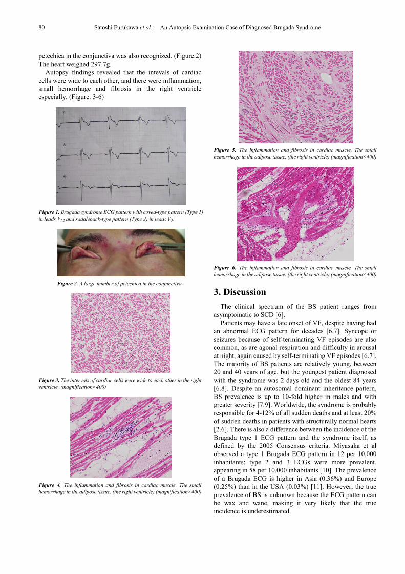
The ECG showed the coved and saddleback type ST
elevation in leads V1 through V3. (Figure.1) His mother
was dead of unknown origin in her thirties. The ECG
classified the Brugada pattern and he diagnosed Brugada
syndrome.
The autopsy was performed on the day following death.
The body was 162 cm in height and 63.8 kg in weight.
Postmortem lividity was large in his back. A large number of
80 Satoshi Furukawa et al.: An Autopsic Examination Case of Diagnosed Brugada Syndrome
petechiea in the conjunctiva was also recognized. (Figure.2)
The heart weighed 297.7g.
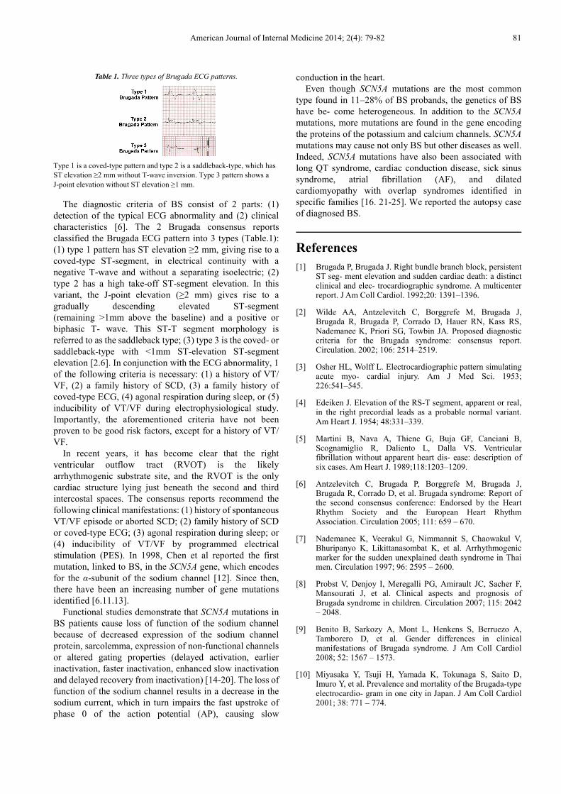
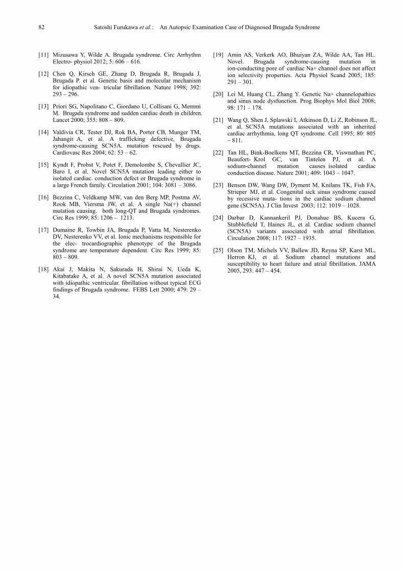
Autopsy findings revealed that the intevals of cardiac
cells were wide to each other, and there were inflammation,
small hemorrhage and fibrosis in the right ventricle
especially. (Figure. 3-6)
Figure 1. Brugada syndrome ECG pattern with coved-type pattern (Type 1)
in leads V1.2 and saddleback-type pattern (Type 2) in leads V3.
Figure 2. A large number of petechiea in the conjunctiva.
Figure 3. The intervals of cardiac cells were wide to each other in the right
ventricle. (magnification×400)
Figure 4. The inflammation and fibrosis in cardiac muscle. The small
hemorrhage in the adipose tissue. (the right ventricle) (magnification×400)
Figure 5. The inflammation and fibrosis in cardiac muscle. The small
hemorrhage in the adipose tissue. (the right ventricle) (magnification×400)
Figure 6. The inflammation and fibrosis in cardiac muscle. The small
hemorrhage in the adipose tissue. (the right ventricle) (magnification×400)
3. Discussion
The clinical spectrum of the BS patient ranges from
asymptomatic to SCD [6].
Patients may have a late onset of VF, despite having had
an abnormal ECG pattern for decades [6.7]. Syncope or
seizures because of self-terminating VF episodes are also
common, as are agonal respiration and difficulty in arousal
at night, again caused by self-terminating VF episodes [6.7].
The majority of BS patients are relatively young, between
20 and 40 years of age, but the youngest patient diagnosed
with the syndrome was 2 days old and the oldest 84 years
[6.8]. Despite an autosomal dominant inheritance pattern,
BS prevalence is up to 10-fold higher in males and with
greater severity [7.9]. Worldwide, the syndrome is probably
responsible for 4-12% of all sudden deaths and at least 20%
of sudden deaths in patients with structurally normal hearts
[2.6]. There is also a difference between the incidence of the
Brugada type 1 ECG pattern and the syndrome itself, as
defined by the 2005 Consensus criteria. Miyasaka et al
observed a type 1 Brugada ECG pattern in 12 per 10,000
inhabitants; type 2 and 3 ECGs were more prevalent,
appearing in 58 per 10,000 inhabitants [10]. The prevalence
of a Brugada ECG is higher in Asia (0.36%) and Europe
(0.25%) than in the USA (0.03%) [11]. However, the true
prevalence of BS is unknown because the ECG pattern can
be wax and wane, making it very likely that the true
incidence is underestimated.
American Journal of Internal Medicine 2014; 2(4): 79-82 81
Table 1. Three types of Brugada ECG patterns.
Type 1 is a coved-type pattern and type 2 is a saddleback-type, which has
ST elevation ≥2 mm without T-wave inversion. Type 3 pattern shows a
J-point elevation without ST elevation ≥1 mm.
The diagnostic criteria of BS consist of 2 parts: (1)
detection of the typical ECG abnormality and (2) clinical
characteristics [6]. The 2 Brugada consensus reports
classified the Brugada ECG pattern into 3 types (Table.1):
(1) type 1 pattern has ST elevation ≥2 mm, giving rise to a
coved-type ST-segment, in electrical continuity with a
negative T-wave and without a separating isoelectric; (2)
type 2 has a high take-off ST-segment elevation. In this
variant, the J-point elevation (≥2 mm) gives rise to a
gradually descending elevated ST-segment
(remaining >1mm above the baseline) and a positive or
biphasic T- wave. This ST-T segment morphology is
referred to as the saddleback type; (3) type 3 is the coved- or
saddleback-type with <1mm ST-elevation ST-segment
elevation [2.6]. In conjunction with the ECG abnormality, 1
of the following criteria is necessary: (1) a history of VT/
VF, (2) a family history of SCD, (3) a family history of
coved-type ECG, (4) agonal respiration during sleep, or (5)
inducibility of VT/VF during electrophysiological study.
Importantly, the aforementioned criteria have not been
proven to be good risk factors, except for a history of VT/
VF.
In recent years, it has become clear that the right
ventricular outflow tract (RVOT) is the likely
arrhythmogenic substrate site, and the RVOT is the only
cardiac structure lying just beneath the second and third
intercostal spaces. The consensus reports recommend the
following clinical manifestations: (1) history of spontaneous
VT/VF episode or aborted SCD; (2) family history of SCD
or coved-type ECG; (3) agonal respiration during sleep; or
(4) inducibility of VT/VF by programmed electrical
stimulation (PES). In 1998, Chen et al reported the first
mutation, linked to BS, in the SCN5A gene, which encodes
for the α-subunit of the sodium channel [12]. Since then,
there have been an increasing number of gene mutations
identified [6.11.13].
Functional studies demonstrate that SCN5A mutations in
BS patients cause loss of function of the sodium channel
because of decreased expression of the sodium channel
protein, sarcolemma, expression of non-functional channels
or altered gating properties (delayed activation, earlier
inactivation, faster inactivation, enhanced slow inactivation
and delayed recovery from inactivation) [14-20]. The loss of
function of the sodium channel results in a decrease in the
sodium current, which in turn impairs the fast upstroke of
phase 0 of the action potential (AP), causing slow
conduction in the heart.
Even though SCN5A mutations are the most common
type found in 11–28% of BS probands, the genetics of BS
have be- come heterogeneous. In addition to the SCN5A
mutations, more mutations are found in the gene encoding
the proteins of the potassium and calcium channels. SCN5A
mutations may cause not only BS but other diseases as well.
Indeed, SCN5A mutations have also been associated with
long QT syndrome, cardiac conduction disease, sick sinus
syndrome, atrial fibrillation (AF), and dilated
cardiomyopathy with overlap syndromes identified in
specific families [16. 21-25]. We reported the autopsy case
of diagnosed BS.
References
[1] Brugada P, Brugada J. Right bundle branch block, persistent ST seg- ment elevation and sudden cardiac death: a distinct clinical and elec- trocardiographic syndrome. A multicenter report. J Am Coll Cardiol. 1992;20: 1391–1396.
[2] Wilde AA, Antzelevitch C, Borggrefe M, Brugada J, Brugada R, Brugada P, Corrado D, Hauer RN, Kass RS, Nademanee K, Priori SG, Towbin JA. Proposed diagnostic criteria for the Brugada syndrome: consensus report. Circulation. 2002; 106: 2514–2519.
[3] Osher HL, Wolff L. Electrocardiographic pattern simulating acute myo- cardial injury. Am J Med Sci. 1953; 226:541–545.
[4] Edeiken J. Elevation of the RS-T segment, apparent or real, in the right precordial leads as a probable normal variant. Am Heart J. 1954; 48:331–339.
[5] Martini B, Nava A, Thiene G, Buja GF, Canciani B, Scognamiglio R, Daliento L, Dalla VS. Ventricular fibrillation without apparent heart dis- ease: description of six cases. Am Heart J. 1989;118:1203–1209.
[6] Antzelevitch C, Brugada P, Borggrefe M, Brugada J, Brugada R, Corrado D, et al. Brugada syndrome: Report of the second consensus conference: Endorsed by the Heart Rhythm Society and the European Heart Rhythm Association. Circulation 2005; 111: 659 – 670.
[7] Nademanee K, Veerakul G, Nimmannit S, Chaowakul V, Bhuripanyo K, Likittanasombat K, et al. Arrhythmogenic marker for the sudden unexplained death syndrome in Thai men. Circulation 1997; 96: 2595 – 2600.
[8] Probst V, Denjoy I, Meregalli PG, Amirault JC, Sacher F, Mansourati J, et al. Clinical aspects and prognosis of Brugada syndrome in children. Circulation 2007; 115: 2042 – 2048.
[9] Benito B, Sarkozy A, Mont L, Henkens S, Berruezo A, Tamborero D, et al. Gender differences in clinical manifestations of Brugada syndrome. J Am Coll Cardiol 2008; 52: 1567 – 1573.
[10] Miyasaka Y, Tsuji H, Yamada K, Tokunaga S, Saito D, Imuro Y, et al. Prevalence and mortality of the Brugada-type electrocardio- gram in one city in Japan. J Am Coll Cardiol 2001; 38: 771 – 774.
82 Satoshi Furukawa et al.: An Autopsic Examination Case of Diagnosed Brugada Syndrome
[11] Mizusawa Y, Wilde A. Brugada syndrome. Circ Arrhythm Electro- physiol 2012; 5: 606 – 616.
[12] Chen Q, Kirsch GE, Zhang D, Brugada R, Brugada J, Brugada P. et al. Genetic basis and molecular mechanism for idiopathic ven-tricular fibrillation. Nature 1998; 392: 293 – 296.
[13] Priori SG, Napolitano C, Giordano U, Collisani G, Memmi M.Brugada syndrome and sudden cardiac death in children. Lancet 2000; 355: 808 – 809.
[14] Valdivia CR, Tester DJ, Rok BA, Porter CB, Munger TM, JahangirA, et al. A trafficking defective, Brugada syndrome-causing SCN5A. mutation rescued by drugs. Cardiovasc Res 2004; 62: 53 – 62.
[15] Kyndt F, Probst V, Potet F, Demolombe S, Chevallier JC, Baro I,et al. Novel SCN5A mutation leading either to isolated cardiac. conduction defect or Brugada syndrome in a large French family. Circulation 2001; 104: 3081 – 3086.
[16] Bezzina C, Veldkamp MW, van den Berg MP, Postma AV, RookMB, Viersma JW, et al. A single Na(+) channel mutation causing. both long-QT and Brugada syndromes. Circ Res 1999; 85: 1206 –1213.
[17] Dumaine R, Towbin JA, Brugada P, Vatta M, Nesterenko DV, Nesterenko VV, et al. Ionic mechanisms responsible for the elec- trocardiographic phenotype of the Brugada syndrome are temperature dependent. Circ Res 1999; 85: 803 – 809.
[18] Akai J, Makita N, Sakurada H, Shirai N, Ueda K, Kitabatake A, etal. A novel SCN5A mutation associated with idiopathic ventricular. fibrillation without typical ECG findings of Brugada syndrome.FEBS Lett 2000; 479: 29 – 34.
[19] Amin AS, Verkerk AO, Bhuiyan ZA, Wilde AA, Tan HL. Novel. Brugada syndrome-causing mutation in ion-conducting pore ofcardiac Na+ channel does not affect ion selectivity properties. ActaPhysiol Scand 2005; 185: 291 – 301.
[20] Lei M, Huang CL, Zhang Y. Genetic Na+ channelopathies and sinus node dysfunction. Prog Biophys Mol Biol 2008; 98: 171 – 178.
[21] Wang Q, Shen J, Splawski I, Atkinson D, Li Z, Robinson JL, et al.SCN5A mutations associated with an inherited cardiac arrhythmia, long QT syndrome. Cell 1995; 80: 805 – 811.
[22] Tan HL, Bink-Boelkens MT, Bezzina CR, Viswnathan PC, Beaufort-Krol GC, van Tintelen PJ, et al. A sodium-channel mutation causesisolated cardiac conduction disease. Nature 2001; 409: 1043 – 1047.
[23] Benson DW, Wang DW, Dyment M, Knilans TK, Fish FA, StrieperMJ, et al. Congenital sick sinus syndrome caused by recessive muta-tions in the cardiac sodium channel gene (SCN5A). J Clin Invest2003; 112: 1019 – 1028.
[24] Darbar D, Kannankeril PJ, Donahue BS, Kucera G, Stubblefield T, Haines JL, et al. Cardiac sodium channel (SCN5A) variants associated with atrial fibrillation. Circulation 2008; 117: 1927 – 1935.
[25] Olson TM, Michels VV, Ballew JD, Reyna SP, Karst ML, HerronKJ, et al. Sodium channel mutations and susceptibility to heart failure and atrial fibrillation. JAMA 2005, 293: 447 – 454.