Embed Size (px)
Citation preview

7/25/2019 Anatomy 5.8 ANS_Javier
http://slidepdf.com/reader/full/anatomy-58-ansjavier 1/6
Anatomy 5.8 January 17, 2012
ANSDr. Javier
Group 6 | Bautista A., Bautista B., Bautista C., Bautista V., Bello C., Bello H., Bernardo, Biag E. Page 1 of 6
OUTLINE
I. Functional components of peripheral nerves
II. Overview of ANS
III. Spinal Cord, Roots & Nerves
IV. Sympathetic Nervous System
V. Parasympathetic Nervous System
VI. ANS Control by the Brain
VII. Visceral Afferents
Objectives:
To describe the general organization of the Autonomic Nervous System,
its functions and principle divisions
To describe the origin and general distribution of each of the principle
divisions
To differentiate the principle divisions by anatomic features, the involved
neurotransmitters and their systemic effects
To describe the higher control of ANS
To briefly touch the Visceral Afferent Pathways
I. FUNCTIONAL COMPONENTS OF PERIPHERAL NERVES
Motor system (general visceral efferent, GVE) which provides for
the innervations of the smooth muscle, cardiac muscle andglandular tissue of the body.
GVE fibers are found in:
1. Body wall structure – Blood vessels of skeletal muscles and skin,
arrector pili muscles and glands of the skin
2.
Internal organs – Blood vessels and glands of these organs
Nervous impulses are conveyed along two-neuron chains from
CNS to target structure
o Preganglionic neurons
Located in the brainstem or the spinal cord
Their axons constitute preganglionic fibers
o Postganglionic neurons
Located in autonomic ganglia (outside CNS)
Their axons constitute post ganglionic fibers
Functional Components
Each nerve can be differentiated according to:
1. Afferent vs Efferent
2. Somatic vs Visceral
3. General vs Special
4. Somatic vs Autonomic
A. AFFERENT VERSUS EFFERENT
Afferent
o Stimulus from the periphery towards the CNS
o Example: Pseudounipolar neurons conducting impulses from
sensory origin to CNS
Efferent
o From CNS (ex. multipolar neurons, muscles and glands
o Located at the ventral horn
o Motor nerve fibers
B. SOMATIC VS. VISCERAL
Attribute Somatic System Visceral System
Embryological
origin of tissue
Derived from the body
wall
Related to somatic
(parietal) mesoderm
o dermatome (skin)
o myotome (muscles)
Derived from
splanchnic (visceral)
mesoderm,
endoderm
Examples of adult
tissues
Dermis of the skin,
skeletal muscles,
connective tissues
Glands, cardiac
muscle, smooth
muscle of GIT and
blood vesselsPerception Conscious, voluntary Unconscious,
involuntary
C. GENERAL VS. SPECIAL
Sensory/Motor + Somatic/Visceral
Somatic Visceral
Sensory
(afferent)
Somatic sensory
General Somatic
Afferent (GSA)
Visceral sensory
General Visceral
Afferent (GVA)
Motor
(efferent)
Somatic motor
General Somatic
Efferent (GSE)
Visceral motor
General Visceral
Efferent (GVE)
Somatic nerves – Somatic Nervous System
Visceral nerves – Autonomic Nervous System
D. SOMATIC VS. AUTONOMIC
Somatic nervous system
o Only one neuron from the ventral horn cells to effector organ
(Skeletal ms) Release of ACh
Autonomic nervous system
o Preganglionic and postganglionic neuron synapse at the
ganglion
o Postganglionic neuron has the nerve cell bodies in the ganglion
o “Location” of neuron is in the nerve cell body of the neuron
o
Preganglionic fiber is myelinated but the postganglionic fibe
is unmyelinated
o Innervated organs: Smooth muscle, cardiac muscle, glands
(myoepithelial cells)

7/25/2019 Anatomy 5.8 ANS_Javier
http://slidepdf.com/reader/full/anatomy-58-ansjavier 2/6
Group 2 | Agustin B, Al-Qaseer, Alegre, Almario, Almazan, Almodiente, Altabano, Alvarez Page 2 of 6
II. OVERVIEW OF THE ANS
Table 1. Differences between sympathetic and parasympathetic
Location of
preganglionic cell
bodies
Sympathetic Parasympathetic
Neurotransmitters
(potential for
pharmacologic
modulation
responses)
*excitation or inhibition
is a receptor-
dependent andreceptor- mediatedresponse
Preganglionic
neurons – release
Ach and are
excitatory (+)
Postganglionic
neurons – NE and
are excitatory* (+) orinhibitory* (-); Ach at
eccrine sweat
glands; nicotinic α
and β receptors
Preganglionic
neurons —release
Ach and are (+)
Postganglionic
neurons – Ach and
are (+)/(-);muscarinic
receptors
Branching of axons
Preganglionic
neurons –More
ganglia from the
same axon
Postganglionic
neurons – Branched
(greater
distribution/
diffused; prolonged
effect)
Preganglionic
neurons – 1 axon
Postganglionic
neurons – Branched
but not as extensive
as in sympathetic n
(more localized
effect)
Target tissues Organs of head,
neck, trunk and
external genitalia
Adrenal medulla
Sweat glands in skin
Arrector muscles of
hair
ALL vascular smooth
muscle
Distributed to
essentially alltissues because of
vascular smooth
muscle
Organs of head,
neck, trunk and
external genitalia
Never reaches
limbs or body wall
except for external
genitalia
Functional
differences
“Fight or flight”
Catabolic (expend
energy)
“Feed and breed”,
“rest and digest”
Maintain
homeostasis
Similarities between Sympathetic and Parasympathetic
o Both are efferent (motor) systems: “Visceromotor”
o Both involve regulation of the internal environment generally
outside of our conscious control: Autonomous
o Both involve 2 neurons that synapse in a peripheral ganglion
o Innervate glands, smooth muscles, cardiac muscles
Differences between Sympathetic and Parasympathetic
o Neurotransmitters and their receptors: basis for pharmacologica
modification (medication or anesthetics)
o Dual innervations of many organs- having a break and an
accelerator provides more control
o Predominance of one over the other

7/25/2019 Anatomy 5.8 ANS_Javier
http://slidepdf.com/reader/full/anatomy-58-ansjavier 3/6
Group 2 | Agustin B, Al-Qaseer, Alegre, Almario, Almazan, Almodiente, Altabano, Alvarez Page 3 of 6
Dual innervation of organs
o having a “break and an accelerator” provides more control
o
Interplay of opposing/antagonistic effects
Exemption to the dual innervations rule:
o Sweat glands and blood vessel smooth muscle are only
innervated by sympathetic
o Rely strictly on up- down control
Higher frequency of stimulation, increased smooth muscle
contraction or increased sweat secretion
Exemption to the antagonism rule:
o Sympathetic and parasympathetic work cooperatively to
achieve male sexual function.
o
Parasympathetic is responsible for erection while sympathetic
is responsible for ejaculation.
o
There’s a similar ANS cooperation in the female sexual
response.
III. SPINAL CORD, ROOTS AND NERVES
Figure 1. Arrangement of spinal cord, spinal nerve and
sympathetic chain ganglion.
Efferent fibers branch out to ventral root, exit IV canal , join with
dorsal root to form the spinal nerve
2 ventral branches of rami communicantes merge to form the
sympathetic chain ganglion
ventral root + dorsal root= spinal nerve → rami communicantes →
sympathetic chain ganglion
Spinal Nerve & Sympathetic Trunk
o Ganglia (pairs)
3 cervical
11-12 thoracic
2-4 lumbar
4 sacral
1 coccygeal (Located at midline, anterior to the vertebral
body of the coccyx; a.k.a. ganglion impar )
o
14 pairs white rami communicantes (T1-L2)
o 31 pairs gray rami communicantes
Note:
Ventral root + dorsal root= Spinal nerve
White matter - More lateral
Gray matter - Medial
For each spinal nerve, there is a gray ramus
Preganglion - Myelinated - White
Postganglion - Unmyelinated - Gray
Somatic Pathways
o Interneuron between GSA and GSE
o
Mixed spinal nerves
Sympathetic pathway
o Nerve cell bodies at intermedial gray column/ lateral horn
IV. SYMPATHETIC NERVOUS SYSTEM
Fight or flight
Afferent fibers will come from the ventral root and then exit the
intervertebral canal and join the dorsal root to form the spina
nerve
Ventrally located branches (rami communicantes; sing
Communicans) join sympathetic chain ganglion
Group of SCG ->becomes sympathetic trunk (both sides of the
vertebral column) -> becomes paravertebral ganglia
Ganglion impar (fuses at midline)
A. PREGANGLIONIC CELL BODIES
Exit spinal cord via ventral root -> spinal nerve -> white ramus
communicans -> synapse at ganglion within sympathetic trunk ->
axon of the ganglion of the post-ganglionic fiber leave the
sympathetic trunk -> spinal nerve via gray ramus communicans
Thoracolumbar area
Preganglionic cell bodies in intermediolateral cell column
(Lamina VII)
Spinal cord level: T1- L2/L3
Somatotrophic organization
Clinical significance:
Dysfunction due to cord injury - Spinal nerve impingement
Referred pain

7/25/2019 Anatomy 5.8 ANS_Javier
http://slidepdf.com/reader/full/anatomy-58-ansjavier 4/6
Group 2 | Agustin B, Al-Qaseer, Alegre, Almario, Almazan, Almodiente, Altabano, Alvarez Page 4 of 6
Figure 1: Sympathetic Pathways
B. POSTGANGLIONIC CELL BODIES
For coccygeal to cervical
1. Paravertebral ganglia
Located just beside the vertebrae
United by preganglionic into sympathetic trunk
Preganglionic neurons are thoracolumbar but postganglionic
neurons span from the cervical area down to coccyx
Some preganglionic fibers ascend or descend in trunk
o Synapse at same level
o Ascend to synapse at higher ganglion
o Descend to synapse at lower ganglion
2. Prevertebral (preaortic) ganglia
Located anterior to abdominal aorta in plexuses surrounding its
major branches Example: Celiac ganglion, superior and inferior mesenteric ganglia
Preganglionic fibers reach prevertebral ganglia via
abdominopelvicsplanchnic nerves
3. Adrenal Medulla
Certain splanchnic nerves synapse on hormone-producing cells of
the adrenal medulla (modified postganglionic neurons)
Cells of the adrenal medulla are derived from the neural crest
Norepinephrine and epinephrine are released to the blood stream
to reach their target organs
C. Sympathetic System: Preganglionic Pathways
1. Synapse at same level2. Synapse above or below spinal level within sympathetic chain
3. Exit through splanchnic nerves to synapse to collateral ganglion or
to adrenal medulla
SUMMARY: SNS
Thoracolumbar preganglionic cell bodies (intermediolateral gray)
Short preganglionic fiber releasing ACh
Ganglia with nicotinic receptors at sympathetic trunk
prevertebral ganglia, adrenal medulla
Long postganglionic fiber releasing NE (+ Epi at adrenal medulla
Ach in sweat glands)
Diffuse and prolonged effect on target organs with α/β receptors
Lansang Notes:
Inferior cervical ganglion + 1st
thoracic ganglion = stellate ganglion
Thoracic splanchnic nerves (greater, lesser, and least; T5 –T12)
o Carry preganglionic fibers (T5-T12) through sympathetic trunk
to postganglionic prevertebral ganglia
preganglionic fibers
o
T5-T9: greater splanchnic nerve
o T10-T11: lesser splanchnic nerve
o
T12: least
postganglionic fiber – reach abdominal viscera
lumbar splanchnic nerves
carry preganglionic fiber from upper lumbar spinal cord (L1-L2)
reach lower abdomen and pelvis
distribution of sympathetic outflow
T1 to T5 – head and neck
T1 to T2 – eye
T2 to T6 – heart and lungs
T6 to L2 – abdominal viscera
L1 to L2 – urinary, genital
Superior cervical ganglion (postganglionic fiber) – form carotid
plexus which innervates head
V. PARASYMPATHETIC NERVOUS SYSTEM
A. CRANIAL OUTFLOW
CN III, VII, IX, X
Four ganglia in head Vagus nerve (CN X) - major preganglionic parasympathetic supply
to thorax and abdomen
synapse in ganglia within wall of target organs (e.g. enteric plexus
B. SACRAL FLOW
S2-S4 via pelvic splanchnic
Hindgut/pelvic viscera distal to the left colonic (colic) flexure, and
external genitalia
As with SNS, have preganglionic bodies located in gray areas o
spinal cord (analogous to lateral horn/IMLC)

7/25/2019 Anatomy 5.8 ANS_Javier
http://slidepdf.com/reader/full/anatomy-58-ansjavier 5/6
Group 2 | Agustin B, Al-Qaseer, Alegre, Almario, Almazan, Almodiente, Altabano, Alvarez Page 5 of 6
C. PARASYMPATHETIC CRANIAL OUTFLOW
CN III Oculomotor
o Nucleus: Edinger-Westphal
o preganglionic fibers: oculomotor nerve
o ganglion: ciliary ganglion
o postganglionic fibers: short ciliary nn
o
target organs:
ciliary muscle - relaxes zonal fibers, making lens more convex
sphincter papillae - constricts pupils (myosis)
CN VII Facial
o
2 sets of nuclei, ganglia, and target organ(s)
o Tears:
nucleus: lacrimal/lacrimatory
preganglionic fibers: nervus intermedius greater petrosal
n. nerve of pterygoid canal
ganglion: pterygopalatine
postganglionic fibers: maxillary n. zygomaticotemporal n.
lacrimal n.
target organ:
lacrimal gland
mucosa of nasal cavity, paranasal sinuses, palate, pharynx
CN IX Glossopharyngeal
o
nucleus: inferior salivatory
o preganglionic fibers: tympanic branch of CN IX lesser
petrosal n.
o ganglion: otic ganglion
o
postganglionic fibers: auriculotemporal nerveo target organ: parotid gland
CN X Vagus
o nucleus: dorsal motor nucleus of CN X
o preganglionic fibers: vagal nerve trunks and branches
o ganglia: on plexuses near or within walls of target organ
o postganglionic fibers: short direct fibers
o
target organ:
cardiac muscle
bronchial smooth muscle
pulmonary blood vessels and glands
stomach and intestines until left colic (splenic) flexure
gallbladder and biliary ducts
kidneys
SUMMARY: PNS
Craniosacral preganglionic cell bodies (cranial nerve nuclei and
sacral gray matter)
Long preganglionic fiber releasing Ach Ganglia with nicotinic receptors near or in walls of target organs
Short postganglionic fibers releasing Ach
In contrast to SNS, has localized and short-lived effect on the
target organ
VI. ANS CONTROL BY THE BRAIN
The hypothalamus is the boss!
Anterior, Medial regions-control Parasympathetic Nervous System
Posterior, Lateral Regions-control Sympathetic Nervous System
These fibers exert direct control via nuclei in the reticular
formations
o Ex: There are respiratory and cardiovascular centers in the
Medulla Oblongata
The Brain controls the ANS by:
1)
Subconscious cerebral input via the limbic lobe connections
influences hypothalamic function
Mediates our “fight or flight” response to emotiona
situations
The relationship between the hypothalamus and the
periaqueductal gray matter and amygdale allow us to
respond to fear
2) Other controls come from the cerebral cortex, reticular
formation and the spinal cord
VII. VISCERAL AFFERENTS
Visceral Sensory Nerves (GVA)
o
Run with sympathetic and parasympathetic nerves
o Cell bodies in dorsal root ganglion (pseudo-unipolar)
o
Nerve ending in viscera
Somatic Sensation
o Conscious, sharp, well-localized
o Touch, pain, temperature, pressure, proprioception
7/25/2019 Anatomy 5.8 ANS_Javier
http://slidepdf.com/reader/full/anatomy-58-ansjavier 6/6
Group 2 | Agustin B, Al-Qaseer, Alegre, Almario, Almazan, Almodiente, Altabano, Alvarez Page 6 of 6
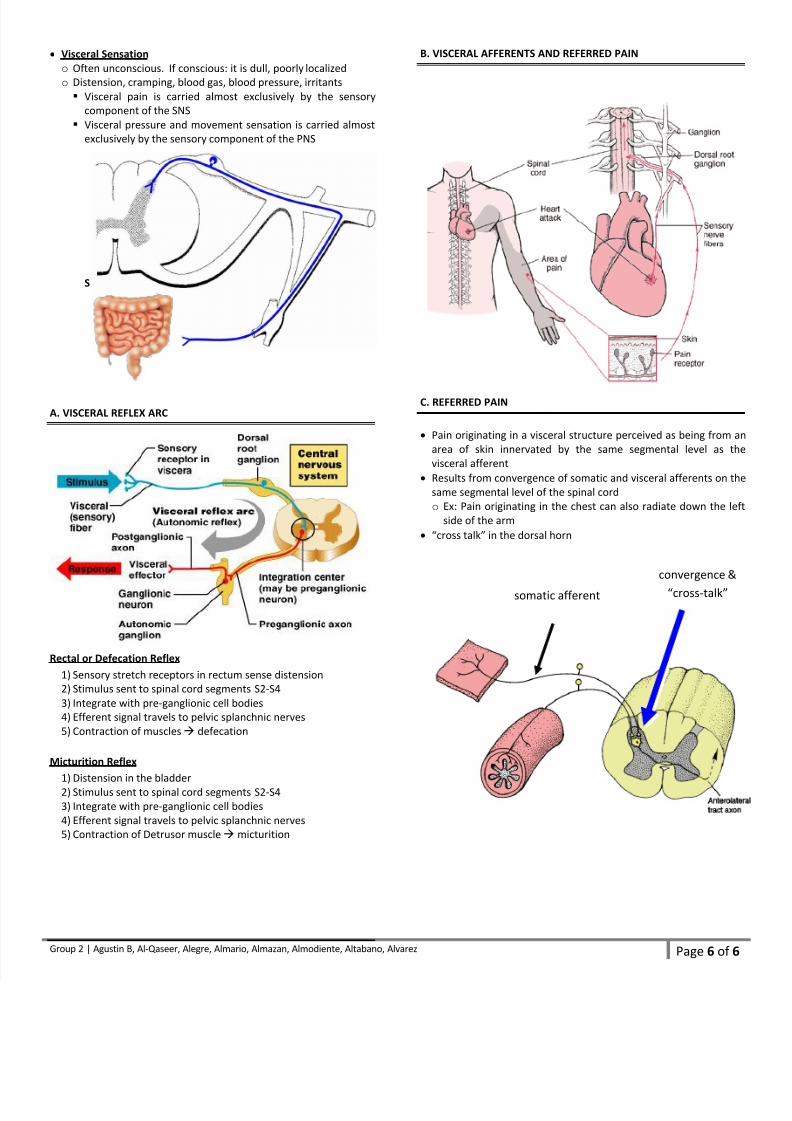
Visceral Sensation
o Often unconscious. If conscious: it is dull, poorly localized
o
Distension, cramping, blood gas, blood pressure, irritants
Visceral pain is carried almost exclusively by the sensory
component of the SNS
Visceral pressure and movement sensation is carried almost
exclusively by the sensory component of the PNS
S
A. VISCERAL REFLEX ARC
Rectal or Defecation Reflex
1) Sensory stretch receptors in rectum sense distension
2) Stimulus sent to spinal cord segments S2-S4
3) Integrate with pre-ganglionic cell bodies
4) Efferent signal travels to pelvic splanchnic nerves
5)
Contraction of muscles defecation
Micturition Reflex
1) Distension in the bladder
2)
Stimulus sent to spinal cord segments S2-S4
3) Integrate with pre-ganglionic cell bodies
4) Efferent signal travels to pelvic splanchnic nerves
5)
Contraction of Detrusor muscle micturition
B. VISCERAL AFFERENTS AND REFERRED PAIN
C. REFERRED PAIN
Pain originating in a visceral structure perceived as being from an
area of skin innervated by the same segmental level as the
visceral afferent
Results from convergence of somatic and visceral afferents on the
same segmental level of the spinal cord
o Ex: Pain originating in the chest can also radiate down the left
side of the arm
“cross talk” in the dorsal horn
convergence &
“cross-talk” somatic afferent