Embed Size (px)
Citation preview
A Pivotal Heme-transfer Reaction Intermediate inCytochrome c Biogenesis*□S
Received for publication, October 17, 2011, and in revised form, November 22, 2011 Published, JBC Papers in Press, November 25, 2011, DOI 10.1074/jbc.M111.313692
Despoina A. I. Mavridou‡1, Julie M. Stevens‡, Leonie Mönkemeyer‡, Oliver Daltrop‡2, Katalin di Gleria§,Benedikt M. Kessler¶3, Stuart J. Ferguson‡4, and James W. A. Allen‡5
From the ‡Department of Biochemistry, University of Oxford, South Parks Road, Oxford OX1 3QU, the §Medical Research CouncilHuman Immunology Unit, Weatherall Institute of Molecular Medicine, Nuffield Department of Medicine, University of Oxford,Oxford OX3 9DS, and the ¶Department of Medicine, Henry Wellcome Building for Molecular Physiology, University of Oxford,Roosevelt Drive, Oxford OX3 7BN, United Kingdom
Background: Heme attachment to cytochrome c is a catalyzed post-translational modification.Results: We identify a ternary complex of the cytochrome c biogenesis protein CcmE, heme, and a cytochrome, and demon-strate its functional significance.Conclusion: The complex is a trapped catalytic intermediate at the point of heme transfer from the cytochrome biogenesisapparatus to the cytochrome.Significance: An insight into biosynthesis of heme proteins.
c-Type cytochromes are widespread proteins, fundamentalfor respiration or photosynthesis in most cells. They containheme covalently bound to protein in a highly conserved, highlystereospecific post-translational modification. In many bacte-ria, mitochondria, and archaea this heme attachment is cata-lyzed by the cytochrome cmaturation (Ccm) proteins. Here weidentify and characterize a covalent, ternary complex betweenthe heme chaperoneCcmE, heme, and cytochrome c. Formationof the complex from holo-CcmE occurs in vivo and in vitro andinvolves the specific heme-binding residues of both CcmE andapocytochrome c. The enhancement and attenuation of theamounts of this complex correlates completelywith known con-sequences of mutations in genes for other Ccm proteins. Wepropose the complex is a trapped catalytic intermediate in thecytochrome c biogenesis process, at the point of heme transferfrom CcmE to the cytochrome, the key step in the maturationpathway.
c-Type cytochromes are widespread proteins, essential forrespiration and/or photosynthesis in mitochondria, chloro-plasts, most bacteria, and some archaea. They contain heme,covalently bound to the polypeptide chain. With very fewexceptions, this attachment is between the vinyl groups of theheme and the cysteine thiols of a Cys-Xxx-Xxx-Cys-His heme-
binding motif (Fig. 1); the histidine becomes a ligand tothe heme iron atom. This heme attachment is a post-transla-tional modification with strictly conserved stereospecificity(1). However, it is catalyzed by multiple, completely distinct,biogenesis proteins in different organisms and organelles(reviewed by Refs. 2–8). The cytochrome cmaturation (Ccm)6system is the biogenesis apparatus ofmanyGram-negative bac-teria, archaea, and the mitochondria of land plants, red algae,and some protozoa. The paradigm Ccm system is from Esche-richia coli, where it consists of eight essential core proteins, allmembrane bound, and several accessory factors (9) (Fig. 1). Thesystem functions in the periplasm where all bacterial c-typecytochromes are located.CcmE is the pivotal protein in the Ccm system. It binds heme
covalently, through a histidine residue, as an intermediate inthe cytochrome c biogenesis pathway (Fig. 1) (10). This hemeattachment is to the �-carbon of the vinyl group of heme,whereas the cysteine thiol of the c-type cytochrome attaches tothe �-carbon (Fig. 1) (11). CcmA, -B, -C, and -D are all involvedin forming holo (i.e. heme-bound) CcmE in a state that cantransfer the heme to apocytochrome c (12–16); CcmF and -Hare involved in heme transfer from CcmE to form the holocy-tochrome (Fig. 1) (10, 17, 18). However, the mechanisms ofmost of these steps remain unresolved. Interactions have beenidentified between apocytochrome c and each of CcmF, CcmG,and CcmH by yeast two-hybrid analysis or in vitro (17, 19, 20).However, no interaction has been observed between CcmE andthe apocytochrome, even though current models of the Ccmpathway predict that such an interaction is crucial.Cytochrome b562 is a soluble, �12-kDa periplasmic E. coli
protein. A variant of this cytochrome containing a CXXCHc-type cytochrome heme-binding motif is matured efficientlyand correctly by the Ccm apparatus (21, 22). Variants contain-
* This work was supported in part by Biotechnology and Biological SciencesResearch Council Grants BB/D019753/1 and BB/H017887/1, WellcomeTrust Grant 092532, and a Value-in-People Award.Author’s Choice—Final version full access.
□S This article contains supplemental Figs. S1 and S2 and Tables S1–S4.1 Supported by the Gustav Born Trust and the Edward Penley Abraham Ceph-
alosporin Fund.2 Junior Research Fellow of Christ Church, Oxford.3 Supported by the Biomedical Research Centre (NIHR), Oxford, UK.4 To whom correspondence may be addressed. Tel.: 44-0-1865-613299;
E-mail: [email protected] Biotechnology and Biological Sciences Research Council David Phillips Fel-
low. To whom correspondence may be addressed. Tel.: 44-0-1865-613330;E-mail: [email protected].
6 The abbreviations used are: Ccm, cytochrome c maturation; cytochromec-b562, c-type variant of cytochrome b562; BisTris, 2-[bis(2-hydroxyethyl)amino]-2-(hydroxymethyl)propane-1,3-diol; DDM, n-dodecyl �-D-maltoside.
THE JOURNAL OF BIOLOGICAL CHEMISTRY VOL. 287, NO. 4, pp. 2342–2352, January 20, 2012Author’s Choice © 2012 by The American Society for Biochemistry and Molecular Biology, Inc. Published in the U.S.A.
2342 JOURNAL OF BIOLOGICAL CHEMISTRY VOLUME 287 • NUMBER 4 • JANUARY 20, 2012
by guest on July 5, 2018http://w
ww
.jbc.org/D
ownloaded from
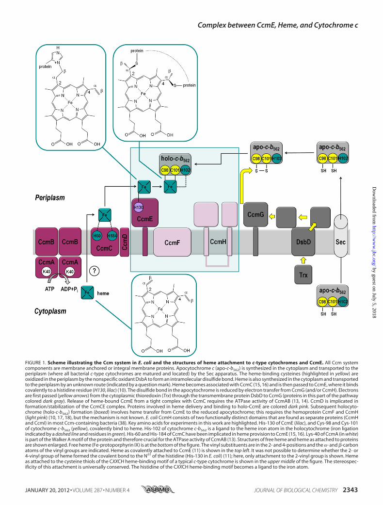
FIGURE 1. Scheme illustrating the Ccm system in E. coli and the structures of heme attachment to c-type cytochromes and CcmE. All Ccm systemcomponents are membrane anchored or integral membrane proteins. Apocytochrome c (apo-c-b562) is synthesized in the cytoplasm and transported to theperiplasm (where all bacterial c-type cytochromes are matured and located) by the Sec apparatus. The heme-binding cysteines (highlighted in yellow) areoxidized in the periplasm by the nonspecific oxidant DsbA to form an intramolecular disulfide bond. Heme is also synthesized in the cytoplasm and transportedto the periplasm by an unknown route (indicated by a question mark). Heme becomes associated with CcmC (15, 16) and is then passed to CcmE, where it bindscovalently to a histidine residue (H130, lilac) (10). The disulfide bond in the apocytochrome is reduced by electron transfer from CcmG (and/or CcmH). Electronsare first passed (yellow arrows) from the cytoplasmic thioredoxin (Trx) through the transmembrane protein DsbD to CcmG (proteins in this part of the pathwaycolored dark gray). Release of heme-bound CcmE from a tight complex with CcmC requires the ATPase activity of CcmAB (13, 14). CcmD is implicated information/stabilization of the CcmCE complex. Proteins involved in heme delivery and binding to holo-CcmE are colored dark pink. Subsequent holocyto-chrome (holo-c-b562) formation (boxed) involves heme transfer from CcmE to the reduced apocytochrome; this requires the hemoprotein CcmF and CcmH(light pink) (10, 17, 18), but the mechanism is not known. E. coli CcmH consists of two functionally distinct domains that are found as separate proteins (CcmHand CcmI) in most Ccm-containing bacteria (38). Key amino acids for experiments in this work are highlighted. His-130 of CcmE (lilac), and Cys-98 and Cys-101of cytochrome c-b562 (yellow), covalently bind to heme. His-102 of cytochrome c-b562 is a ligand to the heme iron atom in the holocytochrome (iron ligationindicated by a dashed line and residues in green). His-60 and His-184 of CcmC have been implicated in heme provision to CcmE (15, 16). Lys-40 of CcmA (in white)is part of the Walker A motif of the protein and therefore crucial for the ATPase activity of CcmAB (13). Structures of free heme and heme as attached to proteinsare shown enlarged. Free heme (Fe-protoporphyrin IX) is at the bottom of the figure. The vinyl substituents are in the 2- and 4-positions and the �- and �-carbonatoms of the vinyl groups are indicated. Heme as covalently attached to CcmE (11) is shown in the top left. It was not possible to determine whether the 2- or4-vinyl group of heme formed the covalent bond to the N�1 of the histidine (His-130 in E. coli) (11); here, only attachment to the 2-vinyl group is shown. Hemeas attached to the cysteine thiols of the CXXCH heme-binding motif of a typical c-type cytochrome is shown in the upper middle of the figure. The stereospec-ificity of this attachment is universally conserved. The histidine of the CXXCH heme-binding motif becomes a ligand to the iron atom.
Complex between CcmE, Heme, and Cytochrome c
JANUARY 20, 2012 • VOLUME 287 • NUMBER 4 JOURNAL OF BIOLOGICAL CHEMISTRY 2343
by guest on July 5, 2018http://w
ww
.jbc.org/D
ownloaded from
ing a single cysteine residue also covalently bind heme (22), butsingle cysteine c-type cytochromes are very poor substrates forthe Ccm system (23, 24). Exceptionally, apocytochrome b562 isalso stable (3, 22); normally apocytochromes c are subject torapid degradation in vivo.In this work, we describe experiments to identify novel inter-
actions between components of the Ccm system and (apo)cy-tochrome c. We have used c-type cytochrome b562 variants tomaximize the chances of isolating such interacting partners.We report a covalent, ternary complex between CcmE, heme,and cytochrome, and propose this represents a critical interme-diate in the Ccm pathway, at the point of heme transfer fromthe biogenesis system to the product cytochrome.
EXPERIMENTAL PROCEDURES
Construction of Plasmids
Plasmids used in this study are listed in supplemental TableS3. Plasmid pb562R98CStrep was produced by site-directedmutagenesis (ExSite, Stratagene) using plasmid pb562R98C(22) as the template. The same method was used to insert athrombin cleavage site, with additional Gly residues(GLVPRGSG) into pE221 (25). The plasmid produced, pE225,expresses the periplasmic region of CcmE (from Ser-32) withan N-terminal cleavable pelB signal sequence. Plasmidspb562R98CH102R and pFR015 were produced by site-direct-ed mutagenesis (QuikChange, Stratagene) using plasmidspb562R98C (22) and pEC86 (9) as templates, respectively. DNAmanipulations were conducted using standard methods. KODHot Start DNA polymerase (Novagen) was used for all PCRsand all constructs were sequenced before use.
Cell Growth and Fractionation
Bacterial strains used in this study are listed in supplementalTable S4. Routine cell growth was conducted in 100 ml of 2�TY medium (16 g liter�1 of peptone, 10 g liter�1 of yeastextract, 5 g liter�1 of NaCl) in 2.5-liter flasks. Cultures wereinoculated from single colonies and incubated at 37 °C for15–18 h with shaking at 200 rpm. Fully aerobic growth condi-tions prevented expression of the endogenous E. coli Ccm sys-tem. 1 mM Isopropyl 1-thio-�-D-galactopyranoside was addedto the cultures from inoculation. 100 �gml�1 of ampicillin and34 �g ml�1 of chloramphenicol were used.For the isolation of the crude membrane fraction a French
press was used. Disruption of the cells was performed at 16,000p.s.i. followed by centrifugation at 257,000 � g for 1 h at 4 °C.The membrane fraction was resuspended in �25 ml of 50 mM
Tris-HCl, 150 mM NaCl (pH 7.5) and was re-centrifuged asabove. The washed crude membrane fraction was resuspendedin 1–2 ml of 50 mM Tris-HCl, 150 mM NaCl (pH 7.5).
SDS-PAGE Analysis
SDS-PAGE analysis was carried out on 10%BisTris NuPAGEgels (Invitrogen) with prestained molecular weight markers(SeeBlue Plus 2, Invitrogen, or ColorPlus Prestained ProteinMarker, New England Biolabs). Samples containing membranefractions were denatured by incubation at 42 °C for 5 min. Allother samples were denatured at 100 °C for 2 min. 2-Mercap-
toethanol and urea were added at final concentrations of 5%(v/v) and 8 M, respectively, where appropriate. Gel loadingswere normalized according to total protein content, and deter-mined using the Pierce BCA Reducing Agent Compatible Pro-tein Assay Kit (ThermoScientific); 5–20 �g of protein wereloaded per lane. Proteins with covalently bound heme weredetected on gels using the method of Goodhew et al. (26) andquantification of heme-bound species was performed by densi-tometry using GeneSnap (SYNGENE).Western blotting was carried out following SDS-PAGE by
transferring onto nitrocellulose (Hybond C-Extra, AmershamBiosciences). Blocking was with 5% (w/v) skimmed milk pow-der in Tris-buffered saline (50 mM Tris-HCl, 120 mM NaCl,0.1% (v/v) Tween 20 (pH 7.5)). The primary antibodies used(Covalab) were rabbit antiserum raised against E. coli cyto-chrome b562 (dilution 1:1000) and rabbit antiserum raisedagainst E. coli CcmE (dilution 1:1000). In the in vitro studiesantibody raised against a CcmE peptide (10) was used. Goatanti-rabbit alkaline phosphatase-conjugated antibody (Sigma)was used as secondary antibody (dilution 1:30000). Develop-ment was carried out using a SigmaFast BCIP/NBT tablet.
Sample Preparation for Proteomics Analysis
Proteomics analysis of the CcmE-heme-cytochrome c-b562R98C complex was carried out on two different samples.Co-immunoprecipitation—Crude membrane fractions from
E. coli JCB387 cells transformed with plasmids pEC86 andpb562R98C were solubilized in 1% (w/v) n-dodecyl �-D-malto-side (DDM) (Melford) under gentle agitation at 4 °C for 1 h.Rabbit antiserum raised against E. coli cytochrome b562 wasbound to beads from the Dynabeads-Protein G Immunopre-cipitation Kit (Invitrogen). The pulled-down product was ana-lyzed by SDS-PAGE. The gel was silver-stained using the Silver-Quest Silver Staining Kit (Invitrogen) and the bandcorresponding to the CcmE-heme-cytochrome c-b562 R98Ccomplex was excised and destained.Purification of CcmE-Heme-Cytochrome c-b562 R98C Com-
plex Using a Streptavidin II Tag—Crude membrane fractionfrom E. coli JCB387 cells, transformed with plasmids pEC86and pb562R98CStrep, was solubilized in 20 mM Tris-HCl, 300mM NaCl, 20% (v/v) glycerol, 1% (w/v) DDM (pH 7.5) undergentle agitation at 4 °C for 1 h at a protein concentration of 5mgml�1. Unsolubilized material was removed by centrifugation at257,000 � g for 30 min at 4 °C. The supernatant was diluted10-fold with 20 mM Tris-HCl, 150 mM NaCl (pH 7.5) andapplied to 7 ml of Strep-Tactin Sepharose (IBA GmbH) pre-equilibrated with 20 mM Tris-HCl, 150 mM NaCl, 0.1% (w/v)DDM (pH 7.5). The column was washed with 20 mM Tris-HCl,1 M NaCl, 0.1% (w/v) DDM (pH 7.5) and protein was elutedusing 20mMTris-HCl, 150mMNaCl, 0.1% (w/v) DDM, 2.5 mM
desthiobiotin (IBA) (pH 7.5). The eluent was exchanged into 20mM Tris-HCl, 50 mM NaCl, 0.03% (w/v) DDM (pH 7.5) andconcentrated. The protein solutionwas analyzed by SDS-PAGEin the presence of dithiothreitol (DTT). The gel was stainedusing SimplyBlue Safestain (Invitrogen) and the band corre-sponding to theCcmE-heme-cytochrome c-b562 R98Ccomplexwas excised.
Complex between CcmE, Heme, and Cytochrome c
2344 JOURNAL OF BIOLOGICAL CHEMISTRY VOLUME 287 • NUMBER 4 • JANUARY 20, 2012
by guest on July 5, 2018http://w
ww
.jbc.org/D
ownloaded from
Proteomics Analysis
Identification of the CcmE-heme-cytochrome c-b562 R98Ccomplex components was achieved by proteomics analysis.Blue-stained gel bands were excised, destained in 25 mM
ammonium bicarbonate in 50% acetonitrile, and then reducedwith DTT and alkylated with iodoacetamide before beingdigested with trypsin overnight. Tryptic peptides wereextracted and then desalted on an in-house manufactured C18tip. Samples were analyzed on a Thermo LTQ XL Orbitrapcoupled to a DionexU3000 nano-LC system run in direct injec-tion configuration. Peptideswere resolved on an in-houseman-ufactured reverse-phase column made by packing a Picotip(New Objective) with C18 resin (Reprosil-Pur, C18-Aq, 3-�mbeads). Samples were typically resolved on 40- or 120-min LC-MS/MS gradients depending on sample complexity. TheOrbitrap was operated in a “Top 5” method in which 1� ionswere not selected for fragmentation and dynamic exclusionwasapplied. Precursormass accuracy tolerance was set at�20 ppmand the MS/MS fragment ion tolerance at �0.5 Da. Data weresearched using Mascot against data base NCBI nr/nr version2010.03.21, specifying E. coli as the taxonomy. The fixed mod-ification was defined as carbamidomethyl cysteine and variablemodifications were oxidized Met, N-terminal acetylation, anddeamidation on Asn and Glu.For peptide mapping, stained gel bands containing CcmE-
heme-cytochrome c-b562 R98C complex were subjected to in-gel digestion with trypsin as described elsewhere (27) or chy-motrypsin (Roche Diagnostics) used under identicalconditions. Digested samples were subjected to analysis bymatrix-assisted laser desorption ionization time-of-flight(MALDI-TOF) using an Ultraflex mass spectrometer (BrukerDaltonics) as described (28). TheMS data (peptidemass finger-printing) was analyzed using FlexAnalysis software version 2.4(Bruker Daltonics) and searched against the SwissProt database combined with the E. coli CcmE and cytochrome c-b562R98C sequences using an in-house Mascot server (Matrix-science). In addition, the same sample was also analyzed byLC-MS/MS using a U3000 nano-LC (Dionex) coupled to a highcapacity trap tandem mass spectrometer (Bruker Daltonics) asdescribed previously (27). MS/MS data were also searchedusing Mascot as described above, allowing for carbamidom-ethylation on Cys, oxidation on Met, deamidation on Glu andAsn, and heme addition on Cys and His, as modifications.
In Vitro Formation and Characterization of CcmE-Heme-Cytochrome c Complex
Soluble holo-CcmE was produced by coexpressing plasmidspE225 and pEC86 in E. coli JM109(DE3) cells. Cultures wereincubated at 37 °C with shaking at 200 rpm in LB medium tomid-exponential phase and induced with 1 mM isopropyl1-thio-�-D-galactopyranoside overnight at 30 °C. Cells wereharvested and resuspended in 50 mM Tris-HCl, 300 mM NaCl(pH 7.5). The periplasmic fraction containing holo-CcmE wasisolated and the protein was purified as described previously(25). Thrombin cleavage of the polyhistidine tagwas performedusing the Sigma CleanCleave Kit.Hydrogenobacter thermophi-lus apocytochrome c552 and variants thereof were produced as
described previously (29, 30). N-terminal sequencing was car-ried out from protein samples in SDS-PAGE gels that wereelectrophoretically transferred to a PVDF membrane andstained with Coomassie Brilliant Blue. The protein bands wereexcised and subjected to automated Edman sequencing usingan Applied Biosystems 494A Procise protein sequencer. APerkinElmer Lambda 2 spectrophotometer was used to collectabsorption spectra. Pyridine hemochrome spectra wereobtained according to the method of Bartsch (31) using 5 �M
protein in 19% (v/v) pyridine and 0.15 M NaOH.H. thermophilus apocytochrome c552, C11A, C14A, and
C11A/C14A apocytochrome variants (15–25 �M) were incu-bated with E. coli holo-CcmE* (5–10 �M) in 50 mM potassiumphosphate buffer (pH 7.0) at 25 °C for up to 20 h. Samples werereduced by the addition of disodium dithionite. Solutions con-tained 10 mM Tris(2-carboxyethyl)phosphine and were thor-oughly sparged with humidified argon. Reactions were carriedout in the dark.
Inhibition of the Ccm System
E. coli JCB387 cells were transformed with the followingcombinations of plasmids (supplemental Table S3): pEG278 �pb562EV; pEG278 � pb562R98C; pRZ001 � pb562EV; andpRZ001� pb562R98C. Cell growth, fractionation, and analysisof the periplasmic fractions was performed as described in Ref.32. Autoinduction was used for expression of cytochromec-b562 R98C and cytochrome cd1. The production of both theendogenous cytochrome NrfA and the exogenous cytochromecd1 was quantified (33).
RESULTS AND DISCUSSION
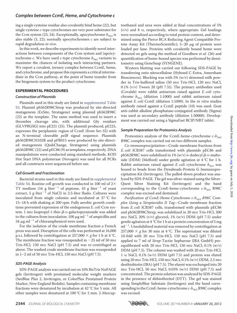
A Complex between CcmE, Heme, and Cytochrome—TheCcm system must interact with apocytochrome c to catalyzeheme attachment and hence holocytochrome formation; we setout to identify such interactions using variants of cytochromeb562 as the test cytochromes. E. coli strain JCB387 cells wereco-transformed with a plasmid from which the Ccm proteinsare constitutively expressed (pEC86), and plasmids encodingvariants of cytochrome b562 with CXXCH (R98C/Y101C),CXXXH (R98C), and XXXCH (Y101C) heme-binding motifs.Similar results to those in this article were also obtained usingE. coli strains MC1000 and MC1061 (supplemental Table S4).In this work, we refer to the c-type cytochrome variants of cyto-chrome b562 as cytochrome c-b562. Cells were fractionated intosoluble and membrane extracts. These fractions were analyzedby Western blotting with a cytochrome b562 antibody. Of par-ticular note was a band in the blot of the membrane fraction,observed at �32 kDa (Fig. 2A, lanes 3–5). This band was notobserved in the soluble fraction (not shown). The cytochromec-b562 variants themselves ran at �12 kDa and a dimer was alsoobserved at �30 kDa (Fig. 2A); these bands arise from cyto-chrome b562 that remains bound to the membranes even afterextensive washing.From themolecularmasses of theCcmproteins, we reasoned
that the 32-kDa band might be a complex between (apo)cyto-chrome c-b562 (molecular mass � 12 kDa) and CcmE (18 kDa),which is a membrane-anchored protein. The band was onlyobserved when both the Ccm system and a cytochrome c-b562
Complex between CcmE, Heme, and Cytochrome c
JANUARY 20, 2012 • VOLUME 287 • NUMBER 4 JOURNAL OF BIOLOGICAL CHEMISTRY 2345
by guest on July 5, 2018http://w
ww
.jbc.org/D
ownloaded from
variant were co-expressed (Fig. 2, compare lanes 3–5with lanes1 and 2). We therefore probed the E. coli membrane fractionusing a CcmE antibody. The 32-kDa band was again observed(Fig. 2B, lanes 3–5), running just below a band corresponding toa CcmE dimer. The CcmEmonomer was observed at �16 kDa.To establish whether heme was involved in the complex withCcmE and cytochrome c-b562, we ran the cell membrane frac-tions on denaturing SDS-PAGE, which was stained for proteinscontaining covalently bound heme (Fig. 2C). Once again, the32-kDa bandwas apparent. Densitometrymeasurements of theheme-stained gel (Fig. 2C), which was normalized for total pro-tein loading, indicate the largest amount of the complex formedwith the R98C (CXXXH) variant cytochrome (lane 4). The rel-ative yield was �70% lower with the Y101C (XXXCH) protein(lane 5). With the CXXCH variant, no band was visible on theheme-stained gel (Fig. 2C, lane 3) (sensitivity � 1 pmol of holo-cytochrome (33)); it was barely detectable in theWestern blots(Fig. 2, A and B, lanes 3). Membranes from cells expressing the
R98C variant cytochrome c-b562 were therefore selected forfurther analysis.The 32-kDa band remained even after treatment of the solubi-
lizedmembrane proteins in harshly denaturing conditions such asboiling (supplemental Fig. S1, lanes 2 and 4) or 8 M urea (data notshown), indicating that theCcmE-heme-cytochrome c-b562 R98Ccomplex is very stable and that the components are covalentlylinked. The complex was insensitive to treatment with 2-mercap-toethanol, which would reduce any disulfide bonds present (sup-plemental Fig. S1, lanes 3 and 4). Following treatmentwith 2-mer-captoethanol, the intensity of the heme-staining bands arisingfromeachof the complex, holo-CcmEandholocytochrome c-b562R98C, decreased (supplemental Fig. S1C, compare lane 1with 3).Such reducing agents cause aportionof the iron todissociate fromheme, which reduces the intrinsic peroxidase activity of the hemethat gives rise to the heme stain. However, it is clear from theWestern blots using both cytochrome b562 and CcmE antibodies(supplemental Fig. S1,A andB, compare lanes 3 and 4with lane 1)that treatment with 2-mercaptoethanol did not decrease the totalamount of complex.To isolate the complex, solubilized membranes were
immunoprecipitated using the cytochrome b562 antibody.The 32-kDa species was visible on a silver-stained SDS-PAGE of the immunoprecipitate. The relevant band wasexcised from the gel, trypsin digested, and assessed by high-resolution mass spectrometry (LC-MS/MS). Peptides fromboth cytochrome c-b562 and CcmE were detected in thisanalysis, including two from cytochrome c-b562 and onefrom CcmE, each of �12 amino acids, and that could beassigned with high confidence (supplemental Table S1A). Toverify this analysis, we co-expressed a C-terminal streptavi-din II-tagged form of cytochrome c-b562 R98C with the Ccmproteins. The tag facilitated small scale purification of theputative complex, which could be seen on protein-stainedSDS-PAGE. Again, bands were excised, trypsin digested andanalyzed by LC-MS/MS. Multiple peptides were identifiedfrom both cytochrome c-b562 R98C and CcmE, each with�95% confidence (but often much higher); sequence cover-age in identified peptides (supplemental Table S1A) was 64%for cytochrome b562 and 44% for CcmE.
We conclude thatwehave resolved a stable complex contain-ing cytochrome c-b562, CcmE, and heme, all linked covalently.This is the first complex to be identified between CcmE anda cytochrome. Such a complex is a critical intermediate incurrent models of the Ccm system-mediated pathway ofcytochrome c biogenesis (10). The small amount of complexformed from CXXCH containing cytochrome c (Fig. 2, A andB, lane 3) presumably represents the steady-state level of thecomplex as a reaction intermediate in the normally function-ing Ccm pathway. In contrast, with single cysteine (XXXCHor CXXXH) cytochromes, the complex accumulated as atrapped intermediate.Organization of the Complex—What is the structural
arrangement of this ternary complex between cytochromec-b562, CcmE, and heme? We investigated its formation usingsite-directed variants of CcmE and cytochrome c-b562 R98C.No complex was observed when wild-type cytochrome b562(which contains no cysteine residues) was co-expressed with
FIGURE 2. Detection of a complex between CcmE, heme, and cytochromec-b562. A, Western blot of cell membranes using a cytochrome b562 antibody.B, Western blot of cell membranes using a CcmE antibody. C, SDS-PAGE of cellmembranes stained for proteins containing covalently bound heme. For eachpanel, the lane order is: M, molecular mass markers (as indicated, in kDa); lanes1, cells expressing the Ccm system from pEC86; 2, cells expressing cyto-chrome c-b562 R98C/Y101C; 3, cells expressing the Ccm system from pEC86and cytochrome c-b562 R98C/Y101C; 4, cells expressing the Ccm system frompEC86 and cytochrome c-b562 R98C; 5, cells expressing the Ccm system frompEC86 and cytochrome c-b562 Y101C. The position of the CcmE-heme-cyto-chrome c-b562 complex, which runs just above a cytochrome c-b562 dimer andjust below the CcmE dimer, is indicated. An equal amount of total protein wasloaded in each lane.
Complex between CcmE, Heme, and Cytochrome c
2346 JOURNAL OF BIOLOGICAL CHEMISTRY VOLUME 287 • NUMBER 4 • JANUARY 20, 2012
by guest on July 5, 2018http://w
ww
.jbc.org/D
ownloaded from
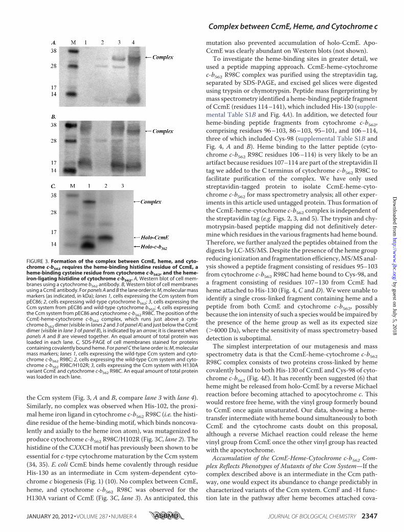
the Ccm system (Fig. 3, A and B, compare lane 3 with lane 4).Similarly, no complex was observed when His-102, the proxi-mal heme iron ligand in cytochrome c-b562 R98C (i.e. the histi-dine residue of the heme-binding motif, which binds noncova-lently and axially to the heme iron atom), was mutagenized toproduce cytochrome c-b562 R98C/H102R (Fig. 3C, lane 2). Thehistidine of the CXXCHmotif has previously been shown to beessential for c-type cytochrome maturation by the Ccm system(34, 35). E. coli CcmE binds heme covalently through residueHis-130 as an intermediate in Ccm system-dependent cyto-chrome c biogenesis (Fig. 1) (10). No complex between CcmE,heme, and cytochrome c-b562 R98C was observed for theH130A variant of CcmE (Fig. 3C, lane 3). As anticipated, this
mutation also prevented accumulation of holo-CcmE. Apo-CcmE was clearly abundant on Western blots (not shown).To investigate the heme-binding sites in greater detail, we
used a peptide mapping approach. CcmE-heme-cytochromec-b562 R98C complex was purified using the streptavidin tag,separated by SDS-PAGE, and excised gel slices were digestedusing trypsin or chymotrypsin. Peptide mass fingerprinting bymass spectrometry identified a heme-binding peptide fragmentof CcmE (residues 114–141), which included His-130 (supple-mental Table S1B and Fig. 4A). In addition, we detected fourheme-binding peptide fragments from cytochrome c-b562,comprising residues 96–103, 86–103, 95–101, and 106–114,three of which included Cys-98 (supplemental Table S1B andFig. 4, A and B). Heme binding to the latter peptide (cyto-chrome c-b562 R98C residues 106–114) is very likely to be anartifact because residues 107–114 are part of the streptavidin IItag we added to the C terminus of cytochrome c-b562 R98C tofacilitate purification of the complex. We have only usedstreptavidin-tagged protein to isolate CcmE-heme-cyto-chrome c-b562 for mass spectrometry analysis; all other exper-iments in this article used untagged protein. Thus formation ofthe CcmE-heme-cytochrome c-b562 complex is independent ofthe streptavidin tag (e.g. Figs. 2, 3, and 5). The trypsin and chy-motrypsin-based peptide mapping did not definitively deter-mine which residues in the various fragments had heme bound.Therefore, we further analyzed the peptides obtained from thedigests by LC-MS/MS. Despite the presence of the heme groupreducing ionization and fragmentation efficiency,MS/MSanal-ysis showed a peptide fragment consisting of residues 95–103from cytochrome c-b562 R98C had heme bound to Cys-98, anda fragment consisting of residues 107–130 from CcmE hadheme attached to His-130 (Fig. 4, C and D). We were unable toidentify a single cross-linked fragment containing heme and apeptide from both CcmE and cytochrome c-b562, possiblybecause the ion intensity of such a species would be impaired bythe presence of the heme group as well as its expected size(�4000 Da), where the sensitivity of mass spectrometry-baseddetection is suboptimal.The simplest interpretation of our mutagenesis and mass
spectrometry data is that the CcmE-heme-cytochrome c-b562R98C complex consists of two proteins cross-linked by hemecovalently bound to both His-130 of CcmE and Cys-98 of cyto-chrome c-b562 (Fig. 4E). It has recently been suggested (6) thatheme might be released from holo-CcmE by a reverse Michaelreaction before becoming attached to apocytochrome c. Thiswould restore free heme, with the vinyl group formerly boundto CcmE once again unsaturated. Our data, showing a heme-transfer intermediate with heme bound simultaneously to bothCcmE and the cytochrome casts doubt on this proposal,although a reverse Michael reaction could release the hemevinyl group from CcmE once the other vinyl group has reactedwith the apocytochrome.Accumulation of the CcmE-Heme-Cytochrome c-b562 Com-
plex Reflects Phenotypes of Mutants of the Ccm System—If thecomplex described above is an intermediate in the Ccm path-way, one would expect its abundance to change predictably incharacterized variants of the Ccm system. CcmF and -H func-tion late in the pathway after heme becomes attached cova-
FIGURE 3. Formation of the complex between CcmE, heme, and cyto-chrome c-b562 requires the heme-binding histidine residue of CcmE, aheme-binding cysteine residue from cytochrome c-b562, and the heme-iron-ligating histidine of cytochrome c-b562. A, Western blot of cell mem-branes using a cytochrome b562 antibody. B, Western blot of cell membranesusing a CcmE antibody. For panels A and B the lane order is: M, molecular massmarkers (as indicated, in kDa); lanes 1, cells expressing the Ccm system frompEC86; 2, cells expressing wild-type cytochrome b562; 3, cells expressing theCcm system from pEC86 and wild-type cytochrome b562; 4, cells expressingthe Ccm system from pEC86 and cytochrome c-b562 R98C. The position of theCcmE-heme-cytochrome c-b562 complex, which runs just above a cyto-chrome b562 dimer (visible in lanes 2 and 3 of panel A) and just below the CcmEdimer (visible in lane 3 of panel B), is indicated by an arrow; it is clearest whenpanels A and B are viewed together. An equal amount of total protein wasloaded in each lane. C, SDS-PAGE of cell membranes stained for proteinscontaining covalently bound heme. For panel C the lane order is: M, molecularmass markers; lanes 1, cells expressing the wild-type Ccm system and cyto-chrome c-b562 R98C; 2, cells expressing the wild-type Ccm system and cyto-chrome c-b562 R98C/H102R; 3, cells expressing the Ccm system with H130Avariant CcmE and cytochrome c-b562 R98C. An equal amount of total proteinwas loaded in each lane.
Complex between CcmE, Heme, and Cytochrome c
JANUARY 20, 2012 • VOLUME 287 • NUMBER 4 JOURNAL OF BIOLOGICAL CHEMISTRY 2347
by guest on July 5, 2018http://w
ww
.jbc.org/D
ownloaded from
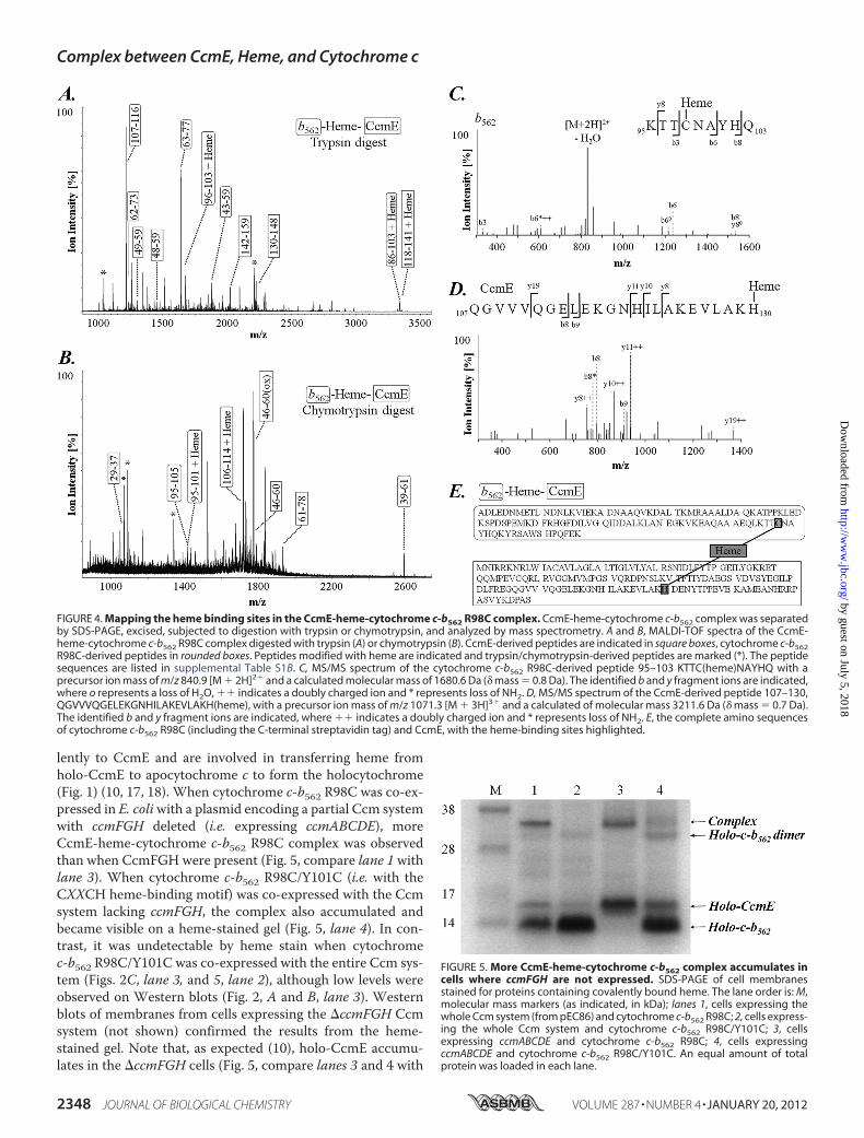
lently to CcmE and are involved in transferring heme fromholo-CcmE to apocytochrome c to form the holocytochrome(Fig. 1) (10, 17, 18). When cytochrome c-b562 R98C was co-ex-pressed in E. coliwith a plasmid encoding a partial Ccm systemwith ccmFGH deleted (i.e. expressing ccmABCDE), moreCcmE-heme-cytochrome c-b562 R98C complex was observedthan when CcmFGH were present (Fig. 5, compare lane 1 withlane 3). When cytochrome c-b562 R98C/Y101C (i.e. with theCXXCH heme-binding motif) was co-expressed with the Ccmsystem lacking ccmFGH, the complex also accumulated andbecame visible on a heme-stained gel (Fig. 5, lane 4). In con-trast, it was undetectable by heme stain when cytochromec-b562 R98C/Y101C was co-expressed with the entire Ccm sys-tem (Figs. 2C, lane 3, and 5, lane 2), although low levels wereobserved on Western blots (Fig. 2, A and B, lane 3). Westernblots of membranes from cells expressing the �ccmFGH Ccmsystem (not shown) confirmed the results from the heme-stained gel. Note that, as expected (10), holo-CcmE accumu-lates in the �ccmFGH cells (Fig. 5, compare lanes 3 and 4 with
FIGURE 5. More CcmE-heme-cytochrome c-b562 complex accumulates incells where ccmFGH are not expressed. SDS-PAGE of cell membranesstained for proteins containing covalently bound heme. The lane order is: M,molecular mass markers (as indicated, in kDa); lanes 1, cells expressing thewhole Ccm system (from pEC86) and cytochrome c-b562 R98C; 2, cells express-ing the whole Ccm system and cytochrome c-b562 R98C/Y101C; 3, cellsexpressing ccmABCDE and cytochrome c-b562 R98C; 4, cells expressingccmABCDE and cytochrome c-b562 R98C/Y101C. An equal amount of totalprotein was loaded in each lane.
FIGURE 4. Mapping the heme binding sites in the CcmE-heme-cytochrome c-b562 R98C complex. CcmE-heme-cytochrome c-b562 complex was separatedby SDS-PAGE, excised, subjected to digestion with trypsin or chymotrypsin, and analyzed by mass spectrometry. A and B, MALDI-TOF spectra of the CcmE-heme-cytochrome c-b562 R98C complex digested with trypsin (A) or chymotrypsin (B). CcmE-derived peptides are indicated in square boxes, cytochrome c-b562R98C-derived peptides in rounded boxes. Peptides modified with heme are indicated and trypsin/chymotrypsin-derived peptides are marked (*). The peptidesequences are listed in supplemental Table S1B. C, MS/MS spectrum of the cytochrome c-b562 R98C-derived peptide 95–103 KTTC(heme)NAYHQ with aprecursor ion mass of m/z 840.9 [M � 2H]2� and a calculated molecular mass of 1680.6 Da (� mass � 0.8 Da). The identified b and y fragment ions are indicated,where o represents a loss of H2O, �� indicates a doubly charged ion and * represents loss of NH2. D, MS/MS spectrum of the CcmE-derived peptide 107–130,QGVVVQGELEKGNHILAKEVLAKH(heme), with a precursor ion mass of m/z 1071.3 [M � 3H]3� and a calculated of molecular mass 3211.6 Da (� mass � 0.7 Da).The identified b and y fragment ions are indicated, where �� indicates a doubly charged ion and * represents loss of NH2. E, the complete amino sequencesof cytochrome c-b562 R98C (including the C-terminal streptavidin tag) and CcmE, with the heme-binding sites highlighted.
Complex between CcmE, Heme, and Cytochrome c
2348 JOURNAL OF BIOLOGICAL CHEMISTRY VOLUME 287 • NUMBER 4 • JANUARY 20, 2012
by guest on July 5, 2018http://w
ww
.jbc.org/D
ownloaded from
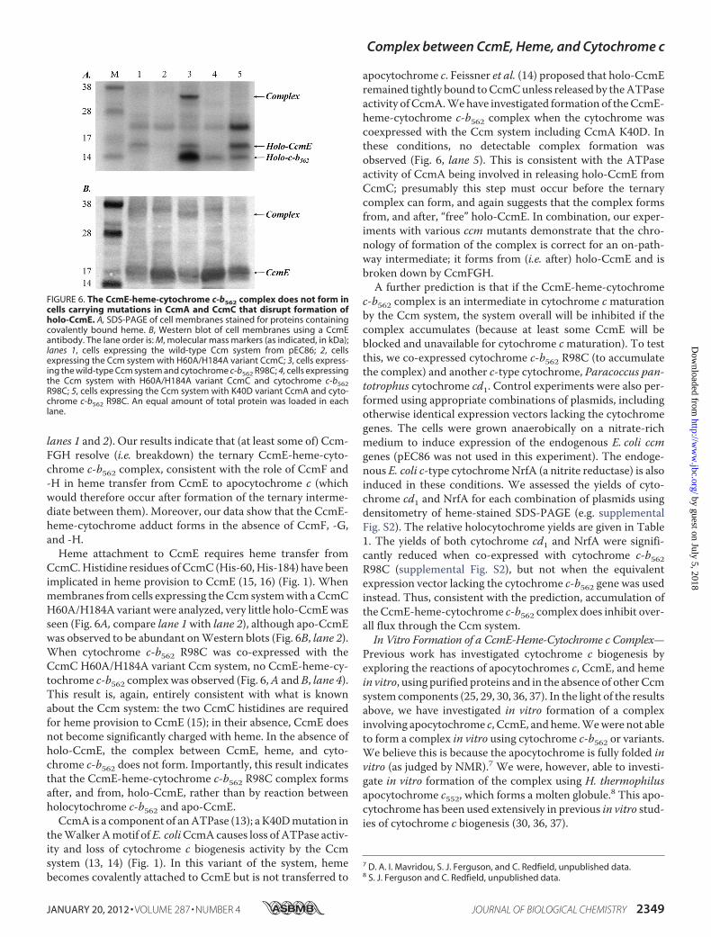
lanes 1 and 2). Our results indicate that (at least some of) Ccm-FGH resolve (i.e. breakdown) the ternary CcmE-heme-cyto-chrome c-b562 complex, consistent with the role of CcmF and-H in heme transfer from CcmE to apocytochrome c (whichwould therefore occur after formation of the ternary interme-diate between them). Moreover, our data show that the CcmE-heme-cytochrome adduct forms in the absence of CcmF, -G,and -H.Heme attachment to CcmE requires heme transfer from
CcmC.Histidine residues of CcmC (His-60,His-184) have beenimplicated in heme provision to CcmE (15, 16) (Fig. 1). Whenmembranes from cells expressing theCcm systemwith aCcmCH60A/H184A variant were analyzed, very little holo-CcmEwasseen (Fig. 6A, compare lane 1with lane 2), although apo-CcmEwas observed to be abundant onWestern blots (Fig. 6B, lane 2).When cytochrome c-b562 R98C was co-expressed with theCcmC H60A/H184A variant Ccm system, no CcmE-heme-cy-tochrome c-b562 complex was observed (Fig. 6,A and B, lane 4).This result is, again, entirely consistent with what is knownabout the Ccm system: the two CcmC histidines are requiredfor heme provision to CcmE (15); in their absence, CcmE doesnot become significantly charged with heme. In the absence ofholo-CcmE, the complex between CcmE, heme, and cyto-chrome c-b562 does not form. Importantly, this result indicatesthat the CcmE-heme-cytochrome c-b562 R98C complex formsafter, and from, holo-CcmE, rather than by reaction betweenholocytochrome c-b562 and apo-CcmE.
CcmA is a component of anATPase (13); a K40Dmutation intheWalkerAmotif ofE. coliCcmAcauses loss of ATPase activ-ity and loss of cytochrome c biogenesis activity by the Ccmsystem (13, 14) (Fig. 1). In this variant of the system, hemebecomes covalently attached to CcmE but is not transferred to
apocytochrome c. Feissner et al. (14) proposed that holo-CcmEremained tightly bound toCcmCunless released by theATPaseactivity ofCcmA.Wehave investigated formation of theCcmE-heme-cytochrome c-b562 complex when the cytochrome wascoexpressed with the Ccm system including CcmA K40D. Inthese conditions, no detectable complex formation wasobserved (Fig. 6, lane 5). This is consistent with the ATPaseactivity of CcmA being involved in releasing holo-CcmE fromCcmC; presumably this step must occur before the ternarycomplex can form, and again suggests that the complex formsfrom, and after, “free” holo-CcmE. In combination, our exper-iments with various ccm mutants demonstrate that the chro-nology of formation of the complex is correct for an on-path-way intermediate; it forms from (i.e. after) holo-CcmE and isbroken down by CcmFGH.A further prediction is that if the CcmE-heme-cytochrome
c-b562 complex is an intermediate in cytochrome cmaturationby the Ccm system, the system overall will be inhibited if thecomplex accumulates (because at least some CcmE will beblocked and unavailable for cytochrome cmaturation). To testthis, we co-expressed cytochrome c-b562 R98C (to accumulatethe complex) and another c-type cytochrome, Paracoccus pan-totrophus cytochrome cd1. Control experiments were also per-formed using appropriate combinations of plasmids, includingotherwise identical expression vectors lacking the cytochromegenes. The cells were grown anaerobically on a nitrate-richmedium to induce expression of the endogenous E. coli ccmgenes (pEC86 was not used in this experiment). The endoge-nous E. coli c-type cytochromeNrfA (a nitrite reductase) is alsoinduced in these conditions. We assessed the yields of cyto-chrome cd1 and NrfA for each combination of plasmids usingdensitometry of heme-stained SDS-PAGE (e.g. supplementalFig. S2). The relative holocytochrome yields are given in Table1. The yields of both cytochrome cd1 and NrfA were signifi-cantly reduced when co-expressed with cytochrome c-b562R98C (supplemental Fig. S2), but not when the equivalentexpression vector lacking the cytochrome c-b562 gene was usedinstead. Thus, consistent with the prediction, accumulation ofthe CcmE-heme-cytochrome c-b562 complex does inhibit over-all flux through the Ccm system.In Vitro Formation of a CcmE-Heme-Cytochrome c Complex—
Previous work has investigated cytochrome c biogenesis byexploring the reactions of apocytochromes c, CcmE, and hemein vitro, using purified proteins and in the absence of other Ccmsystem components (25, 29, 30, 36, 37). In the light of the resultsabove, we have investigated in vitro formation of a complexinvolving apocytochrome c, CcmE, andheme.Wewere not ableto form a complex in vitro using cytochrome c-b562 or variants.We believe this is because the apocytochrome is fully folded invitro (as judged by NMR).7 We were, however, able to investi-gate in vitro formation of the complex using H. thermophilusapocytochrome c552, which forms a molten globule.8 This apo-cytochrome has been used extensively in previous in vitro stud-ies of cytochrome c biogenesis (30, 36, 37).
7 D. A. I. Mavridou, S. J. Ferguson, and C. Redfield, unpublished data.8 S. J. Ferguson and C. Redfield, unpublished data.
FIGURE 6. The CcmE-heme-cytochrome c-b562 complex does not form incells carrying mutations in CcmA and CcmC that disrupt formation ofholo-CcmE. A, SDS-PAGE of cell membranes stained for proteins containingcovalently bound heme. B, Western blot of cell membranes using a CcmEantibody. The lane order is: M, molecular mass markers (as indicated, in kDa);lanes 1, cells expressing the wild-type Ccm system from pEC86; 2, cellsexpressing the Ccm system with H60A/H184A variant CcmC; 3, cells express-ing the wild-type Ccm system and cytochrome c-b562 R98C; 4, cells expressingthe Ccm system with H60A/H184A variant CcmC and cytochrome c-b562R98C; 5, cells expressing the Ccm system with K40D variant CcmA and cyto-chrome c-b562 R98C. An equal amount of total protein was loaded in eachlane.
Complex between CcmE, Heme, and Cytochrome c
JANUARY 20, 2012 • VOLUME 287 • NUMBER 4 JOURNAL OF BIOLOGICAL CHEMISTRY 2349
by guest on July 5, 2018http://w
ww
.jbc.org/D
ownloaded from
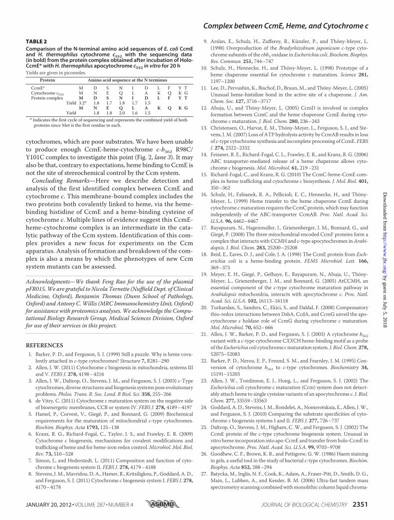
Holo-CcmE lacking its membrane anchor andwith its C-ter-minal polyhistidine purification tag cleaved (denoted CcmE* inthis work) was mixed with H. thermophilus apocytochromec552, which has a typical CXXCHheme-bindingmotif, in reduc-ing conditions. Over several hours, a change in the visibleabsorption spectrum of the mixture was observed, indicatingthat a reaction was taking place (Fig. 7A and supplementalTable S2). The pyridine hemochrome and absorption maximablue-shifted (supplemental Table S2), as would be expected forsaturation of the free vinyl group of the heme bound to CcmE(Fig. 1) if it were forming an adduct with cytochrome c552.
Supporting this interpretation, when the mixture was ana-lyzed by denaturing SDS-PAGE stained for proteins with cova-lently bound heme, an additional band appeared comparedwith that from holo-CcmE* alone, its mass (�26 kDa) corre-sponding to that of holo-CcmE* (16 kDa) plus apocytochrome c(8 kDa) (Fig. 7B, lane 2). The additional band (from an equiva-lent protein-stained gel) was subjected to N-terminal sequenc-ing; the band contained the sequences of bothH. thermophiluscytochrome and E. coliCcmE* in a 1:1 ratio (Table 2). The pres-ence of CcmE* in this complex was further confirmed byWest-ern blotting (Fig. 7C).The results from the optical spectroscopy, SDS-PAGE,
Western blotting, and Edman sequencing therefore all indicateformation in vitro of a covalent, ternary complex betweenCcmE, heme, and cytochrome. Surprisingly, in these experi-ments little heme transfer to the apocytochrome c (to produceholocytochrome) was observed. This is in contrast to the obser-vation that a His-tagged version of holo-CcmE used in previousstudies was capable of transferring most of its heme to yieldholocytochrome c (25). Isolation of this stable CcmE-heme-cytochrome c552 complex in vitro is also consistentwith the roleof CcmF, -G and/or -H in breaking down the complex, whichwas apparent in our experiments in vivo (Fig. 5).
No complex formed when a C11A/C14A variant of H. ther-mophilus apocytochrome c552, which lacks the heme-bindingcysteines, was used in experiments similar to those above (Fig.7, B and C, lane 8). However, a complex did form when eitherthe C11A (XXXCH heme-binding motif) or C14A (CXXXH)variants were used (Fig. 7, B and C, lanes 4 and 6), indicatingthat both cysteines of the apocytochrome are capable of bind-ing covalently to CcmE* in vitro. In structurally characterizedc-type cytochromes, the stereochemistry of heme attachment isinvariant; the N-terminal cysteine of the CXXCH motif isbound covalently to the 2-vinyl group of the heme, and theC-terminal cysteine to the 4-vinyl group (1) (Fig. 1). The struc-ture of heme attachment to His-130 of CcmE has been deter-
mined from a heme-containing fragment of the protein (Fig. 1),but the heme vinyl group forming the covalent bond was notidentified (11). It is often assumed that heme binding to CcmEis stereo- and regiospecific, leaving one heme vinyl group freeand oriented for stereospecific attachment to cytochrome c.However, the CcmE-heme-cytochrome complex reported hereformed from apocytochromes with both XXXCH and CXXXHheme-bindingmotifs, both in vivo and in vitro (Figs. 2 and 7). Itmay be that stereochemical control is favored in vivo for a cyto-chromewith aCXXCHheme-bindingmotif, i.e. for the genuinesubstrate of the Ccm system, but not for the single cysteine
TABLE 1Inhibition of the Ccm system by expression of cytochrome c-b562 R98C, which forms a cytochrome c-b562-heme-CcmE complexVarious combinations of cytochromes were expressed as shown. E. coli NrfA is endogenous and was expressed in all cases. Where a cytochrome was not expressed, anequivalent expression vector lacking the cytochrome gene was expressed instead, so all cells were grown in identical conditions under the same antibiotic selection. Relativeholocytochrome yields were assessed by densitometry of heme-stained SDS-PAGE (e.g. supplemental Fig. S2). Values are the mean of at least 6 biological replicates. Yieldsof cytochrome cd1 and NrfA are given relative to that particular cytochrome and cannot be compared with each other.
Combination of cytochromes expressed Relative yield of NrfA Relative yield of cytochrome cd1NrfA only (dummy cytochrome c-b562 and cytochrome cd1 expression vectors) 100% NAa
NrfA and cytochrome cd1 (dummy cytochrome c-b562 expression vector) 88% 100%NrfA and cytochrome c-b562 R98C (dummy cytochrome cd1 expression vector) 56% NANrfA, cytochrome cd1 and cytochrome c-b562 R98C 40% 18%
a NA � not applicable.
FIGURE 7. The in vitro reaction between E. coli holo-CcmE* and H. thermo-philus apocytochrome c552. A, visible absorption spectra of reduced holo-CcmE* with and without addition of apocytochrome c552. Dashed line,reduced holo-CcmE*. Solid line, the spectrum of the holo-CcmE*-cytochromec mixture obtained after a 10-min incubation in reducing conditions. Dottedline, the reaction product from holo-CcmE* and apocytochrome after incuba-tion for 10 h. All samples were in 50 mM potassium phosphate buffer (pH 7.0)containing 10 mM Tris(2-carboxyethyl)phosphine and disodium dithionite.B, SDS-PAGE stained for proteins containing covalently bound heme, andC, Western blot using a CcmE antibody showing various forms of H. thermo-philus cytochrome c with and without incubation with E. coli holo-CcmE*. Thelane order for both panels is: M, molecular mass markers (as indicated, in kDa);lanes 1, wild-type H. thermophilus apocytochrome c552; 2, the reaction mixtureof wild-type apocytochrome c552 with holo-CcmE*; 3, C11A variant apocyto-chrome c552; 4, the reaction mixture of C11A variant apocytochrome c552 withholo-CcmE*; 5, C14A variant apocytochrome c552; 6, the reaction mixture ofC14A variant apocytochrome c552 with holo-CcmE*; 7, C11A/C14A variantapocytochrome c552; 8, the reaction mixture of C11A/C14A variant apocyto-chrome c552 with holo-CcmE*; 9, holo-CcmE* incubated by itself under other-wise identical reaction conditions. All reactions were carried out in 50 mM
potassium phosphate buffer (pH 7.0) containing 10 mM Tris(2-carboxyethyl)phosphine and disodium dithionite. The incubation time was 20 h.
Complex between CcmE, Heme, and Cytochrome c
2350 JOURNAL OF BIOLOGICAL CHEMISTRY VOLUME 287 • NUMBER 4 • JANUARY 20, 2012
by guest on July 5, 2018http://w
ww
.jbc.org/D
ownloaded from
cytochromes, which are poor substrates. We have been unableto produce enough CcmE-heme-cytochrome c-b562 R98C/Y101C complex to investigate this point (Fig. 2, lane 3). It mayalso be that, contrary to expectations, heme binding to CcmE isnot the site of stereochemical control by the Ccm system.Concluding Remarks—Here we describe detection and
analysis of the first identified complex between CcmE andcytochrome c. This membrane-bound complex includes thetwo proteins both covalently linked to heme, via the heme-binding histidine of CcmE and a heme-binding cysteine ofcytochrome c. Multiple lines of evidence suggest this CcmE-heme-cytochrome complex is an intermediate in the cata-lytic pathway of the Ccm system. Identification of this com-plex provides a new focus for experiments on the Ccmapparatus. Analysis of formation and breakdown of the com-plex is also a means by which the phenotypes of new Ccmsystem mutants can be assessed.
Acknowledgments—We thank Feng Rao for the use of the plasmidpFR015.We are grateful to Nicola Ternette (Nuffield Dept. of ClinicalMedicine, Oxford), Benjamin Thomas (Dunn School of Pathology,Oxford) and Antony C.Willis (MRC Immunochemistry Unit, Oxford)for assistance with proteomics analyses.We acknowledge the Compu-tational Biology Research Group, Medical Sciences Division, Oxfordfor use of their services in this project.
REFERENCES1. Barker, P. D., and Ferguson, S. J. (1999) Still a puzzle. Why is heme cova-
lently attached in c-type cytochromes? Structure 7, R281–2902. Allen, J. W. (2011) Cytochrome c biogenesis in mitochondria, systems III
and V. FEBS J. 278, 4198–42163. Allen, J. W., Daltrop, O., Stevens, J. M., and Ferguson, S. J. (2003) c-Type
cytochromes, diverse structures and biogenesis systems pose evolutionaryproblems. Philos. Trans. R. Soc. Lond. B Biol. Sci. 358, 255–266
4. de Vitry, C. (2011) Cytochrome cmaturation system on the negative sideof bioenergetic membranes, CCB or system IV. FEBS J. 278, 4189–4197
5. Hamel, P., Corvest, V., Giegé, P., and Bonnard, G. (2009) Biochemicalrequirements for the maturation of mitochondrial c-type cytochromes.Biochim. Biophys. Acta 1793, 125–138
6. Kranz, R. G., Richard-Fogal, C., Taylor, J. S., and Frawley, E. R. (2009)Cytochrome c biogenesis, mechanisms for covalent modifications andtrafficking of heme and for heme-iron redox control.Microbiol. Mol. Biol.Rev. 73, 510–528
7. Simon, J., and Hederstedt, L. (2011) Composition and function of cyto-chrome c biogenesis system II. FEBS J. 278, 4179–4188
8. Stevens, J.M.,Mavridou, D. A., Hamer, R., Kritsiligkou, P., Goddard, A. D.,and Ferguson, S. J. (2011) Cytochrome c biogenesis system I. FEBS J. 278,4170–4178
9. Arslan, E., Schulz, H., Zufferey, R., Künzler, P., and Thöny-Meyer, L.(1998) Overproduction of the Bradyrhizobium japonicum c-type cyto-chrome subunits of the cbb3 oxidase in Escherichia coli. Biochem. Biophys.Res. Commun. 251, 744–747
10. Schulz, H., Hennecke, H., and Thöny-Meyer, L. (1998) Prototype of aheme chaperone essential for cytochrome c maturation. Science 281,1197–1200
11. Lee, D., Pervushin, K., Bischof, D., Braun,M., and Thöny-Meyer, L. (2005)Unusual heme-histidine bond in the active site of a chaperone. J. Am.Chem. Soc. 127, 3716–3717
12. Ahuja, U., and Thöny-Meyer, L. (2005) CcmD is involved in complexformation between CcmC and the heme chaperone CcmE during cyto-chrome cmaturation. J. Biol. Chem. 280, 236–243
13. Christensen, O., Harvat, E. M., Thöny-Meyer, L., Ferguson, S. J., and Ste-vens, J. M. (2007) Loss of ATP hydrolysis activity by CcmAB results in lossof c-type cytochrome synthesis and incomplete processing of CcmE. FEBSJ. 274, 2322–2332
14. Feissner, R. E., Richard-Fogal, C. L., Frawley, E. R., and Kranz, R. G. (2006)ABC transporter-mediated release of a heme chaperone allows cyto-chrome c biogenesis.Mol. Microbiol. 61, 219–231
15. Richard-Fogal, C., and Kranz, R. G. (2010) The CcmC-heme-CcmE com-plex in heme trafficking and cytochrome c biosynthesis. J. Mol. Biol. 401,350–362
16. Schulz, H., Fabianek, R. A., Pellicioli, E. C., Hennecke, H., and Thöny-Meyer, L. (1999) Heme transfer to the heme chaperone CcmE duringcytochrome cmaturation requires the CcmCprotein, whichmay functionindependently of the ABC-transporter CcmAB. Proc. Natl. Acad. Sci.U.S.A. 96, 6462–6467
17. Rayapuram, N., Hagenmuller, J., Grienenberger, J. M., Bonnard, G., andGiegé, P. (2008) The three mitochondrial encoded CcmF proteins form acomplex that interacts with CCMH and c-type apocytochromes inArabi-dopsis. J. Biol. Chem. 283, 25200–25208
18. Reid, E., Eaves, D. J., and Cole, J. A. (1998) The CcmE protein from Esch-erichia coli is a heme-binding protein. FEMS Microbiol. Lett. 166,369–375
19. Meyer, E. H., Giegé, P., Gelhaye, E., Rayapuram, N., Ahuja, U., Thöny-Meyer, L., Grienenberger, J. M., and Bonnard, G. (2005) AtCCMH, anessential component of the c-type cytochrome maturation pathway inArabidopsis mitochondria, interacts with apocytochrome c. Proc. Natl.Acad. Sci. U.S.A. 102, 16113–16118
20. Turkarslan, S., Sanders, C., Ekici, S., and Daldal, F. (2008) Compensatorythio-redox interactions between DsbA, CcdA, and CcmG unveil the apo-cytochrome c holdase role of CcmG during cytochrome c maturation.Mol. Microbiol. 70, 652–666
21. Allen, J. W., Barker, P. D., and Ferguson, S. J. (2003) A cytochrome b562variant with a c-type cytochrome CXXCH heme-binding motif as a probeof theEscherichia coli cytochrome cmaturation system. J. Biol. Chem. 278,52075–52083
22. Barker, P. D., Nerou, E. P., Freund, S. M., and Fearnley, I. M. (1995) Con-version of cytochrome b562 to c-type cytochromes. Biochemistry 34,15191–15203
23. Allen, J. W., Tomlinson, E. J., Hong, L., and Ferguson, S. J. (2002) TheEscherichia coli cytochrome cmaturation (Ccm) system does not detect-ably attach heme to single cysteine variants of an apocytochrome c. J. Biol.Chem. 277, 33559–33563
24. Goddard, A. D., Stevens, J.M., Rondelet, A., Nomerotskaia, E., Allen, J.W.,and Ferguson, S. J. (2010) Comparing the substrate specificities of cyto-chrome c biogenesis systems I and II. FEBS J. 277, 726–737
25. Daltrop, O., Stevens, J. M., Higham, C. W., and Ferguson, S. J. (2002) TheCcmE protein of the c-type cytochrome biogenesis system. Unusual invitro heme incorporation into apo-CcmE and transfer fromholo-CcmE toapocytochrome. Proc. Natl. Acad. Sci. U.S.A. 99, 9703–9708
26. Goodhew, C. F., Brown, K. R., and Pettigrew, G. W. (1986) Haem stainingin gels, a useful tool in the study of bacterial c-type cytochromes. Biochim.Biophys. Acta 852, 288–294
27. Batycka, M., Inglis, N. F., Cook, K., Adam, A., Fraser-Pitt, D., Smith, D. G.,Main, L., Lubben, A., and Kessler, B. M. (2006) Ultra-fast tandem massspectrometry scanning combinedwithmonolithic column liquid chroma-
TABLE 2Comparison of the N-terminal amino acid sequences of E. coli CcmEand H. thermophilus cytochrome c552 with the sequencing data(in bold) from the protein complex obtained after incubation of Holo-CcmE* with H. thermophilus apocytochrome c552 in vitro for 20 hYields are given in picomoles.
Protein Amino acid sequence at the N terminus
CcmE* M D S N I D L F Y TCytochrome c552 M N E Q L A K Q K GProtein complex M D S N I D L F Y T
Yield 3.2a 1.8 1.7 1.8 1.7 1.5M N E Q L A K Q K G
Yield 1.8 1.8 2.0 1.6 1.5a Indicates the first cycle of sequencing and represents the combined yield of bothproteins since Met is the first residue in each.
Complex between CcmE, Heme, and Cytochrome c
JANUARY 20, 2012 • VOLUME 287 • NUMBER 4 JOURNAL OF BIOLOGICAL CHEMISTRY 2351
by guest on July 5, 2018http://w
ww
.jbc.org/D
ownloaded from
tography increases throughput in proteomic analysis. Rapid Commun.Mass Spectrom. 20, 2074–2080
28. Kramer,H. B., Lavender, K. J., Qin, L., Stacey, A. R., Liu,M.K., di Gleria, K.,Simmons, A., Gasper-Smith, N., Haynes, B. F., McMichael, A. J., Borrow,P., and Kessler, B. M. (2010) Elevation of intact and proteolytic fragmentsof acute phase proteins constitutes the earliest systemic antiviral responsein HIV-1 infection. PLoS Pathog. 6, e1000893
29. Daltrop, O., and Ferguson, S. J. (2003) Cytochrome c maturation. The invitro reactions of horse heart apocytochrome c and Paracoccus dentrifi-cans apocytochrome c550 with heme. J. Biol. Chem. 278, 4404–4409
30. Daltrop,O., Smith, K.M., and Ferguson, S. J. (2003) Stereoselective in vitroformation of c-type cytochrome variants from Hydrogenobacter thermo-philus containing only a single thioether bond. J. Biol. Chem. 278,24308–24313
31. Bartsch, R. G. (1971) Cytochromes: Bacterial. Methods Enzymol. 23,344–363
32. Mavridou, D. A., Saridakis, E., Kritsiligkou, P., Goddard, A. D., Stevens,J. M., Ferguson, S. J., and Redfield, C. (2011) Oxidation state-dependentprotein-protein interactions in disulfide cascades. J. Biol. Chem. 286,24943–24956
33. Goddard, A. D., Stevens, J. M., Rao, F., Mavridou, D. A., Chan, W.,
Richardson, D. J., Allen, J. W., and Ferguson, S. J. (2010) c-Type cyto-chrome biogenesis can occur via a natural Ccm system lacking CcmH,CcmG, and the heme-binding histidine of CcmE. J. Biol. Chem. 285,22882–22889
34. Allen, J. W., and Ferguson, S. J. (2003) Variation of the axial heme ligandsand heme-binding motif as a probe of the Escherichia coli c-type cyto-chrome maturation (Ccm) system. Biochem. J. 375, 721–728
35. Allen, J. W., Leach, N., and Ferguson, S. J. (2005) The histidine of thec-type cytochrome CXXCH heme-binding motif is essential for heme at-tachment by the Escherichia coli cytochrome cmaturation (Ccm) appara-tus. Biochem. J. 389, 587–592
36. Daltrop, O., Allen, J. W., Willis, A. C., and Ferguson, S. J. (2002) In vitroformation of a c-type cytochrome. Proc. Natl. Acad. Sci. U.S.A. 99,7872–7876
37. Daltrop, O., and Ferguson, S. J. (2004) In vitro studies on thioetherbond formation between Hydrogenobacter thermophilus apocyto-chrome c(552) with metalloprotoporphyrin derivatives. J. Biol. Chem.279, 45347–45353
38. Robertson, I. B., Stevens, J. M., and Ferguson, S. J. (2008) Dispensableresidues in the active site of the cytochrome c biogenesis protein CcmH.FEBS Lett. 582, 3067–3072
Complex between CcmE, Heme, and Cytochrome c
2352 JOURNAL OF BIOLOGICAL CHEMISTRY VOLUME 287 • NUMBER 4 • JANUARY 20, 2012
by guest on July 5, 2018http://w
ww
.jbc.org/D
ownloaded from
Katalin di Gleria, Benedikt M. Kessler, Stuart J. Ferguson and James W. A. AllenDespoina A. I. Mavridou, Julie M. Stevens, Leonie Mönkemeyer, Oliver Daltrop,
BiogenesiscA Pivotal Heme-transfer Reaction Intermediate in Cytochrome
doi: 10.1074/jbc.M111.313692 originally published online November 25, 20112012, 287:2342-2352.J. Biol. Chem.
10.1074/jbc.M111.313692Access the most updated version of this article at doi:
Alerts:
When a correction for this article is posted•
When this article is cited•
to choose from all of JBC's e-mail alertsClick here
Supplemental material:
http://www.jbc.org/content/suppl/2011/11/25/M111.313692.DC1
http://www.jbc.org/content/287/4/2342.full.html#ref-list-1
This article cites 38 references, 16 of which can be accessed free at
by guest on July 5, 2018http://w
ww
.jbc.org/D
ownloaded from


![Modèles structuraux et fonctionnels du site actif des … · 2014. 10. 4. · Ni Cys-S S S Cys-S Fe CN CN CO Cys Cys Site actif des H2ases [NiFe] Dérivés du [Ni(xbsms)]: 1. Synthèse](https://img.pdfslide.tips/doc/110x75/5fc526ed9695db7c55538df1/modles-structuraux-et-fonctionnels-du-site-actif-des-2014-10-4-ni-cys-s-s.jpg)