Embed Size (px)
Citation preview
8/7/2019 articole autism
http://slidepdf.com/reader/full/articole-autism 1/29
Neural Pathways for Language in Autism:
The Potential for Music-based Treatments
Catherine Y Wan; Gottfried Schlaug
Abstract and Introduction
Abstract
Language deficits represent the core diagnostic characteristics of autism, and some of theseindividuals never develop functional speech. The language deficits in autism may be due tostructural and functional abnormalities in certain language regions (e.g., frontal and temporal),or due to altered connectivity between these brain regions. In particular, a number of anatomical pathways that connect auditory and motor brain regions (e.g., the arcuatefasciculus, the uncinate fasciculus and the extreme capsule) may be altered in individuals withautism. These pathways may also provide targets for experimental treatments to facilitatecommunication skills in autism. We propose that music-based interventions (e.g., auditory± motor mapping training) would take advantage of the musical strengths of these children, andare likely to engage, and possibly strengthen, the connections between frontal and temporalregions bilaterally. Such treatments have important clinical potential in facilitating expressivelanguage in nonverbal children with autism.
Introduction
Impairments in language and communication skills represent the core diagnostic features of
autism or autism spectrum disorders.
[1]
The linguistic ability of individuals on the autismspectrum varies greatly. Up to 25% of individuals with autism spectrum disorders lack theability to communicate with others using speech sounds.[101] Others have adequate linguisticknowledge coupled with abnormalities of nonliteral language, such as the comprehension of idioms,[2] and some individuals display impairments in the understanding of language incontext.[3,4] At present, there appears to be no evidence-based intervention that consistentlyproduces significant improvements in expressive language in individuals with autism.[5] Deficits in communication thus present a persistent and life-long challenge for individualswith autism and their families.
To elucidate the language deficits in autism, researchers have used structural and functionalimaging and neurophysiological techniques to examine potential abnormalities in classical
language areas in the brain, such as the posterior inferior frontal gyrus (pIFG; i.e., Broca'sregion) and the posterior superior and middle temporal gyri (i.e., Wernicke's region). In thisarticle, we review studies that have reported abnormalities in these key brain regions and theconnections between them, and present a new experimental intervention that may provide analternative medium to engage a network that might be abnormal, impaired or underdeveloped.It is inevitable that verbal individuals will be over-represented in this literature, so the work reviewed here may not be ideal for illuminating the mechanisms underlying the completeabsence of speech, as is observed in some individuals with autism. We argue thatinterventions that engage the network of frontal and temporal brain regions bilaterally, such as
8/7/2019 articole autism
http://slidepdf.com/reader/full/articole-autism 2/29
using alternative methods, may have important clinical potential, specifically in facilitatingexpressive language in otherwise nonverbal individuals, as well as in strengthening theunderlying connections. Finally, we present a music-based intervention (termed auditory± motor mapping training) and provide a rationale of why it may serve as a viable therapeutictool in assisting individuals with autism to develop speech.
Language Processing in Typically Developing Individuals
An investigation of language processing in autism requires an understanding of the corelanguage regions and the underlying neural mechanisms in typically developing individuals.The two core regions of language consists of an anterior 'expressive' language region with acenter in the left pIFG, which may serve as a coordinating center for motor planning andexecution regions in the adjacent premotor and motor regions, and a posterior 'receptive'language region with a center in the left posterior superior temporal and middle temporalgyrus, which may have different subregions that deal with auditory feedback, matching of auditory perceptions to formed templates and a lexicon. Most of what we know about thesebrain regions, their interactions and their hemispheric laterality is derived from observationsof patients with acquired brain lesions. Functional imaging studies have demonstrated that thecognitive processes that emphasize temporal features, such as speech perception, activate theleft hemisphere more than the right hemisphere, whereas the opposite pattern of lateralizationhas been observed when the emphasis is on spectral or pitch information.[6,7]
As a complement to these two classically defined language areas, it has been proposed thatthe putative human mirror neuron system (MNS) plays an important role in the acquisition of language. Originally discovered in area F5 of the macaque monkey, neurons in this region firein response to both observed and performed actions.[8±10] A homolog area is believed to existin the human brain with its hub in the inferior frontal gyrus, which overlaps with Broca's area.Other regions, such as the inferior parietal lobule and the superior temporal sulcus, are alsobelieved to contain mirror neurons.[9±11] The shared representations of observed and executed
actions in these neurons may serve as a foundation for our capacity to understand theexperiences of other people, which is crucial for effective communication and socialinteractions. Accordingly, it has been hypothesized that an intact MNS might underlie normallanguage functions in humans,[12,13] and that language comprehension may be achievedthrough action understanding and mental simulations of sensory motor structures.[13±15] Asillustrated below, components of the putative MNS are often abnormal in individuals withautism, which may account for some of their behavioral deficits, such as those related tolanguage.[12,13]
Structural Abnormalities in Autism
Neuroimaging studies reported structural differences in language-related regions between
individuals with autism and controls. A larger total brain volume has been consistentlyreported in children with autism,[16±19] with some studies showing that this overall volumedifference may persist through to adulthood.[20,21]
Abnormal asymmetry in frontal and temporal areas has been reported by a number of studies,although the direction of regional abnormalities is somewhat inconsistent. For example, areversal of the usual left±right asymmetry (in typically developing individuals) has beenfound in the right inferior frontal gyrus, with larger volumes in the right hemisphere of individuals with autism.[22,23] By contrast, a smaller right volume in autism has also been
8/7/2019 articole autism
http://slidepdf.com/reader/full/articole-autism 3/29
reported.[24] Using structural MRI, smaller volumes of the left planum temporale have beenobserved.[25,26] However, other research has reported a reduction in both hemispheres.[27] Theinconsistent findings reported by these structural imaging studies may be attributable, in part,to the complexity of the disorder, which may have different etiologies, as well as intrinsicheterogeneity in linguistic abilities among individuals on the autism spectrum. In particular,individuals with Asperger's syndrome with no language delay should be separated from
individuals with autism who display atypical language development. Indeed, McAlonan et al. found gray matter differences between these two groups;[28] children with autism had smaller gray matter volumes in posterior cingulate and precuneus regions compared with theAsperger's group. Therefore, this finding highlights the importance of language skills as adifferentiating variable.
A recent study compared a relatively homogenous group of participants (atypical languagedevelopment with average IQ) with matched controls.[29] Increases in cortical thickness werefound in the autism group in areas that are implicated in social cognition and communication,such as the inferior frontal gyrus, superior temporal sulcus, inferior parietal lobule andfusiform gyrus. Thus, it appears that structural abnormalities are apparent in brains of individuals with autism, particularly in areas that underlie core features such as
communication problems of the disorder.
Aberrant Connectivity in Autism
To fully characterize the neural underpinnings of autism, it may be necessary to view it as adisorder of connections between brain regions rather than at the level of a single region. Fromthis perspective, the language deficits in autism may be due to problems integrating a set of brain functions into a coherent concept even though the ability to execute individual functionsmay be relatively preserved. Indeed, it has been reported that some high-functioning childrenwith autism have unusual strengths in processing single words, whereas their ability toprocess the meaning of complex sentences is significantly impaired.[30]
Connectivity across brain regions can be examined using functional and structural imagingtechniques. Functional connectivity examines the extent to which the activation levels withinspecified regions of interest are correlated with each other. Using functional MRI (fMRI), Justet al. compared the activation patterns between high-functioning individuals with autism andcontrols on a written sentence comprehension task.[31] The autism group demonstratedincreased activation in Wernicke's area but decreased activation in Broca's area. Despite theenhanced activation in Wernicke's area, there was reduced functional connectivity (lesscorrelation in activity) across the two areas in the autism group, supporting the idea thatlanguage functions may be poorly integrated in autism.
In addition to functional connectivity, researchers have also investigated abnormalities in
brain networks using a structural imaging method known as diffusion tensor imaging (DTI).DTI enables the delineation of white matter tract structure based on the degree of restriction towater diffusion and the direction of water diffusion (fractional anisotropy [FA]). Low FAimplies less organized diffusion of water molecules along axons or in a certain direction,which reflects lower white matter integrity and possibly less efficient transmission of information. To date, only a handful of DTI studies in autism have been conducted, and lowFA has been found in a number of key brain regions; the corpus callosum, [32] which is criticalfor interhemispheric communication; the white matter of the superior temporal gyrus and thetemporal stem, which includes portions of the uncinate fasciculus and inferior occipitofrontal
8/7/2019 articole autism
http://slidepdf.com/reader/full/articole-autism 4/29
fasciculus,[32] which are important for language and sound processing and comprehension;and the ventromedial prefrontal cortices, the anterior cingulated gyri and the temporoparietaljunction,[33] which are critical for social cognitive processing. Recent research has alsoreported abnormality in the corpus callosum and frontal lobe tracts, such as the arcuatefasciculus, in children with autism.[34]
In addition to abnormal long-range connectivity across brain regions, researchers suggestedthat there may be increased short-range connectivity in autism. [35,36] Post-mortem studiesreported increased density of cortical mini-columns in brains of individuals with autism,suggesting a greater proportion of short range (as opposed to long-range) fibers. [36] Similarly,Herbert and colleagues[35] used a white matter parcellation technique and found increasedradiate white matter in the autism group, which contains predominately short associationfibers. Thus, these findings indicate abnormal microstructure of white matter in autism.
Language-related Anatomical Pathways
A number of tracts in the human brain are believed to be involved in language and speechprocessing, and possibly in the integration of auditory and motor functions. They are thearcuate fasciculus (AF), extreme capsule (EmC) and the uncinate fasciculus (UF). The tractthat has received the most attention is the AF, which is a bundle of arched fibers thatsupposedly reciprocally connects the frontal motor coordinating and planning centers with theposterior temporal comprehension and auditory feedback regions. The AF may overlap withparts of the superior longitudinal fascicle.[37,38] Patients with isolated lesion of just the AF,known as conduction aphasics, have difficulty with aspects of language functions, such aspoor word and phrase repetition and problems with naming, but relatively intact spontaneousspeech and comprehension. The function of the AF can also be inferred from the structuralasymmetry of the tracts across the two hemispheres, which may be either the cause or theconsequence of hemispheric language specialization.[39] Indeed, a number of studies havereported left hemispheric dominance of the AF with a larger volume and a more elaborate
connection pattern,
[38,40,41]
which is consistent with its hypothesized function of languageprocessing.
Although there is widespread support for the Broca±Wernicke connection of the AF, recentfindings have also implicated the involvement of the precentral gyrus, the premotor andprimary motor areas.[38,39] This has led to the suggestion that the AF connects the Broca's andWernicke's area through a relay station located in the premotor and motor cortex,[42] whichhighlights the importance of this auditory-motor feedforward and feedback loop through theAF in coordinating and planning the motor actions of speech production, as well as themonitoring of speech production and language learning.[42] In particular, the connectionbetween the postcentral gyrus and the inferior frontal gyrus may underlie imitation andprogramming of speech, which is important for language acquisition. This idea fits well with
the clinical view that speech apraxia may underlie some of the deficits associated withconduction aphasia.[42] More importantly, the complete absence of speech in some individualswith autism, and the speech±motor planning difficulties of these individuals observed in our own laboratory,[43] highlights the possible involvement of the AF in accounting for thecommunication deficits in autism.
Beyond the AF, recent research has implicated two other frontotemporal tracts, the UF andEmC, which may underlie language functions in humans. The UF is a hook-shaped fiber bundle that links the anterior temporal lobe to the orbitofrontal area, including the inferior
8/7/2019 articole autism
http://slidepdf.com/reader/full/articole-autism 5/29
front l rus.[38]
Some of its hypothesi ed functions include lexical retr ieval semanticassociations and naming of actions.[38] The EmC is a f i er bundle that interconnects the
prefrontal cor tex/infer ior frontal cor tex and the super ior temporal gyrus extending into the
infer ior par ietal lobule. The EmC has not been studied extensively; however, it is believed toplay a role in language processing and possibly even auditory±motor mapping owing to the
f iber's course connecting par ts of both Broca's and Wernicke's areas.[44]
Given the connections between frontal and temporal regions, these anatomical pathways mayserve to integrate sensory information with motor planning, preparation and action areas that is crucial for language representation and operations. Hickok and Poeppel proposed a dualstream framework in which phonological and semantic processing occurs in two separatepathways.
[45]The dorsal stream, which connects the temporal lobe with the infer ior
motor/premotor and pIFG via the AF, is responsible for the mapping of sound ontoar ticulatory-based representations. By contrast, the ventral stream connects the temporal lobe
with the anter ior infer ior frontal gyrus and the infer ior/ventral prefrontal cor tex via the UF
and EmC tracts, and is involved in the mapping of sound onto meaning.
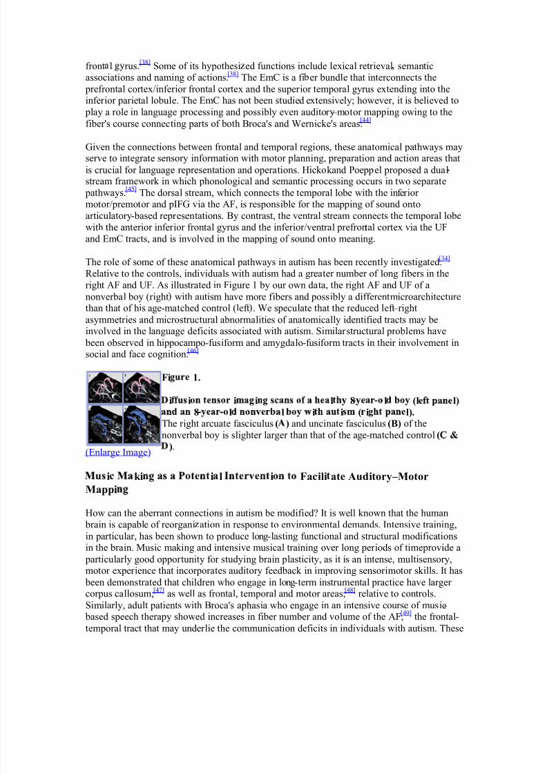
The role of some of these anatomical pathways in autism has been recently investigated.[34]
R elative to the controls, individuals with autism had a greater number of long f ibers in ther ight AF and UF. As illustrated in Figure 1 by our own data, the r ight AF and UF of a
nonverbal boy (r ight with autism have more f ibers and possibly a differentmicroarchitecturethan that of his age-matched control (lef t . We speculate that the reduced lef t±r ight asymmetr ies and microstructural abnormalities of anatomically identif ied tracts may beinvolved in the language def icits associated with autism. Similar structural problems have
been observed in hippocampo-fusiform and amygdalo-fusiform tracts in their involvement insocial and face cognition.[46]
(Enlarge Image)
Fi 1.
i i i i l 8 l (l l)
8 l l i i ( i l). The r ight arcuate fasciculus ( ) and uncinate fasciculus (B) of the
nonverbal boy is slighter larger than that of the age-matched control ( &
).
i ki i l i Facili ate Auditory±Motor
Mappi
How can the aberrant connections in autism be modif ied? It is well known that the human
brain is capable of reorgani ation in response to environmental demands. Intensive training,
in par ticular, has been shown to produce long-lasting functional and structural modif ications
in the brain. Music mak ing and intensive musical training over long per iods of time provide a
par ticular ly good oppor tunity for studying brain plasticity, as it is an intense, multisensory,motor exper ience that incorporates auditory feedback in improving sensor imotor sk ills. It has
been demonstrated that children who engage in long-term instrumental practice have larger corpus callosum,[47] as well as frontal, temporal and motor areas,[48] relative to controls.
Similar ly, adult patients with Broca's aphasia who engage in an intensive course of music-based speech therapy showed increases in f iber number and volume of the AF,[49] the frontal-temporal tract that may under lie the communication def icits in individuals with autism. These
8/7/2019 articole autism
http://slidepdf.com/reader/full/articole-autism 6/29
structural changes are consistent with a large body of literature suggesting training-inducedplasticity, such as in jugglers,[50,51] taxi drivers[52] and foreign language learners.[53] A recentstudy using DTI also showed structural changes following training with a complex visual± motor task.[54]
Given the potential benefits of music making in producing plastic changes in the brain, it is
conceivable that a music-based intervention can be used to engage and strengthen theconnections between frontal and temporal regions that are abnormal in autism, thuspotentially enabling affected individuals to develop their language skills. One suchintervention is auditory±motor mapping training (AMMT),[43] which utilizes the musicalstrengths of individuals with autism, many of whom exhibit superior music perceptionabilities[55±57] and thoroughly enjoy music making (through singing and/or playing aninstrument).[58±60] In addition, they tend to focus more on the perceptual (e.g., prosodic)information rather than the linguistic information of speech compared to typically developingindividuals, which may contribute to their language and communication deficits.[61±65] Moreover, listening to music can evoke a great intensity of emotions in individuals withautism, who typically have difficulty processing emotions, a condition known asalexithymia.[66±68] The potential utility of music interventions in autism has been
reported.[69,70] Musical stimuli have been shown to activate brain regions associated with theprocessing of emotions, such as the insular and cingulate cortex, hypothalamus, hippocampus,amygdala and prefrontal cortex,[71] thus further highlighting the therapeutic potential of musical activities in autism.
Auditory±motor mapping training involves three main components: singing, motor activityand imitation. This training contains features of MIT,[72] but also uses a set of tuned drums toengage both hands in rhythmic motor activity and to facilitate auditory±motor mapping.Singing (more than speaking) is known to engage a bilateral reciprocal network betweenfrontal and temporal regions, which contain some components of the putative MNS.[73,74] Critically, it has been proposed that a dysfunctional MNS underlies some of the languagedeficits in autism,[75] although some researchers have argued that the mirror neuronexplanation may not account for all of the deficits in autism.[76] Motor activity (throughplaying an instrument) not only captures the child's interest, but also engages a sensorimotor network that controls orofacial and articulatory movements in speech.[77] The sound producedby the instrument may also facilitate the auditory±motor mapping that is critical for meaningful vocal communication.[78] Imitation through repetitive training facilitates learningand alters the responses in the MNS.[79]
The potential utility of AMMT in ameliorating the language deficits in autism is reinforced byneuroimaging research showing overlapping responses to music and language stimuli.[74,80±83] In particular, fMRI studies have reported activation of the inferior frontal regions duringmusic perception tasks,[80,84] active music tasks such as singing[74] and imagining playing an
instrument.
[85,86]
Research has also shown that the dopaminergic system plays an importantrole in some aspects of language processing (e.g., grammar)[87] and that this system alsomediates musical pleasure in individuals with autism.[88] Moreover, a common network appears to support the sensorimotor components for both speaking and singing,[74,86,89] andengaging in musical activities has been shown to improve verbal abilities in language-delayedchildren.[90]
8/7/2019 articole autism
http://slidepdf.com/reader/full/articole-autism 7/29
Conclusion
Taken together, therapies that incorporate elements of music making (e.g., AMMT) may offer a viable approach to facilitate social skills and communication ± including expressivelanguage ± in otherwise nonverbal individuals with autism. More importantly, as evidencedby the literature on training-induced plasticity, an intensive course of music-based or
auditory-motor intervention, such as AMMT, may create a situation in which long-rangeconnections between auditory and motor regions could be particularly engaged and possiblystrengthened, such as those observed following intensive music training in children,[47] or melodic intonation therapy in aphasic patients.[49] Given the aberrant connectivity betweenfrontal and temporal regions in autism, and the abnormalities within these two regions, theAF, the UF and the EmC may be some of the long-range tracts that serve as targets for experimental treatments to facilitate communication skills in autism.
Future Perspective
Over the past decade, research on autism has focused on its behavioral manifestations, neuralunderpinning, and more recently, possible candidate genes. Although the mechanismsunderlying autism remain elusive, the considerable body of research conducted to date haslaid a foundation for the development of new and innovative interventions. Theoreticallygrounded music-based interventions have been underutilized, which is unfortunate becausemusic perception and music making is known to be a relative strength of individuals withautism. In particular, no study has systematically investigated the efficacy of a music-basedintervention in facilitating speech output, and whether an intensive program can induce plasticchanges in the brains of these individuals. On the basis of previous and current research, wehope that such specialized treatments for autism will be developed in the near future.Ultimately, such treatments should maximize the individual's potential for developing or relearning expressive language function and, thus, improve the quality of life for people withautism and their families.
References
1. American Psychiatric Association:Diagnost ic and Stat ist ical Manual of Mental
Disorders ( DSM-IVTR): 4th Ed it ion, Text Revision Ed it ion. American PsychiatricPress, Washington, DC, USA (2000).
2. K erbel D, Grunwell P: A study of idiom comprehension in children with semantic-pragmatic difficulties. Part II: between-groups results and discussion. J . Commun.
Disord. 33(1), 23±44 (1998).3.
Tager-Flusberg H: Language impairments in children with complex
neurodevelopmental disorders: the case of autism. In: Language Competence AcrossPopulat ions: Toward a Def init ion of Specif ic Language Impairment . Levy Y,Schaeffer JC (Eds). Lawrence Erlbaum Associates, NJ, USA, 297±321 (2003).
4. Tager-Flusberg H: Language and communicative deficits and their effects on learningand behavior. In: Asperger Syndrome: Behavioral and Educat ional Aspects . Prior M(Ed.). Guilford Press, NY, USA, 85±103 (2004).
5. Francis K : Autism interventions: a critical update. Dev. Med. Child Neurol. 47 (7),493±499 (2005).

8/7/2019 articole autism
http://slidepdf.com/reader/full/articole-autism 8/29
6. Zatorre R, Belin P: Spectral and temporal processing in human auditory cortex.Cereb.
Cortex 11(10) 946±953 (2001).7. Zatorre RJ, Gandour JT: Neural specializations for speech and pitch: moving beyond
the dichotomies. Philos. Trans. R. Soc. Lond. B Biol. S ci. 363(1493), 1087±1104(2008).
8.
Dipellegrino G, Fadiga L, Fogassi L, Gallese V, Rizzolatti G: Understanding motor
events ± a neurophysiological study. Exp. Brain Res. 91(1), 176±180 (1992).9. Buccino G, Binkofski F, Fink GR et al.: Action observation activates premotor and
parietal areas in a somatotopic manner: an fMRI study. Eur. J . Neurosci. 13(2), 400± 404 (2001).
10. Rizzolatti G, Fadiga L, Gallese V, Fogassi L: Premotor cortex and the recognition of motor actions. Brain Res. Cogn. Brain Res. 3(2), 131±141 (1996).� Landmark article on the mirror neuron system.
11. Buccino G, Lui F, Canessa N et al.: Neural circuits involved in the recognition of actions performed by nonconspecifics: an fMRI study. J . Cogn. Neurosci. 16(1), 114± 126 (2004).
12. Arbib MA: From grasp to language: embodied concepts and the challenge of abstraction. J . Physiol. Par is 102(1±3), 4±20 (2008).
13. Rizzolatti G, Arbib MA: Language within our grasp. Trends Neurosci. 21(5), 188±194(1998).
14. Barsalou LW: Perceptions of perceptual symbols. Behav. Brain S ci. 22 (4), 637±660(1999).
15. Gallese V, Lakoff G: The brain's concepts: the role of the sensory±motor system inconceptual knowledge. Cogn. Neuropsychol. 22(3±4), 455±479 (2005).
16. Sparks BF, Friedman SD, Shaw DW et al.: Brain structural abnormalities in youngchildren with autism spectrum disorder. Neurology 59 (2), 184±192 (2002).
17. Stanfield AC, McIntosh AM, Spencer MD, Philip R, Gaur S, Lawrie SM: Towards aneuroanatomy of autism: a systematic review and meta-analysis of structural magneticresonance imaging studies. Eur. Psychiatry 23(4), 289±299 (2008).
18. Courchesne E, K arns CM, Davis HR et al.: Unusual brain growth patterns in early lifein patients with autistic disorder: an MRI study. Neurology 57(2), 245±254 (2001).
19. Hazlett HC, Poe M, Gerig G et al.: Magnetic resonance imaging and headcircumference study of brain size in autism: birth through age 2 years. Ar ch. Gen.
Psychiatry 62(12), 1366±1376 (2005).20. Freitag CM, Luders E, Hulst HE et al.: Total brain volume and corpus callosum size in
medication-naive adolescents and young adults with autism spectrum disorder. Biol.
Psychiatry 66(4), 316±319 (2009).21. Hazlett HC, Poe MD, Gerig G, Smith RG, Piven J: Cortical gray and white brain
tissue volume in adolescents and adults with autism. Biol. Psychiatry 59(1), 1±6(2006).
22. De Fosse L, Hodge SM, Makris N et al.: Language-association cortex asymmetry in
autism and specific language impairment. Ann. Neurol. 56(6), 757±766 (2004).23. Herbert MR, Harris GJ, Adrien K T et al.: Abnormal asymmetry in languageassociation cortex in autism. Ann. Neurol. 52(5), 588±596 (2002).�� Important imaging study on the language abilities in autism.
24. McAlonan GM, Cheung V, Cheung C et al.: Mapping the brain in autism: a voxel-based MRI study of volumetric differences and intercorrelations in autism.Brain 128,268±276 (2005).
8/7/2019 articole autism
http://slidepdf.com/reader/full/articole-autism 9/29
25. Rojas DC, Bawn SD, Benkers TL, Reite ML, Rogers SJ: Smaller left hemisphereplanum temporale in adults with autistic disorder. Neurosci. Lett. 328(3), 237±240(2002).
26. Rojas DC, Camou SL, Reite ML, Rogers SJ: Planum temporale volume in childrenand adolescents with autism. J . Aut ism Dev. Disord. 35(4), 479±486 (2005).
27.
Boddaert N, Chabane N, Gervais H et al.: Superior temporal sulcus anatomical
abnormalities in childhood autism: a voxel-based morphometry MRI study.Neuroimage 23(1), 364±369 (2004).
28. McAlonan GM, Suckling J, Wong N et al.: Distinct patterns of grey matter abnormality in high-functioning autism and Asperger's syndrome. J . Child Psychol.
Psychiatry 49(12), 1287±1295 (2008).29. Hyde K L, Samson F, Evans AC, Mottron L: Neuroanatomical differences in brain
areas implicated in perceptual and other core features of autism revealed by corticalthickness analysis and voxel-based morphometry. H um. Brain Mapp. 31(4), 556±566(2010).
30. Goldstein G, Minshew NJ, Siegel DJ: Age differences in academic achievement inhigh-functioning autistic individuals. J . Cl in. Exp. Neuropsychol. 16(5), 671±680(1994).
31. Just MA, Cherkassky VL, K eller TA, Minshew NJ: Cortical activation andsynchronization during sentence comprehension in high-functioning autism: evidenceof underconnectivity. Brain 127, 1811±1821 (2004).
32. Alexander AL, Lee JE, Lazar M et al.: Diffusion tensor imaging of the corpuscallosum in autism. Neuroimage 34(1), 61±73 (2007).
33. Barnea-Goraly N, K won H, Menon V, Eliez S, Lotspeich L, Reiss AL: White matter structure in autism: preliminary evidence from diffusion tensor imaging. Biol.
Psychiatry 55(3), 323±326 (2004).34. K umar A, Sundaram SK , Sivaswamy L et al.: Alterations in frontal lobe tracts and
corpus callosum in young children with autism spectrum disorder. Cereb. Cortex (2009).
35. Herbert MR, Ziegler DA, Makris N et al.: Localization of white matter volumeincrease in autism and developmental language disorder. Ann. Neurol. 55(4), 530±540(2004).
36. Casanova MF, Buxhoeveden DP, Switala AE, Roy E: Minicolumnar pathology inautism. Neurology 58(3), 428±432 (2002).
37. Saur D, K reher BW, Schnell S et al.: Ventral and dorsal pathways for language. Proc.
Natl. Acad. S ci. USA 105(46), 18035±18040 (2008).38. Catani M, Thiebaut de Schotten M: A diffusion tensor imaging tractography atlas for
virtual in vivo dissections. Cortex 44(8), 1105±1132 (2008).39. Glasser MF, Rilling JK : DTI tractography of the human brain's language pathways.
Cereb. Cortex 18(11), 2471±2482 (2008).�� Excellent description of the language pathways in humans.
40. Parker GJ, Luzzi S, Alexander DC, Wheeler-K
ingshott CA, Ciccarelli O, LambonRalph MA: Lateralization of ventral and dorsal auditory-language pathways in thehuman brain. Neuroimage 24(3), 656±666 (2005).
41. Powell HW, Parker GJ, Alexander DC et al.: Hemispheric asymmetries in language-related pathways: a combined functional MRI and tractography study. Neuroimage 32(1), 388±399 (2006).
42. Bernal B, Ardila A: The role of the arcuate fasciculus in conduction aphasia. Brain 132(Pt 9), 2309±2316 (2009).
8/7/2019 articole autism
http://slidepdf.com/reader/full/articole-autism 10/29
43. Wan CY, Demaine K , Zipse L, Norton A, Schlaug G: From music making tospeaking: engaging the mirror neuron system in autism. Brain Res. Bull. 82(3±4),161±168 (2010).�� Theoretical rational of auditory±motor mapping training.
44. Makris N, Pandya DN: The extreme capsule in humans and rethinking of the languagecircuitry. Brain Struct. Funct. 213(3), 343±358 (2009).
45. Hickok G, Poeppel D: Dorsal and ventral streams: a framework for understandingaspects of the functional anatomy of language. Cognit ion 92(1±2), 67±99 (2004).� Excellent description of the dorsal and ventral language streams.
46. Conturo TE, Williams DL, Smith CD, Gultepe E, Akbudak E, Minshew NJ: Neuronalfiber pathway abnormalities in autism: an initial MRI diffusion tensor tracking studyof hippocampo-fusiform and amygdalo-fusiform pathways. J . Int. Neuropsychol. Soc. 14(6), 933±946 (2008).
47. Schlaug G, Forgeard M, Zhu L, Norton A, Winner E: Training-induced neuroplasticityin young children. Neurosciences and Music III: Disorders and Plast icity 205±208(2009).
48. Hyde K L, Lerch J, Norton A et al.: Musical training shapes structural braindevelopment. J . Neurosci. 29(10), 3019±3025 (2009).
49. Schlaug G, Marchina S, Norton A: Evidence for plasticity in white matter tracts of chronic aphasic patients undergoing intense intonation-based speech therapy. Ann. NY Acad. S ci. 1169, 385±394 (2009).
50. Draganski B, Gaser C, Busch V, Schuierer G, Bogdahn U, May A: Neuroplasticity:changes in grey matter induced by training ± newly honed juggling skills show up as atransient feature on a brain-imaging scan. Nature 427(6972), 311±312 (2004).
51. Draganski B, Gaser C, K empermann G et al.: Temporal and spatial dynamics of brainstructure changes during extensive learning. J . Neurosci. 26(23), 6314±6317 (2006).
52. Maguire EA, Gadian DG, Johnsrude IS et al.: Navigation-related structural change inthe hippocampi of taxi drivers. Proc. Natl Acad. S ci. USA 97(8), 4398±4403 (2000).
53. Golestani N, Paus T, Zatorre R: Anatomical correlates of learning novel speechsounds 35, 997±1010 (2002).
54. Scholz J, K lein MC, Behrens TE, Johansen-Berg H: Training induces changes inwhite-matter architecture. Nat. Neurosci. 12(11), 1370±1371 (2009).
55. Bonnel A, Mottron L, Peretz I, Trudel M, Gallun E, Bonnel AM: Enhanced pitchsensitivity in individuals with autism: a signal detection analysis. J . Cogn. Neurosci. 15(2), 226±235 (2003).
56. Heaton P: Pitch memory, labelling and disembedding in autism. J . Child Psychol.
Psychiatry 44(4), 543±551 (2003).57. Heaton P, Hermelin B, Pring L: Autism and pitch processing: a precursor for savant
musical ability? Music Per cept. 15(3), 291±305 (1998).58. Allen R, Hill E, Heaton P: 'Hath charms to soothe.' An exploratory study of how high-
functioning adults with ASD experience music. Aut ism 13(1), 21±41 (2009).
59. Bhatara AK
, Quintin EM, Heaton P, Fombonne E, Levitin DJ: The effect of music onsocial attribution in adolescents with autism spectrum disorders. Child Neuropsychol. 15(4), 375±396 (2009).
60. Bonoldi I, Emanuele E, Politi P: A piano composer with low-functioning severeautism. Acta Neuropsychiatr. 21(1), 2±3 (2009).
61. Jarvinen-Pasley A, Heaton P: Evidence for reduced domain-specificity in auditoryprocessing in autism.Dev. S ci. 10(6), 786±793 (2007).
8/7/2019 articole autism
http://slidepdf.com/reader/full/articole-autism 11/29
62. Mottron L, Peretz I, Menard E: Local and global processing of music in high-functioning persons with autism: beyond central coherence? J . Child Psychol.Psychiatry 41(8), 1057±1065 (2000).
63. Jarvinen-Pasley A, Pasley J, Heaton P: Is the linguistic content of speech less salientthan its perceptual features in autism? J . Aut ism Dev. Disord. 38(2), 239±248 (2008).
64.
Jarvinen-Pasley A, Peppe S, K ing-Smith G, Heaton P: The relationship between form
and function level receptive prosodic abilities in autism. J . Aut ism Dev. Disord. 38(7),1328±1340 (2008).
65. Jarvinen-Pasley A, Wallace GL, Ramus F, Happe F, Heaton P: Enhanced perceptualprocessing of speech in autism.Dev. S ci. 11(1), 109±121 (2008).
66. Bird G, Silani G, Brindley R, White S, Frith U, Singer T: Empathic brain responses ininsula are modulated by levels of alexithymia but not autism. Brain 133(Pt 5), 1515± 1525 (2010).
67. Allen R, Heaton P: Autism, music, and the therapeutic potential of music inalexithymia. Music Per cept. 27(4), 251±261 (2010).
68. Hill E, Berthoz A, Frith C: Brief report: cognitive processing of own emotions inindividuals with autistic spectrum disorder and in their relatives. J . Aut ism Dev.
Disord. 34(2), 229±235 (2004).
69. Boso M, Emanuele E, Minazzi V, Abbamonte M, Politi P: Effect of long-terminteractive music therapy on behavior profile and musical skills in young adults withsevere autism. J . Altern. Complement Med. 13(7), 709±712 (2007).
70. Gold C, Wigram T: Music therapy in the assessment and treatment of autisticspectrum disorder: clinical application and research evidence. Child Care H ealth Dev. 32(5), 535±542 (2006).
71. Boso M, Politi P, Barale F, Enzo E: Neurophysiology and neurobiology of the musicalexperience. Funct. Neurol. 21(4), 187±191 (2006).
72. Norton A, Zipse L, Marchina S, Schlaug G: Melodic intonation therapy: how it is doneand why it might work. Ann. NY Acad. S ci. 1169, 431±436 (2009).
73. Brown S, Martinez MJ, Hodges DA, Fox PT, Parsons LM: The song system of thehuman brain. Brain Res. Cogn. Brain Res. 20, 363±375 (2004).
74. Ozdemir E, Norton A, Schlaug G: Shared and distinct neural correlates of singing andspeaking. Neuroimage 33(2), 628±635 (2006).
75. Hadjikhani N, Joseph RM, Snyder J, Tager-Flusberg H: Anatomical differences in themirror neuron system and social cognition network in autism. Cereb. Cortex 16(9),1276±1282 (2006).
76. Hamilton AFD, Brindley RM, Frith U: Imitation and action understanding in autisticspectrum disorders: how valid is the hypothesis of a deficit in the mirror neuronsystem? Neuropsycholog ia 45(8), 1859±1868 (2007).
77. Meister IG, Buelte D, Staedtgen M, Boroojerdi B, Sparing R: The dorsal premotor cortex orchestrates concurrent speech and fingertapping movements. Eur. J . Neurosci. 29, 2074±2082 (2009).
78. Lahav A, Saltzman E, Schlaug G: Action representation of sound: audiomotor recognition network while listening to newly acquired actions. J . Neurosci. 27(2),308±314 (2007).
79. Catmur C, Walsh V, Heyes C: Sensorimotor learning configures the human mirror system. Curr. Biol. 17(17), 1527±1531 (2007).
80. K oelsch S, Gunter TC, von Cramon DY, Zysset S, Lohmann G, Friederici AD: Bachspeaks: a cortical 'language-network' serves the processing of music. Neuroimage 17(2), 956±966 (2002).
8/7/2019 articole autism
http://slidepdf.com/reader/full/articole-autism 12/29
81. K oelsch S, Gunter TC, Wittfoth M, Sammler D: Interaction between syntaxprocessing in language and in music: an ERP study. J . Cogn. Neurosci. 17(10), 1565± 1577 (2005).
82. Patel AD, Gibson E, Ratner J, Besson M, Holcomb PJ: Processing syntactic relationsin language and music: an event-related potential study. J . Cogn. Neurosci. 10(6),717±733 (1998).
83. Schon D, Magne C, Besson M: The music of speech: music training facilitates pitchprocessing in both music and language. Psychophysiology 41(3), 341±349 (2004).
84. Tillmann B, Janata P, Bharucha JJ: Activation of the inferior frontal cortex in musicalpriming. Brain Res. Cogn. Brain Res. 16(2), 145±161 (2003).
85. Meister IG, K rings T, Foltys H et al.: Playing piano in the mind-an fMRI study onmusic imagery and performance in pianists. Brain Res. Cogn. Brain Res. 19(3), 219± 228 (2004).
86. K leber B, Veit R, Birbaumer N, Gruzelier J, Lotze M: The brain of opera singers:experience-dependent changes in functional activation. Cereb. Cortex 20(5), 1144± 1152 (2009).
87. Tettamanti M, Moro A, Messa C et al.: Basal ganglia and language: phonologymodulates dopaminergic release. Neuroreport 16(4), 397±401 (2005).
88. Emanuele E, Boso M, Cassola F et al.: Increased dopamine DRD4 receptor mRNAexpression in lymphocytes of musicians and autistic individuals: bridging the music-autism connection. Neuro. Endocr inol. Lett. 31(1), 122±125 (2010).
89. Pulvermuller F: Brain mechanisms linking language and action. Nature Rev. Neurosci. 6(7), 576±582 (2005).
90. Hoskins C: Use of music to increase verbal response and improve expressive languageabilities of preschool language delayed children. J . Music Ther. 25, 73±84 (1988).
Website 101. Autism Speaks homepage www.autismspeaks.org
8/7/2019 articole autism
http://slidepdf.com/reader/full/articole-autism 13/29
Advanced Parental Age and the Ris of
Autism Spectrum Disorder
Maureen S. Durkin; Matthew J. Maenner; Craig J. Newschaffer; Li-Ching Lee; Christopher
M. Cunniff; Julie L. Daniels; Russell S. K irby; Lewis Leavitt; Lisa Miller; Walter Zahorodny;Laura A. Schieve
Abstract and Introduction
Abstract
This study evaluated independent effects of maternal and paternal age on risk of autismspectrum disorder. A case-cohort design was implemented using data from 10 US study sitesparticipating in the Centers for Disease Control and Prevention's Autism and DevelopmentalDisabilities Monitoring Network. The 1994 birth cohort included 253,347 study-site birthswith complete parental age information. Cases included 1,251 children aged 8 years withcomplete parental age information from the same birth cohort and identified as having anautism spectrum disorder based on Diagnostic and Statistical Manual of Mental
Disorders, Fourth Edition, Text Revision criteria. After adjustment for the other parent'sage, birth order, maternal education, and other covariates, both maternal and paternal agewere independently associated with autism (adjusted odds ratio for maternal age � 35 vs. 25-29 years = 1.3, 95% confidence interval: 1.1, 1.6; adjusted odds ratio for paternal age � 40years vs. 25-29 years = 1.4, 95% confidence interval: 1.1, 1.8). Firstborn offspring of 2 older parents were 3 times more likely to develop autism than were third- or later-born offspring of mothers aged 20-34 years and fathers aged <40 years (odds ratio 3.1, 95% confidenceinterval: 2.0, 4.7). The increase in autism risk with both maternal and paternal age haspotential implications for public health planning and investigations of autism etiology.
Introduction
This paper examines the relation between parental age at delivery and the prevalence of autism spectrum disorder (ASD). The possibility that autism is more common in offspring of older parents has generated considerable interest.[1-6] Confirmation of such an associationcould have important public health implications in light of increasing trends in recent decadesregarding both maternal and paternal age.[7] In addition, evidence of paternal and maternal ageeffects on autism risk may provide clues to the etiology of a class of neurodevelopmentaldisorder that is still poorly understood and thought to be complex and multifactoral.
In evaluating the association between parental age and autism risk, it is important to account
for other variables related to both parental age and autism or that may modify the association.Birth order is a potentially confounding factor because it is positively associated with parentalage and has been reported in some studies to be associated with autism risk, with at least 3studies reporting firstborn children to be at increased risk of autism.[1,2,4] The goal of thisstudy was to determine, in a large, population-based cohort of US children, whether advancing maternal and paternal age each independently increase a child's risk of developingautism after controlling for the other parent's age, birth order, and other risk factors.
Materials and Methods
8/7/2019 articole autism
http://slidepdf.com/reader/full/articole-autism 14/29
Study Design and Sample
We implemented a population-based, case-cohort design in which the comparison group wasa cohort of all livebirths in 1994 in 10 geographically defined study areas participating in theCenters for Disease Control and Prevention's Autism and Developmental DisabilitiesMonitoring Network.[8] The 10 areas are all sites with deidentified birth certificate
information on parental age and other relevant variables included in the Network database andinclude sites in Alabama, Arizona, Arkansas, Colorado, Georgia, Maryland, Missouri, NewJersey, North Carolina, and Wisconsin.
The cohort serving as the comparison group includes all livebirths to mothers residing in any1 of the study areas in 1994, with complete information available from birth certificates onmaternal and paternal age, birth order, and other variables. We used 2 data sources toconstruct the cohort: 1994 deidentified birth records for the Wisconsin study area provided bythe Wisconsin Department of Health and Family Services and, for the remaining sites, theNational Center for Health Statistics public use natality data files.[9] The public use fileincludes county of residence for births in densely populated counties, which enabled us toascertain deidentified birth information for all births in most of the counties. We were unable
to precisely obtain counts of births occurring in sparsely populated counties in which 13,043(4.1%) of the study-area births occurred in 1994. For these counties, we obtained county-levelaggregate information on the total number of births in 1994 and their distribution by variablessuch as maternal marital status, ethnicity, and age and selected a stratified random sample of deidentified birth records (equal in number and similar in distribution by maternal maritalstatus, ethnicity, and age to all livebirths occurring in the respective counties in 1994) fromsparsely populated counties of the state in which the study area was located. The full cohortincluded 326,785 livebirths, of which 73,438 (22.5%) were excluded because of missingpaternal age. The cohort serving as the comparison group thus included the 253,347 livebirthswith complete information on parental age and other key variables ( Table 1 ).
The total number of children aged 8 years residing in the study areas in 2002 determined bythe Autism and Developmental Disabilities Monitoring Network surveillance system to havean ASD was 2,142. Birth certificate information was available for 1,517 (70.8%) of thesechildren, who were born in the same state as their state of residence in 2002. The remaining29.2% of cases were excluded from this analysis because of missing birth certificateinformation. The case group for the present analysis was further restricted to the 1,251children (58.4% of the total ASD case group) for whom information on both parents' age aswell as birth order and gestational age was available. Our final sample was comparable to thetotal population of ASD cases regarding demographic factors and ASD case characteristics (Table 1 ).
Case Definition
ASDs include behaviorally defined neurodevelopmental disorders diagnosed through clinicalobservation, and they encompass impairments in social, communicative, and behavioraldevelopment, often accompanied by abnormalities in intellectual functioning, learning,attention, and sensory processing. For this study, children with ASD included members of thebirth cohort residing in the study area in 2002 who met Diagnost ic and Stat ist ical Manual of
Mental Disorders, Fourth Edition, Text Revision criteria for autistic disorder; pervasivedevelopmental disorders-not otherwise specified (PDD-NOS(http://www.cdc.gov/ncbddd/autism/overview_diagnostic_criteria.htm ), including atypical
8/7/2019 articole autism
http://slidepdf.com/reader/full/articole-autism 15/29
autism); or Asperger's disorder [10] based on a comprehensive review of educational andclinical records by trained clinicians. Children were classified by clinician reviewers ashaving an ASD if they had either a documented previous classification of ASD (65%) or anevaluation record from an educational or medical setting indicating unusual behaviorsconsistent with ASD (35%). For children previously identified as having an ASD, case statuswas confirmed on the basis of evaluation records. For children without a documented ASD
classification, data were abstracted on all relevant ASD and developmental behaviors fromeducation or medical evaluations to determine whether behaviors described in the evaluationsby clinical reviewers were consistent with the diagnostic criteria. Because case status wasdetermined solely on the basis of information contained in evaluation records, and becauseDiagnost ic and Stat ist ical Manual of Mental Disorders, Fourth Edition, Text Revision criteriaare less well defined for PDD-NOS than for autistic disorder, the surveillance protocol for determining whether a child could be classified as having PDD-NOS required documentationof at least 1 behavior considered to be an ASD discriminator, such as being oblivious to otherswhen there is a clear social opportunity or demonstrating atypical and persistent focus onsensory input.[11]
Of the 1,251 ASD cases, 80.7% were determined to meet criteria for autistic disorder, while
there was insufficient information for those remaining to distinguish between autisticdisorder, Asperger's disorder, or PDD-NOS. Information from standardized intelligence testswas available for approximately 75% of the ASD cases. On the basis of this information,children with ASD were classified as having intellectual impairment (an IQ of < 70) versusnormal intelligence. Further details regarding the 2002 Autism and DevelopmentalDisabilities Monitoring Network sample and methodology have been reported previously.[8,11]
Analytic Strategy and Statistical Methods
Potential for confounding effects of birth order, gender, and other variables was evaluated byfirst examining unadjusted associations between each potential confounder and theindependent variables of maternal and paternal age as well as the dependent variable, ASDcase status. Variables were considered to be potentially confounding factors if they wereassociated with both parental age and ASD. Unadjusted odds ratios with confidence intervalswere computed to evaluate the magnitude of these associations, and unconditional logisticregression models were fit to estimate adjusted odds ratios and 95% confidence intervals.Statistical significance was evaluated by using chi-square tests for categorical variables andanalysis of variance for continuous variables.
To enhance the comparability of our findings with those from other studies, we fit 2 types of models, 1 with parental ages categorized into 6 categories: < 20, 20-24, 25-29, 30-34, 35-39,� 40 years; and the other with parental age as a continuous variable with the odds ratio scaledto reflect a 10-year difference in age.[4] Although we found the association between parental
age and autism risk to be similar across the 10 sites, to adjust for potential site-to-sitevariability we included site dummy variables in all multivariable models. To evaluateinteraction or modifying effects of each covariate and of ASD subtypes on the associationsbetween parental age and ASD, we performed stratified analyses. We also tested interactionterms for maternal age by paternal age and 2-way and 3-way interaction terms for eachparent's age by the other covariates in the regression models, but we identified no significantinteractions. SAS version 9.1.3 software (SAS Institute, Inc., Cary, North Carolina) was usedfor all statistical analyses.
8/7/2019 articole autism
http://slidepdf.com/reader/full/articole-autism 16/29
This research involved secondary analysis of deidentified data and was approved by theUniversity of Wisconsin Institutional Review Board.
Results
In unadjusted analyses, both mean maternal age and mean paternal age were significantly
higher for ASD cases than for the birth cohort as a whole ( Table 2 ). Table 2 also shows thatmean parental ages differed significantly in unadjusted analyses across categories of birthorder, maternal education, ethnicity, multiple birth, gestational age, and birth weight for gestational age, but not for gender. With parental age 25-29 years as the reference group, theodds of developing ASD was significantly reduced for parental age < 20 years and increasedfor maternal age � 35 and paternal age � 40 years ( Table 3 , unadjusted odds ratios). We therefore used these age cutoffs (maternal age � 35, paternal age � 40 years) to classify eachparent's age as "older" versus "younger." Other significant predictors of ASD in unadjustedanalyses included low birth order, male gender, advanced maternal education, and pretermbirth ( Table 3 ).
Multivariable Analysis of Parental Ages Modeled as Categorical Variables
After we adjusted for the other parent's age and other covariates, the increases in ASD risk associated with maternal age � 35 years and paternal age � 40 years (relative to age 25-29years) were slightly reduced compared with the unadjusted analysis ( Table 3 ). In contrast,the results for birth order suggest that the decline in ASD risk associated with increasing birthorder is somewhat stronger in the adjusted analysis than in the unadjusted analysis ( Table 3 ).In addition, the apparent increase in ASD risk associated with higher levels of maternaleducation in the unadjusted analysis is no longer evident in the adjusted model, suggestingthat the apparent maternal education effect is due to its association with parental age ( Table 3 ).
Parental Ages Modeled as Continuous Variables
In unadjusted analyses, the risk of developing ASD increased significantly with each 10-year increase in both maternal age and paternal age. After adjustment for age of the other parentand other covariates, each 10-year increase in maternal age was associated with a 20%increase in ASD risk (odds ratio = 1.2, 95% confidence interval: 1.1, 1.4) while each 10-year increase in paternal age was associated with a 30% increase in ASD risk (odds ratio = 1.3,95% confidence interval: 1.1, 1.5).
Combined Effects of Parental Age and Birth Order
The risk of ASD within each of 3 parental age categories (both parents "younger," 1 parent
"older," and both parents "older") was highest among firstborn children and declined withincreasing birth order ( Table 4 ). Considering the combined effects of parental age and birthorder, we excluded from the analysis births to mothers aged < 20 years and found the lowestrisk group to be third- or later-born offspring of mothers aged 20-34 years and fathers aged< 40 years. Compared with that for this group, the risk of ASD increased with both decliningbirth order and increasing number of older parents. The highest risk group included firstbornoffspring of mothers aged � 35 years and fathers aged � 40 years, with a risk 3 times that of the reference group ( Table 4 ).
8/7/2019 articole autism
http://slidepdf.com/reader/full/articole-autism 17/29
Discussion
Our findings are consistent with those recently reported from a large study of members of aCalifornia health maintenance organization[4] that found the risk of ASD to be positively andindependently associated with both maternal and paternal age, with adjusted odds ratiosnearly identical to those reported here. These findings contrast somewhat with 5 other recent
epidemiologic studies that found only 1 or neither parent's age to be associated with ASD risk after controlling for the other parent's age.[2,3,12-14]
The lack of consistency across studies could be due to limitations of sample size and of population representation of previous studies as well as other methodological differences,including autism case definitions and inclusion criteria and the ability to control for importantvariables. The present study included a large sample of children with sufficient information toenable evaluation of separate and combined effects of each parent's age as well as birth order and other variables. With more than 1,200 cases, it included over 50% more cases and thusmore statistical power than any of the previous studies examining independent effects of maternal and paternal age on ASD risk.
Another advantage of this study is the population-based nature and diversity of the cohort,allowing control for factors that may confound the association between parental age and ASD.Maternal education is 1 variable we considered to be a potentially confounding factor becauseit is associated with maternal age and has been observed to be related to ASD risk.[15] Our results, however, suggest that the association between advanced maternal education and ASDrisk observed in unadjusted analysis may be spurious and due to confounding by parental age.
The results of this study also demonstrate the importance of controlling for birth order inevaluating independent effects of parental age on ASD risk. Because birth order increaseswith parental age and, in this and other studies, has been found to be negatively associatedwith ASD risk, failure to control for birth order may mask a positive association between
parental age and ASD risk. Two of the previous studies reporting an association betweenadvancing maternal age and ASD[2,4] also had adjusted for birth order and, similar to thepresent study, found birth order to be negatively associated with ASD.
An additional advantage of this study is its restriction to a single birth year, therebycontrolling for temporal trends in recent decades in both ASD prevalence and parental ages atthe birth of their children. This feature of the study allows estimation of the associationbetween parental age and ASD risk independently of temporal trends in diagnostic practicesor other factors.
Public Health Implications
The strength of the independent associations between maternal and paternal age and ASDrisk, as indicated by the odds ratios in the range of 1.2-1.4 reported here, is modest. However,the observation that these effects are independent of each other and of low birth order raisesthe likelihood that the combined effects of parental age and birth order may have importantpublic health implications. Mean maternal age in the United States has increased steadilysince the 1970s, particularly for firstborn children, for whom mean maternal age at deliveryincreased by 3.8 years between 1970 and 2004.[16] In addition, the proportion of births towomen aged � 35 years began increasing in the United States after 1980, when it was 5%; by2004, it had increased to 14.2%.[17,18] During this same period, fertility rates for men aged
8/7/2019 articole autism
http://slidepdf.com/reader/full/articole-autism 18/29
� 40 years also increased each year, while fertility among men age d < 30 years declined. [16] With the decline in average family size in recent decades, we would also expect theproportion of children who are firstborn to have increased. Similar trends are occurring inother developed countries.[7] The results of this study raise the question of whether someportion of the recent rise in ASD prevalence[19] may be linked to recent trends in parental ageand family size. A furthe r question is whethe r a modest increase in prevalence associated
with advancing parental age and low birth order may have contributed to a greater awarenessof ASD and, in turn, increases in measured prevalence. The tendency for older parents of firstborn children to have higher levels of educational achievement and resources than other parents could further contribute to increased awareness and an expansion of the diagnosis of ASD.
Potential Etiologic Implications of Parental Age Effects
Because we observed independent effects of the age of each parent on ASD risk, the possiblemechanisms for these effects could include a broad range of processes associated with either or both maternal and paternal age. The observed paternal age effect independent of maternalage could point to a causal role of gene mutations in male germ cells, because the probability
or selection of these mutations increases as men age.[20,21] The independent effect of maternalage, on the other hand, may point to age -related chromosome changes, pregnancycomplications, or environmental exposures during pregnancy. Independent effects of 1 or bothparents' ages also could point to a role of accumulated environmental exposures that may havemutagenic effects on gametes or could result from a combination of mechanisms.[21,22]
The association between advanced maternal and paternal age and ASD is also consistent witha potential role of infertility treatments or assisted reproductive technologies, the uses of which have increased in the past decade, especially by women and men of advancedreproductive age.[23] Numerous studies have found associations between these technologiesand adverse pregnancy outcomes, including those due to epigenetic effects (24-27), althougha recent review found no evidence of elevated rates of autism among children born after invitro fertilization techniques.[28] Even though we have no information about exposure to thesetreatments in our cohort, the observation that firstborn children of older parents had thehighest ASD risk is consistent with a possible infertility treatment effect because women whogive birth after infertility treatment are more likely to be primiparous than those representedin the general birth cohort. However, the association between multiple birth and ASD in thisstudy was weak and not statistically significant ( Table 3 , unadjusted odds ratio), whereasassisted reproduction technologies are strongly associated with multiple birth.[23]
Another unmeasured factor in the present study potentially associated with both advancedparental age and ASD risk in offspring is psychopathology or behavioral traits of parents thatmay result in both delayed parenthood and genetic susceptibility to autism in offspring.[14]
Birth-order Effects
The observation in this and at least 2 previous studies[2,4] that the risk of developing ASD washighest for firstborn children and declined with increasing birth order is a pattern alsoobserved for other childhood disorders, including type I diabetes and atopy, and is cited assupport for the "hygiene hypothesis." According to this hypothesis, firstborn children areexposed to fewer infections from other children early in childhood and, because of delayedimmunologic challenge, may be more likely to develop autoimmune responses including
8/7/2019 articole autism
http://slidepdf.com/reader/full/articole-autism 19/29
those that may adversely affect neurodevelopment.[29] Another possible factor that could leadto the observed birth-order effect is exposure to potentially neurotoxic, fat-soluble chemicalsaccumulated in maternal tissue that have been passed to offspring transplacentally or throughbreast milk.[30] Because of accumulation over a lifetime, the load of such neurotoxinstransmitted might be expected to be highest for firstborn children, particularly when combinedwith advanced maternal age. Another possible explanation for the observed birth order effect
is "stoppage" or a tendency for parents of 1 child with ASD not to have subsequent childrenbecause of the demand s of parenting a child with a disability or concerns about geneticsusceptibility,[31] thus increasing the likelihood in the cohort as a whole that a child with ASDwill have a low birth order. Information available for the present study did not allowexamination of these hypotheses.
Another important limitation of this study is that the cohort available for analysis excludesbirths with missing paternal age information. Because this exclusion applied to both the ASDcases and the comparison group ( Table 1 ), we would not expect it to have resulted in biasedestimates of the association between ASD and parental age. In a separate analysis, weexamined the association between maternal age and ASD without adjusting for paternal ageand including the full birth cohort, and we found the association between maternal age and
ASD to be the same as that observed in the subcohort with paternal age.
Another limitation is that the birth cohort comparison group includes about 1% of births of children who died postnatally in addition to an undetermined number who moved out of thestudy area between birth and the age of 8 years, whereas children who died postnatally andthose moving out of the study area after birth are excluded from the case group. Because of this limitation, we could not estimate cumulative incidence of ASD. Nonetheless, thislimitation is unlikely to have biased the estimated odds ratios reported in this study,particularly those adjusted for factors such as gestational age and birth weight for gestationalage, which are strongly associated with postnatal mortality. Another possible explanation for the increase in ASD among offspring of older parents, but one we cannot evaluate with thedata available, is that, compared with younger parents, older parents may be more aware of developmental abnormalities and better able to access diagnostic and special educationalservices. other limitations are that parity pertains to only mothers and does not take intoaccount the number of previous births fathered by the fathers represented in the cohort,potential for residual confounding by factors not measured in the present study, possiblemisclassification of ASD case status, and missing information on paternal education.
Conclusion
The results of this study provide the most compelling evidence to date that ASD risk increaseswith both maternal and paternal age and decreases with birth order. Further research involvinglarge, well-characterized birth cohorts followed longitudinally will be required to confirm
these findings and adequately evaluate the range of alternative genetic and environmentalhypotheses that this and other studies raise regarding parental age and birth-order effects onASD risk. Smaller, focused studies may also be useful, such as Crow's idea to look for mutations responsible for complex disorders of unknown etiology and with parental ageeffects by studying affected families with older parents.[20]

8/7/2019 articole autism
http://slidepdf.com/reader/full/articole-autism 20/29
References
1. Tsai LY, Stewart MA. Etiological implication of maternal age and birth order ininfantile autism. J Aut ism Dev Disord . 1983;13(1):57-65.
2. Glasson EJ, Bower C, Petterson B, et al. Perinatal factors and the development of autism. Ar ch Gen Psychiatry. (2004;61(6):618-627.
3. Reichenberg A, Gross R, Weiser M, et al. Advancing paternal age and autism. Ar chGen Psychiatry. 2006;63(9):1026-1032.
4. Croen LA, Najjar DV, Fireman B, et al. Maternal and paternal age and risk of autismspectrum disorder. Ar ch Ped iatr Adolesc Med . 2007;161(4):334-340.
5. Cantor RM, Yoon JL, Furr J, et al. Paternal age and autism are associated in a family-based sample. Mol Psychiatry. 2007;12(5):419-421.
6. K oyama T, Miyake Y, K urita H. Parental age s at birth of children with pervasivedevelopmental disorders are higher than those of children in the general population.Psychiatry Cl in Neurosci. 2007;61(2):200-202.
7. Bray I, Gunnell D, Davey Smith G. Advanced paternal age : how old is too old? J
Epidemiol Community H ealth. 2006;60(10):851-853.8. Autism and Developmental Disabilities Monitoring Network Surveillance Year 2002
Principal Investigators; Centers for Disease Control and Prevention. 14 sites, UnitedStates, 2002. MMWR Surveill Summ. 2007;56(1):12-28.
9. National Center for Health Statistics, natality data, public-use data files.(http://www.cdc.gov/nchs/products/elec_prods/subject/natality.htm) (AccessedNovember 2007).
10. American Psychiatric Association. Diagnost ic and Stat ist ical Manual of Mental
Disorders: DSM-IV-TR. Fourth Ed it ion, Text Revision. Arlington, VA: AmericanPsychiatric Association; 2000.
11. Rice CE, Baio JL, Van Naarden Braun K , et al. A public health collaboration for thesurveillance of autism spectrum disorders. Paed iatr Per inat Epidemiol .2007;21(2):179-190.
12. Lauritsen MB, Pedersen CB, Mortensen PB. Effects of familial risk factors and placeof birth on the risk of autism: a nationwide register-based study. J Child Psychol Psychiatry. 2005;46(9):963-971.
13. Maimburg RD, Vaeth M. Perinatal risk factors for infantile autism. Acta Psychiatr S cand . 2006;114(4):257-264.
14. Larsson HJ, Eaton WW, Madsen K M, et al. Risk factors for autism: perinatal factors,parental psychiatric history, and socioeconomic status.Am J Epidemiol .2005;161(10):916-925.
15. Treffert DA. Epidemiology of infantile autism. Ar ch Gen Psychiatry. 1970;22(5):431-438.
16. Martin JA, Hamilton BE, Sutton PD. Births: final data for 2004. Natl V ital Stat Rep. 2006;55(1):1-102.
17. Mathe ws TJ, Hamilton BE. Mean age of mother, 1970-2000. Natl V ital Stat Rep. 2002;51(1):1-14.
18. CDC. National Vital Statistics System.(http://www.cdc.gov/nchs/births.htm#Tabulated) (Accessed September 11, 2008).
19. Yeargin-Allsopp M, Rice C, K arapurkar T, et al. Prevalence of autism in a USmetropolitan area. J AMA 2003;289(1):49-55.
20. Crow JF. The high spontaneous mutation rate: is it a health risk? Proc Natl Acad S ci U
S A. 1997;94(16):8380-8386.
8/7/2019 articole autism
http://slidepdf.com/reader/full/articole-autism 21/29
21. Crow JF. Age and sex effects on new mutation rates: an old problem with newcomplexities. J Rad iat Res (Tokyo). 2006;47(suppl B):B75-B82.
22. Penrose LS. Parental age and mutation. Lancet . 1955;269(6885):312-313.23. Wright VC, Chang J, Jeng G, et al. Assisted reproductive technology surveillance²
United States, 2004. MMWR Surveill Summ. 2007;56(6):1-22.24.
Ombelet W, Martens G, De Sutter P, et al. Perinatal outcome of 12,021 singleton and
3108 twin births after non-IVF-assisted reproduction: a cohort study. H um Reprod .2006;21(4):1025-1032.
25. Schieve LA, Rasmussen SA, Reefhuis J. Risk of birth defects among childrenconceived with assisted reproductive technology: providing an epidemiologic contextto the data. Fert il Ster il . 2005;84(5):1320-1324.
26. DeBaun MR, Niemitz EL, Feinberg AP. Association of in vitro fertilization withBeckwith-Wiedemann syndrome and epigenetic alterations of LIT1 and H19. Am J
H um Genet . 2003;72(1):156-160.27. Sato A, Otsu E, Negishi H, et al. Aberrant DNA methylation of imprinted loci in
superovulated oocytes. H um Reprod . 2007;22(1):26-35.28. Newschaffer CJ, Croen LA, Daniels J, et al. The epidemiology of autism spectrum
disorders. Annu Rev Publ ic H ealth. 2007;28:235-258.
29. Rook GA. The hygiene hypothe sis and the increasing prevalence of chronicinflammatory disorders. Trans R Soc Trop Med H yg . 2007;101(11):1072-1074.
30. Iida T, Hirakawa H, Matsueda T, et al. Polychlorinated dibenzo-P -dioxins and relatedcompounds in breast milk of Japanese primiparas and multiparas. Chemosphere.1999;38(11):2461-2466.
31. Jones MB, Szatmari P. Stoppage rules and genetic studies of autism. J Aut ism DevDisord . 1988;18(1):31-40.
8/7/2019 articole autism
http://slidepdf.com/reader/full/articole-autism 22/29
Mental Health of Adults with Autism
Spectrum Disorders and Intellectual
Disability
Lisa Underwood; Jane McCarthy; Elias Tsakanikos
Abstract and Introduction
Abstract
Purpose of review The literature has often suggested that individuals with intellectualdisability who have an autism spectrum disorder (ASD) experience higher rates of mentalhealth problems than those without ASD. This finding has been challenged in recent years andso the purpose of this article was to critically review relevant studies since March 2009. Thereview focuses on studies specifically about the mental health of adults with intellectualdisability who have ASD.Recent findings Recent studies do not support the hypothesis that adults with intellectualdisability and ASD are more vulnerable to psychiatric disorders than those without ASD.Factors found to be associated with poorer mental health include severity of intellectualdisability, adaptive behaviour skills and social skills.Summary The evidence base on the mental health of adults with intellectual disability andASD is small but rapidly increasing. Studies tend to have relatively small sample sizes andthere remain difficulties in accurately assessing ASD and psychopathology in adults withintellectual disability.
Introduction
Autism spectrum disorders (ASDs) are neurodevelopmental conditions characterized by life-long impairments in social interaction, communication and imagination.[1] Depending ondiagnostic criteria and methodological instruments, the rate of ASD in those with intellectualdisability has been reported to be between 8 and 20%.[2,3,4�] In adults with intellectualdisability who live in community settings, presence of ASD is the strongest predictor of hospital admission, psychotropic medication and problem behaviours.[5,6] Although presenceof ASD in children and young adults increases the likelihood of mental health problems[7,8] itis less clear if this is also the case in adults with intellectual disability. Studies that employedclinical[9] and population-based[10] samples showed that the presence of ASD in adults doesnot increase the likelihood of mental-health problems.
The Evidence Base
Research with a specific focus on comorbid psychopathology in adults with intellectualdisability and ASD has increased dramatically in recent years. Five years ago only one study,on rates of psychiatric disorder, had been published. [11] Between then and the current reviewperiod another five reports emerged.[9,10,12±14] The most recently published studies aredescribed below. Table 1 [4�,15�,16�,17�] describes relevant reviews published since March 2009.
Assessment of Psychopathology
8/7/2019 articole autism
http://slidepdf.com/reader/full/articole-autism 23/29
A review of measures for assessing psychopathology in people with ASD identified severaltools for adults with intellectual disability.[15�] The authors concluded that, as a number of instruments have been developed for people with intellectual disability and ASD, there iscurrently insufficient evidence on their accuracy and that tools need to be further developedand tested. The review looked at recent reports on the Autism Spectrum Disorders ± Comorbidity for Adults scale (ASD-CA;)[18�] and the Psychopathology in Autism Checklist
(PAC;).[19]
Prevalence and Patterns of Psychopathology
The prevalence of psychiatric disorder in people with ASD and intellectual disability wasdiscussed in a review on the mental health needs of people with ASD.[16�] The authorsreported mixed results from the studies discussed and highlighted the difficulties in carryingout epidemiological studies, including referral bias (particularly in clinic-based samples),small sample sizes and problems differentiating between symptoms of psychiatric disordersand features of ASD. The authors point to a range of risk factors for psychiatric disorder including genetic factors, communication problems, loneliness and low self-esteem. Cooper and van der Speck [17��] reviewed studies on the epidemiology of mental health problems inadults with intellectual disability. Their section on ASD covered three papers from 2008 withmixed results: a low rate of psychiatric disorder, a higher rate of schizophrenia thanindividuals without ASD and no difference in prevalence rates of mental ill health. Theauthors did not draw any conclusions on the epidemiology of mental ill health in adults withASD and intellectual disability.
A Canadian study looked at the clinical characteristics of adults with ASD and intellectualdisability.[20�] More than half of those with ASD also had a psychiatric disorder; 26.1% had amood disorder, another 26.1% had a psychotic disorder and one person (4.3%) had apersonality disorder. When rates of these disorders were compared with those in individualswithout ASD, those with ASD were significantly less likely to have a psychotic disorder.
There were no other statistically significant differences between the groups; however, thisshould be taken with caution given the very small sample sizes in some of the analyses.Furthermore, the study was unable to match the groups with intellectual disability on severitydue to a lack of available information.
A report on older adults with intellectual disability and ASD combined data from four UK studies of participants in staffed group homes.[21��] Two analyses were carried out; firstly,participants with ASD were compared with all those without ASD, then a subsample of participants with ASD were compared with a matched sample without ASD on the basis of their level of adaptive skills. In the comparison of the unmatched groups there was nostatistically significant difference in the rate of psychiatric disorder, which was 31.7% for people with ASD and 23.3% for those without ASD. The analysis of the matched groups
found no statistically significant differences between the groups with and without ASD. Thefindings of the study are discussed with caution, given the way in which presence of ASD wasdefined in this study, with the authors suggesting that '«the presence of the triad of impairment typically observed in ASD is not associated with differential outcomes amongolder adults with an ID [intellectual disability].' Information on the severity of intellectualdisability of the study's participants was not available. Therefore, it is unclear whether differences in psychopathology, behaviour problems and quality of life are associated with astable characteristic such as level of intellectual disability or adaptive skills, which the authorssuggest could be improved thus perhaps leading to better outcomes.
8/7/2019 articole autism
http://slidepdf.com/reader/full/articole-autism 24/29
A study in the United States investigated the frequency of symptoms in adults withintellectual disability, ASD and diagnosed psychiatric disorder.[18�] Psychopathology wasassessed using the ASD-CA. Differences were found between those with intellectualdisability only and those with intellectual disability and ASD. However, expected differencesbetween those with intellectual disability and ASD and those with additional psychopathologywere not found. A more recent study from the same research group employed the ASD-CA to
look at rates of psychopathology in adults with combinations of intellectual disability, ASDand epilepsy.[22]
A small number of individuals with ASD (N ¼25, 4.4% of the sample) were included in astudy of factors associated with mental disorders in adults with intellectual disability.[23�] Thepresence of autism was associated with higher levels of psychosis, anxiety, problembehaviour and overall mental ill health. However, after controlling for age, sex, presence of autism and other factors, only severity of intellectual disability remained a statisticallysignificant predictor of psychopathology.
The relationship between intellectual disability and ASD was reviewed by Matson andShoemaker.[4�] A section on comorbid psychopathology looked at a number of studies
published since 2006. Their finding of higher rates of psychopathology in those withintellectual disability and ASD was mainly supported by studies on children and adolescents.The authors call for more work on the development of tools for accurately assessingpsychopathology in people with intellectual disability and ASD of all ages. A review of studies on psychosis, affective disorders and anxiety in ASD found a wide range in the ratesof these disorders.[24] Anxiety disorders were most common (5±35% for generalized anxiety,10±64% for phobias and 1±37% for OCD), followed by affective disorders (0±50%) andschizophrenia (0±6%). Aside from methodological issues, the authors found it difficult to tellwhether studies included individuals with intellectual disability and ASD. In addition, for many studies comorbidity was not the primary aim.
Characteristics Associated with Psychopathology
The relationship between psychopathology and challenging behaviour in adults withintellectual disability and ASD was investigated in a large sample of patients from a specialistmental health service in the UK .[25��] The 124 participants with intellectual disability and ASDwere more likely to exhibit challenging behaviour than those without ASD (N ¼562). Inparticipants with challenging behaviour there were significant differences in psychopathologybetween those with and without ASD. Participants with ASD and challenging behaviour wereless likely to have schizophrenia and less likely to have any additional psychiatric disorder.However, when level of intellectual disability, sex and age were controlled for presence of challenging behaviour was not associated with psychopathology; only the presence of ASDand severe intellectual disability predicted challenging behaviour. The authors concluded that,
in adults with intellectual disability and ASD, challenging behaviour and mental healthproblems are independent conditions. This finding could have implications for the diagnosisof psychopathology ± which often relies on the assessment of behavioural symptoms ± and for the management of challenging behaviour in individuals with intellectual disability and ASD.
A study on the relationship between psychopathology and social skills in adults withintellectual disability and ASD was carried out in the United States.[26��] There weresignificant variations in the pattern of associations between individual psychopathologysymptoms and social skills between those with and without ASD. Only the ASD group had
8/7/2019 articole autism
http://slidepdf.com/reader/full/articole-autism 25/29
positive skills associated with psychopathology as well as negative social behaviours. Socialskill items were a predictor of the severity of psychiatric symptoms for both groups. Therelationship between adaptive behaviour skills, psychopathology and ASD was investigated ina study of adults with intellectual disability.[27�] Half of the participants with intellectualdisability and ASD had an additional mental health problem compared with only 13% of theparticipants with intellectual disability. Those with intellectual disability, ASD and a
psychiatric disorder had the lowest level of adaptive skills, significantly lower than those withintellectual disability and ASD or intellectual disability alone. Presence of a psychiatricdisorder appeared to have no effect on adaptive behaviour, and there did not seem to be anyinteractional effect between psychopathology and ASD. There were no differences betweenthe groups with and without ASD on sex or severity of intellectual disability. This studyfound that a higher proportion of those without ASD were African±American than those withASD.
A study of adults with intellectual disability receiving psychiatric treatment from a specialistmental health service in the UK looked at the role of ethnic differences in clinicalpsychopathology. [28�] Among the participants were 144 people with intellectual disability andASD (constituting 19.1% of the total sample). A diagnosis of ASD was more likely in black
participants than in white or other ethnic group participants. However, this may be accountedfor by age differences between the groups studied and the authors state that there is 'a lack of substantial supporting evidence [for] an association between ethnicity and autistic spectrumdisorders'.
Services and Intervention for Mental Health Problems
In June 2009, the National Audit Office published its report on services for adults withautism.[29�] Although much of the report focused on people with 'high-functioning' autism,several of the recommendations were relevant to mental health services for people withintellectual disability and ASD. During 2009 the Autism Act became law in the UK in
November.
[30]
The Government is required to publish mandatory guidance for health andsocial care services to support implementation of the Act by end of 2010.
K napp et al. [31��] estimated that the economic cost of ASD in the UK is £27.7 billion a year.They carried out separate analyses for adults with and without intellectual disability, findingthat annual costs for adults with intellectual disability are £16.9 billion. Several of the datasetsused in the analyses for those with ASD and intellectual disability included participants withadditional mental health problems. However the exact number of people with intellectualdisability, ASD and mental health problems was not clear, separate analyses for those withand without mental health problems were not carried out and the study focused on servicecosts specifically attributable to ASD so it was not clear whether there was any variation inmental health service use. A survey of systematic reviews on psychosocial interventions for
adults with intellectual disability and/or ASD with mental health problems found only threestudies that met their inclusion criteria.[32] Unfortunately, neither the two reviews onpsychotherapy nor the review on integrated care appeared to include studies on participantswith ASD. It was not clear how many of the 123 excluded reviews, which covered housingsupport, social skills training and psychotherapy, included studies on people with ASD.
Additional Studies
8/7/2019 articole autism
http://slidepdf.com/reader/full/articole-autism 26/29
Other potentially relevant studies published since March 2009 include those on psychiatricand psychosocial problems in adults with 'normal-intelligence' ASD;[33] psychiatric andpsychosocial problems in young adults with autistic traits;[34] medication use among peoplewith ASD (the majority of whom had intellectual disability);[35] psychiatric symptoms amonghigh-functioning adolescents with ASD;[36] and challenging behaviour in adults withintellectual disability with and without ASD.[37] In addition to the studies reviewed above
there have been a number of recent studies on children with ASD, some of which includedparticipants with intellectual disability.[38±43]
The primary studies included in this review vary in purpose, methodology and quality. Table2 summarizes the issues which are important when judging the strength of studies and theextent to which they add to the evidence base.
Conclusion
At present there is no consensus on the best way to assess psychopathology in adults withASD and intellectual disability. More population-based and clinic-based studies are neededbefore a clear picture of the prevalence, course and outcome of mental health problems inadults with intellectual disability and ASD can emerge. In addition to ensuring that presenceof ASD is attributed accurately, studies should collect data on a broader range of measuresincluding health, social functioning and service users' experience with mental health services.These recent findings show how important it is that studies match groups with and withoutASD on age, sex and severity of intellectual disability. Investigating whether predictors of outcome are fixed or possibly subject to intervention (e.g. adaptive behaviour skills) would bea valuable addition to the evidence base.
References
1. American Psychiatric Association. Diagnostic and statistical manual of mental
disorders. 4th edition. Washington, DC: American Psychiatric Association; 1994.2. Bhaumik S, Tyrer F, McGrother C, Ganghadaran SK . Psychiatric service use andpsychiatric disorders in adults with intellectual disability. J Intellect Disabil Res 2008;52:986±995.
3. Cooper S-A, Smiley E, Morrison J, et al. Mental ill-health in adults with intellectualdisabilities: prevalence and associated factors. Br J Psychiatry 2007; 190:27±35.
4. Matson JL, Shoemaker M. Intellectual disability and its relationship to autismspectrum disorders. Res Dev Disabil 2009; 30:1107±1114.� This is an in-depth review covering the nosology and prevalence of ASD in peoplewith intellectual disability. This report critiques recent research on adaptive behaviour,challenging behaviour and comorbid psychopathology.
5. Cowley A, Newton J, Sturmey P, et al. Psychiatric inpatient admissions of adults with
intellectual disabilities: predictive factors. Am J Ment Retard 2005; 110:216±225.6. Tsakanikos E, Costello H, Holt G, et al. Behaviour management problems aspredictors of psychotropic medication and use of psychiatric services in adults withautism. J Autism Dev Disord 2007; 37:1080±1085.
7. Bradley EA, Summers JA,Wood HL, Bryson SE.Comparing rates of psychiatric andbehaviour disorders in adolescents and young adults with severe intellectual disabilitywith and without autism. J AutismDev Disord 2004; 34:151±161.
8/7/2019 articole autism
http://slidepdf.com/reader/full/articole-autism 27/29
8. Brereton AV, Tonge BJ, Einfeld SL. Psychopathology in children and adolescentswith autism compared to young people with intellectual disability. J Autism DevDisord 2006; 36:863±870.
9. Tsakanikos E, Costello H, Holt G, et al. Psychopathology in adults with autism andintellectual disability. J Autism Dev Disord 2006; 36:1123±1129.
10.
Melville CA, Cooper S-A, Morrison J, et al. The prevalence and incidence of mental
ill-health in adults with autism and intellectual disabilities. J Autism Dev Disord 2008;38:1676±1688.
11. Morgan CN, Roy M, Chance P. Psychiatric comorbidity and medication use in autism:a community survey. Psychiatric Bull 2003; 27:378±381.
12. Tsakanikos E, Sturmey P, Costello H, et al. Referral trends in mental health servicesfor adults with intellectual disability and autism spectrum disorders. Autism 2007;11:9±17.
13. La Malfa G, Lassi S, Salvini R, et al. The relationship between autism and psychiatricdisorders in intellectually disabled adults. Res Autism Spectr Disord 2007; 1:218±228.
14. Matson JL, Boisjoli JA. Autism spectrum disorders in adults with intellectualdisability and comorbid psychopathology: scale development and reliability of theASD-CA. Res Autism Spectr Disord 2008; 2:276±287.
15. Underwood LA, McCarthy J, Tsakanikos, E. Assessment of co-morbidpsychopathology. In: Matson JL, Sturmey P, editors. International handbook of autismand pervasive developmental disorders. New York: Springer (in press).� Extensive critical review of the tools available to assess psychopathology in childrenand adolescents with ASD. Looks at issues of reliability and validity, as well as theevidence available for each measure.
16. K annabiran M, McCarthy J. The mental health needs of people with autism spectrumdisorders. Psychiatry 2009; 8:398±401.� This is a comprehensive review focusing on issues of diagnosis and risk factors for psychopathology in people with ASD. Covers studies on those with additionalintellectual disability.
17. Cooper S-A, van der Speck R. Epidemiology of mental ill health in adults withintellectual disabilities. Curr Opin Psychiatry 2009; 22:431±436.�� Useful critique of recent research, which highlights a number of important studiesand suggests directions for future research.
18. LoVullo SV, Matson JL. Comorbid psychopathology in adults with autism spectrumdisorders and intellectual disabilities. Res Dev Disabil 2009; 30:1288±1296.� There is a great need for tools that can accurately assess psychopathology in adultswith intellectual disability and ASD. This study reports on the development of such ameasure and provides evidence on its psychometric properties.
19. Helvershou SB, Bakken TL, Martinsen H. The Psychopathology in Autism (PAC): apilot study. Res Autism Spectr Disord 2009; 3:179±195.
20. Lunsky Y, Gracey C, Bradley E. Adults with autism spectrum disorders using
psychiatric hospitals in Ontario: clinical profile and service needs. Res Autism Spectr Disord 2009; 3:1006±1013.� One of the few studies specifically on the mental health service needs of adults withintellectual disability and ASD. Interesting differences in participants' clinicalcharacteristics were also reported.
21. Totsika V, Felce D, K err M, Hastings RP. Behavior problems, psychiatric symptomsand quality of life for older adults with intellectual disability with and without autism.J Autism Dev Disord 2010. [Epub ahead of print]�� The first study to look at the experiences of older adults with intellectual disability
8/7/2019 articole autism
http://slidepdf.com/reader/full/articole-autism 28/29
and ASD. With a large sample size, the analyses point to the effect of differences inadaptive skills on mental health outcomes.
22. Smith K RM, Matson JL. Psychopathology: differences among adults withintellectually disabled comorbid autism spectrum disorders and epilepsy. Res DevDisabil 2010; 31:743±749.
23.
Hove O, Havik OE. Developmental level and other factors associated with symptoms
of mental disorders and problem behaviour in adults with intellectual disabilities livingin the community. Soc Psychiat Epidemiol 2010; 45:105±113.� This is an important attempt to explore the effect of the clinical characteristics of adults with intellectual disability on their mental health. This study includes separateanalyses on a sub-sample of individuals with ASD.
24. Skokauskas N, Gallagher L. Psychosis, affective disorders and anxiety in autisticspectrum disorder: prevalence and nosological considerations. Psychopathology 2010;43:8±16.
25. McCarthy J, Hemmings C, K ravariti E, et al. Challenging behaviour and co-morbidpsychopathology in adults with intellectual disability and autism spectrum disorders.Res Dev Disabil 2010; 31:362±366.�� One of the largest studies specifically on the mental health of adults with
intellectual disability and ASD. Adds valuable evidence on the (apparent lack of)association between psychopathology and challenging behaviour.
26. Matson JL, Dempsey T, Rivet TT. The interrelationships of psychopathologysymptoms on social skills in adults with autism or PDD-NOS and intellectualdisability. J Dev Phys Disabil 2009; 21:39±55.�� Significant study on adults with intellectual disability, ASD and mental healthproblems, which explores the association between symptom severity and social skills.The consideration of a wider range of factors than age, sex and severity of intellectualdisability marks an important development in the evidence base.
27. Matson JL, Rivet TT, Fodstad JC, et al. Examination of adaptive behaviour differences in adults with autism spectrum disorders and intellectual disability. ResDev Disabil 2009; 30:1317±1325.� This large study on adults with intellectual disability, ASD and mental healthproblems provides additional evidence on the complex relationship betweenindividuals' cognitive impairments, skills and symptoms of psychopathology.
28. Tsakanikos E, McCarthy J, K ravariti E, et al. The role of ethnicity in clinicalpsychopathology and care pathways of adults with intellectual disabilities. Res DevDisabil 2010; 31:410±415.� One of the very few studies to investigate the impact of ethnicity on mental healthservice users with intellectual disability. This report also includes analyses on thosewith ASD.
29. National Audit Office. Supporting people with autism through adulthood. London:National Audit Office; 2009.
� Government commissioned review of services for adults with ASD, which exploredthe impacts of providing specialized support for those with high-functioning ASD.The recommendations made by the report informed the Autism Bill, which was passedin November 2009.
30. Great Britain. Autism Act 2009. Chapter 15, London: The Stationery Office; 2009.31. K napp M, Romeo R, Beecham J. Economic cost of autism in the UK . Autism 2009;
13:317±336.�� This is the only study to estimate the number of people with ASD and intellectual
8/7/2019 articole autism
http://slidepdf.com/reader/full/articole-autism 29/29
disability in the UK . This is the very important finding on the costs of autism in theUK and how they vary between groups.
32. Gustafsson C, Öjehagen A, Hansson L, et al. Effects of psychosocial interventions for people with intellectual disabilities and mental health problems: a survey of systematicreviews. Res Soc Work Pract 2009; 19:281± 290.
33.
Hofvander B, Delorme R, Chaste P, et al. Psychiatric and psychosocial problems in
adults with normal-intelligence autism spectrum disorders. BMC Psychiatry 2009;9:35.
34. K anne SM, Christ SE, Reiersen AM. Psychiatric symptoms and psychosocialdifficulties in young adults with autistic traits. J Autism Dev Disord 2009; 39:827± 833.
35. Esbensen AJ, Greenberg JS, Seltzer MM, Aman MG. A longitudinal investigation of psychotropic and nonpsychotropic medication use among adolescents and adults withautism spectrum disorders. J Autism Dev Disord 2009; 39:1339±1349.
36. Hurtig T, K uusikko S, Mattila ML, et al. Multiinformant reports of psychiatricsymptoms among high-functioning adolescents with Asperger syndrome or autism.Autism 2009; 13:583±598.
37. Rojahn J, Wilkins J, Matson JL, Boisjoli J. A comparison of adults with intellectual
disabilities with and without ASD on parallel measures of challenging behaviour: thebehavior problems inventory-01 (BPI-01) and autism spectrum disorders-behavior problems for intellectually disabled adults (ASDBPA). J Appl Res Intellect Disabil2009; 23:179±185.
38. MacNeil BM, Lopes VA, Minnes PM. Anxiety in children and adolescents withautism spectrum disorders. Res Autism Spectr Disord 2009; 3:1±21.
39. K anne SM, Abbacchi AM, Constantino JN. Multiinformant ratings of psychiatricsymptom severity in children with autism spectrum disorders: the importance of environmental context. J Autism Dev Disord 2009; 39:856± 864.
40. Matson JL, LoVullo SV, Rivet TT, Boisjoli JA. Validity of the autism spectrumdisorder-comorbid for children (ASD-CC). Res Autism Spectr Disord 2009; 3:345± 357.
41. White SW, Oswald D, Ollendick T, Scahill L. Anxiety in children and adolescentswith autism spectrum disorders. Clin Psychol Rev 2009; 29:216±229.
42. Matson JL, Hess JA, Boisjoli JA. Comorbid psychopathology in infants and toddlerswith autism and pervasive developmental disorders-not otherwise specified (PDD-NOS). Res Autism Spectr Disord 2010; 4:300±304.
43. Howlin P. Evaluating psychological treatments for children with autismspectrumdisorders. Adv Psychiatr Treat 2010; 16:133±140.
Additional references related to this topic can also be found in the Current WorldLiterature section in this issue (pp. 482±483).