Embed Size (px)
Citation preview
Basic Nervous System
Anatomy and Physiology
Kent Rice, MS, REPT, CNIM, D-ABNM
Director of Education and Training
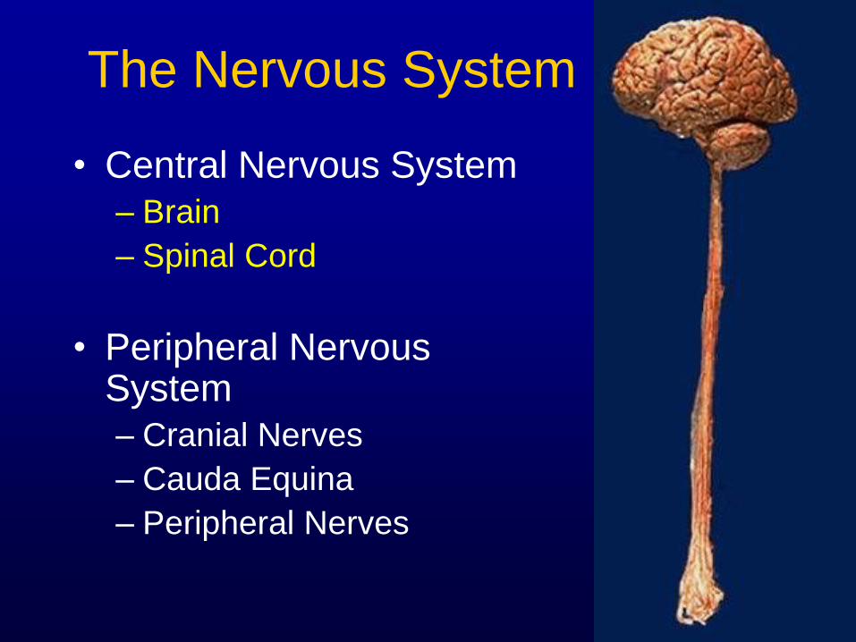
The Nervous System
• Central Nervous System
– Brain
– Spinal Cord
• Peripheral Nervous System
– Cranial Nerves
– Cauda Equina
– Peripheral Nerves
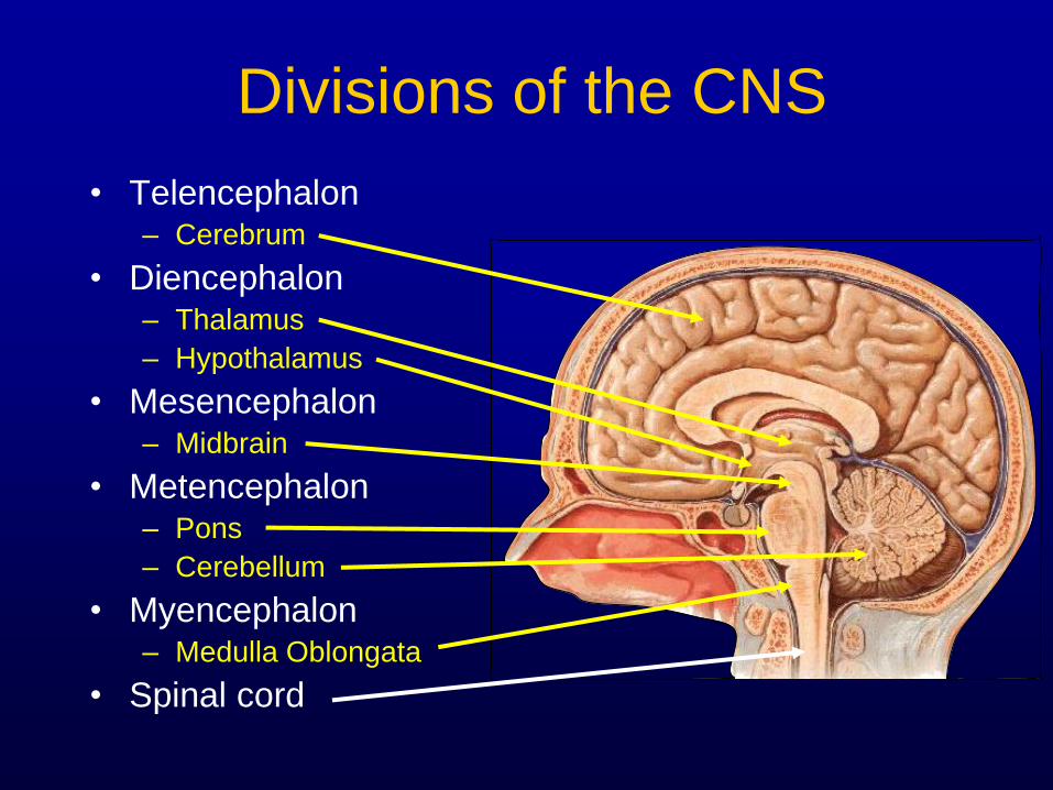
Divisions of the CNS
• Telencephalon – Cerebrum
• Diencephalon – Thalamus
– Hypothalamus
• Mesencephalon – Midbrain
• Metencephalon – Pons
– Cerebellum
• Myencephalon – Medulla Oblongata
• Spinal cord
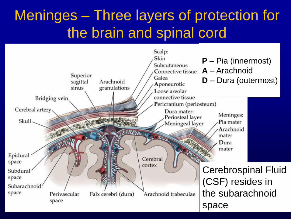
Meninges – Three layers of protection for
the brain and spinal cord
P – Pia (innermost)
A – Arachnoid
D – Dura (outermost)
Cerebrospinal Fluid
(CSF) resides in
the subarachnoid
space
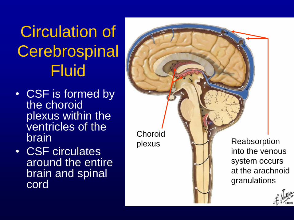
Circulation of
Cerebrospinal
Fluid
• CSF is formed by the choroid plexus within the ventricles of the brain
• CSF circulates around the entire brain and spinal cord
Reabsorption
into the venous
system occurs
at the arachnoid
granulations
Choroid
plexus
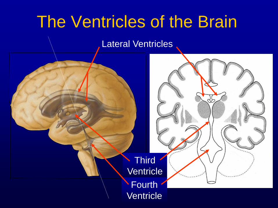
The Ventricles of the Brain Lateral Ventricles
Third
Ventricle
Fourth
Ventricle
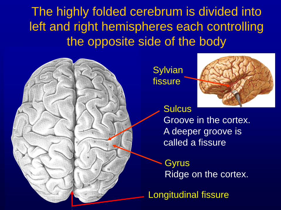
The highly folded cerebrum is divided into
left and right hemispheres each controlling
the opposite side of the body
Sulcus
Groove in the cortex.
A deeper groove is
called a fissure
Gyrus
Ridge on the cortex.
Longitudinal fissure
Sylvian
fissure
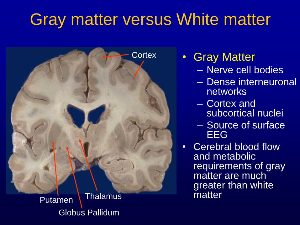
Gray matter versus White matter
• Gray Matter – Nerve cell bodies
– Dense interneuronal networks
– Cortex and subcortical nuclei
– Source of surface EEG
• Cerebral blood flow and metabolic requirements of gray matter are much greater than white matter
Cortex
Putamen Thalamus
Globus Pallidum
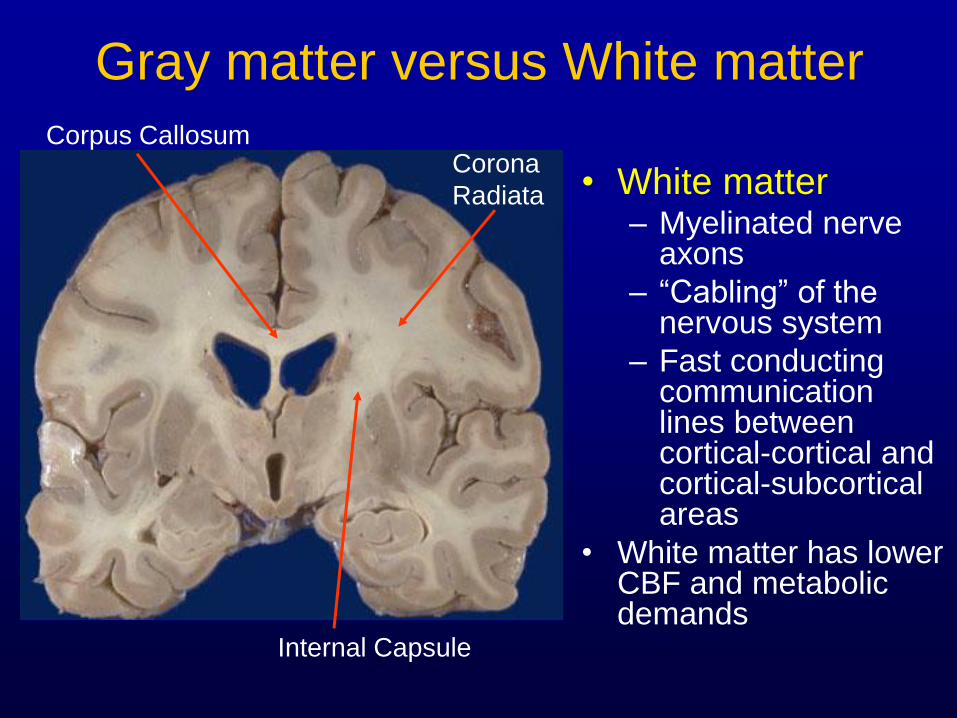
Gray matter versus White matter
• White matter – Myelinated nerve
axons
– “Cabling” of the nervous system
– Fast conducting communication lines between cortical-cortical and cortical-subcortical areas
• White matter has lower CBF and metabolic demands
Corpus Callosum
Internal Capsule
Corona
Radiata
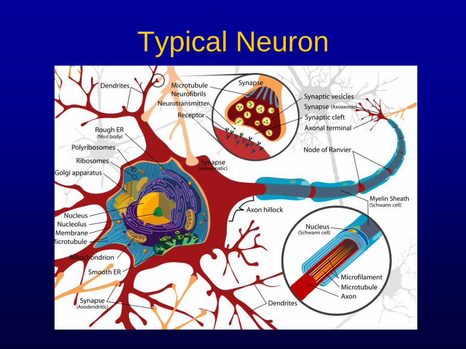
Typical Neuron
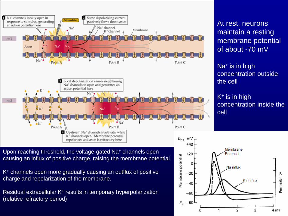
Upon reaching threshold, the voltage-gated Na+ channels open
causing an influx of positive charge, raising the membrane potential.
K+ channels open more gradually causing an outflux of positive
charge and repolarization of the membrane.
Residual extracellular K+ results in temporary hyperpolarization
(relative refractory period)
At rest, neurons
maintain a resting
membrane potential
of about -70 mV
Na+ is in high
concentration outside
the cell
K+ is in high
concentration inside the
cell
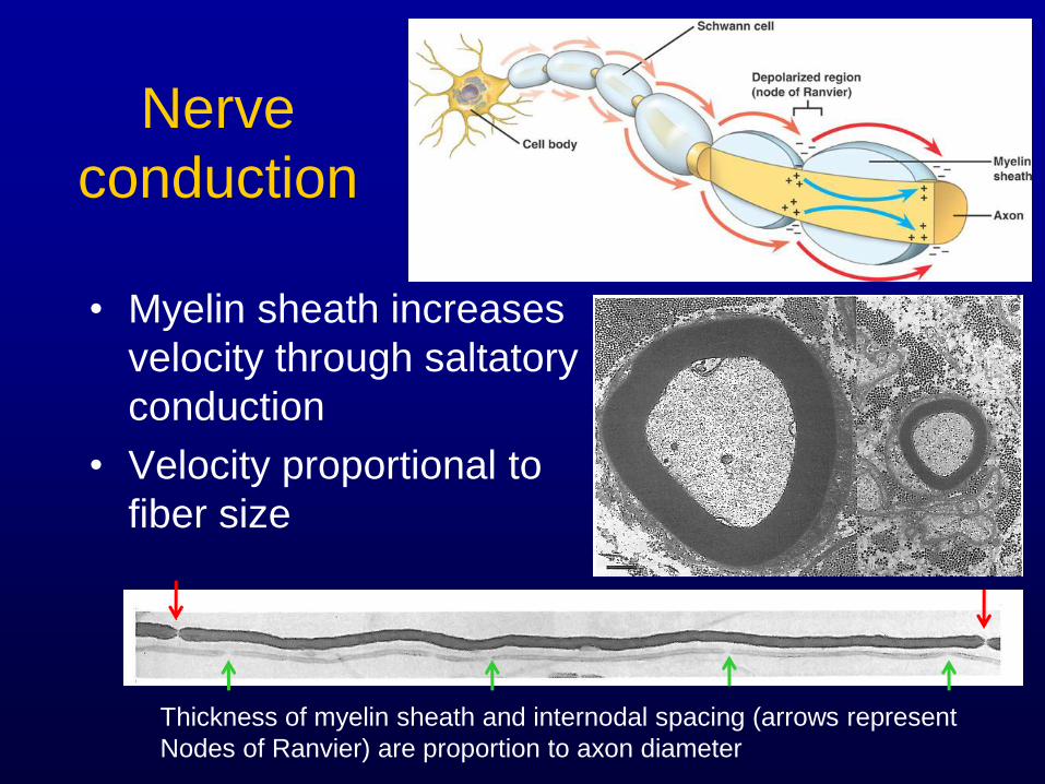
Nerve
conduction
• Myelin sheath increases
velocity through saltatory
conduction
• Velocity proportional to
fiber size
Thickness of myelin sheath and internodal spacing (arrows represent
Nodes of Ranvier) are proportion to axon diameter
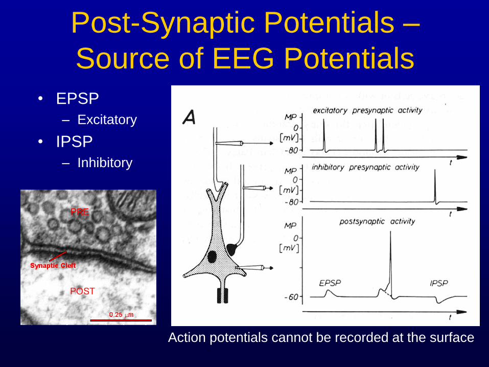
Post-Synaptic Potentials –
Source of EEG Potentials
• EPSP
– Excitatory
• IPSP
– Inhibitory
Action potentials cannot be recorded at the surface
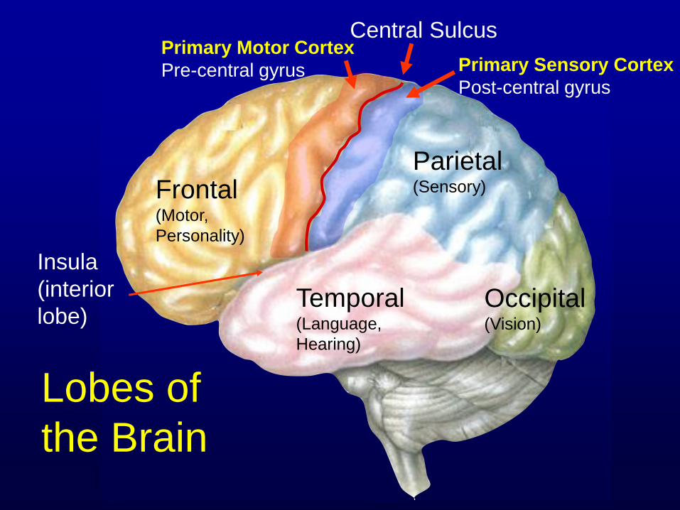
Frontal (Motor,
Personality)
Temporal (Language,
Hearing)
Parietal (Sensory)
Occipital (Vision)
Lobes of
the Brain
Central Sulcus
Insula
(interior
lobe)
Primary Motor Cortex
Pre-central gyrus Primary Sensory Cortex
Post-central gyrus
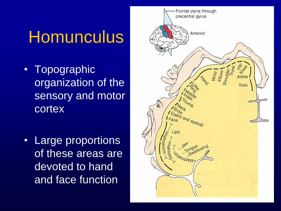
Homunculus
• Topographic
organization of the
sensory and motor
cortex
• Large proportions
of these areas are
devoted to hand
and face function
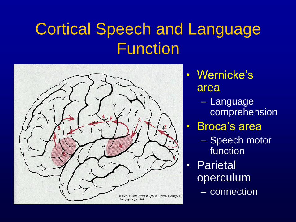
Cortical Speech and Language
Function
• Wernicke’s area – Language
comprehension
• Broca’s area – Speech motor
function
• Parietal operculum – connection
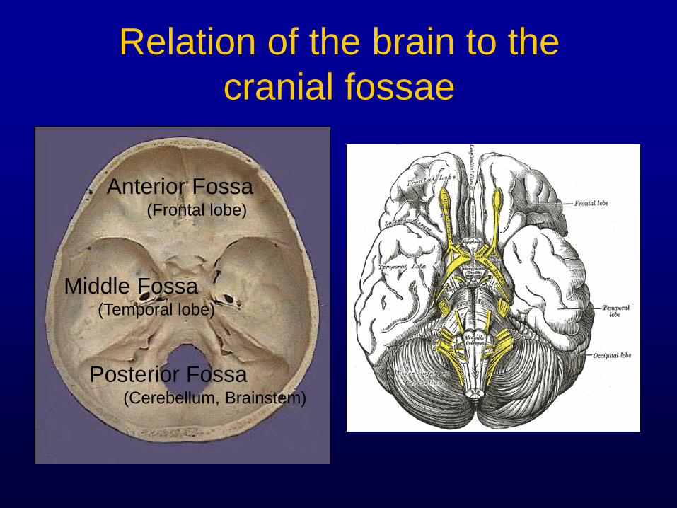
Relation of the brain to the
cranial fossae
Anterior Fossa (Frontal lobe)
Middle Fossa (Temporal lobe)
Posterior Fossa (Cerebellum, Brainstem)
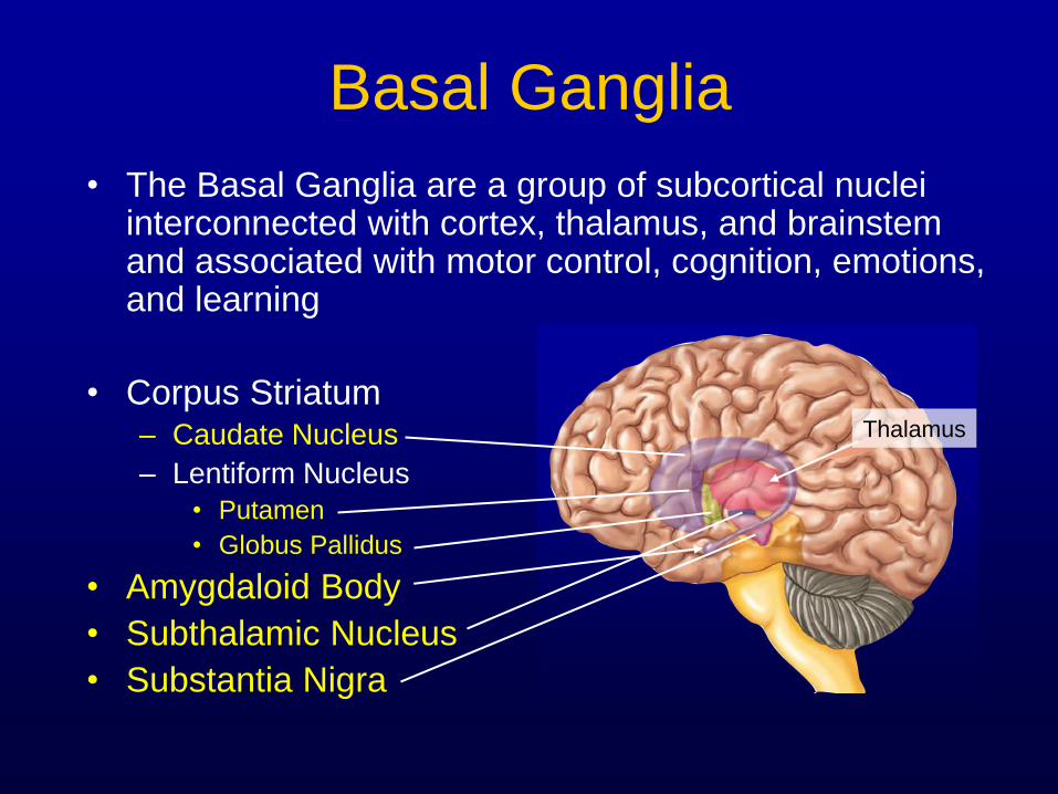
Basal Ganglia
• The Basal Ganglia are a group of subcortical nuclei interconnected with cortex, thalamus, and brainstem and associated with motor control, cognition, emotions, and learning
• Corpus Striatum – Caudate Nucleus
– Lentiform Nucleus
• Putamen
• Globus Pallidus
• Amygdaloid Body
• Subthalamic Nucleus
• Substantia Nigra
Thalamus
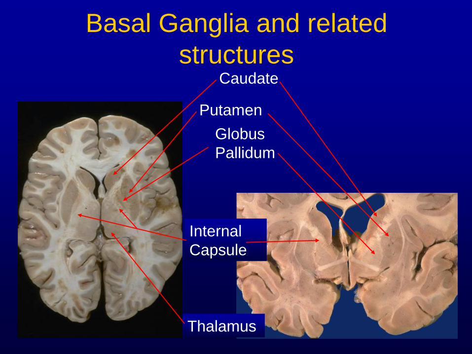
Basal Ganglia and related
structures Caudate
Putamen
Globus
Pallidum
Internal
Capsule
Thalamus
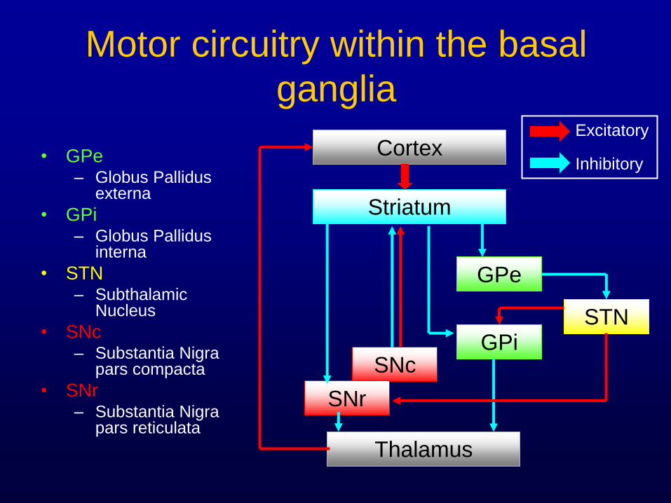
Motor circuitry within the basal
ganglia
• GPe – Globus Pallidus
externa
• GPi – Globus Pallidus
interna
• STN – Subthalamic
Nucleus
• SNc – Substantia Nigra
pars compacta
• SNr – Substantia Nigra
pars reticulata
Cortex
Striatum
GPe
GPi STN
SNc
SNr
Thalamus
Excitatory
Inhibitory
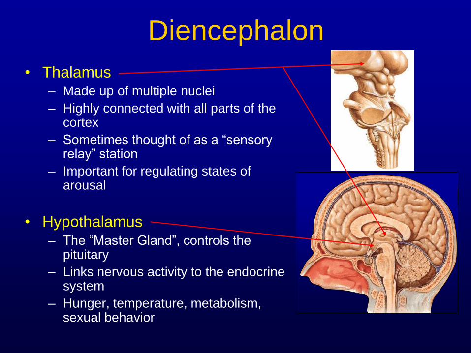
Diencephalon
• Thalamus – Made up of multiple nuclei
– Highly connected with all parts of the cortex
– Sometimes thought of as a “sensory relay” station
– Important for regulating states of arousal
• Hypothalamus – The “Master Gland”, controls the
pituitary
– Links nervous activity to the endocrine system
– Hunger, temperature, metabolism, sexual behavior
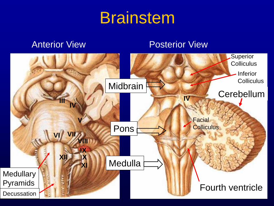
Brainstem
Midbrain
Pons
Medulla
Fourth ventricle
III IV
V
VI VII VIII
IX X
XI
XII
Cerebellum
Anterior View Posterior View
Medullary
Pyramids
Decussation
IV
Superior
Colliculus
Inferior
Colliculus
Facial
Colliculus
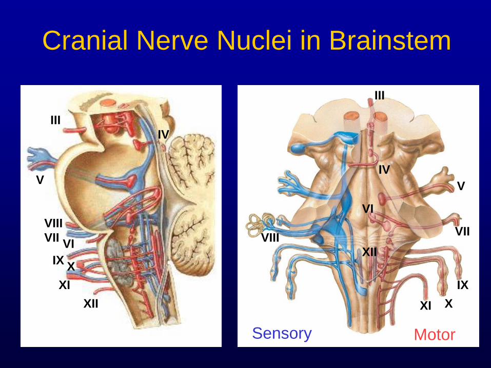
Cranial Nerve Nuclei in Brainstem
Sensory Motor
III
III
IV
IV V
V
VI
VI
VII VII
VIII
VIII
IX
IX
X
X
XI
XI XII
XII
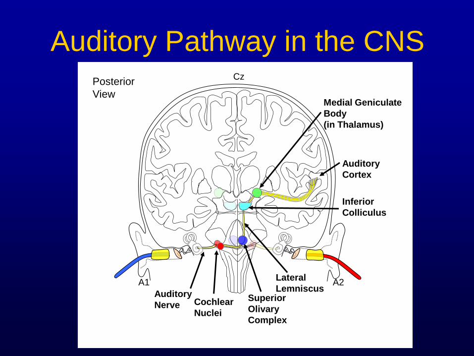
A1 A2
Cz
Lateral
Lemniscus
Inferior
Colliculus
Medial Geniculate
Body
(in Thalamus)
Auditory
Cortex
Posterior
View
Cochlear
Nuclei
Superior
Olivary
Complex
Auditory
Nerve
Auditory Pathway in the CNS
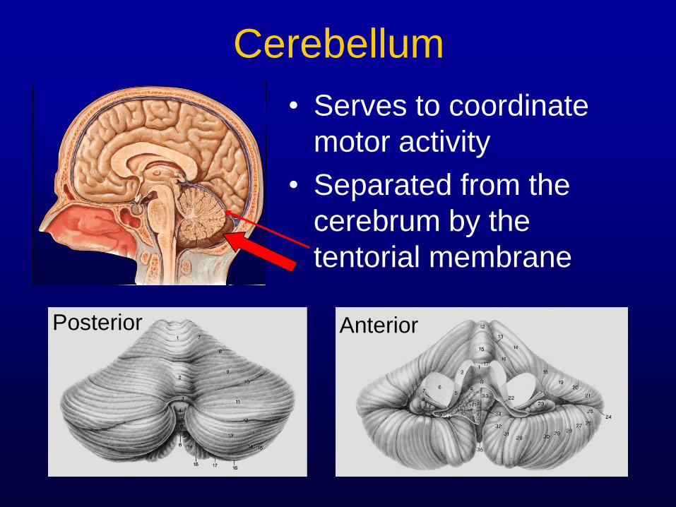
Cerebellum
• Serves to coordinate
motor activity
• Separated from the
cerebrum by the
tentorial membrane
Posterior Anterior
Neurovascular Supply
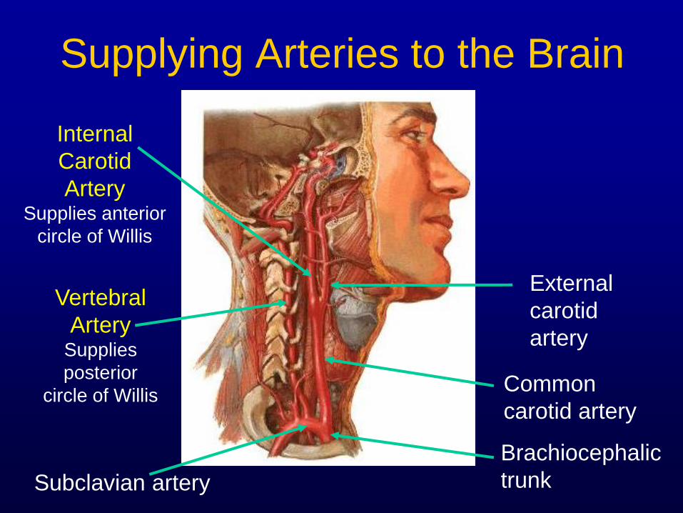
Supplying Arteries to the Brain
Common
carotid artery
Subclavian artery
Brachiocephalic
trunk
Internal
Carotid
Artery Supplies anterior
circle of Willis
External
carotid
artery
Vertebral
Artery Supplies
posterior
circle of Willis
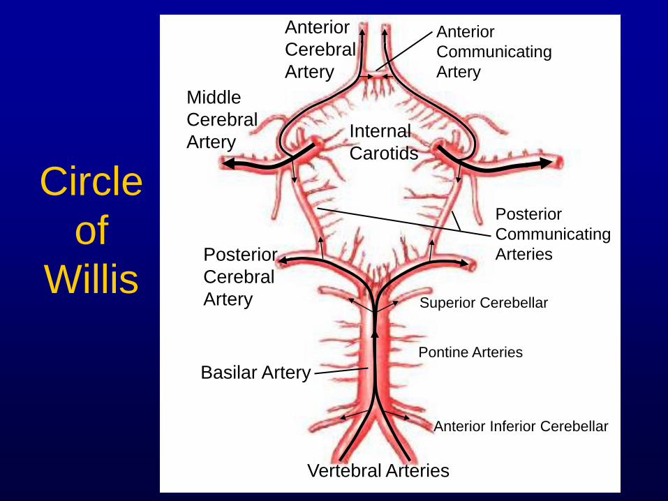
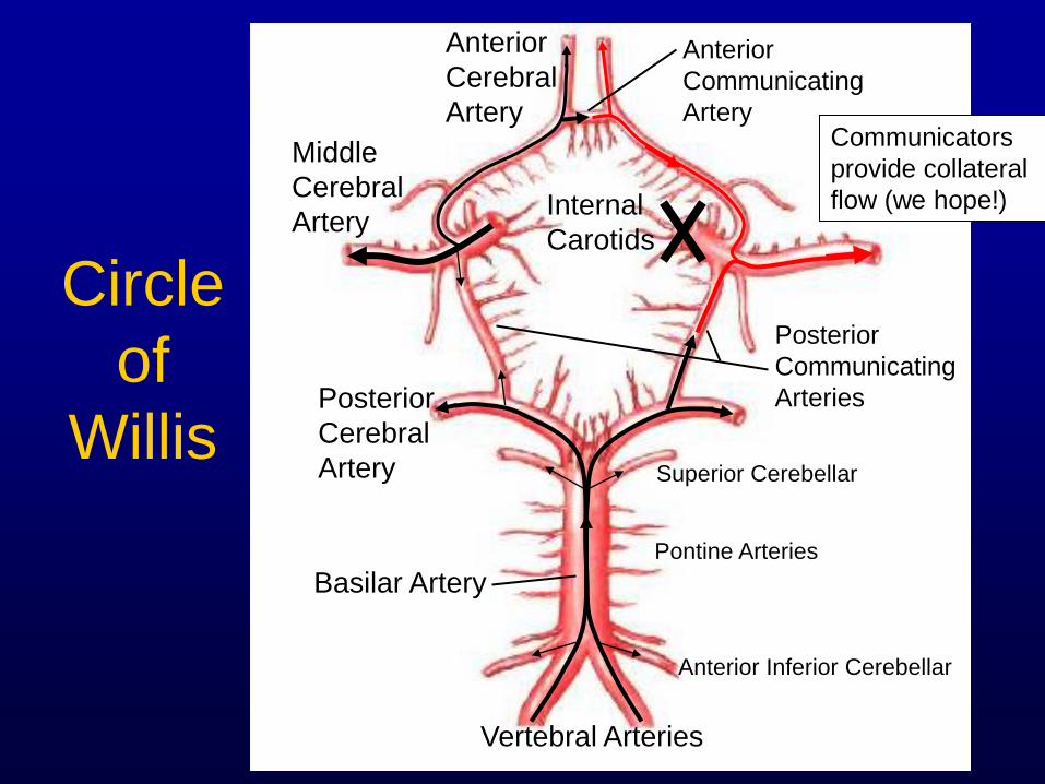
Circle
of
Willis
Internal
Carotids
Middle
Cerebral
Artery
Anterior
Cerebral
Artery
Anterior
Communicating
Artery
Posterior
Cerebral
Artery
Vertebral Arteries
Posterior
Communicating
Arteries
Basilar Artery
Superior Cerebellar
Pontine Arteries
Anterior Inferior Cerebellar
Circle
of
Willis
Internal
Carotids
Middle
Cerebral
Artery
Anterior
Cerebral
Artery
Anterior
Communicating
Artery
Posterior
Cerebral
Artery
Vertebral Arteries
Posterior
Communicating
Arteries
Basilar Artery
Superior Cerebellar
Pontine Arteries
Anterior Inferior Cerebellar
Communicators
provide collateral
flow (we hope!)
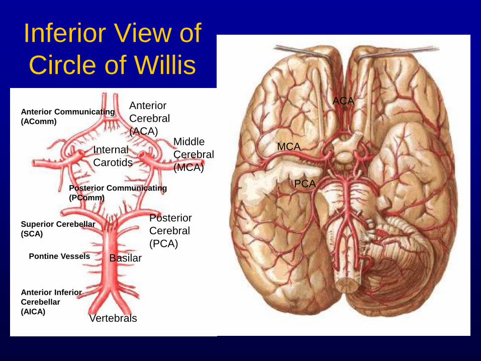
Inferior View of
Circle of Willis
Vertebrals
Basilar
Internal
Carotids
Posterior
Cerebral
(PCA)
Middle
Cerebral
(MCA)
Anterior
Cerebral
(ACA)
Posterior Communicating
(PComm)
Anterior Communicating
(AComm)
Superior Cerebellar
(SCA)
Anterior Inferior
Cerebellar
(AICA)
Pontine Vessels
MCA
ACA
PCA
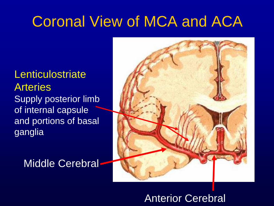
Coronal View of MCA and ACA
Anterior Cerebral
Middle Cerebral
Lenticulostriate
Arteries Supply posterior limb
of internal capsule
and portions of basal
ganglia
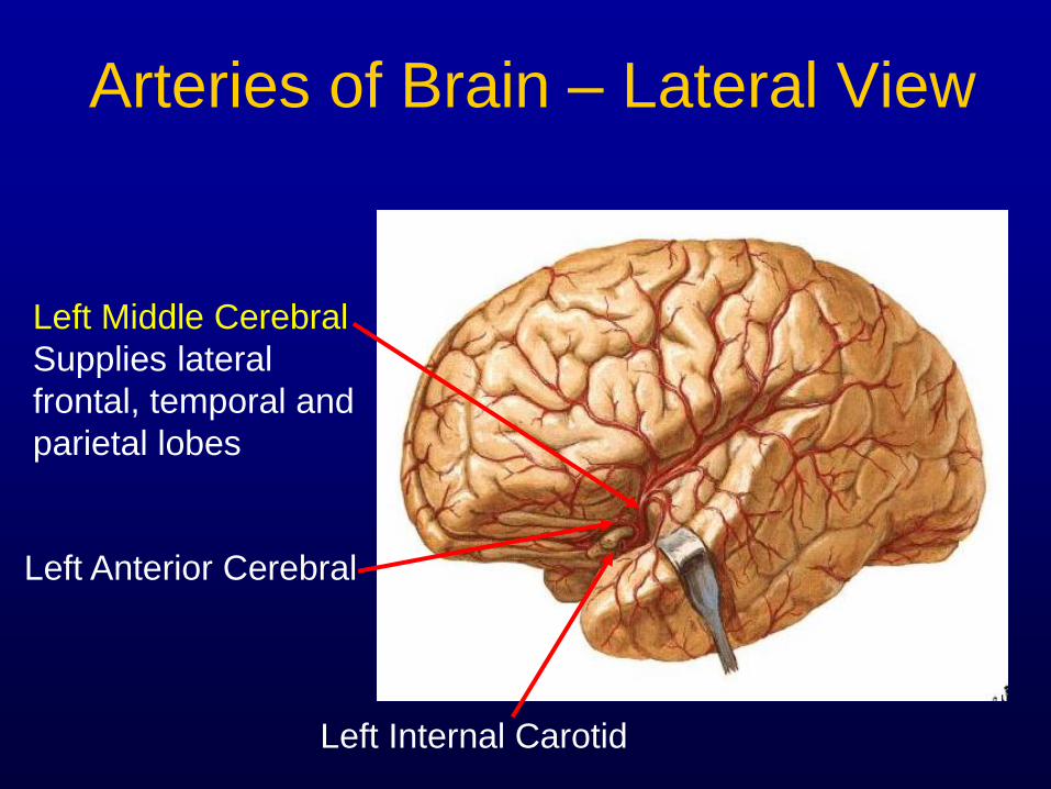
Arteries of Brain – Lateral View
Left Middle Cerebral
Supplies lateral
frontal, temporal and
parietal lobes
Left Anterior Cerebral
Left Internal Carotid
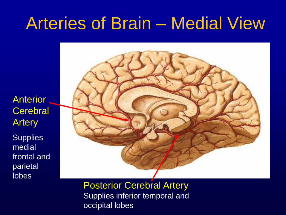
Arteries of Brain – Medial View
Posterior Cerebral Artery Supplies inferior temporal and
occipital lobes
Anterior
Cerebral
Artery
Supplies
medial
frontal and
parietal
lobes
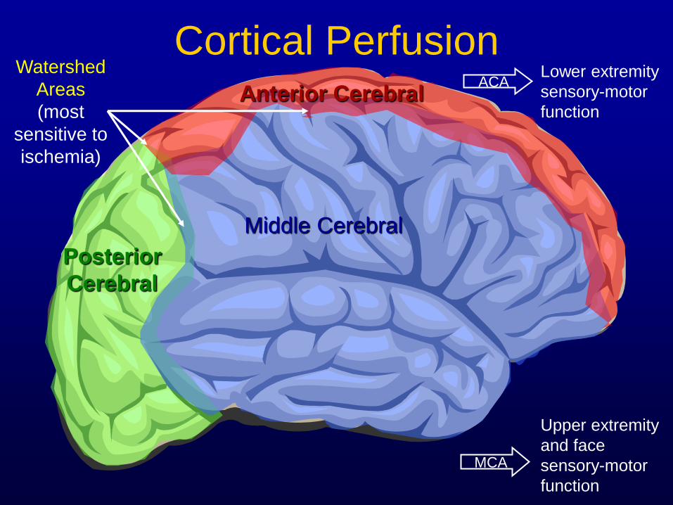
Cortical Perfusion
Middle Cerebral
Anterior Cerebral
Posterior
Cerebral
Watershed
Areas
(most
sensitive to
ischemia)
Lower extremity
sensory-motor
function
ACA
Upper extremity
and face
sensory-motor
function
MCA
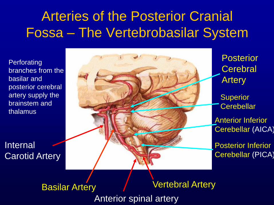
Arteries of the Posterior Cranial
Fossa – The Vertebrobasilar System
Internal
Carotid Artery
Basilar Artery
Anterior spinal artery
Vertebral Artery
Posterior
Cerebral
Artery
Superior
Cerebellar
Anterior Inferior
Cerebellar (AICA)
Posterior Inferior
Cerebellar (PICA)
Perforating
branches from the
basilar and
posterior cerebral
artery supply the
brainstem and
thalamus
Spinal Cord Anatomy
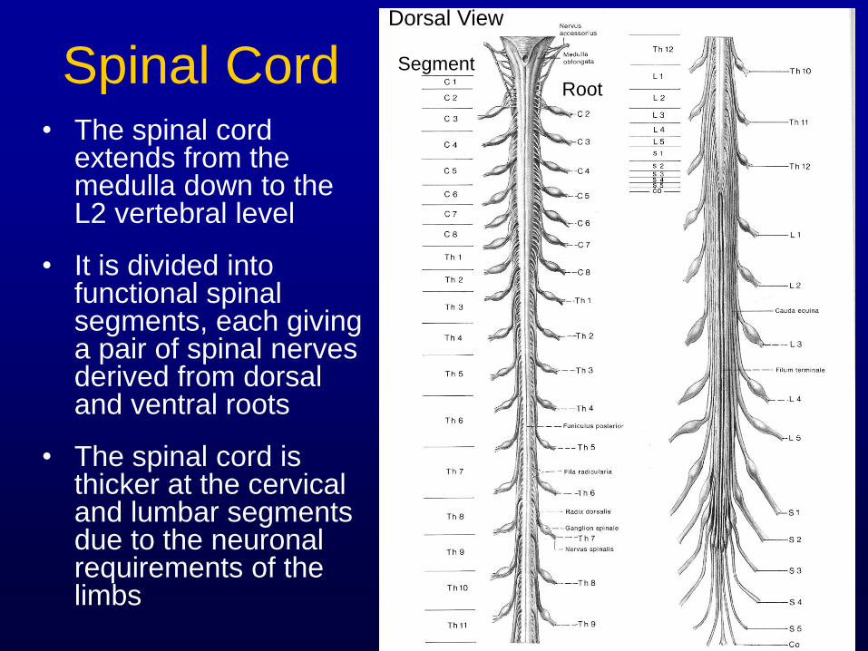
Spinal Cord • The spinal cord
extends from the medulla down to the L2 vertebral level
• It is divided into functional spinal segments, each giving a pair of spinal nerves derived from dorsal and ventral roots
• The spinal cord is thicker at the cervical and lumbar segments due to the neuronal requirements of the limbs
Dorsal View
Segment
Root
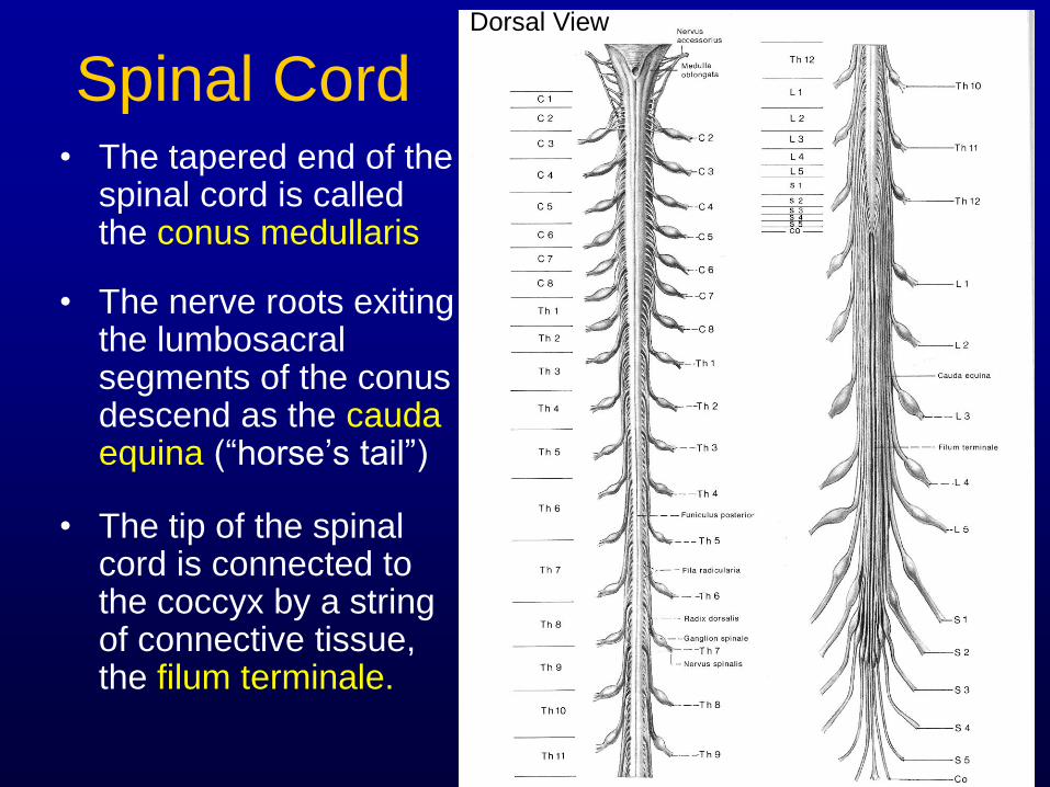
Spinal Cord • The tapered end of the
spinal cord is called the conus medullaris
• The nerve roots exiting the lumbosacral segments of the conus descend as the cauda equina (“horse’s tail”)
• The tip of the spinal cord is connected to the coccyx by a string of connective tissue, the filum terminale.
Dorsal View
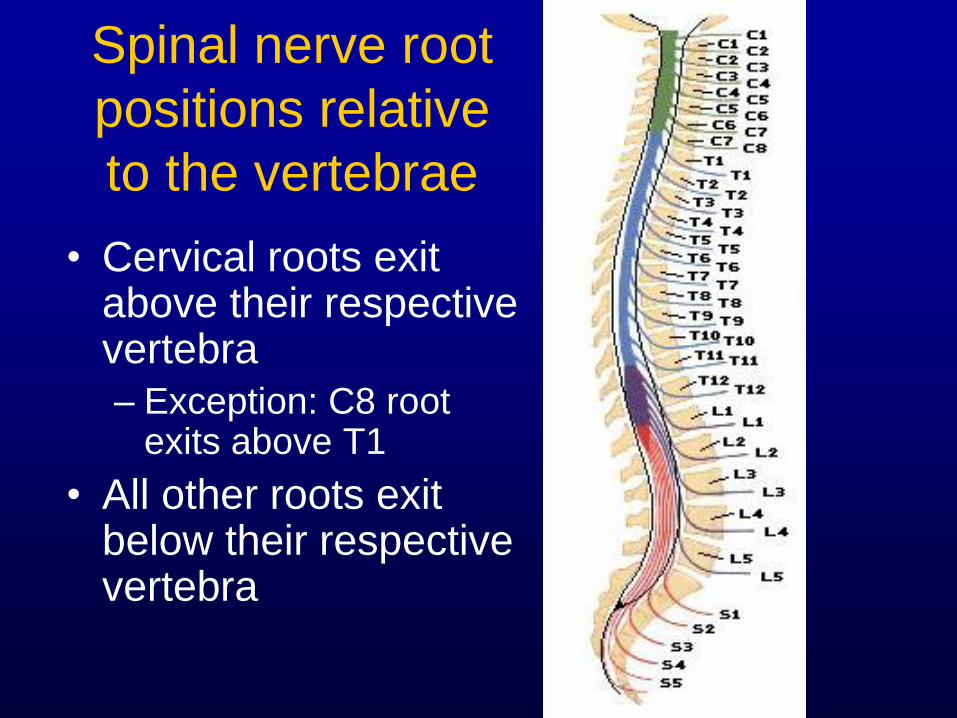
Spinal nerve root
positions relative
to the vertebrae
• Cervical roots exit above their respective vertebra
– Exception: C8 root exits above T1
• All other roots exit below their respective vertebra
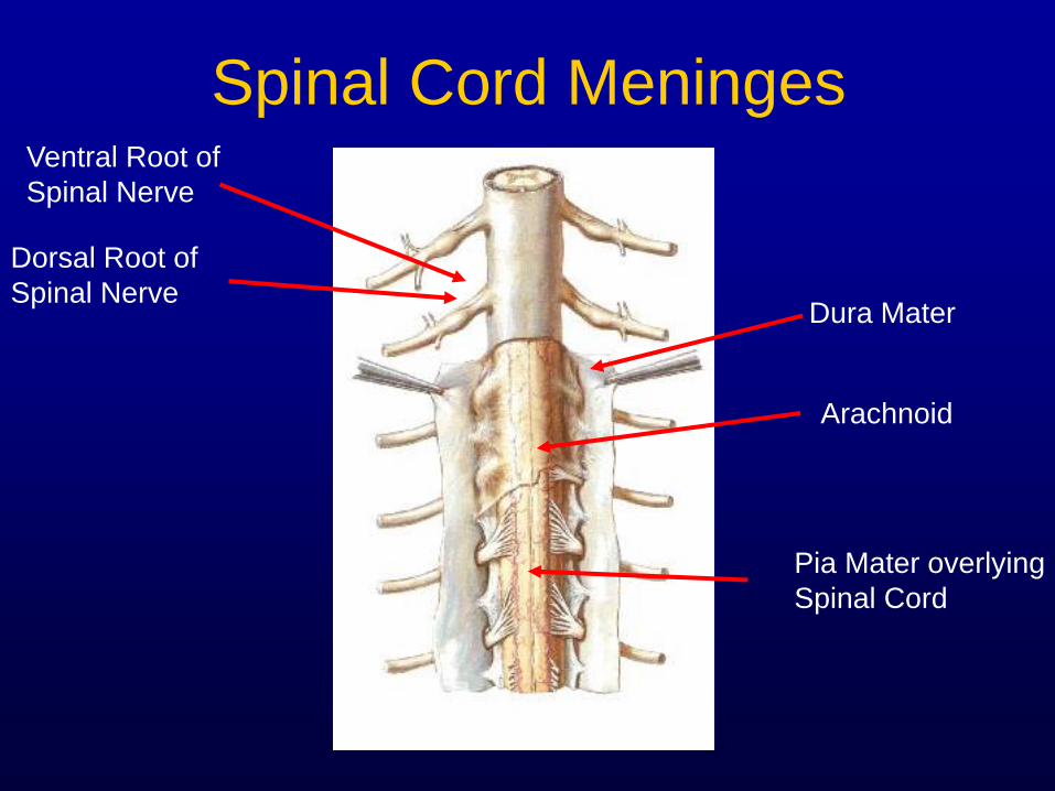
Spinal Cord Meninges
Arachnoid
Pia Mater overlying
Spinal Cord
Dura Mater
Ventral Root of
Spinal Nerve
Dorsal Root of
Spinal Nerve
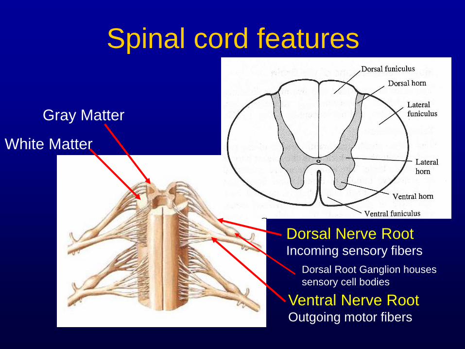
Spinal cord features
Gray Matter
White Matter
Ventral Nerve Root Outgoing motor fibers
Dorsal Nerve Root Incoming sensory fibers
Dorsal Root Ganglion houses
sensory cell bodies
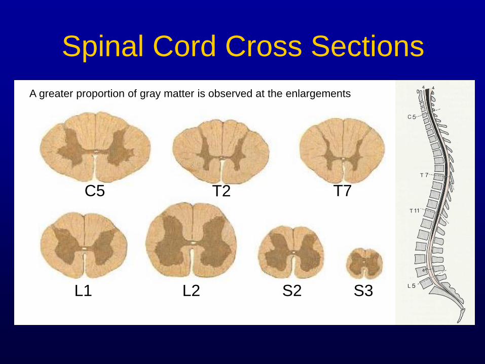
Spinal Cord Cross Sections
C5
L1 L2 S2 S3
T2 T7
A greater proportion of gray matter is observed at the enlargements
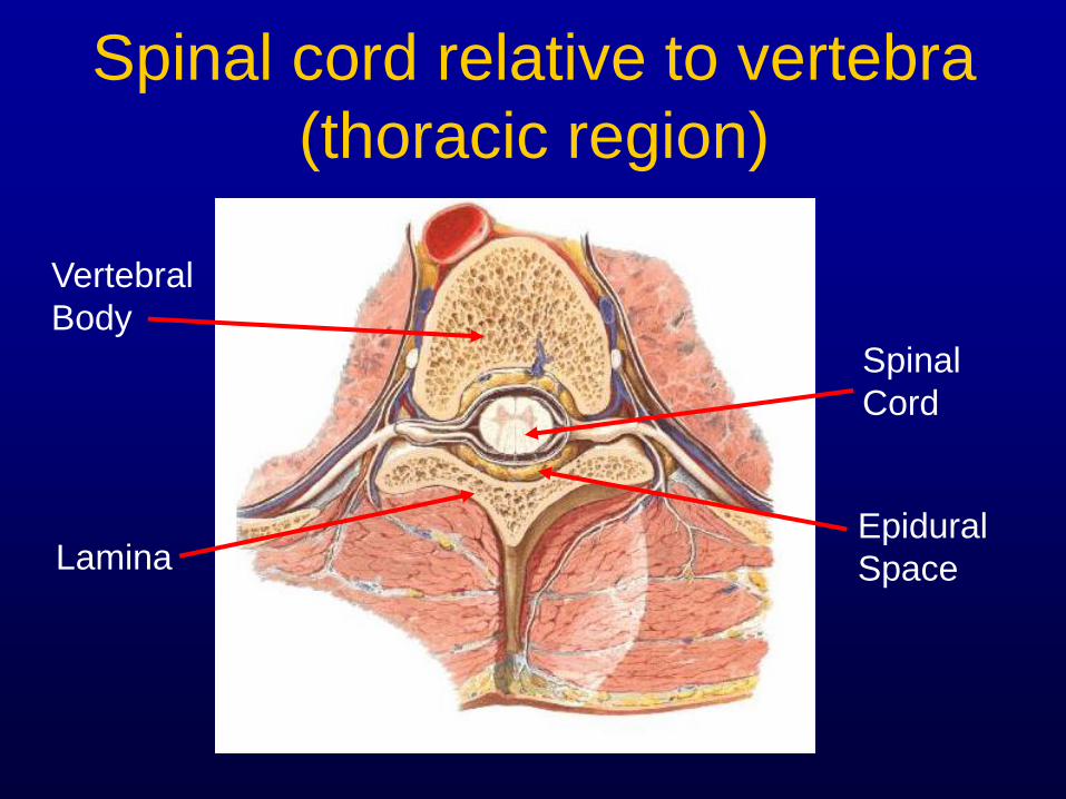
Spinal cord relative to vertebra
(thoracic region)
Vertebral
Body
Epidural
Space
Spinal
Cord
Lamina
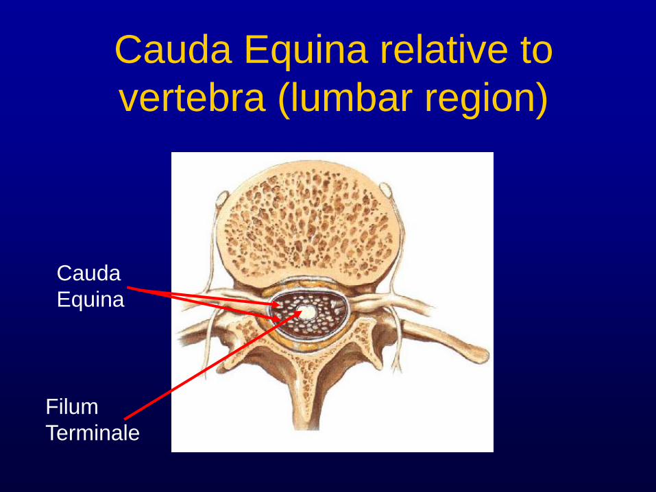
Cauda Equina relative to
vertebra (lumbar region)
Cauda
Equina
Filum
Terminale
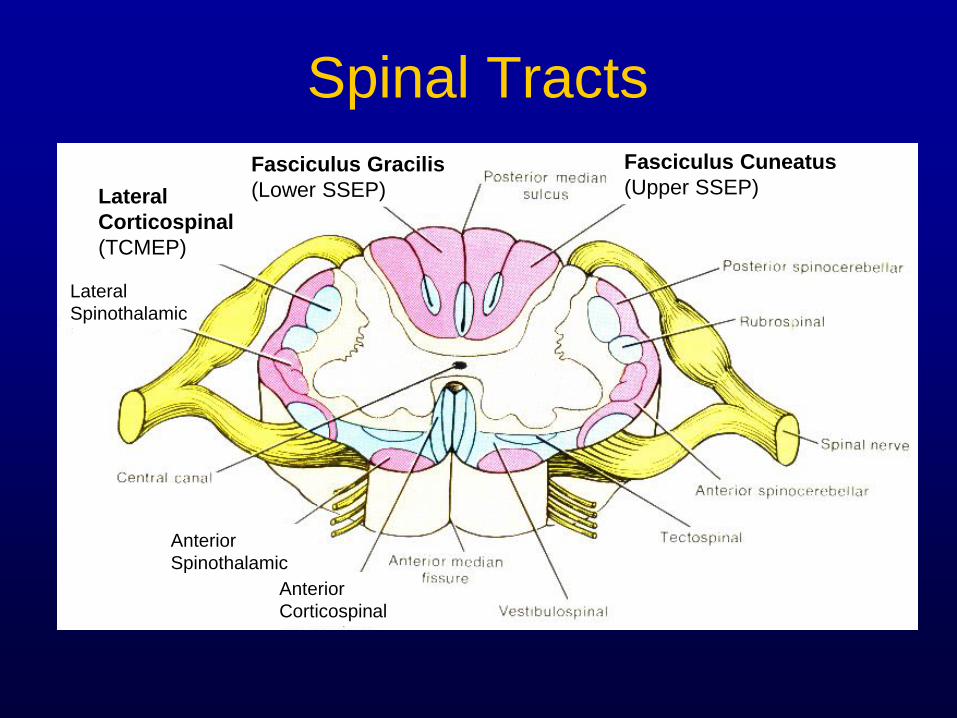
Spinal Tracts
Fasciculus Gracilis
(Lower SSEP)
Fasciculus Cuneatus
(Upper SSEP) Lateral
Corticospinal
(TCMEP)
Lateral
Spinothalamic
Anterior
Spinothalamic
Anterior
Corticospinal
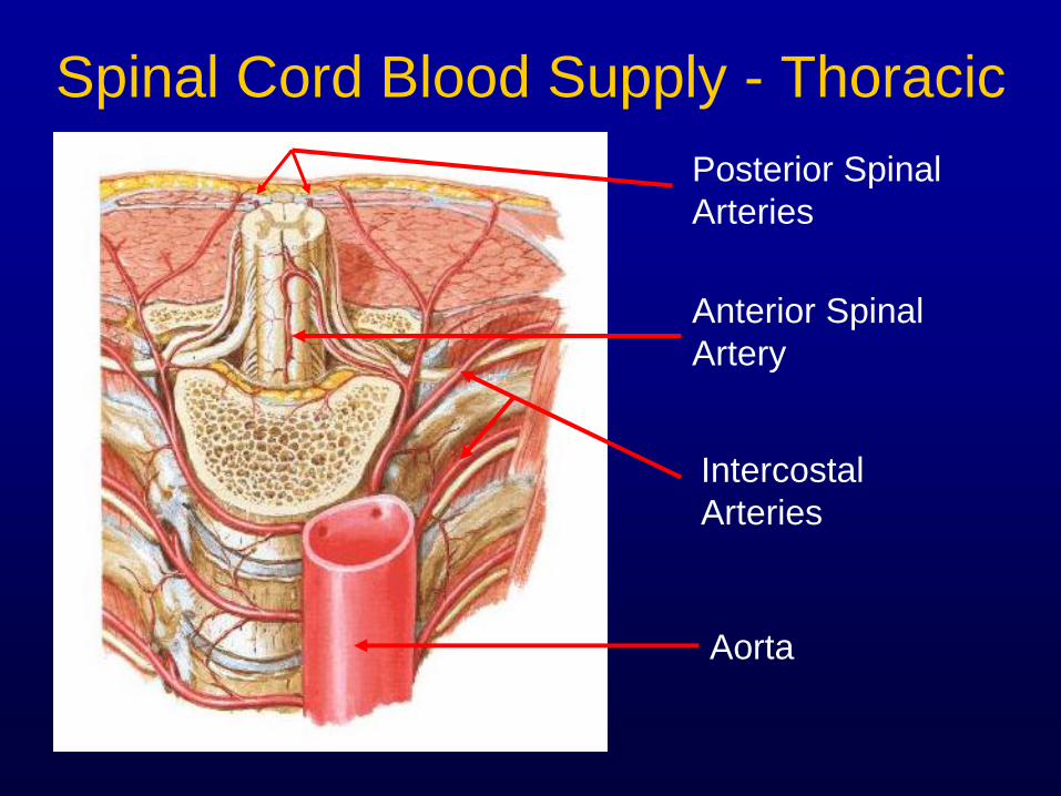
Spinal Cord Blood Supply - Thoracic
Aorta
Anterior Spinal
Artery
Posterior Spinal
Arteries
Intercostal
Arteries
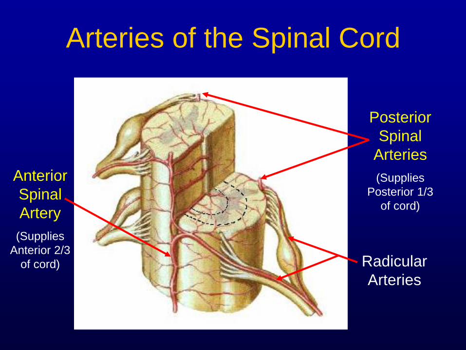
Arteries of the Spinal Cord
Anterior
Spinal
Artery
(Supplies
Anterior 2/3
of cord) Radicular
Arteries
Posterior
Spinal
Arteries
(Supplies
Posterior 1/3
of cord)
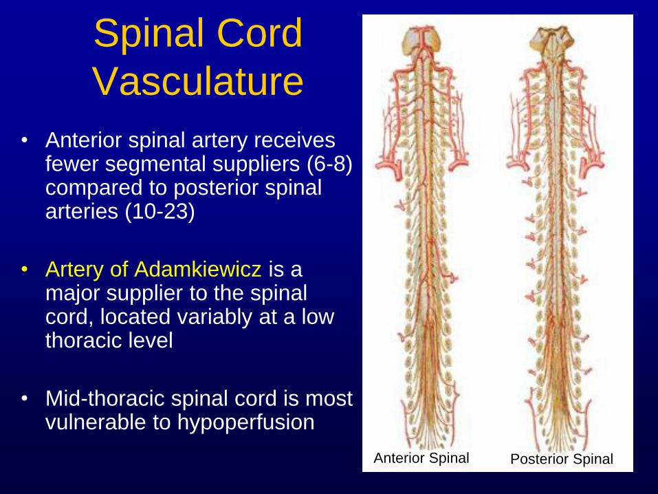
Spinal Cord
Vasculature
• Anterior spinal artery receives fewer segmental suppliers (6-8) compared to posterior spinal arteries (10-23)
• Artery of Adamkiewicz is a major supplier to the spinal cord, located variably at a low thoracic level
• Mid-thoracic spinal cord is most vulnerable to hypoperfusion
Anterior Spinal Posterior Spinal
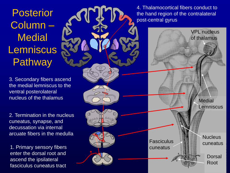
Posterior
Column –
Medial
Lemniscus
Pathway
1. Primary sensory fibers
enter the dorsal root and
ascend the ipsilateral
fasciculus cuneatus tract
2. Termination in the nucleus
cuneatus, synapse, and
decussation via internal
arcuate fibers in the medulla
3. Secondary fibers ascend
the medial lemniscus to the
ventral posterolateral
nucleus of the thalamus
4. Thalamocortical fibers conduct to
the hand region of the contralateral
post-central gyrus
Nucleus
cuneatus
Dorsal
Root
Medial
Lemniscus
VPL nucleus
of thalamus
Fasciculus
cuneatus
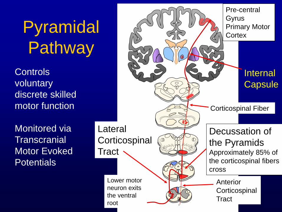
Pyramidal
Pathway
Internal
Capsule
Pre-central
Gyrus
Primary Motor
Cortex
Decussation of
the Pyramids Approximately 85% of
the corticospinal fibers
cross
Controls
voluntary
discrete skilled
motor function
Monitored via
Transcranial
Motor Evoked
Potentials
Lateral
Corticospinal
Tract
Anterior
Corticospinal
Tract
Lower motor
neuron exits
the ventral
root
Corticospinal Fiber

THANK YOU!!