Embed Size (px)
Citation preview
7/23/2019 benign tumor
http://slidepdf.com/reader/full/benign-tumor 1/4
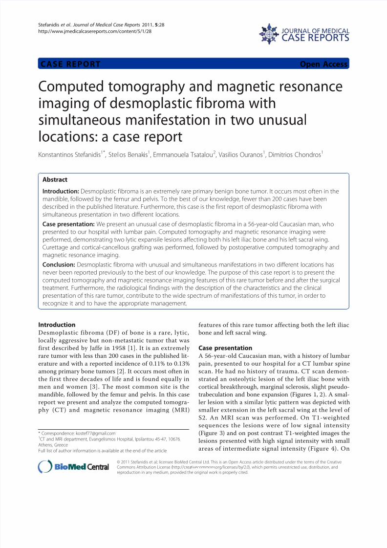
C A S E R E P O R T Open Access
Computed tomography and magnetic resonanceimaging of desmoplastic fibroma withsimultaneous manifestation in two unusuallocations: a case reportKonstantinos Stefanidis1*, Stelios Benakis1, Emmanouela Tsatalou2, Vasilios Ouranos1, Dimitrios Chondros1
Abstract
Introduction: Desmoplastic fibroma is an extremely rare primary benign bone tumor. It occurs most often in themandible, followed by the femur and pelvis. To the best of our knowledge, fewer than 200 cases have been
described in the published literature. Furthermore, this case is the first report of desmoplastic fibroma with
simultaneous presentation in two different locations.
Case presentation: We present an unusual case of desmoplastic fibroma in a 56-year-old Caucasian man, who
presented to our hospital with lumbar pain. Computed tomography and magnetic resonance imaging were
performed, demonstrating two lytic expansile lesions affecting both his left iliac bone and his left sacral wing.
Curettage and cortical-cancellous grafting was performed, followed by postoperative computed tomography and
magnetic resonance imaging.
Conclusion: Desmoplastic fibroma with unusual and simultaneous manifestations in two different locations has
never been reported previously to the best of our knowledge. The purpose of this case report is to present the
computed tomography and magnetic resonance imaging features of this rare tumor before and after the surgical
treatment. Furthermore, the radiological findings with the description of the characteristics and the clinicalpresentation of this rare tumor, contribute to the wide spectrum of manifestations of this tumor, in order to
recognize it and to have the appropriate management.
Introduction
Desmoplastic fibroma (DF) of bone is a rare, lytic,
locally aggressive but non-metastatic tumor that was
first described by Jaffe in 1958 [1]. It is an extremely
rare tumor with less than 200 cases in the published lit-
erature and with a reported incidence of 0.11% to 0.13%
among primary bone tumors [2]. It occurs most often in
the first three decades of life and is found equally in
men and women [3]. The most common site is themandible, followed by the femur and pelvis. In this case
report we present and analyze the computed tomogra-
phy (CT) and magnetic resonance imaging (MRI)
features of this rare tumor affecting both the left iliac
bone and left sacral wing.
Case presentation
A 56-year-old Caucasian man, with a history of lumbar
pain, presented to our hospital for a CT lumbar spine
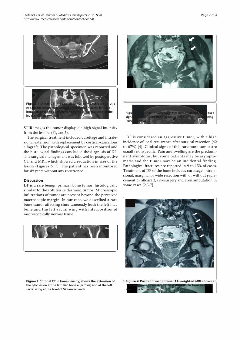
scan. He had no history of trauma. CT scan demon-
strated an osteolytic lesion of the left iliac bone with
cortical breakthrough, marginal sclerosis, slight pseudo-trabeculation and bone expansion (Figures 1, 2). A smal-
ler lesion with a similar lytic pattern was depicted with
smaller extension in the left sacral wing at the level of
S2. An MRI scan was performed. On T1-weighted
sequences the lesions were of low signal intensity
(Figure 3) and on post contrast T1-weighted images the
lesions presented with high signal intensity with small
areas of intermediate signal intensity (Figure 4). On
* Correspondence: [email protected] and MRI department, Evangelismos Hospital, Ipsilantou 45-47, 10676,
Athens, Greece
Full list of author information is available at the end of the article
Stefanidis et al . Journal of Medical Case Reports 2011, 5 :28
http://www.jmedicalcasereports.com/content/5/1/28 JOURNAL OF MEDICALCASE REPORTS
© 2011 Stefanidis et al; licensee BioMed Central Ltd. This is an Open Access article distributed under the terms of the CreativeCommons Attribution License (http://creativecommons.org/licenses/by/2.0), which permits unrestricted use, distribution, andreproduction in any medium, provided the original work is properly cited.
7/23/2019 benign tumor
http://slidepdf.com/reader/full/benign-tumor 2/4
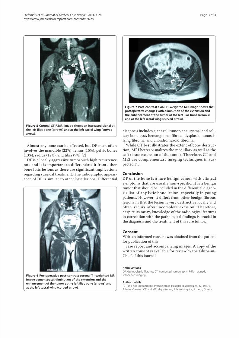
STIR images the tumor displayed a high signal intensity
from the lesions (Figure 5).
The surgical treatment included curettage and intrale-
sional extension with replacement by cortical-cancellous
allograft. The pathological specimen was reported and
the histological findings concluded the diagnosis of DF.
The surgical management was followed by postoperative
CT and MRI, which showed a reduction in size of the
lesion (Figures 6, 7). The patient has been monitored
for six years without any recurrence.
Discussion
DF is a rare benign primary bone tumor, histologically
similar to the soft tissue desmoid tumor. Microscopic
infiltrations of tumor are present beyond the perceived
macroscopic margin. In our case, we described a rare
bone tumor affecting simultaneously both the left iliac
bone and the left sacral wing with interposition of
macroscopically normal tissue.
DF is considered an aggressive tumor, with a high
incidence of local recurrence after surgical resection (42
to 67%) [4]. Clinical signs of this rare bone tumor are
usually nonspecific. Pain and swelling are the predomi-
nant symptoms, but some patients may be asympto-
matic and the tumor may be an incidental finding.
Pathological fractures are reported in 9 to 15% of cases.
Treatment of DF of the bone includes curettage, intrale-
sional, marginal or wide resection with or without repla-
cement by allograft, cryosurgery and even amputation insome cases [2,5-7].
Figure 1 Axial CT in bone density, demonstrates a lytic
expansible lesion with cortical breakthrough at the left iliac
bone (arrows) and a smaller one at the left sacral wing at the
level of S2 (arrowhead).
Figure 2 Coronal CT in bone density, shows the extension of
the lytic lesion at the left iliac bone e (arrows) and at the left
sacral wing at the level of S2 (arrowhead) .
Figure 3 Coronal T1-weighted MRI demonstrates decreased
signal at the left iliac bone (arrows) and at the left sacral wing
(curved arrow).
Figure 4 Post contrast coronal T1-weighted MRI shows a
heterogeneous increased signal at the left iliac bone (arrows)
and at the left sacral wing .
Stefanidis et al . Journal of Medical Case Reports 2011, 5 :28
http://www.jmedicalcasereports.com/content/5/1/28
Page 2 of 4
7/23/2019 benign tumor
http://slidepdf.com/reader/full/benign-tumor 3/4
Almost any bone can be affected, but DF most often
involves the mandible (22%), femur (15%), pelvic bones
(13%), radius (12%), and tibia (9%) [2]
DF is a locally aggressive tumor with high recurrence
rate and it is important to differentiate it from other
bone lytic lesions as there are significant implications
regarding surgical treatment. The radiographic appear-
ance of DF is similar to other lytic lesions. Differential
diagnosis includes giant cell tumor, aneurysmal and soli-
tary bone cyst, hemangioma, fibrous dysplasia, nonossi-
fying fibroma, and chondromyxoid fibroma.
While CT best illustrates the extent of bone destruc-
tion, MRI better visualizes the medullary as well as the
soft tissue extension of the tumor. Therefore, CT and
MRI are complementary imaging techniques in sus-
pected DF.
Conclusion
DF of the bone is a rare benign tumor with clinicalsymptoms that are usually non-specific. It is a benign
tumor that should be included in the differential diagno-
sis list of any lytic bone lesion, especially in young
patients. However, it differs from other benign fibrous
lesions in that the lesion is very destructive locally and
often recurs after incomplete excision. Therefore,
despite its rarity, knowledge of the radiological features
in correlation with the pathological findings is crucial in
the diagnosis and the treatment of this rare tumor.
Consent
Written informed consent was obtained from the patientfor publication of this
case report and accompanying images. A copy of the
written consent is available for review by the Editor-in-
Chief of this journal.
Abbreviations
DF: desmoplastic fibroma; CT: computed tomography; MRI: magnetic
resonance imaging
Author details1CT and MRI department, Evangelismos Hospital, Ipsilantou 45-47, 10676,
Athens, Greece. 2CT and MRI department, 7thIKA Hospital, Athens, Greece.
Figure 5 Coronal STIR.MRI image shows an increased signal at
the left iliac bone (arrows) and at the left sacral wing (curved
arrow).
Figure 6 Postoperative post-contrast coronal T1-weighted MR
image demonstrates diminution of the extension and the
enhancement of the tumor at the left iliac bone (arrows) and
at the left sacral wing (curved arrow) .
Figure 7 Post-contrast axial T1-weighted MR image shows the
postoperative changes with diminution of the extension and
the enhancement of the tumor at the left iliac bone (arrows)
and at the left sacral wing (curved arrow) .
Stefanidis et al . Journal of Medical Case Reports 2011, 5 :28
http://www.jmedicalcasereports.com/content/5/1/28
Page 3 of 4
7/23/2019 benign tumor
http://slidepdf.com/reader/full/benign-tumor 4/4
Authors’ contributions
KS prepared the case report and reviewed the literature. SB, ET and DC
participated in the conception, design, and data collection and
interpretation, analyzed the article and made necessary corrections. Allauthors read and approved the final manuscript.
Competing interests The authors declare that they have no competing interests.
Received: 16 December 2009 Accepted: 24 January 2011
Published: 24 January 2011
References1. Jaffe HL: Tumors and tumorous conditions of the bones and joints
Philadelphia: Lea & Febiger; 1958, 298-303.
2. Bohm P, Krober S, Greschniok A, Laniado M, Kaiserling E: Desmoplasticfibroma of the bone. A report of two patients, review of the literature
and therapeutic implications. Cancer 1996, 78:1011-1023.
3. Crim JR, Gold RH, Mirra JM, Eckardt JJ, Bassett LW: Desmoplastic fibroma
of bone: radiographic analysis. Radiology 1989, 172:827-832.
4. Gebhardt MC, Campbell CJ, Schiller AL, Mankin HJ: Desmoplastic fibroma
of bone. J Bone Joint Surg 1985, 67A:732-747.
5. Rabin D, Ang LC, Megyesi J, Lee DH, Duggal N: Desmoplastic fibroma of
the cranium: case report and review of the literature. Neurosurgery 2003,
52:950-954.
6. Daneyemez M, Akay KM, Izci Y: Desmoplastic fibroma of the cervical
spine. Eur Spine J 2005, 14:799-802.7. Muramatsu K, Ihara K, Azuma E, Orui R, Goto Y, Shigetomi M, Doi K : Free
vascularized fibula grafting for reconstruction of the wrist following
wide tumor excision. Microsurgery 2005, 25 :101-106.
doi:10.1186/1752-1947-5-28Cite this article as: Stefanidis et al .: Computed tomography andmagnetic resonance imaging of desmoplastic fibroma with
simultaneous manifestation in two unusual locations: a case report. Journal of Medical Case Reports 2011 5 :28.
Submit your next manuscript to BioMed Centraland take full advantage of:
• Convenient online submission
• Thorough peer review
• No space constraints or color figure charges
• Immediate publication on acceptance
• Inclusion in PubMed, CAS, Scopus and Google Scholar
• Research which is freely available for redistribution
Submit your manuscript atwww.biomedcentral.com/submit
Stefanidis et al . Journal of Medical Case Reports 2011, 5 :28
http://www.jmedicalcasereports.com/content/5/1/28
Page 4 of 4