Embed Size (px)
Citation preview
Biliary Anatomy and Physiology
Dr Amir Ashrafi
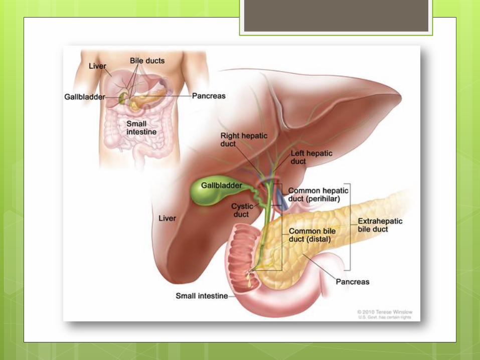
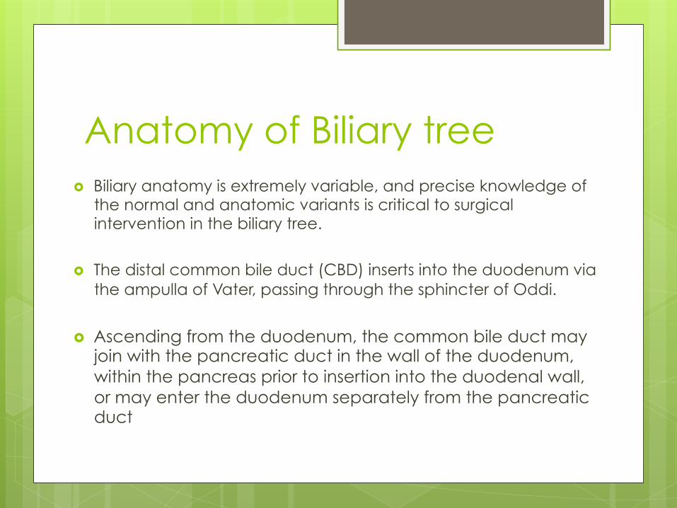
Anatomy of Biliary tree � Biliary anatomy is extremely variable, and precise knowledge of
the normal and anatomic variants is critical to surgical intervention in the biliary tree.
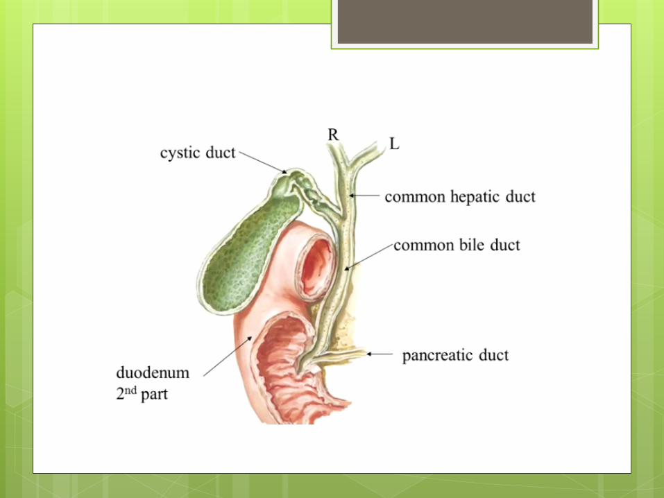
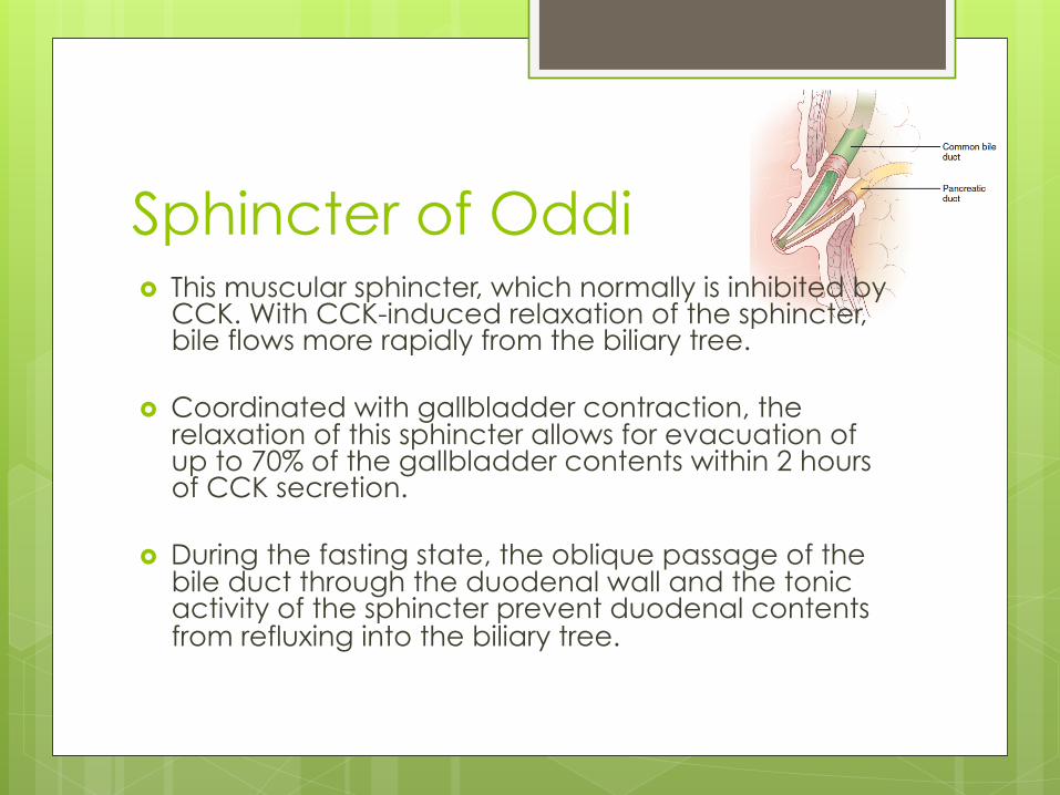
� The distal common bile duct (CBD) inserts into the duodenum via the ampulla of Vater, passing through the sphincter of Oddi.
� Ascending from the duodenum, the common bile duct may join with the pancreatic duct in the wall of the duodenum, within the pancreas prior to insertion into the duodenal wall, or may enter the duodenum separately from the pancreatic duct
� The most inferior portion of the CBD is encompassed by the head of the pancreas.
� Superior to this portion, the common bile duct is divided into retroduodenal and supraduodenal segments.
� The insertion of the cystic duct marks the differentiation of the common hepatic duct above and the common bile duct below.
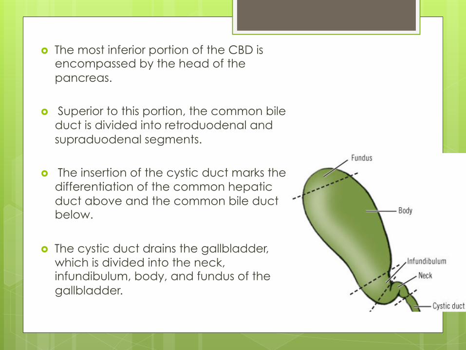
� The cystic duct drains the gallbladder,
which is divided into the neck, infundibulum, body, and fundus of the gallbladder.
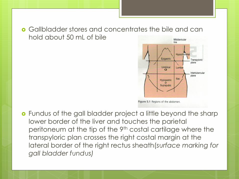
� Gallbladder stores and concentrates the bile and can hold about 50 mL of bile
� Fundus of the gall bladder project a little beyond the sharp lower border of the liver and touches the parietal peritoneum at the tip of the 9th costal cartilage where the transpyloric plan crosses the right costal margin at the lateral border of the right rectus sheath(surface marking for gall bladder fundus)
� gall bladder fossa roughly identifies the right and left lobe of the liver and is Where the gallbladder attaches to the liver, Glisson’s capsule does not form, and this common surface provides the venous and lymphatic drainage of the gallbladder.
� Also small bile ducts may pass from the liver to the gall bladder in this surface and cause bile leak post cholecystectomy if undetected.
� The cystic duct drains at an acute angle into the common bile duct and is normally 2-3cm in length and 2-3 mm in diameter. It runs backwards and and downwards and joins the Common hepatic duct
� Seeing a small diverticulum in the neck of the gall bladder (Hartman’s pouch) is could be sign of a pathological problem such as impacted stone. There is a significant association between the presence of Hartmann's pouch and stones (p < 0.05). Adhesions between the cystic duct and the neck of the gallbladder are responsible for Hartmann's pouch.
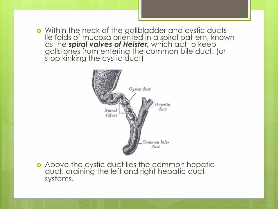
� Within the neck of the gallbladder and cystic ducts lie folds of mucosa oriented in a spiral pattern, known as the spiral valves of Heister, which act to keep gallstones from entering the common bile duct. (or stop kinking the cystic duct)
� Above the cystic duct lies the common hepatic duct, draining the left and right hepatic duct systems.
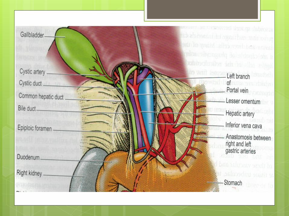
Vascular Anatomy � As opposed to the liver, where most perfusion
comes from portal venous flow, the entire biliary tree is supplied solely by the arterial anatomy.
� The inferior bile duct, below the level of the duodenal bulb, receives its perfusion from posterosuperior pancreaticoduodenal and gastroduodenal arteries
� The superior common bile duct, from the duodenal bulb to the cystic duct, and common hepatic ducts receive their blood supply from the right hepatic and cystic arteries.
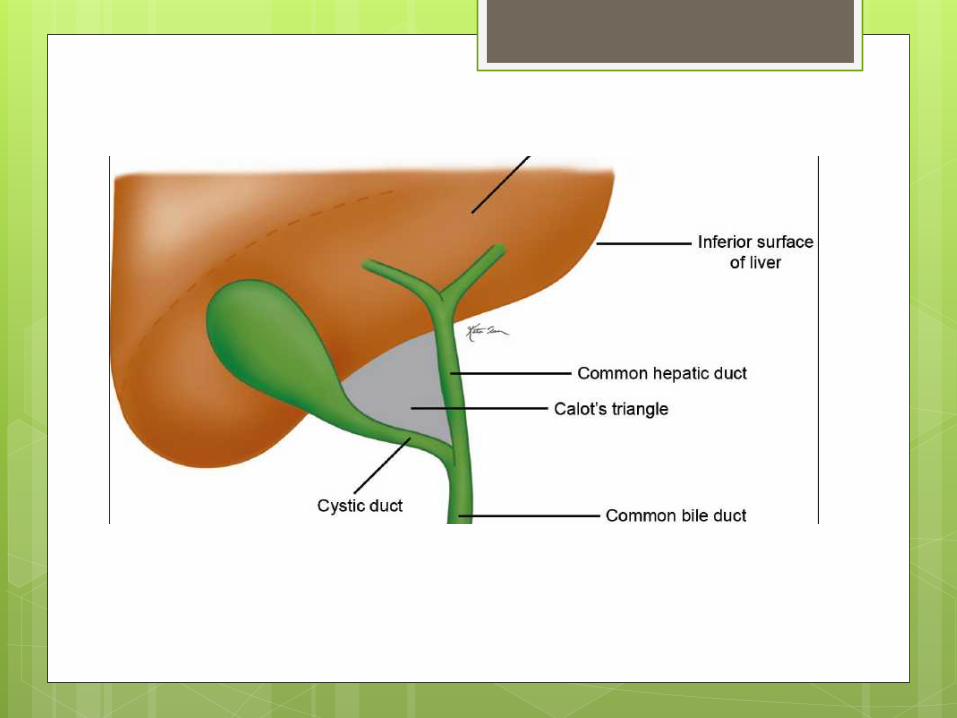
� In most cases, the right hepatic artery passes posterior to the common hepatic duct to supply the right lobe of the liver.
� After crossing the duct, the right hepatic artery passes
through the triangle of Calot bordered by the cystic duct, common hepatic duct and edge of liver.
� In this triangle, the right hepatic artery gives off the cystic artery to the gallbladder and is at risk for injury during a cholecystectomy.

� Both within the liver and immediately outside the parenchyma, the bile ducts generally lie superior to the corresponding portal veins, which in turn are superior to the arterial supply.
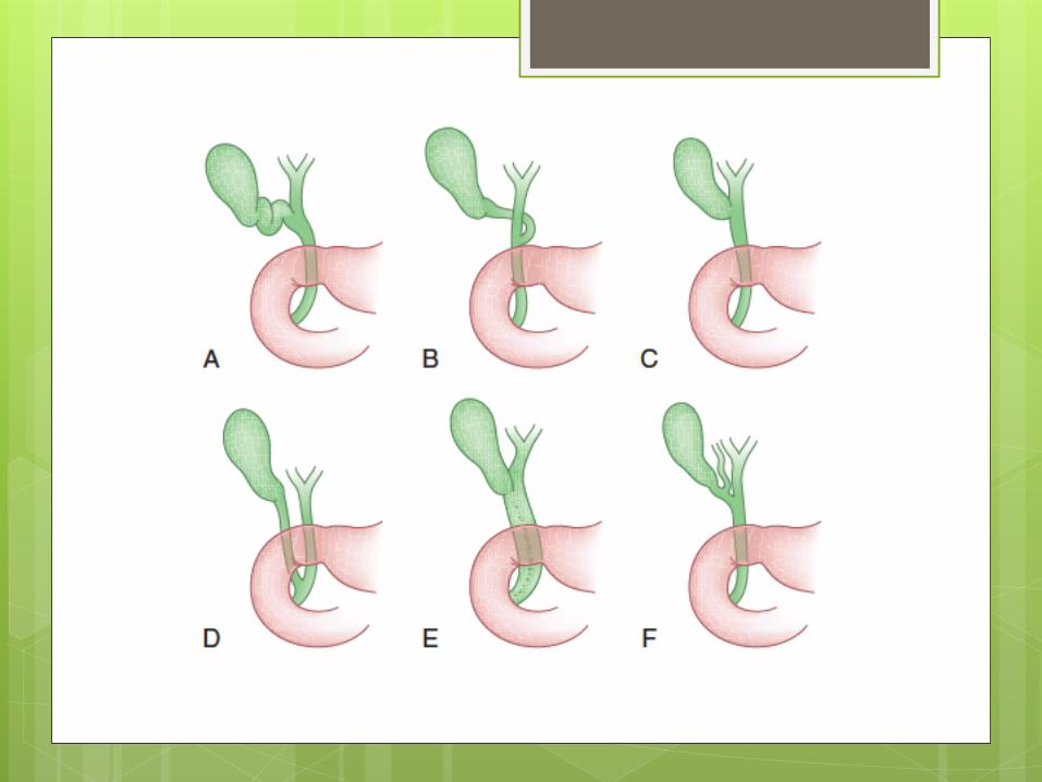
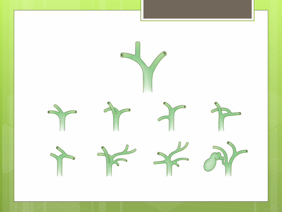
� Cystic duct unites with common hepatic duct 1-2 cm above the duodenum But can be :
- Parallel to the and RIGHT side of the hepatic duct for a
variable distance before joining it
- Spiral round behind the hepatic duct before joining it on the left side
- Rarely gall bladder directly drains into hepatic duct with no
cystic duct
- Also an ACCESSORY RIGHT HEPATIC DUCT joining hepatic duct, cystic duct or even gall bladder
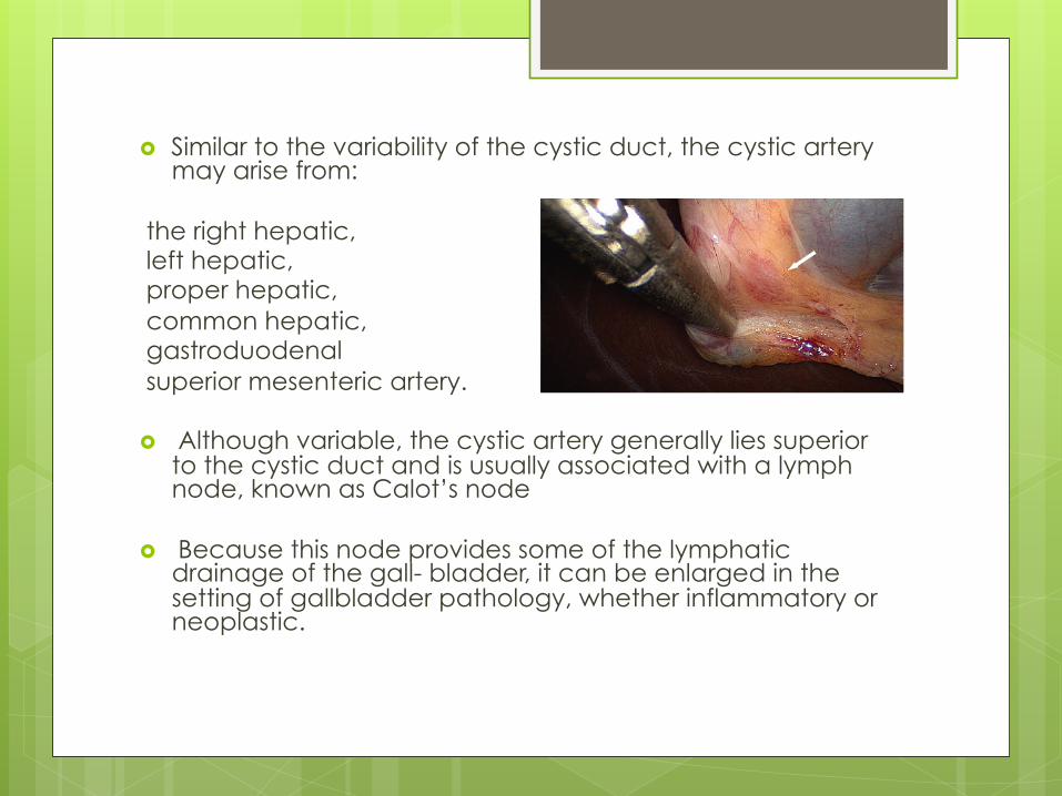
� Similar to the variability of the cystic duct, the cystic artery may arise from:
the right hepatic, left hepatic, proper hepatic, common hepatic, gastroduodenal superior mesenteric artery. � Although variable, the cystic artery generally lies superior
to the cystic duct and is usually associated with a lymph node, known as Calot’s node
� Because this node provides some of the lymphatic drainage of the gall- bladder, it can be enlarged in the setting of gallbladder pathology, whether inflammatory or neoplastic.
� Venous return by multiple small veins in the gall bladder vein.
One or more cystic veins may be present but its uncommon. If present they run from the neck of the gall bladder.
� Cystic veins do not accompany cystic artery.
Common Bile Duct � 6-8 cm long and normal diameter less than 8mm - Supraduodenal (in free edge of the lesser omentum in
front of the portal vein and right side of hepatic artery)
- Retroduodenal (behind the 1st part of the duodenum) Portal vein is on the left side of the duct with gastroduodenal artery and IVC is in behind.
- Paraduodenal: back of the head of the pancreas (can get obstructed by pancreatic Ca)
- Ampula vater opens to the posteromedial wall of the 2nd part of the duodenum at the major duodenal paplia 10cm from pylorus
PHYSIOLOGY � bile transport allows excretion of toxins
and normal cellular metabolites � bile salts have a critical role in the
absorption of most lipids
� The secretion of bile components into the biliary tree form a major stimulus to bile flow, and the volume of bile flow is an osmotic process.
� bile maintains an osmolality approximately comparable
to that of plasma.
� much of the flow is dependent on neural, humoral, and chemical stimuli.
� Vagal activity induces bile secretion, as does the gastrointestinal hormone secretin.
� Cholecystokinin (CCK), secreted by the intestinal mucosa, serves to induce biliary tree secretion and gallbladder wall contraction, thereby augmenting excretion of bile into the intestines.
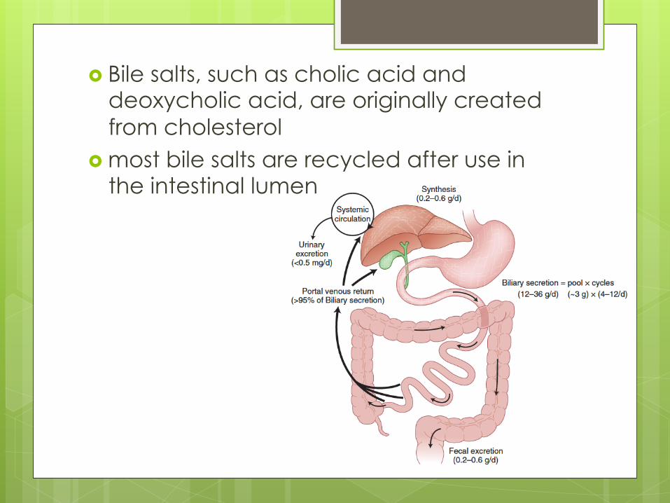
� Bile salts, such as cholic acid and deoxycholic acid, are originally created from cholesterol
� most bile salts are recycled after use in the intestinal lumen
� After passage into the intestinal tract and reabsorption by the terminal ileum, bile acids are transported back to the liver for recycling bound to albumin. transport of bile up an extreme concentration gradient is ATP dependent
� Less than 5% of bile salts are lost each day in the stool.
� When sufficient quantities of bile salts reach the colonic lumen, the powerful detergent activity of the bile salts can cause inflammation and diarrhea.
� Bile pigments, such as bilirubin, are breakdown products of hemoglobin and myoglobin.
Gall bladder physiology
� fills through a retrograde mechanism. With an increase in the tonic activity of the sphincter of Oddi in the fasting state, pressure increases in the common bile duct.
� The passage of fat, protein, and acid into the duodenum induces CCK secretion from duodenal epithelial cells. Cholecystokinin then causes gallbladder contraction, with intraluminal pressures up to 300 mm Hg.
� Vagal activity also induces gallbladder emptying, but is a less powerful stimulus to gallbladder contraction than CCK.
Sphincter of Oddi � This muscular sphincter, which normally is inhibited by
CCK. With CCK-induced relaxation of the sphincter, bile flows more rapidly from the biliary tree.
� Coordinated with gallbladder contraction, the relaxation of this sphincter allows for evacuation of up to 70% of the gallbladder contents within 2 hours of CCK secretion.
� During the fasting state, the oblique passage of the bile duct through the duodenal wall and the tonic activity of the sphincter prevent duodenal contents from refluxing into the biliary tree.