Embed Size (px)
Citation preview
8/6/2019 biofilme Campylobacter
http://slidepdf.com/reader/full/biofilme-campylobacter 1/10
Biofilm formation in Campylobacter jejuni
G. W. P Joshua, C. Guthrie-Irons, A. V. Karlyshev and B. W. Wren
Correspondence
B. W. Wren
Department of Infectious and Tropical Diseases, London School of Hygiene and Tropical
Medicine, Keppel Street, London WC1E 7HT, UK
Received 19 July 2005
Revised 21 October 2005
Accepted 10 November 2005
The major gastrointestinal pathogen Campylobacter jejuni is shown to exist as three forms of
monospecies biofilm in liquid culture. It attaches to a glass surface; forms an unattached aggregate
(floc); and forms a pellicle at the liquid–gas interface. The three forms of biofilm resemble each
other when examined by scanning electron microscopy. The biofilm mode of growth confers
protection against environmental stress, the microaerobic bacteria in flocs surviving up to 24 days
at ambient temperature and atmosphere compared to 12 days survival by planktonic bacteria.
The wild-type strains C. jejuni 33106, 32799, 33084 and 31485 did not form flocs, and floc
formation was reduced in strains mutant in a putative flagellar protein (FliS) and in a phosphate
acetyltransferase (Cj0688). All other strains tested,including strains withmutationsaffecting capsular
polysaccharide ( kpsM ), flagella ( maf5), protein glycosylation ( pglH ) and lipo-oligosaccharide( neuB1) formed flocs. Similarly, all strains tested formed a pellicle and attached to glass except the
aflagellate mutant maf5; pellicle formation was reduced in fliS and cj0688 mutants. Different
mechanisms, therefore, may control formation of different forms of biofilm. It is proposed that
these poorly characterized forms of growth are important for the persistence of C. jejuni in the
environment and may in part explain the high incidence of Campylobacter-associated food borne
disease.
INTRODUCTION
Campylobacter jejuni , a Gram-negative, microaerophilicbacterium, is the most frequently isolated foodborne patho-
gen worldwide. The annual reported incidence in Englandand Wales is around 50 000, which is thought to be a grossunderestimate (CPLS, 2000). C. jejuni is also associated withneuropathies such as Guillain–Barre and Miller–Fishersyndromes (Salloway et al ., 1996), where it is postulatedthat there is molecular mimicry between C. jejuni lipo-oligosaccharide and human gangliosides, leading to anauto-immune response (Yuki, 1997). The primary sourceof human infection is thought to be the consumption orhandling of contaminated chicken and therefore the survivalof C. jejuni in poultry house water systems is particularly significant (Pearson et al ., 1996; Zimmer et al ., 2003).
There is a paradox in that although C. jejuni is extremely widespread, its microaerobic growth requirements meanthat the organism does not multiply in the natural aerobicenvironment. Although not all ‘natural environments’ thatC. jejuni inhabits may necessarily be aerobic, it is still diffi-cult to explain the high incidence of infection, particularly as in contrast to several other gastrointestinal pathogens, C. jejuni is rarely transmitted between humans. It has beensuggested that C. jejuni maintains itself in the environment
by forming a biofilm (Buswell et al ., 1998). Biofilms arecommonly defined as matrix-enclosed bacterial populationsadherent to each other and/or to surfaces or interfaces
(Costerton et al ., 1995). Aggregates of bacteria not attachedto a surface are commonly termed flocs and have many of the characteristics of a biofilm (Hall-Stoodley et al ., 2004).Pellicles are aggregates of bacterial cells that form at an air–liquid interface (Friedman & Kolter, 2004).
The biofilm mode of growth is widespread and is of econo-mic importance in diverse ecological niches (Costerton etal .,1995). Monospecies biofilms are formed by many humanpathogens such as Pseudomonas aeruginosa (Whiteley et al .,2001), Staphylococcus epidermidis (Mack et al ., 2000),Salmonella enteritidis (Solano et al ., 2002), Vibrio cholerae (Nesper et al ., 2001), Streptococcus gordonii (Loo et al ., 2000)
and Burkholderia cepacia (Huber et al ., 2001). In humaninfection, biofilms are often less susceptible to antibiotics(Nichols, 1991).
C. jejuni has been observed to autoagglutinate, in Mueller–Hinton broth, minimal essential medium (Golden & Acheson, 2002) and phosphate-buffered saline (Misawa & Blaser, 2000). However, it is unclear if autoagglutinationrepresents a biofilm mode of growth in this species. C. jejuni has been found in preformed biofilms of other bacterialspecies (Trachoo et al ., 2002; Keevil, 2003). Buswell et al .(1998) postulated that C. jejuni in autochthonous biofilmshad enhanced survival. C. jejuni adherent to stainless steel
Abbreviations: CLSM, confocal laser scanning microscopy; EPM,extracellular polymeric matrix; SEM, scanning electron microscopy.
0002-8358 G 2006 SGM Printed in Great Britain 387
Microbiology (2006), 152, 387–396 DOI 10.1099/mic.0.28358-0
8/6/2019 biofilme Campylobacter
http://slidepdf.com/reader/full/biofilme-campylobacter 2/10
8/6/2019 biofilme Campylobacter
http://slidepdf.com/reader/full/biofilme-campylobacter 3/10
SYTO 16 (Molecular Probes). For indirect immunofluorescence tests(IFATs), Penner 2 antiserum was used at 1 : 100 and visualized withAlexa 546 (Molecular Probes). For SEM, aggregates were transferredto cover slips with a wide-bore pipette; pellicles were transferred by dipping cover slips through the surface of standing cultures. Attachedbacteria were obtained by breaking the glass test tube and processingbacteria in situ . Bacteria were fixed for 2 h in 3 % glutaraldehydemade up in 0?2 M sodium cacodylate buffer pH 7?4. The samples
were then washed in the same buffer for 75 min prior to post-fixingin 1 % osmium tetroxide in the 0?2 M sodium cacodylate buffer for105 min. Subsequent washing in 0?2 M buffer for a further 25 minwas followed by the storage at 4 uC i n 0?2 M sucrose in 0?2 Msodium cacodylate overnight. The samples were then washed in twochanges of MilliQ water over 145 min and dehydrated through agraduated series of ethanol (15–30 min per step; 30 %, 50 %, 70 %,90%, 26100 %). They were subsequently air-dried in a ventilatedcovered container and sputter-coated in an Edwards S150 sputtercoater. The samples were examined on a JEOL JSM25III scanningelectron microscope and photographed using Kodak 120 TP black and white film. Kodak D19 developer was used for developing therolls of film and the images were printed on Agfa multi-grade paperusing an Agfa Print Processor and Agfa chemistry.
Hydrophobicity assay. The relative hydrophobicity of cells wasdetermined using the ‘salting out’ method of Misawa & Blaser(2000). Hydrophobicity was assessed on the minimum concentrationof ammonium sulphate permitting aggregation of cells using a serialdoubling dilution of a 4 M solution.
Survival assay. For comparison of survival of aggregate and plank-tonic bacteria, 10 ml cultures in Mueller–Hinton liquid mediumwere grown at 50 r.p.m. for 3 days at 37 uC and transferred to testtubes. Aggregates were allowed to settle under gravity for 10 min,washed three times in broth and resuspended in 10 ml freshMueller–Hinton liquid medium. Similarly, planktonic bacteria in thesupernatant were centrifuged at 4000 r.p.m., washed three times inbroth and resuspended in 10 ml fresh Mueller–Hinton liquid medium.Cultures were then stored at ambient temperature and atmospheric
conditions for the duration of the assay. At 3 day intervals viablebacterial counts were determined on Columbia blood-agar plates.Experiments were performed in triplicate.
RESULTS
In our initial studies we attempted to grow biofilms of thesequenced strain of C. jejuni (NCTC 11168) on glass coverslips or on cellulose acetate filters, either in tissue cultureflasks, in 24-well tissue culture plates or in a modifiedRobbins device (Tyler Engineering) (Domingue etal ., 1994).We found that in the modified Robbins device, no bacteriaattached to either cellulose acetate filters or glass with a flow
rate of 300 ml h
21
, as used by Domingue et al . (1994) orwith various flow rates down to 10 ml h21. Nor did C. jejuni NCTC 11168 attach to polystyrene, as assessed by crystalviolet staining,in tests to quantify biofilm formation(Nesperet al ., 2001) (data not shown). Similarly, in liquid culturewith normal shaking (80–100 r.p.m.) there was no growthof bacteria on the plastic of the tissue culture flask, or on thesurface of glass cover slips or cellulose acetate filters, andbacteria in the medium were planktonic. However, withshaking at a lower speed (50 r.p.m.) we noted the formationof bacterial aggregates in culture. We also observed theformation of a pellicle at the liquid–gas interface of culturesgrown without shaking after 3–6 days, with cells impinging
on the walls of the test tubes at the liquid–gas interfacestrongly attached.
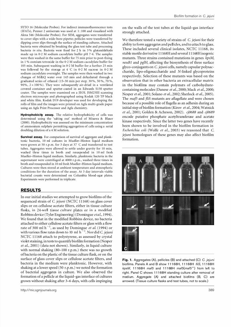
We therefore tested a variety of strains of C. jejuni for theirability to form aggregates and pellicles, and to attach to glass.These included several clinical isolates, NCTC 11168, itsstable motile derivative 11168H and several 11168H isogenic
mutants. Three strains contained mutations in genes kpsM ,neuB1 and pglH , affecting the biosynthesis of three surfaceglyco-conjugants on C. jejuni cells, namely capsular polysac-charide, lipo-oligosaccharide and N -linked glycoproteinsrespectively. Selection of these mutants was based on theobservation that in other bacteria an extracellular matrix in the biofilms may contain polymers of carbohydrate-containing molecules (Danese et al ., 2000; Mack et al ., 2000;Nesper et al ., 2001; Solano et al ., 2002; Sherlock et al ., 2005).The maf5 and fliS mutants are aflagellate and were chosenbecause of a possible role of flagella as an adhesin during aninitial step of biofilm formation (Kirov et al ., 2004; Watnick et al ., 2001; Golden & Acheson, 2002). cj0688 and cj0689 encode putative phosphate acetyltransferase and acetatekinase respectively. Since the latter two genes have recently been shown to be involved in the biofilm formation inEscherichia coli (Wolfe et al ., 2003) we reasoned that C. jejuni homologues of these genes may also affect biofilmformation.
Fig. 1. Aggregates (A), pellicles (B) and attached (C) C. jejuni
biofilms. Panels A and B show 11168H, 11168H fliS , 11168HkpsM , 11168H maf5 and 11168H maf5( maf5+) from left to
right. Panel C shows 11168H standing culture after removal ofmedium. Aggregate (A) and attached biofilms (B, C) arearrowed. (Tissue culture flasks and test tubes, not to scale.)
http://mic.sgmjournals.org 389
Biofilm formation in C. jejuni
8/6/2019 biofilme Campylobacter
http://slidepdf.com/reader/full/biofilme-campylobacter 4/10
Excepting C. jejuni wild-type strains 33106, 32799, 33084and 31485, all strains tested formed aggregates, includingthe isogenic mutants pglH , kpsM , neuB1 and maf5 . Aggre-gates in fliS and in cj0688 mutants were reduced. All wild-type strains also formed pellicles and attached to the surfaceof glass test tubes, including strains 33106, 32799, 33084 and31485. The pglH mutant formed a pellicle and attached toglass in a similar way to wild-type strains; the kpsM andneuB1 mutants formed a noticeably larger pellicle than all
other strains. The maf5 mutant did not form a pellicle orattach to glass but this ability was restored in the com-plemented strains. Pellicle formation was also reduced in the fliS and cj0688 mutants. Fig. 1 shows examples of the variousforms of biofilm. Note that attached bacteria were con-tiguous with the pellicle, and thus are not easily seen inintact standing cultures (Fig. 1B); Fig. 1C shows attached11168H bacteria after removal of a portion of medium. Theresults are summarized in Table 1; all strains were tested
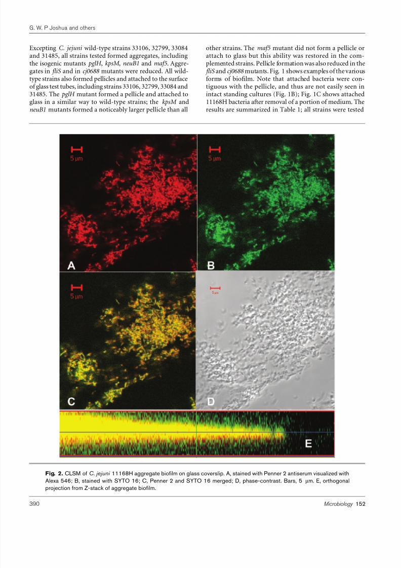
Fig. 2. CLSM of C. jejuni 11168H aggregate biofilm on glass coverslip. A, stained with Penner 2 antiserum visualized withAlexa 546; B, stained with SYTO 16; C, Penner 2 and SYTO 16 merged; D, phase-contrast. Bars, 5 mm. E, orthogonalprojection from Z-stack of aggregate biofilm.
390 Microbiology 152
G. W. P Joshua and others
8/6/2019 biofilme Campylobacter
http://slidepdf.com/reader/full/biofilme-campylobacter 5/10
in triplicate (mutant strains using three clonal isolates).Additionally, to provide information on the potential phy-sical forces responsible for cell–cell interactions, Table 1includes the relative hydrophobicity values for each strainand mutant strain studied.
We examined aggregates, pellicles and attached forms of
bacteria in 11168H and selected mutants. Fig. 2 showsCLSM images of C. jejuni 11168H aggregates on glass coverslips. Under phase-contrast (Fig. 2D) a mass of bacteria canbe seen as curved rods which were morphologically identi-fiable as C. jejuni . These were stained by the nucleic acidspecific stain SYTO 16 (Fig. 2B) and Penner 2 antiserum,specific to 11168H, (Fig. 2A, and shown merged in Fig. 2C).The three-dimensional nature of the bacterial mass,
characteristic of a biofilm, is shown by orthogonal pro- jection from a Z-stack (Fig. 2E). However, CLSM does notshow connection of the bacteria by an extracellularpolymeric matrix (EPM), which is a defining characteristicof a biofilm. This is seen by SEM (Figs 3–5).
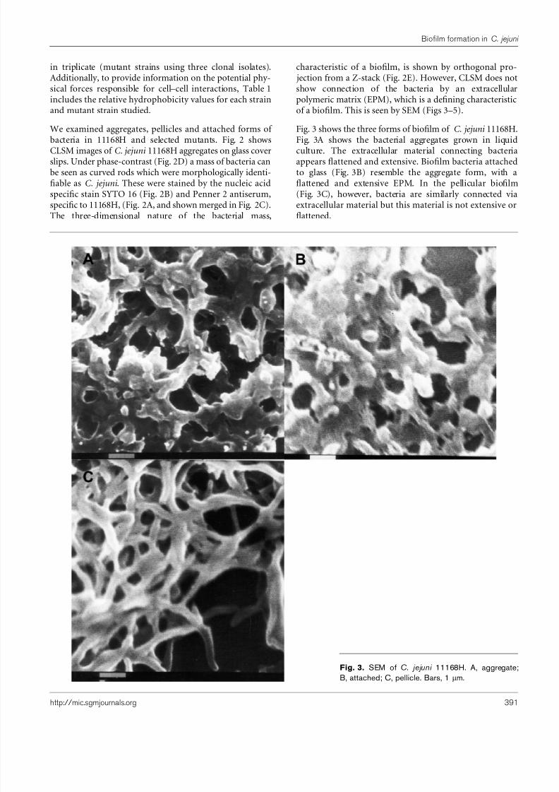
Fig. 3 shows the three forms of biofilm of C. jejuni 11168H.
Fig. 3A shows the bacterial aggregates grown in liquidculture. The extracellular material connecting bacteriaappears flattened and extensive. Biofilm bacteria attachedto glass (Fig. 3B) resemble the aggregate form, with aflattened and extensive EPM. In the pellicular biofilm(Fig. 3C), however, bacteria are similarly connected viaextracellular material but this material is not extensive orflattened.
Fig. 3. SEM of C. jejuni 11168H. A, aggregate;B, attached; C, pellicle. Bars, 1 mm.
http://mic.sgmjournals.org 391
Biofilm formation in C. jejuni
8/6/2019 biofilme Campylobacter
http://slidepdf.com/reader/full/biofilme-campylobacter 6/10
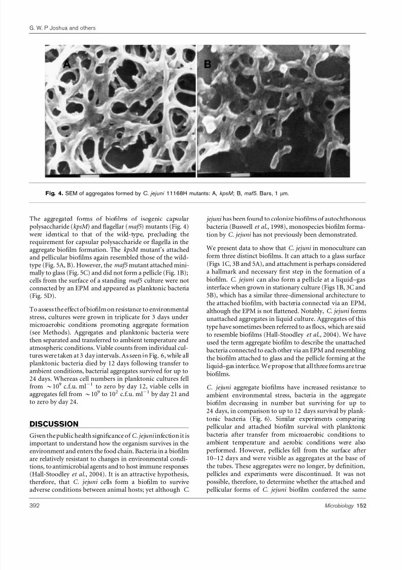
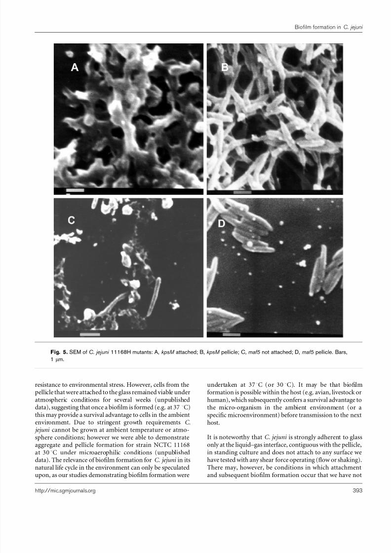
The aggregated forms of biofilms of isogenic capsularpolysaccharide (kpsM ) and flagellar (maf5 ) mutants (Fig. 4)were identical to that of the wild-type, precluding therequirement for capsular polysaccharide or flagella in theaggregate biofilm formation. The kpsM mutant’s attachedand pellicular biofilms again resembled those of the wild-type (Fig. 5A, B). However, the maf5 mutant attached mini-mally to glass (Fig. 5C) and did not form a pellicle (Fig. 1B);cells from the surface of a standing maf5 culture were notconnected by an EPM and appeared as planktonic bacteria(Fig. 5D).
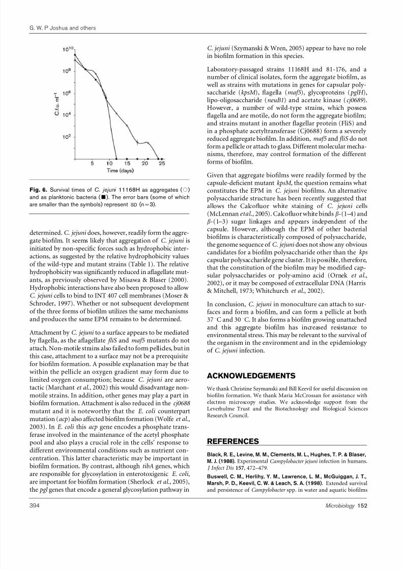
To assess the effect of biofilm on resistance to environmentalstress, cultures were grown in triplicate for 3 days undermicroaerobic conditions promoting aggregate formation(see Methods). Aggregates and planktonic bacteria werethen separated and transferred to ambient temperature andatmospheric conditions. Viable counts from individual cul-tures were taken at 3 day intervals. As seen in Fig. 6, while allplanktonic bacteria died by 12 days following transfer toambient conditions, bacterial aggregates survived for up to24 days. Whereas cell numbers in planktonic cultures fellfrom ~109 c.f.u. ml21 to zero by day 12, viable cells inaggregates fell from~109 to 102 c.f.u. ml21 by day 21 and
to zero by day 24.
DISCUSSION
Given the public health significance of C. jejuni infection it isimportant to understand how the organism survives in theenvironment and enters the food chain. Bacteria in a biofilmare relatively resistant to changes in environmental condi-tions, to antimicrobial agents and to host immune responses(Hall-Stoodley et al ., 2004). It is an attractive hypothesis,therefore, that C. jejuni cells form a biofilm to surviveadverse conditions between animal hosts; yet although C.
jejuni has been found to colonize biofilms of autochthonousbacteria (Buswell et al ., 1998), monospecies biofilm forma-tion by C. jejuni has not previously been demonstrated.
We present data to show that C. jejuni in monoculture canform three distinct biofilms. It can attach to a glass surface(Figs 1C, 3B and 5A), and attachment is perhaps considereda hallmark and necessary first step in the formation of abiofilm. C. jejuni can also form a pellicle at a liquid–gasinterface when grown in stationary culture (Figs 1B, 3C and5B), which has a similar three-dimensional architecture tothe attached biofilm, with bacteria connected via an EPM,although the EPM is not flattened. Notably, C. jejuni formsunattached aggregates in liquid culture. Aggregates of thistype have sometimes been referred to as flocs, which are saidto resemble biofilms (Hall-Stoodley et al ., 2004). We haveused the term aggregate biofilm to describe the unattachedbacteria connected to each other via an EPM and resemblingthe biofilm attached to glass and the pellicle forming at theliquid–gas interface. We propose that all three forms are truebiofilms.
C. jejuni aggregate biofilms have increased resistance toambient environmental stress, bacteria in the aggregatebiofilm decreasing in number but surviving for up to24 days, in comparison to up to 12 days survival by plank-tonic bacteria (Fig. 6). Similar experiments comparingpellicular and attached biofilm survival with planktonicbacteria after transfer from microaerobic conditions toambient temperature and aerobic conditions were alsoperformed. However, pellicles fell from the surface after10–12 days and were visible as aggregates at the base of the tubes. These aggregates were no longer, by definition,pellicles and experiments were discontinued. It was notpossible, therefore, to determine whether the attached andpellicular forms of C. jejuni biofilm conferred the same
Fig. 4. SEM of aggregates formed by C. jejuni 11168H mutants: A, kpsM ; B, maf5. Bars, 1 mm.
392 Microbiology 152
G. W. P Joshua and others
8/6/2019 biofilme Campylobacter
http://slidepdf.com/reader/full/biofilme-campylobacter 7/10
resistance to environmental stress. However, cells from thepellicle that were attached to the glass remained viable underatmospheric conditions for several weeks (unpublisheddata), suggesting that once a biofilm is formed (e.g. at 37 uC)this may provide a survival advantage to cells in the ambientenvironment. Due to stringent growth requirements C. jejuni cannot be grown at ambient temperature or atmo-sphere conditions; however we were able to demonstrateaggregate and pellicle formation for strain NCTC 11168at 30 uC under microaerophilic conditions (unpublisheddata). The relevance of biofilm formation for C. jejuni in itsnatural life cycle in the environment can only be speculatedupon, as our studies demonstrating biofilm formation were
undertaken at 37u
C (or 30u
C). It may be that biofilmformation is possible within the host (e.g. avian, livestock orhuman), which subsequently confers a survival advantage tothe micro-organism in the ambient environment (or aspecific microenvironment) before transmission to the nexthost.
It is noteworthy that C. jejuni is strongly adherent to glassonly at the liquid–gas interface, contiguous with the pellicle,in standing culture and does not attach to any surface wehave tested with any shear force operating (flow or shaking).There may, however, be conditions in which attachmentand subsequent biofilm formation occur that we have not
Fig. 5. SEM of C. jejuni 11168H mutants: A, kpsM attached; B, kpsM pellicle; C, maf5 not attached; D, maf5 pellicle. Bars,
1 mm.
http://mic.sgmjournals.org 393
Biofilm formation in C. jejuni
8/6/2019 biofilme Campylobacter
http://slidepdf.com/reader/full/biofilme-campylobacter 8/10
determined. C. jejuni does, however, readily form the aggre-gate biofilm. It seems likely that aggregation of C. jejuni isinitiated by non-specific forces such as hydrophobic inter-actions, as suggested by the relative hydrophobicity valuesof the wild-type and mutant strains (Table 1). The relativehydrophobicity was significantly reduced in aflagellate mut-ants, as previously observed by Misawa & Blaser (2000).Hydrophobic interactions have also been proposed to allow C. jejuni cells to bind to INT 407 cell membranes (Moser & Schroder, 1997). Whether or not subsequent developmentof the three forms of biofilm utilizes the same mechanismsand produces the same EPM remains to be determined.
Attachment by C. jejuni to a surface appears to be mediatedby flagella, as the aflagellate fliS and maf5 mutants do notattach. Non-motile strains also failed to form pellicles, but inthis case, attachment to a surface may not be a prerequisitefor biofilm formation. A possible explanation may be thatwithin the pellicle an oxygen gradient may form due tolimited oxygen consumption; because C. jejuni are aero-tactic (Marchant et al ., 2002) this would disadvantage non-motile strains. In addition, other genes may play a part inbiofilm formation. Attachment is also reduced in the cj0688 mutant and it is noteworthy that the E. coli counterpartmutation (acp ) also affected biofilm formation (Wolfe et al .,2003). In E. coli this acp gene encodes a phosphate trans-ferase involved in the maintenance of the acetyl phosphatepool and also plays a crucial role in the cells’ response todifferent environmental conditions such as nutrient con-centration. This latter characteristic may be important inbiofilm formation. By contrast, although tibA genes, whichare responsible for glycosylation in enterotoxigenic E. coli ,are important for biofilm formation (Sherlock et al ., 2005),the pgl genes that encode a general glycosylation pathway in
C. jejuni (Szymanski & Wren, 2005) appear to have no rolein biofilm formation in this species.
Laboratory-passaged strains 11168H and 81-176, and anumber of clinical isolates, form the aggregate biofilm, aswell as strains with mutations in genes for capsular poly-saccharide (kpsM ), flagella (maf5 ), glycoproteins ( pglH ),
lipo-oligosaccharide (neuB1) and acetate kinase (cj0689 ).However, a number of wild-type strains, which possessflagella and are motile, do not form the aggregate biofilm;and strains mutant in another flagellar protein (FliS) andin a phosphate acetyltransferase (Cj0688) form a severely reduced aggregate biofilm. In addition, maf5 and fliS do notform a pellicle or attach to glass. Different molecular mecha-nisms, therefore, may control formation of the differentforms of biofilm.
Given that aggregate biofilms were readily formed by thecapsule-deficient mutant kpsM , the question remains whatconstitutes the EPM in C. jejuni biofilms. An alternative
polysaccharide structure has been recently suggested thatallows the Calcofluor white staining of C. jejuni cells(McLennan etal ., 2005). Calcofluor white binds b-(1–4) andb-(1–3) sugar linkages and appears independent of thecapsule. However, although the EPM of other bacterialbiofilms is characteristically composed of polysaccharide,the genome sequence of C. jejuni does not show any obviouscandidates for a biofilm polysaccharide other than the kps capsular polysaccharide gene cluster. It is possible, therefore,that the constitution of the biofilm may be modified cap-sular polysaccharides or poly-amino acid (Ornek et al .,2002), or it may be composed of extracellular DNA (Harris& Mitchell, 1973; Whitchurch et al ., 2002).
In conclusion, C. jejuni in monoculture can attach to sur-faces and form a biofilm, and can form a pellicle at both37 uC and 30 uC. It also forms a biofilm growing unattachedand this aggregate biofilm has increased resistance toenvironmental stress. This may be relevant to the survival of the organism in the environment and in the epidemiology of C. jejuni infection.
ACKNOWLEDGEMENTS
We thank Christine Szymanski and Bill Keevil for useful discussion onbiofilm formation. We thank Maria McCrossan for assistance withelectron microscopy studies. We acknowledge support from theLeverhulme Trust and the Biotechnology and Biological SciencesResearch Council.
REFERENCES
Black, R. E., Levine, M. M., Clements, M. L., Hughes, T. P. & Blaser,
M. J. (1988). Experimental Campylobacter jejuni infection in humans. J Infect Dis 157, 472–479.
Buswell, C. M., Herlihy, Y. M., Lawrence, L. M., McGuiggan, J. T.,
Marsh, P. D., Keevil, C. W. & Leach, S. A. (1998). Extended survivaland persistence of Campylobacter spp. in water and aquatic biofilms
Fig. 6. Survival times of C. jejuni 11168H as aggregates ( #)
and as planktonic bacteria ( &
). The error bars (some of whichare smaller than the symbols) represent SD (n=3).
394 Microbiology 152
G. W. P Joshua and others
8/6/2019 biofilme Campylobacter
http://slidepdf.com/reader/full/biofilme-campylobacter 9/10
and their detection by immunofluorescent-antibody and -rRNA
staining. Appl Environ Microbiol 64, 733–741.
Costerton, J. W., Lewandowski, Z., Caldwell, D. E., Korber, D. R. &
Lappin-Scott, H. M. (1995). Microbial biofilms. Annu Rev Microbiol
49, 711–745.
CPLS (2000). Common gastrointestinal infections. Communicable
Diseases Report Weekly England and Wales 10, 9–12.
Danese, P. N., Pratt, L. A. & Kolter, R. (2000). Exopolysaccharideproduction is required for development of Escherichia coli K-12
biofilm architecture. J Bacteriol 182, 3593–3596.
Domingue, G., Ellis, B., Dasgupta, M. & Costerton, J. W. (1994).
Testing antimicrobial susceptibilities of adherent bacteria by amethod that incorporates guidelines of the National Committee forClinical Laboratory Standards. J Clin Microbiol 32, 2564–2568.
Friedman, L. & Kolter, R. (2004). Genes involved in matrix formationin Pseudomonas aeruginosa PA14 biofilms. Mol Microbiol 51,675–690.
Golden, N. J. & Acheson, D. W. (2002). Identification of motility andautoagglutination Campylobacter jejuni mutants by random trans-
poson mutagenesis. Infect Immun 70, 1761–1771.
Hall-Stoodley, L., Costerton, J. W. & Stoodley, P. (2004).Bacterialbiofilms: from the natural environment to infectious diseases. Nat
Rev Microbiol 2, 95–108.
Harris, R. H. & Mitchell, R. (1973). The role of polymers in microbialaggregation. Annu Rev Microbiol 27, 27–50.
Huber, B., Riedel, K., Hentzer, M., Heydorn, A., Gotschlich, A.,
Givskov, M., Molin, S. & Eberl, L. (2001). The cep quorum-sensingsystem of Burkholderia cepacia H111 controls biofilm formation andswarming motility. Microbiology 147, 2517–2528.
Karlyshev, A. V. & Wren, B. W. (2005). Development and application
of an insertional system for gene delivery and expression in Campylo-bacter jejuni . Appl Environ Microbiol 71, 4004–4013.
Karlyshev, A. V., Linton, D., Gregson, N. A., Lastovica, A. J. & Wren,
B. W. (2000). Genetic and biochemical evidence of a Campylobacter
jejuni capsular polysaccharide that accounts for Penner serotypespecificity. Mol Microbiol 35, 529–541.
Karlyshev, A. V., Linton, D., Gregson, N. A. & Wren, B. W. (2002).
A novel paralogous gene family involved in phase-variable flagella-mediated motility in Campylobacter jejuni . Microbiology 148,
473–480.
Keevil, C. W. (2003). Rapid detection of biofilms and adherent
pathogens using scanning confocal laser microscopy and episcopicdifferential interference contrast microscopy. Water Sci Technol 47,105–116.
Kirov, S. M., Castrisios, M. & Shaw, J. G. (2004). Aeromonas flagella(polar and lateral) are enterocyte adhesins that contribute to biofilmformation on surfaces. Infect Immun 72, 1939–1945.
Linton, D., Karlyshev, A. V., Hitchen, P. G., Morris, H. R., Dell, A.,
Gregson, N. A. & Wren, B. W. (2000). Multiple N -acetyl neuraminicacid synthetase (neuB ) genes in Campylobacter jejuni : identificationand characterization of the gene involved in sialylation of lipo-oligosaccharide. Mol Microbiol 35, 1120–1134.
Linton, D., Allan, E., Karlyshev, A. V., Cronshaw, A. D. & Wren, B. W.
(2002). Identification of N -acetylgalactosamine-containing glyco-
proteins PEB3 and CgpA in Campylobacter jejuni . Mol Microbiol 43,497–508.
Loo, C. Y., Corliss, D. A. & Ganeshkumar, N. (2000). Streptococcus gordonii biofilm formation: identification of genes that code for
biofilm phenotypes. J Bacteriol 182, 1374–1382.
Mack, D., Rohde, H., Dobinsky, S., Riedewald, J., Nedelmann, M.,
Knobloch, J. K., Elsner, H. A. & Feucht, H. H. (2000). Identification
of three essential regulatory gene loci governing expression of Staphylococcus epidermidis polysaccharide intercellular adhesin andbiofilm formation. Infect Immun 68, 3799–3807.
Marchant, J., Wren, B. & Ketley, J. (2002). Exploiting genomesequence: predictions for mechanisms of Campylobacter chemotaxis.Trends Microbiol 10, 155–159.
McLennan, M., Ringoir, D., Jarrell, H. C. Szymanski, C. & Gaynor, E.
(2005). Characterisation of Campylobacter jejuni surface moiety thatcross-reacts with calcofluor white: implications for surface carbohy-drates, stress response, and pathogenesis. Abstracts of CHRO Meeting F32, 87.
Misawa, N. & Blaser, M. J. (2000). Detection and characterization of autoagglutination activity by Campylobacter jejuni . Infect Immun 68,6168–6175.
Moser, I. & Schroder, W. (1997). Hydrophobic characterization of thermophilic Campylobacter species and adhesion to INT 407 cellmembranes and fibronectin. Microb Pathog 22, 155–164.
Nesper, J., Lauriano, C. M., Klose, K. E., Kapfhammer, D., Kraiss, A.
& Reidl, J. (2001). Characterization of Vibrio cholerae O1 El tor galU and galE mutants: influence on lipopolysaccharide structure, coloni-zation, and biofilm formation. Infect Immun 69, 435–445.
Nichols, W. W. (1991). Biofilms, antibiotics and penetration. Rev Med Microbiol 2, 177–181.
Ornek, D., Jayaraman, A., Syrett, B. C., Hsu, C. H., Mansfeld, F. B. &
Wood, T. K. (2002). Pitting corrosion inhibition of aluminum 2024by Bacillus biofilms secreting polyaspartate or gamma-polyglutamate.Appl Microbiol Biotechnol 58, 651–657.
Parkhill, J., Wren, B. W., Mungall, K. & 18 other authors (2000). Thegenome sequence of the food-borne pathogen Campylobacter jejuni reveals hypervariable sequences. Nature 403, 665–668.
Pearson, A. D., Greenwood, M. H., Feltham, R. K., Healing, T. D.,
Donaldson, J., Jones, D. M. & Colwell, R. R. (1996). Microbialecology of Campylobacter jejuni in a United Kingdom chicken supply chain: intermittent common source, vertical transmission, andamplification by flock propagation. Appl Environ Microbiol 62,
4614–4620.
Salloway, S., Mermel, L. A., Seamans, M., Aspinall, G. O., Nam Shin,
J. E., Kurjanczyk, L. A. & Penner, J. L. (1996). Miller-Fisher syndromeassociated with Campylobacter jejuni bearing lipopolysaccharidemolecules that mimic human ganglioside GD3. Infect Immun 64,2945–2949.
Sherlock, O., Vejborg, R. M. & Klemm, P. (2005). The TibA adhesin/invasin from enterotoxigenic Escherichia coli is self recognizing andinduces bacterial aggregation and biofilm formation. Infect Immun 73, 1954–1963.
Solano, C., Garcia, B., Valle, J., Berasain, C., Ghigo, J. M.,
Gamazo, C. & Lasa, I. (2002). Genetic analysis of Salmonella enteritidis biofilm formation: critical role of cellulose. Mol Microbiol 43, 793–808.
Somers, E. B., Schoeni, J. L. & Wong, A. C. (1994). Effect of trisodium phosphate on biofilm and planktonic cells of Campylobacter jejuni , Escherichia coli O157: H7, Listeria monocyto-genes and Salmonella typhimurium. Int J Food Microbiol 22,269–276.
Szymanski, C. M. & Wren, B. W. (2005). Protein glycosylation inbacterial mucosal pathogens. Nat Rev Microbiol 3, 225–237.
Trachoo, N., Frank, J. F. & Stern, N. J. (2002). Survival of Campylo-bacter jejuni in biofilms isolated from chicken houses. J Food Prot 65,1110–1116.
van Vliet, A. H., Wooldridge, K. G. & Ketley, J. M. (1998). Iron-responsive gene regulation in a Campylobacter jejuni fur mutant. J Bacteriol 180, 5291–5298.
http://mic.sgmjournals.org 395
Biofilm formation in C. jejuni
8/6/2019 biofilme Campylobacter
http://slidepdf.com/reader/full/biofilme-campylobacter 10/10
Watnick, P. I., Lauriano, C. M., Klose, K. E., Croal, L. & Kolter, R.
(2001). The absence of a flagellum leads to altered colony mor-phology, biofilm development and virulence in Vibrio cholerae O139.Mol Microbiol 39, 223–235.
Whitchurch, C. B., Tolker-Nielsen, T., Ragas, P. C. & Mattick, J. S.
(2002). Extracellular DNA required for bacterial biofilm formation.Science 295, 1487.
Whiteley, M., Ott, J. R., Weaver, E. A. & McLean, R. J. (2001). Effectsof community composition and growth rate on aquifer biofilmbacteria and their susceptibility to betadine disinfection. Environ Microbiol 3, 43–52.
Wolfe, A. J., Chang, D. E., Walker, J. D. & 10 other authors (2003).
Evidence that acetyl phosphate functions as a global signal duringbiofilm development. Mol Microbiol 48, 977–988.
Yuki, N. (1997). Molecular mimicry between gangliosides andlipopolysaccharides of Campylobacter jejuni isolated from patientswith Guillain-Barre syndrome and Miller Fisher syndrome. J Infect Dis 176, Suppl 2, S150–S153.
Zimmer, M., Barnhart, H., Idris, U. & Lee, M. D. (2003). Detection of Campylobacter jejuni strains in the water lines of a commercialbroiler house and their relationship to the strains that colonized thechickens. Avian Dis 47, 101–107.
396 Microbiology 152
G. W. P Joshua and others