Embed Size (px)
Citation preview
Cover Page
The handle http://hdl.handle.net/1887/66270 holds various files of this Leiden University dissertation. Author: Bouwens, J.A. Title: Neuropsychiatric symptoms and immune system parameters in Huntington's disease Issue Date: 2018-10-18

Neuropsychiatric symptoms and immune system parameters in Huntington’s disease

ISBN: 978-94-6361-150-3Cover design by: Optima Grafische Communicatie, Rotterdam, The NetherlandsPrinting: Optima Grafische Communicatie, Rotterdam, The Netherlands
Neuropsychiatric symptoms and immune system parameters in Huntington’s disease
Proefschrift
ter verkrijging vande graad van Doctor aan de Universiteit Leiden,
op gezag van Rector Magnificus prof. Mr. C.J.J.M. Stolker,volgens besluit van het College voor Promotieste verdedigen op donderdag 18 oktober 2018
klokke 16.15 uur
door
Joseph Arnoldus Bouwensgeboren te Rotterdam
in 1985

promotor
prof. dr. R.C. van der Mast
copromotoren
dr. E. van Duijndr. E.J. Giltay
leden promotiecommissie
prof. dr. N.J.A. van der Wee dr. W.M.C. van Roon-Momprof. dr. A. Tibbenprof. dr. I. Tendolkar, UMC Radboud, Nijmegenprof. dr. H.P.H. Kremer, UMC Groningen, Groningen

Contents
Chapter 1 Introduction 7Chapter 2 Irritability in Huntington’s disease 17Chapter 3 Irritability in a prospective cohort of Huntington’s disease mutation
carriers33
Chapter 4 Acute-phase proteins in relation to neuropsychiatric symptoms and use of psychotropic medication in Huntington’s disease
47
Chapter 5 Disease stage and plasma levels of cyotkines in Huntington’s disease: a 2-year follow-up study
65
Chapter 6 Plasma cytokine levels in relation to neuropsychiatric symptoms and cognitive dysfunction in Hungtington’s disease
69
Chapter 7 General discussion and summary 85
Nederlandse samenvatting 97Dankwoord 99Curriculum vitae 101Publicaties 103


Chapter 1
Introduction

9
Introduction
huntIngton’s dIsease
Clinical features of Huntington’s disease (HD) include motor, neuropsychiatric and cognitive symptoms[1]. HD is an autosomal dominant inheritable neurodegenerative disorder. This im-plies that, if one of the parents is afflicted, their offspring has a 50% probability of developing HD. Although neuropsychiatric and cognitive symptoms often precede the manifestation of motor abnormalities of HD, the definite diagnosis of HD is usually made when the first motor symp-toms occur. Although the age of onset is wide (ranging from early childhood to senescence), it is generally between 30 and 50 years of age. The duration between the time of diagnosis and death is around 20 years[2]. Motor symptoms may initially be subtle and might, at first, escape the aware-ness of the patient. Chorea is the most prominent motor symptom in HD and is characterized by involuntary irregular movements of the head, trunk and limbs. Other movement disorders, such as dystonia, bradykinesia, hypokinesia and postural instability, also occur[3]. In advanced stages of the disease, dysphagia and dysarthria may also develop[4].
neuropsyChIatrIC symptoms In hd
In HD, the most prevalent neuropsychiatric symptoms are depressed mood, irritability and apathy. The prevalence of depressed mood ranges from 33-69%. Apathy and irritability occur fre-quently in HD with a reported prevalence of 34-76% for apathy and of 33-73% for irritability[1].
Apathy is defined as a disorder of motivation with diminished goal-directed behavior, cognitions and emotions[5]. Apathy is positively correlated with cognitive decline, male sex, the presence of depression, and the use of psychotropic medication[6]. Longitudinally, although new-onset apathy was shown to occur in 14% of HD gene expansion carriers over a two-year study period, apathy also remitted in 14% during that same time period[7], indicating the poten-tial for remittance. This suggests that HD patients with apathy should be evaluated for treatable causes of apathy.
Irritability can be characterized as a dysphoric mood state that predisposes toward both verbal and non-verbal expressions of aggression in a non-adaptive manner that complicates the relationship between the patient and the caregiver and/or partner[8]. Even before motor symptoms are present, irritability can cause severe distress to HD patients and their caregivers.
In pre-motor symptomatic disease stages, cognitive dysfunction mostly comprises deficits in attentional and executive functions, semantic verbal fluency and visual working memory. Deficits in memory become apparent around the time of onset of motor symptomatic disease. In the advanced disease stage, patients often exhibit severe impairments, particularly in executive functioning[9].
Chapter 1
10
pathophysIology
The causal genetic mutation was discovered in 1993 and comprises an expanded cytosine-adenine-guanine (CAG) trinucleotide repeat coding for the huntingtin (htt) protein on the short arm of chromosome 4 (4p16.3)[10]. Persons with an expansion of >39 CAG repeats will develop the disease, although an expansion of 36-39 CAG repeats may also result in symptomatic HD (‘reduced penetrance’). The expanded gene codes for mutant huntingtin (mhtt)[11]. A greater CAG repeat length is associated with an earlier age of onset of symptoms.
Neurodegeneration is a prominent feature of HD. In HD, there is neuronal cell loss in the brain, particularly in the caudate nucleus and the putamen, but also in other brain regions, including the cortex. This loss of neurons is already detectable in pre-motor symptomatic HD gene expansion carriers[12]. In physiological circumstances htt is expressed in all cells (with the highest concentrations in the brain and testes) and plays a key role in transcription, cell signaling and intracellular transporting[13]. However, much remains unknown about the physiological function of htt as well as the exact pathophysiology by which mhtt causes cerebral tissue damage. Several mechanisms are likely to play a role in the neurodegenerative process, including increased excitotoxicity, mitochondrial damage, free radical formation, and immune activation[14].
Immune system and InflammatIon In hd
Aberrant immune activation is one of the proposed underlying mechanisms for neurodegenera-tion and, subsequently, the development of the characteristic symptoms in HD[14]. Microglia, the main immunocompetent cells in the central nervous system, play a key role in immune pro-cesses in the brain[15]. Activated microglia have been demonstrated in post-mortem samples of patients with HD and on cerebral positron emission tomography (PET) scans[16, 17]. Mhtt may play an important role in the activation of microglia by directly activating the nuclear transcrip-tion factor-kB (NF-kB), thereby initiating the first steps of the acute-phase response [18]. Also, mhtt can activate the IkB kinase complex, thereby enhancing activity of the nuclear transcription factor NF-kB[18]. NF-kB plays a key role in the regulation of the immune response to infec-tion[19]. Alternatively, cell damage caused by mhtt through other mechanisms may activate the acute-phase response. The NF-kB pathway is a major inducer of the pro-inflammatory cytokines interleukin (IL)-1β, IL-6 and tumor necrosis factor (TNF)-α. Among these cytokines, IL-6 is the most potent inductor of the acute-phase response[20], a systemic reaction aimed at restoring physiological homeostasis under physiologic circumstances[21]. This response is regulated by several other pro-inflammatory and anti-inflammatory cytokines such as IL-8 and IL1-ra, IL-5 and IL-10, respectively. As part of this systemic reaction, the production of certain proteins is upregulated or downregulated to the benefit of the injured organism[22, 23]. As such, the pro-duction of C-reactive protein (CRP), that plays a prominent role in the complement cascade, is
11
Introduction
upregulated and its circulating level can be thought of as a positive acute-phase protein, whereas the production of albumin is downregulated and its circulating level can therefore be thought of as a negative acute-phase protein.
BIomarkers of dIsease state In hd
Biomarkers are important because they may provide an early indication as to whether (or not) a potential therapeutic intervention is beneficial. A useful biomarker needs to be readily quantifiable, robust and reproducible [12, 24, 25]. In HD, biomarkers of several modalities have been employed to observe differences between HD gene expansion carriers and controls, and to observe differences between HD gene expansion carriers at different disease stages, or even to observe differences in HD gene expansion carriers before disease onset[12].
Clinical measures are regularly used as markers of disease stage in HD investigations. One of the most frequently used clinical rating scales is the Unified Huntington’s Disease Rating Scale (UHDRS) which comprises subscales on several domains, such as daily functioning and mo-tor symptoms. In general, the UHDRS scale measures changes in symptomatic HD over time, but is not able to detect changes in pre-motor symptomatic HD. In addition, neuropsychiatric measures, such as the Problem Behaviours Assessment (PBA) scale, are used to assess specific neuropsychiatric features of the disease.
Structural magnetic resonance imaging (MRI) is widely used to define cerebral biomarkers in HD gene expansion carriers. Cross-sectionally, MRI can show disease-related atrophy of the striatum and white matter of HD gene expansion carriers compared with controls. This atrophy can be demonstrated years before the onset of clinical disease. In addition, using MRI, progres-sive neurodegenerative changes can be demonstrated in early HD and pre-motor symptomatic HD gene expansion carriers.
However, information from biochemical biomarkers might be obtained more rapidly (and at less cost) compared with imaging markers. In addition, biochemical markers that are in close proximity to the underlying pathology may be more sensitive to disease progression and might show reversal in response to therapeutic interventions. Cytokine levels in the plasma are poten-tial biochemical biomarkers in HD. Increased levels of several cytokines have been reported in cross-sectional studies among HD gene expansion carriers[26-30]. Most consistently, plasma IL-6 levels were higher in motor symptomatic HD gene expansion carriers than in matched controls. Also, increased plasma levels of sIL-2R, sTNF-α and IL-8 were found compared with levels in controls.
Cytokines and acute-phase proteins in relation to neuropsychiatric symptoms in HD
The acute-phase response involves a systemic reaction of the immune system as reflected in activation of monocytes, which is regulated by several cytokines[20, 21]. In addition, metabolic

Chapter 1
12
changes occur, such as the production of certain acute-phase proteins and the inhibition of pro-duction of other molecules [22, 23]. These changes, which are readily quantifiable in the blood, may be accompanied by behavioral changes that favor the survival of the organism when the ho-meostasis is disturbed. In mouse models, behavioral changes (such as psychomotor retardation, anorexia, sleep disturbances and lethargy) have been called ‘sickness behavior’. Changed levels of cytokines in the brain may also cause part of these behavioral effects in humans[31]. Reciprocal connections exist between the peripheral immune system and the brain[31-34]. Cytokines in the peripheral plasma can pass the blood-brain barrier under certain conditions and can activate nerve cells that stimulate immune cells in the brain to produce cytokines.
Given this bidirectional association between plasma and brain cytokines and behavior, sev-eral studies have examined the connection between cytokines and neuropsychiatric symptoms in non-HD populations. The association between cytokines, acute-phase proteins and depression has been investigated the most extensively[35-37]. Consistently increased levels of IL-6 and CRP were found in (non-HD) depressed patients. In addition, the association between cytokines and cognitive decline, as well as dementia, yielded similar results[38]. The association between cytokines and irritability and apathy has also been investigated in non-HD populations, but with inconsistent results[37, 39-44].
In HD, neuropsychiatric symptoms (in particular psychosis and irritability) and motor symptoms are frequently treated with antipsychotic medications[45]. However, antipsychot-ics can induce symptoms that can mimic apathy, depressive mood and cognitive decline[46]. Through several (hepatic) mechanisms, antipsychotics may adversely influence plasma levels of cytokines and acute-phase proteins[47-49]. In addition, metabolic disturbances are a well-known side-effect of antipsychotics, which is associated with low-grade inflammation[50]. Therefore, the use of antipsychotics is a potential confounder of the relationship between cytokine levels and acute-phase proteins on the one hand and neuropsychiatric symptoms on the other, and should be taken into account when investigating these relationships.
aIms and outlIne of thIs thesIs
The aim of this thesis was to get a better understanding of the incidence and course of neuro-psychiatric symptoms in HD, particularly irritability that is a core behavioral symptom. Also, we aimed to investigate the relationship between activity of the immune system and presence of neuropsychiatric symptoms. We expected to find a relation between neuropsychiatric symptoms and increased pro-inflammatory activity of the immune system, and, given the findings of ear-lier studies, we expected this activity to further increase in more advanced disease stages. This aberrant immune state drives neuroinflammation and in turn causes neuronal dysfunction and in the end, cell-death. We hypothesized that these pathological changes would be reflected in occurrence of neuropsychiatric symptoms. Therefore, we expected to find that neuropsychiatric

13
Introduction
symptoms would increase as activity of the immune system increased as measured by levels of cytokines in the plasma. In Chapter 2, the psychometric properties of the Irritability Scale were assessed in order to reliably measure and detect irritability in HD. Also, because environ-mental factors play an important role in HD psychopathology, in Chapter 2 and Chapter 3, we investigated sociodemographic and clinical characteristics that correlated with irritability, or could predict irritability. The role of some parts of the innate immune system was investigated using both plasma acute-phase proteins and cytokines in relation to neuropsychiatric symptoms and cognitive dysfunction in HD. In Chapter 4, the relation between acute-phase proteins and neuropsychiatric symptoms, and the use of psychotropic medication in HD, were examined. The studies in Chapter 5 investigated the role and usefulness of immune system parameters as biomarkers in HD. In a longitudinal study design, we aimed to investigate whether cytokine levels correlated with disease stage and whether cytokine levels changed in conjunction with changes in disease stage. In Chapter 6, plasma cytokine levels were investigated in relation to neuropsychiatric symptoms and cognitive dysfunction in HD. Finally, in Chapter 7, our findings are summarized and considered within the current perspective, methodological and clinical implications are discussed, and suggestions are made for further research.
Chapter 1
14
referenCes
[1] van Duijn E Kingma EM, van der Mast RC. Psychopathology in verified Huntington’s disease gene carriers. JNeu-
ropsychiatry ClinNeurosci. 2007;19(4):441-8.
[2] Walker FO. Huntington’s Disease. SeminNeurol. 2007;27(2):143-50.
[3] Reedeker N, Van Der Mast RC, Giltay EJ, Van Duijn E, Roos RA. Hypokinesia in Huntington’s disease co-occurs
with cognitive and global dysfunctioning. Mov Disord. 2010;25(11):1612-8.
[4] de Tommaso M, Nuzzi A, Dellomonaco AR, Sciruicchio V, Serpino C, Cormio C, et al. Dysphagia in Huntington’s
Disease: Correlation with Clinical Features. Eur Neurol. 2015;74(1-2):49-53.
[5] Starkstein SE, Fedoroff JP, Price TR, Leiguarda R, Robinson RG. Apathy following cerebrovascular lesions. Stroke.
1993;24(11):1625-30.
[6] van Duijn E, Reedeker N, Giltay EJ, Roos RA, van der Mast RC. Correlates of apathy in Huntington’s disease. J
Neuropsychiatry Clin Neurosci. 2010;22(3):287-94.
[7] Reedeker N, Bouwens JA, van Duijn E, Giltay EJ, Roos RA, van der Mast RC. Incidence, course, and predictors of
apathy in Huntington’s disease: a two-year prospective study. J Neuropsychiatry Clin Neurosci. 2011;23(4):434-41.
[8] Chatterjee A, Anderson KE, Moskowitz CB, Hauser WA, Marder KS. A comparison of self-report and care-
giver assessment of depression, apathy, and irritability in Huntington’s disease. JNeuropsychiatry ClinNeurosci.
2005;17(3):378-83.
[9] Montoya A, Price BH, Menear M, Lepage M. Brain imaging and cognitive dysfunctions in Huntington’s disease.
Journal of Psychiatry and Neuroscience. 2006;31(1):21.
[10] A novel gene containing a trinucleotide repeat that is expanded and unstable on Huntington’s disease chromo-
somes. The Huntington’s Disease Collaborative Research Group. Cell. 1993;72(6):971-83.
[11] Hoogeveen AT, Willemsen R, Meyer N, de Rooij KE, Roos RA, van Ommen GJ, et al. Characterization and localiza-
tion of the Huntington disease gene product. Hum Mol Genet. 1993;2(12):2069-73.
[12] Tabrizi SJ, Scahill RI, Owen G, Durr A, Leavitt BR, Roos RA, et al. Predictors of phenotypic progression and
disease onset in premanifest and early-stage Huntington’s disease in the TRACK-HD study: analysis of 36-month
observational data. Lancet Neurol. 2013;12(7):637-49.
[13] Cattaneo E, Zuccato C, Tartari M. Normal huntingtin function: an alternative approach to Huntington’s disease. Nat
Rev Neurosci. 2005;6(12):919-30.
[14] Ellrichmann G, Reick C, Saft C, Linker RA. The Role of the Immune System in Huntington‘s Disease. ClinDevIm-
munol. 2013;2013:541259.
[15] Aguzzi A, Barres BA, Bennett ML. Microglia: scapegoat, saboteur, or something else? Science. 2013;339(6116):156-
61.
[16] Tai YF, Pavese N, Gerhard A, Tabrizi SJ, Barker RA, Brooks DJ, et al. Microglial activation in presymptomatic
Huntington‘s disease gene carriers. Brain. 2007;130(Pt 7):1759-66.
[17] Sapp E, Kegel KB, Aronin N, Hashikawa T, Uchiyama Y, Tohyama K, et al. Early and progressive accumulation of
reactive microglia in the Huntington disease brain. JNeuropatholExpNeurol. 2001;60(2):161-72.
[18] Khoshnan A, Ko J, Watkin EE, Paige LA, Reinhart PH, Patterson PH. Activation of the IkappaB kinase complex and
nuclear factor-kappaB contributes to mutant huntingtin neurotoxicity. JNeurosci. 2004;24(37):7999-8008.
[19] Gilmore TD. Introduction to NF-kappaB: players, pathways, perspectives. Oncogene. 2006;25(51):6680-4.
[20] Nishimoto N. Interleukin-6 as a therapeutic target in candidate inflammatory diseases. ClinPharmacolTher.
2010;87(4):483-7.
[21] Baumann H, Gauldie J. The acute phase response. Immunol Today. 1994;15(2):74-80.
[22] Ganapathi MK, Schultz D, Mackiewicz A, Samols D, Hu SI, Brabenec A, et al. Heterogeneous nature of the acute
phase response. Differential regulation of human serum amyloid A, C-reactive protein, and other acute phase
proteins by cytokines in Hep 3B cells. J Immunol. 1988;141(2):564-9.

15
Introduction
[23] Moshage H. Cytokines and the hepatic acute phase response. JPathol. 1997;181(3):257-66.
[24] Andre R, Scahill RI, Haider S, Tabrizi SJ. Biomarker development for Huntington‘s disease. Drug DiscovToday.
2014;19(7):972-9.
[25] Ross CA, Aylward EH, Wild EJ, Langbehn DR, Long JD, Warner JH, et al. Huntington disease: natural history,
biomarkers and prospects for therapeutics. NatRevNeurol. 2014;10(4):204-16.
[26] Bjorkqvist M, Wild EJ, Thiele J, Silvestroni A, Andre R, Lahiri N, et al. A novel pathogenic pathway of immune
activation detectable before clinical onset in Huntington‘s disease. JExpMed. 2008;205(8):1869-77.
[27] Chang KH, Wu YR, Chen YC, Chen CM. Plasma inflammatory biomarkers for Huntington‘s disease patients and
mouse model. Brain BehavImmun. 2014.
[28] Dalrymple A, Wild EJ, Joubert R, Sathasivam K, Bjorkqvist M, Petersen A, et al. Proteomic profiling of
plasma in Huntington‘s disease reveals neuroinflammatory activation and biomarker candidates. JProteomeRes.
2007;6(7):2833-40.
[29] Leblhuber F, Walli J, Jellinger K, Tilz GP, Widner B, Laccone F, et al. Activated immune system in patients with
Huntington‘s disease. ClinChemLab Med. 1998;36(10):747-50.
[30] Sanchez-Lopez F, Tasset I, Aguera E, Feijoo M, Fernandez-Bolanos R, Sanchez FM, et al. Oxidative stress and
inflammation biomarkers in the blood of patients with Huntington‘s disease. NeurolRes. 2012;34(7):721-4.
[31] Konsman JP, Parnet P, Dantzer R. Cytokine-induced sickness behaviour: mechanisms and implications. Trends
Neurosci. 2002;25(3):154-9.
[32] Banks WA. Blood-brain barrier transport of cytokines: a mechanism for neuropathology. CurrPharmDes.
2005;11(8):973-84.
[33] Licinio J, Wong ML. Pathways and mechanisms for cytokine signaling of the central nervous system. JClinInvest.
1997;100(12):2941-7.
[34] Rosano C, Marsland AL, Gianaros PJ. Maintaining brain health by monitoring inflammatory processes: a mecha-
nism to promote successful aging. Aging Dis. 2012;3(1):16-33.
[35] Dowlati Y, Herrmann N, Swardfager W, Liu H, Sham L, Reim EK, et al. A meta-analysis of cytokines in major
depression. BiolPsychiatry. 2010;67(5):446-57.
[36] Howren MB, Lamkin DM, Suls J. Associations of depression with C-reactive protein, IL-1, and IL-6: a meta-analysis.
PsychosomMed. 2009;71(2):171-86.
[37] Spalletta G, Cravello L, Imperiale F, Salani F, Bossu P, Picchetto L, et al. Neuropsychiatric symptoms and interleu-
kin-6 serum levels in acute stroke. JNeuropsychiatry ClinNeurosci. 2013;25(4):255-63.
[38] Michaud M, Balardy L, Moulis G, Gaudin C, Peyrot C, Vellas B, et al. Proinflammatory cytokines, aging, and
age-related diseases. JAmMedDirAssoc. 2013;14(12):877-82.
[39] Kiecolt-Glaser JK, Loving TJ, Stowell JR, Malarkey WB, Lemeshow S, Dickinson SL, et al. Hostile marital interac-
tions, proinflammatory cytokine production, and wound healing. ArchGenPsychiatry. 2005;62(12):1377-84.
[40] Kraus MR, Schafer A, Faller H, Csef H, Scheurlen M. Psychiatric symptoms in patients with chronic hepatitis C
receiving interferon alfa-2b therapy. JClinPsychiatry. 2003;64(6):708-14.
[41] Lotrich FE, Sears B, McNamara RK. Anger induced by interferon-alpha is moderated by ratio of arachidonic acid
to omega-3 fatty acids. JPsychosomRes. 2013;75(5):475-83.
[42] Preau M, Marcellin F, Spire B, Ravaux I, Dellamonica P, Blanc D, et al. Impaired anger control as an underap-
preciated side effect of treatments for chronic HCV infection in HIV-HCV coinfected patients. JClinGastroenterol.
2008;42(1):92-6.
[43] Suarez EC, Lewis JG, Krishnan RR, Young KH. Enhanced expression of cytokines and chemokines by blood mono-
cytes to in vitro lipopolysaccharide stimulation are associated with hostility and severity of depressive symptoms in
healthy women. Psychoneuroendocrinology. 2004;29(9):1119-28.
[44] Suarez EC, Lewis JG, Kuhn C. The relation of aggression, hostility, and anger to lipopolysaccharide-stimulated
tumor necrosis factor (TNF)-alpha by blood monocytes from normal men. Brain BehavImmun. 2002;16(6):675-84.

Chapter 2
16
[45] Venuto CS, McGarry A, Ma Q, Kieburtz K. Pharmacologic approaches to the treatment of Huntington’s disease.
Movement disorders : official journal of the Movement Disorder Society. 2012;27(1):31-41.
[46] Artaloytia JF, Arango C, Lahti A, Sanz J, Pascual A, Cubero P, et al. Negative signs and symptoms secondary to
antipsychotics: a double-blind, randomized trial of a single dose of placebo, haloperidol, and risperidone in healthy
volunteers. The American journal of psychiatry. 2006;163(3):488-93.
[47] Diaz FJ, Perez-Iglesias R, Mata I, Martinez-Garcia O, Vazquez-Barquero JL, de Leon J, et al. Possible effects of some
antipsychotic drugs on C-reactive protein in a drug-naive psychotic sample. Schizophrenia research. 2010;121(1-
3):207-12.
[48] Maes M, Wauters A, Neels H, Scharpe S, Van Gastel A, D’Hondt P, et al. Total serum protein and serum protein frac-
tions in depression: relationships to depressive symptoms and glucocorticoid activity. Journal of affective disorders.
1995;34(1):61-9.
[49] Meyer JM, McEvoy JP, Davis VG, Goff DC, Nasrallah HA, Davis SM, et al. Inflammatory markers in schizophrenia:
comparing antipsychotic effects in phase 1 of the clinical antipsychotic trials of intervention effectiveness study.
Biological psychiatry. 2009;66(11):1013-22.
[50] Newcomer JW. Second-generation (atypical) antipsychotics and metabolic effects: a comprehensive literature
review. CNS drugs. 2005;19 Suppl 1:1-93.

Chapter 2
Irritability in huntington’s disease
W. Reedeker, J.A. Bouwens, E.J. Giltay, S.E. Le Mair, R.A.C. Roos, R.C. van der Mast, E. van Duijn
Psychiatry Research 2012 Dec 30;200(2-3):813-8
Chapter 2
18
aBstraCt
Irritability is a frequent neuropsychiatric symptom in patients with Huntington’s disease (HD). The Irritability Scale (IS) and the irritability factor of the Problem Behaviours Assessment (PBA) was used to assess irritability among 130 HD mutation carriers and 43 verified non-carriers. The IS was tested using receiver operating characteristic analysis against different cut-offs of the PBA irritability factor. A robust IS cut-off score of ≥14 points was found indicating that 45 (35%) of the 130 mutation carriers were irritable vs. 4 (9%) of the 43 non-carriers (P=0.001). The level of agreement between self-report and informant-report IS was of moderate strength (intraclass correlation=0.61). Using univariate and multivariate regression analyses, independent correlates of irritability were: being married/living together (P=0.02), CAG repeat length (P=0.01), and use of benzodiazepines (P=0.008). Using the same model with the informant’s irritability score, use of benzodiazepines was the only significant independent correlate of irritability (P=0.005). Irritability is a prominent symptom of HD and can be reliably assessed with the IS using a cut-off score ≥14 points. Although it is unclear whether benzodiazepine use causes irritability, or irritability leads to the prescription of benzodiazepines, clinical evaluation with respect to the use of benzodiazepines in HD warrants attention.

19
Irritability in Huntington’s disease
IntroduCtIon
Huntington’s disease (HD) is a progressive autosomal dominant neurodegenerative disorder resulting from an expanded trinucleotide cytosine-adenine-guanine (CAG) repeat in the hun-tingtin (HTT) gene on chromosome 4 (Huntington’s Disease Collaborative Research Group, 1993). Clinical features of HD include motor disturbances, cognitive deterioration and a variety of psychiatric symptoms (Walker, 2007). Psychiatric symptoms such as depressed mood, perse-verative behaviour, and irritability are frequently reported, and often precede the manifestation of motor abnormalities of HD (Paulsen, 2001; Duff, 2007; van Duijn, 2007).
Despite its frequent occurrence, negative clinical consequences for mutation carriers and its heavy burden for caregivers, irritability has scarcely been studied in HD. The term ‘irritability’ has been used as a description of behavior ranging from bad temper to violent outbursts, but is also defined as a mood state characterized by a reduction in control over temper possibly but not necessarily resulting in verbal or behavioral outbursts (Snaith, 1985; Craig, 2008).
Few reliable data on the true prevalence of irritability in HD exist due to small sample sizes, use of different methodologies, and lack of control groups. Reported prevalence rates for ir-ritability range from 38–73% (van Duijn, 2007). This variation in prevalence can be explained by the use of different assessment methods with varying definitions and different study populations. No follow-up studies have covered a long period of time. Irritability may occur in all stages of HD, even before motor symptoms are present, and may cause severe distress to mutation carriers and their families, determining the need for admittance to a nursing home.
The instruments used to assess the presence and severity of irritability in HD include the Neuropsychiatric Inventory (NPI) (Paulsen, 2001; Kulisevsky, 2001). The behavioural section of the Unified Huntington’s Disease Rating Scale (UHDRS-b) (Murgod, 2001), the Irritability Scale (IS) (Chatterjee 2005; Klöppel, 2010), and the Problem Behaviours Assessment (PBA) (Craufurd, 2001; Kingma 2008). However, no gold standard (cut-off) for assessing the presence of irritability is available.
The present study uses the IS and the PBA to assess irritability in HD. The aim was to in-vestigate the psychometric properties of the IS against the irritability factor of the PBA, in order to establish reliable cut-off scores for irritability with high sensitivity and specificity. Prevalence rates of both self-reported and informant-reported irritability in HD and their correlates were assessed.
methods
Participants
Between May 2004 and August 2006, HD mutation carriers and first-degree non-carriers were recruited from the outpatient department of Neurology and Clinical Genetics of the Leiden
Chapter 2
20
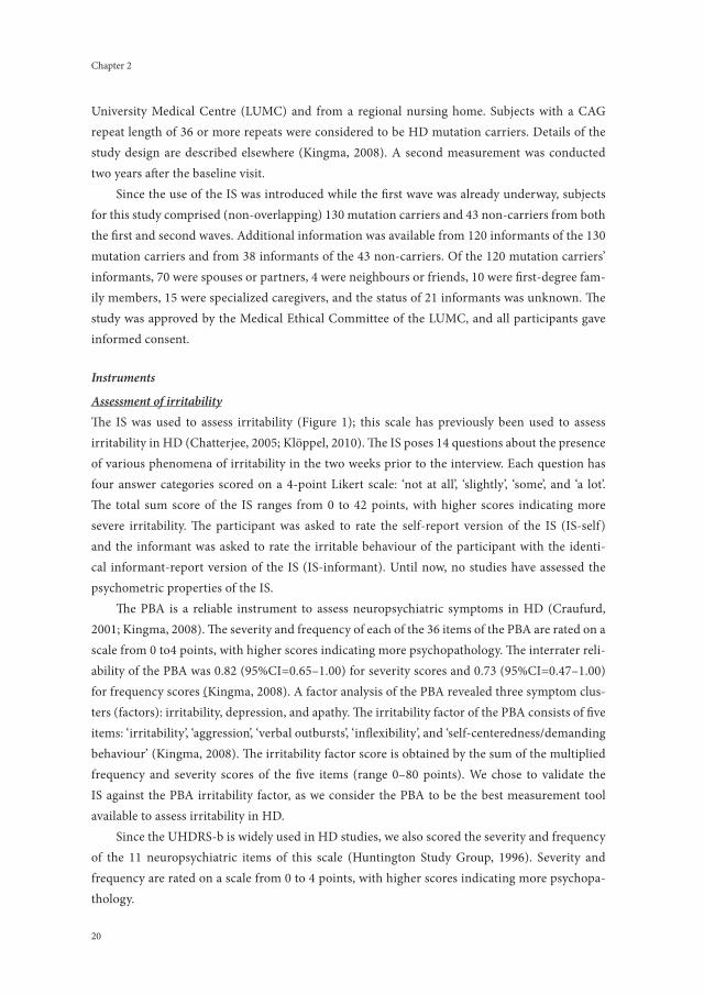
University Medical Centre (LUMC) and from a regional nursing home. Subjects with a CAG repeat length of 36 or more repeats were considered to be HD mutation carriers. Details of the study design are described elsewhere (Kingma, 2008). A second measurement was conducted two years after the baseline visit.
Since the use of the IS was introduced while the first wave was already underway, subjects for this study comprised (non-overlapping) 130 mutation carriers and 43 non-carriers from both the first and second waves. Additional information was available from 120 informants of the 130 mutation carriers and from 38 informants of the 43 non-carriers. Of the 120 mutation carriers’ informants, 70 were spouses or partners, 4 were neighbours or friends, 10 were first-degree fam-ily members, 15 were specialized caregivers, and the status of 21 informants was unknown. The study was approved by the Medical Ethical Committee of the LUMC, and all participants gave informed consent.
Instruments
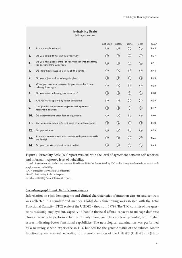
Assessment of irritabilityThe IS was used to assess irritability (Figure 1); this scale has previously been used to assess irritability in HD (Chatterjee, 2005; Klöppel, 2010). The IS poses 14 questions about the presence of various phenomena of irritability in the two weeks prior to the interview. Each question has four answer categories scored on a 4-point Likert scale: ‘not at all’, ‘slightly’, ‘some’, and ‘a lot’. The total sum score of the IS ranges from 0 to 42 points, with higher scores indicating more severe irritability. The participant was asked to rate the self-report version of the IS (IS-self) and the informant was asked to rate the irritable behaviour of the participant with the identi-cal informant-report version of the IS (IS-informant). Until now, no studies have assessed the psychometric properties of the IS.
The PBA is a reliable instrument to assess neuropsychiatric symptoms in HD (Craufurd, 2001; Kingma, 2008). The severity and frequency of each of the 36 items of the PBA are rated on a scale from 0 to4 points, with higher scores indicating more psychopathology. The interrater reli-ability of the PBA was 0.82 (95%CI=0.65–1.00) for severity scores and 0.73 (95%CI=0.47–1.00) for frequency scores (Kingma, 2008). A factor analysis of the PBA revealed three symptom clus-ters (factors): irritability, depression, and apathy. The irritability factor of the PBA consists of five items: ‘irritability’, ‘aggression’, ‘verbal outbursts’, ‘inflexibility’, and ‘self-centeredness/demanding behaviour’ (Kingma, 2008). The irritability factor score is obtained by the sum of the multiplied frequency and severity scores of the five items (range 0–80 points). We chose to validate the IS against the PBA irritability factor, as we consider the PBA to be the best measurement tool available to assess irritability in HD.
Since the UHDRS-b is widely used in HD studies, we also scored the severity and frequency of the 11 neuropsychiatric items of this scale (Huntington Study Group, 1996). Severity and frequency are rated on a scale from 0 to 4 points, with higher scores indicating more psychopa-thology.
21
Irritability in Huntington’s disease
Sociodemographic and clinical characteristicsInformation on sociodemographic and clinical characteristics of mutation carriers and controls was collected in a standardized manner. Global daily functioning was assessed with the Total Functional Capacity (TFC) scale of the UHDRS (Shoulson, 1979). The TFC consists of five ques-tions assessing employment, capacity to handle financial affairs, capacity to manage domestic chores, capacity to perform activities of daily living, and the care level provided, with higher scores indicating better functional capabilities. The neurological examination was performed by a neurologist with experience in HD, blinded for the genetic status of the subject. Motor functioning was assessed according to the motor section of the UHDRS (UHDRS-m) (Hun-
Are you easily irritated?
Do you pout if things don't go your way?
Do you have good control of your temper with the family (or persons living with you)?
Do little things cause you to fly off the handle?
Do you adjust well to a change in plans?
When you lose your temper, do you have a hard time calming down again?
Do you insist on having your own way?
Are you easily agitated by minor problems?
Irritability ScaleSelf-report version
1.
2.
3.
4.
5.
6.
7.
8.
9.
10.
11.
12.
13.
14.
Can you discuss problems together and agree to a reasonable solution?
Do disagreements often lead to arguments?
Can you appreciate a different point of view from yours?
Do you yell a lot?
Are you able to control your temper with persons outside the family?
0 1 2 3
0 1 2 3
3 2 1 0
0 1 2 3
3 2 1 0
0 1 2 3
0 1 2 3
0 1 2 3
3 2 1 0
0 1 2 3
3 2 1 0
0 1 2 3
3 2 1 0
0 1 2 3Do you consider yourself to be irritable?
not at all slightly some a lot
0.49
0.37
ICC*
0.31
0.44
0.43
0.38
0.28
0.38
0.47
0.40
0.30
0.59
0.25
0.45
figure 1 Irritability Scale (self-report version) with the level of agreement between self-reported and informant-reported level of irritability.* Level of agreement for each score between IS-self and IS-inf as determined by ICC with a 1-way random effects model with single-measure reliability. ICC = Intraclass Correlation Coefficients;IS-self = Irritability Scale self-report;IS-inf = Irritability Scale informant-report.

Chapter 2
22
tington Study Group, 1996). Mutation carriers with UHDRS Confidence Level score 0 or 1 were considered pre-motor symptomatic and mutation carriers with Confidence Level score >1 were considered motor symptomatic. Estimated duration of disease was calculated by the estimated age of onset according to the equation of Vassos et al.: ln [age of onset (years)] = 6.18 – 0.054 * [CAG repeats (number)] minus the current age(Vassos, 2008).
Assessment of cognitive functioningThe Mini-Mental Status Examination (MMSE) (Folstein, 1975), Symbol Digit Modalities Test (SDMT) (Smith, 1968), Verbal Fluency Test (VFT) (Hodges, 2009), and Stroop tests (Stroop, 1935) were administered to assess both global and frontal executive cognitive functioning.
Assessment of psychiatric disordersThe Dutch translation of the computerised version of the Composite International Diagnostic Interview (CIDI, version 2.1) (Robins, 1989) was used to assess the presence of a depressive dis-order according to the criteria of the Diagnostic and Statistical Manual of mental disorders, 4th edition (DSM-IV) (American Psychiatric Association, 2000). The CIDI was not administered in subjects with MMSE score <18 points, since the CIDI cannot be reliably administered to patients with severe cognitive dysfunction.
Statistical analyses
Data are presented as n (%), mean (± standard deviation (S.D.)), or median (inter-quartile range (IQR)) when appropriate. Chi-square tests for categorical data, t-tests for independent samples with normal distribution, or nonparametric Mann-Whitney U tests were conducted to compare mutation carriers and non-carriers. Convergent validity was assessed by Spearman’s correlation coefficient. Kruskal-Wallis tests were conducted to compare IS-self and IS-informant scores among the three groups of non-carriers, pre-motor symptomatic carriers, and motor symptom-atic mutation carriers. Cronbach’s alpha was assessed as a measure for internal consistency of the IS-self and IS-informant.
Because no known cut-off score exists for the presence of irritability as assessed with the IS, Receiver Operator Characteristic (ROC) analyses were performed against different cut-offs (i.e. 10, 15, and 20 points) of the irritability factor of the PBA that yielded optimal sensitivity and specificity. The area under the ROC curve (AUC) was used as an indicator of the discriminatory power of the IS to distinguish between irritable and non-irritable subjects according to the ir-ritability factor of the PBA.
Using univariate logistic regression analyses, mutation carriers with IS score ≥14 points were compared to those with IS score <14 points to determine correlates of irritability. Odds ratios (OR) and their corresponding 95% confidence interval (CI) were computed. Because of a non-normal distribution of TFC, UHDRS-m and MMSE scores, these data were dichotomized using a median split. To yield the independent correlates of irritability (IS-self), multiple lo-

23
Irritability in Huntington’s disease
gistic regression analysis with a forward selection procedure was used, selecting the following univariate correlates with P<0.10: being married/living together with a partner, CAG repeat length, TFC, use of benzodiazepines, and Stroop interference test. This model was adjusted for age and sex (i.e., forced into the model). In addition, the same variables were entered using the IS-informant score as the dependent variable. In sensitivity analyses, models were repeated using different cut-off scores of the IS.
Agreement between IS-self and IS-informant scores was assessed using one-way random, single measure intraclass correlation coefficients (ICCs). The same analysis was performed to assess the level of agreement on each of the 14 items of the IS. All tests were two-tailed with P<0.05 denoting statistical significance. The SPSS version 16.0 (SPSS Inc., Chicago, USA) was used for the analyses.
results
Sociodemographic, clinical, and neuropsychiatric characteristics
Table 1 presents the sociodemographic, clinical and neuropsychiatric characteristics of the 130 (39 pre-motor symptomatic and 91 motor symptomatic) HD mutation carriers and the 43 non-carriers. Mutation carriers were older and less often married/living together with a partner than non-carriers. Mutation carriers had significantly higher irritability scores (both IS-self and IS-informant) than non-carriers, and 45 (35%) of the 130 mutation carriers were irritable ac-cording to an IS-self score ≥14 points, compared to 4 (9%) of the 43 non-carriers. Although the CIDI assessment was not possible in 11 mutation carriers due to severe cognitive impairment (MMSE<18 points), there were 13 (10%) mutation carriers, of whom 8 had a major depressive disorder vs. one (2%) non-carrier with a psychiatric disorder.
Psychometric properties of the Irritability Scale
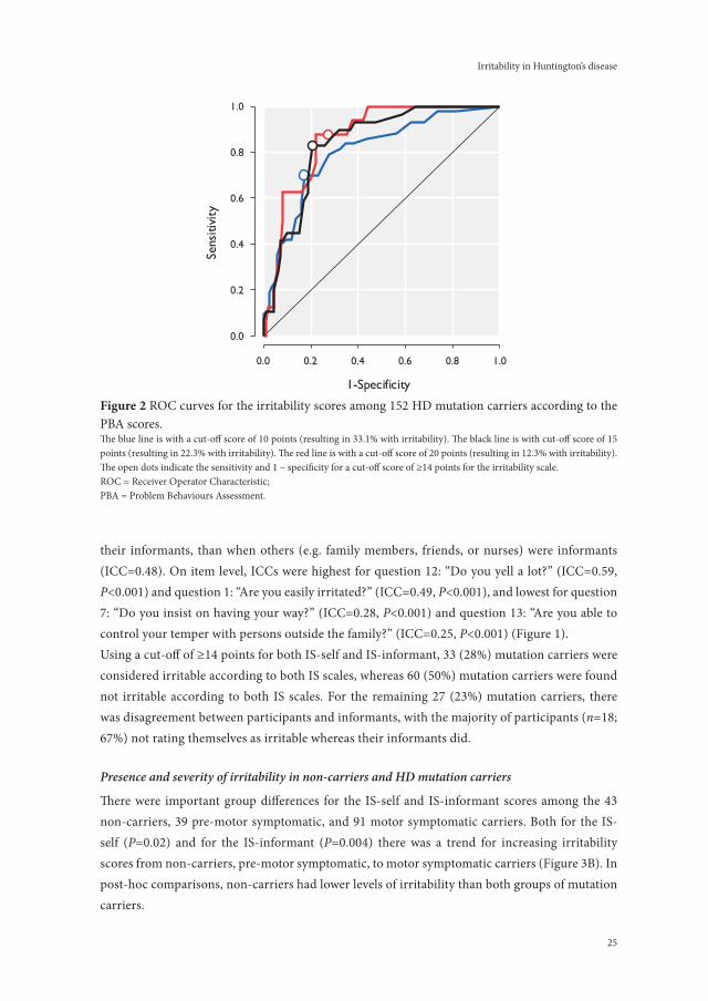
Cronbach’s alphas were 0.90 for the IS-self and 0.93 for the IS-informant. There was some evi-dence for convergent validity with the irritability item of the UHDRS-b indicated by Spearman’s correlation coefficient of 0.56 (P=0.001, n=32 with complete data for both scales). Using ROC analysis, a score of ≥14 points on the IS-self was identified as a robust indicator for irritability according to all three different cut-off points (10, 15, and 20 points) of the irritability factor of the PBA; the three cut-off points corresponded to prevalence rates of 33%, 22% and 12% of irritability (Table 2; Figure 2). The IS cut-off score of ≥14 points yielded an acceptable sensitivity and high specificity for all three cut-off points.
Level of agreement between the IS-self and IS-informant scores
The overall ICC for IS-self and IS-informant scores was 0.61 (95%CI=0.50–0.72, P<0.001) (Figure 3A). The ICC for IS-self and IS-informant was higher (ICC=0.75) when spouses/partners were

Chapter 2
24
table 1. Sociodemographic and clinical characteristics of HD mutation carriers and non-carriers.mutation carriers non-carriers p-value*(n = 130) (n = 43)
sociodemographic and clinical characteristicsMale (n, %) 58 (45) 20 (47) 0.83Age, years (mean ± SD) 49 ± 11 41 ± 11 < 0.001Higher level of education a (n, %) 78 (60) 31 (72) 0.17Married/living together (n, %) 81 (62) 35 (81) 0.02Excessive use of alcohol (n,%) 13 (10) 1 (2) 0.14CAG repeat length (mean ± SD) 44 ± 3 22 ± 4 < 0.001
neuropsychiatric characteristicsIS-self (median, IQR) 9 (3 – 17) 5 (2 – 9) 0.01IS-self with cut-off ≥ 14 (n, %) 45 (35) 4 (9) 0.001IS-inf (median, IQR) 11 (5 – 19) 4 (2 – 10) 0.01IS-inf with cut-off ≥ 14 (n, %) 51 (39) 5 (12) 0.001PBA irritability (IQR) 7 (1 – 16) 1 (0 – 5) < 0.001Any psychiatric disorder b (n, %) 13 (10) 1 (2) 0.10Major depressive disorder b (n, %) 8 (6) 0 (0) 0.09
Data are presented as n (%), mean (± Standard Deviation (SD)), or median (Inter Quartile Range (IQR)) when appropriate. * P-values by chi-square tests for catechorical data, by t-test for independent samples with normal distributions, or non-para-metric Whitney-U tests.a Higher education ≥ 12 years of education;b The presence of psychiatric disorders in the last two weeks are diagnosed with the Composite International Diagnostic In-terview;IS-self = Irritability Scale self report; IS-inf = Irritability Scale informant report;PBA = Problem Behaviours Assessment.
table 2. ROC analysis for IS-self scores among 152 HD mutation carriers against three different cut-off scores for PBA irritability factor.
PBA irritability cut-off score
> 10 points > 15 points > 20 points
Prevalence of irritability 33% 22% 12%
Optimal IS-self cut-off 13 – 14 13 – 14 13 – 14
Sensitivity 0.58 0.69 0.88
Specificity 0.84 0.81 0.78
AUC 0.80 0.84 0.87
ROC = Receiver Operator Characteristic;IS-self = Irritability Scale self-report;PBA = Problem Behaviours Assessment;AUC = Area Under the Curve.
25
Irritability in Huntington’s disease
their informants, than when others (e.g. family members, friends, or nurses) were informants (ICC=0.48). On item level, ICCs were highest for question 12: “Do you yell a lot?” (ICC=0.59, P<0.001) and question 1: “Are you easily irritated?” (ICC=0.49, P<0.001), and lowest for question 7: “Do you insist on having your way?” (ICC=0.28, P<0.001) and question 13: “Are you able to control your temper with persons outside the family?” (ICC=0.25, P<0.001) (Figure 1).Using a cut-off of ≥14 points for both IS-self and IS-informant, 33 (28%) mutation carriers were considered irritable according to both IS scales, whereas 60 (50%) mutation carriers were found not irritable according to both IS scales. For the remaining 27 (23%) mutation carriers, there was disagreement between participants and informants, with the majority of participants (n=18; 67%) not rating themselves as irritable whereas their informants did.
Presence and severity of irritability in non-carriers and HD mutation carriers
There were important group differences for the IS-self and IS-informant scores among the 43 non-carriers, 39 pre-motor symptomatic, and 91 motor symptomatic carriers. Both for the IS-self (P=0.02) and for the IS-informant (P=0.004) there was a trend for increasing irritability scores from non-carriers, pre-motor symptomatic, to motor symptomatic carriers (Figure 3B). In post-hoc comparisons, non-carriers had lower levels of irritability than both groups of mutation carriers.
1-Specificity
0.0 0.2 0.4 0.6 0.8 1.0
Sensitivity
0.0
0.2
0.4
0.6
0.8
1.0
figure 2 ROC curves for the irritability scores among 152 HD mutation carriers according to the PBA scores.The blue line is with a cut-off score of 10 points (resulting in 33.1% with irritability). The black line is with cut-off score of 15 points (resulting in 22.3% with irritability). The red line is with a cut-off score of 20 points (resulting in 12.3% with irritability). The open dots indicate the sensitivity and 1 – specificity for a cut-off score of ≥14 points for the irritability scale.ROC = Receiver Operator Characteristic;PBA = Problem Behaviours Assessment.

Chapter 2
26
Correlates of irritability in HD mutation carriers
Table 3 shows that mutation carriers with IS-self ≥14 points had a higher mean CAG repeat length (OR=1.16 per CAG triplet, 95%CI=1.02–1.30, P=0.02), a lower TFC score (OR=2.12, 95%CI=1.02–4.48, P=0.04), and more often used benzodiazepines (OR=2.67, 95%CI=1.20–5.92, P=0.02) compared to those with IS-self score <14 points.
In the multivariate logistic regression model, being married/living together (OR=2.85, 95%CI=1.19–6.83, P=0.02), CAG repeat length (OR=1.20 per CAG triplet, 95%CI=1.04–1.39, P=0.01), and the use of benzodiazepines (OR=3.28, 95%CI=1.36–7.89, P=0.008) were indepen-dent correlates of self-reported irritability (Table 4). Using the same model with the dichotomized IS-informant score as the dependent variable, the use of benzodiazepines was the only significant independent correlate of irritability (OR=3.54, 95%CI=1.45–8.64, P=0.005).
In sensitivity analyses, being married/living together and the use of benzodiazepines re-mained independent correlates of self-reported irritability when cut-off scores of IS-self ≥12 points and ≥16 points were used. However, CAG repeat length was no longer an independent correlate.
Irrit
abilit
y sc
ale:
self-
repo
rted
sco
re (p
oint
s)
0
10
20
30
40
Overall P=0.02
Irrit
abilit
y sc
ale:
info
rman
t-re
port
ed s
core
(po
ints
)
0
10
20
30
40
Overall P=0.004
Noncarriers Presymptomatic SymptomaticNoncarriers Presymptomatic Symptomatic
**
***B
0 10 14 20 30 42
0
10
14
20
30
42
ICC = 0.61P < 0.001
Irrit
abilit
y sc
ale:
info
rman
t-re
port
ed s
core
(poi
nts)
Irritability scale:self-reported score (points)
AN.S. N.S.
figure 3 Scatter plot representing the intercorrelation between IS-self and IS-inf scores; B. IS-self and IS-inf scores (median, IQR) in non-carriers, pre-motor symptomatic carriers and motor symp-tomatic mutation carriersA. Univariate regression line is shown, with the intraclass-correlation coefficient (ICC). Dotted lines indicate the cut-off score of ≥14 points being indicative of the presence of irritability.B. The line within the box represents the median and the boundaries of the box represent the interquartile range, while the error bars represent the 10th and 90th percentile values (P10 and P90).* p < 0.05; ** p < 0.005, n.s. = non-significant.

27
Irritability in Huntington’s disease
table 3. Sociodemographic, clinical, and neuropsychiatric characteristics as correlates of irritabil-ity in HD mutation carriers.
No irritability
(n = 85)
Irritability*
(n = 45)
Univariate logistic regressionOR (95%CI) p-value
Sociodemographic characteristics
Male (n, %) 38 (45) 20 (44) 0.99 (0.48 – 2.05) 0.98
Age, years (mean ± SD) 49 ± 11 48 ± 12 1.00 (0.97 – 1.03) 0.80
Higher level of education a (n, %) 53 (62) 26 (58) 0.83 (0.40 – 1.73) 0.61
Married/living together (n, %) 48 (57) 33 (73) 2.12 (0.96 – 4.66) 0.06
Clinical characteristics
Excessive use of alcohol (n, %) 6 (7) 6 (13) 2.03 (0.61 – 6.69) 0.25
CAG repeat length (mean ± SD) 44 ± 3 45 ± 3 1.16 (1.02 – 1.30) 0.02
TFC < 8.5 points (n, %) 37 (44) 28 (62) 2.12 (1.02 – 4.48) 0.04
UHDRS-m > 19 points (n, %) 38 (45) 26 (58) 1.69 (0.82 – 3.51) 0.16
Use of psychotropics (n, %) 39 (46) 29 (64) 2.14 (1.02 – 3.51) 0.16
- Antidepressants (n, %) 27 (32) 18 (40) 1.43 (0.68 – 3.04) 0.35
- Antipsychotics (n, %) 22 (26) 10 (22) 0.82 (0.35 – 1.92) 0.65
- Benzodiazepines (n, %) 17 (20) 18 (40) 2.67 (1.20 – 5.92) 0.02
Neuropsychiatric characteristics
IS-self (median, IQR) 5 (1 – 8.5) 20 (16.5 – 25) - -
IS-inf (median, IQR) 7 (3 – 12) 19 (12.5 – 27.5) - -
Major depressive disorder b (n, %) 3 (7) 5 (6) 1.15 (0.26 – 5.08) 0.85
MMSE < 28 points (n, %) 46 (54) 30 (67) 1.82 (0.85 – 3.90) 0.13
SDMT 0.08 (1.05) -0.12 (0.90) 0.80 (0.55 – 1.15) 0.23
VFT 0.01 (0.94) -0.02 (1.12) 0.98 (0.68 – 1.40) 0.89
Stroop word 0.14 (1.04) -0.26 (0.86) 0.66 (0.45 – 0.97) 0.11
Stroop colour 0.10 (1.05) -0.19 (0.88) 0.75 (0.51 – 1.08) 0.12
Stroop interference 0.11 (1.06) -0.21 (0.86) 0.73 (0.50 – 1.05) 0.09
Data are presented as n (%), mean (± Standard Deviation (SD)), or median (Inter Quartile Range (IQR)) when appropriate. Odds Ratios (ORs) with 95% Confidence Intervals (CI) and p-values by univariate logistic regression. *Irritability was considered present if IS-self ≥ 14 points, range 0 – 42 points. a Higher education ≥ 12 years of education;b The presence of major depressive disorder in the last two weeks are diagnosed with the Composite International Diagnostic Interview+TFC = Total Functioning Capacity;UHDRS-m = Unified Huntington’s Disease Rating Scale motor section;IS-self = Irritability Scale self-report;IS-inf = Irritability Scale informant-report;MMSE = Mini-Mental State Examination;SDMT = Symbol Digit Modality Test;VFT = Verbal Fluency Test;SDMT, VFT, and Stroop tests scores are in standardized z-scores.

Chapter 2
28
dIsCussIon
Using the optimal cut off score of IS ≥14 points, the prevalence of irritability in HD mutation carriers was 35%. There was a moderate level of agreement between mutation carriers and their informants in reporting irritability, with a tendency for mutation carriers to underestimate their level of irritability. Being married/living together, a higher CAG repeat length, and the use of benzodiazepines were independent correlates of self-reported irritability, whereas the use of ben-zodiazepines was the only independent correlate of both self-reported and informant-reported irritability.
Since there is no gold standard or formal criteria for the assessment of irritability, any cut-off point remains somewhat arbitrary. Therefore, we investigated the psychometric properties of the IS against the irritability factor of the PBA, an instrument especially developed for the assessment of behavioural problems in HD. ROC analysis showed that a cut-off score of ≥14 points was robust over three PBA cut-off scores. This cut-off score had face validity, since we considered it likely that irritable subjects would score at least 1 point on each of the 14 questions of the IS. In an earlier study using the IS (n=53), the median IS-self score of 15 points was used as a cut-off, defining irritability by IS>15 points; however, that study did not perform a ROC analysis (Chatterjee, 2005).
Whereas other (smaller) studies found prevalence rates between 38% and 73% for irritability in HD (van Duijn, 2007), we found a rather low prevalence. This may partly be explained by the different assessment tools used: the other studies all used non-specific measures for neuropsy-chiatric symptoms. Besides differences in methodology, sociodemographic and clinical factors – like the use of medication – may have contributed to the variation in prevalence of irritability. Unfortunately, the only two studies that used the IS do not report prevalence rates of irritability; however, these two studies report mean IS-self score of 14 points (Chatterjee, 2005), and 12 points (Klöppel, 2010), respectively. Furthermore, high levels of hostility may already be present before motor symptoms occur (Vassos, 2007), but prevalence of irritability may vary between
table 4. Independent correlates of self-reported and informant-reported irritability in HD muta-tion carriers.
Self-reported(n = 130)
Informant-reported(n = 120)
OR (95%CI) p-value OR (95%CI) p-value
Age 1.01 (0.98 – 1.06) 0.48 1.00 (0.96 – 1.04) 0.87
Male 0.98 (0.43 – 2.21) 0.96 1.73 (0.77 – 3.87) 0.18
Married/living together 2.85 (1.19 – 6.83) 0.02 1.75 (0.75 – 4.06) 0.19
CAG repeat length 1.20 (1.04 – 1.39) 0.01 1.14 (0.99 – 1.32) 0.06
Use of benzodiazepines 3.28 (1.36 – 7.89) 0.008 3.54 (1.45 – 8.64) 0.005
Odds Ratios (ORs) with 95% Confidence Intervals (CI) and p-values by multivariate logistic regression.

29
Irritability in Huntington’s disease
disease stages. So far, no significant differences between different disease stages have been found. In line with our results
Both previous studies using the IS assessed self-reported and informant-reported irritability. In the first study, agreement between (motor symptomatic) mutation carriers and informants re-garding the presence of irritability (median IS-self >15 points, median IS-informant >16 points) was moderate to poor (Chatterjee, 2005). Disagreement was stronger among mutation carriers with more intact cognition. The second study assessed irritability in 15 pre-motor symptomatic mutation carriers and found no significant differences between self-reported and informant-reported irritability (Klöppel, 2010). In the present study, we found moderate agreement between self-reported and informant-reported irritability. Mutation carriers tended to underestimate their level of irritability compared to their informants, since in 18 of the 27 cases with discor-dant scores, mutation carriers rated themselves as non-irritable (IS <14 points), whereas their informants scored above the cut-off. This may indicate denial or a lack of awareness by muta-tion carriers of their level of irritability. Since we did not ask informants of non-carriers to rate the level of irritability, we cannot conclude whether or not this is related to the disease itself. On the other hand, caregiver burden may be a source of disagreement between self-reported and informant-reported irritability, contributing to a possible overestimation of irritability by informants. However, there was a higher level of agreement between IS-self and IS-informant scores when spouses/partners rated the IS, than when other informants did so, suggesting a more correct estimation by the most intimate informants.
Of all the sociodemographic and clinical characteristics, being married/living together, CAG repeat length, and use of benzodiazepines were independent correlates of self-reported irritabil-ity. While most partners and other caregivers are extremely helpful and important for mutation carriers, a higher level of irritability may become more pronounced in intimate relationships that may comprise more potential triggers of increased irritability.
The CAG repeat length of mutation carriers was also independently correlated with self-reported irritability, but not with informant-reported irritability, whereas sensitivity analysis also showed that CAG repeat length was not an independent correlate. This is in line with studies that found no relationship between CAG repeat length of the HTT gene and any kind of psychopa-thology (Weigell-Weber, 1996; Zappacosta, 1996; Ravina, 2008; Vassos, 2008).
In the present study use of benzodiazepines was independently correlated to both self-re-ported and informant-reported irritability. Although benzodiazepines may have been prescribed more often to irritable mutation carriers, this cross-sectional study does not allow to draw conclusions about causality. Even if the occurrence of irritability in HD is (in part) iatrogenic and induced by the use of benzodiazepines, no longitudinal studies have examined the use of benzodiazepines and their effects on irritability in HD or other neurodegenerative diseases. Nevertheless, it is established that some patients may show paradoxical ‘aggressive’ behaviour with benzodiazepines, or behavioural disinhibition (Bond, 1995; Weisman, 1998; Lader, 1991).

Chapter 2
30
The strength of this study is the use of three different assessment methods for irritabil-ity, with standardized interviews, in a relatively large HD study population. However, some limitations also need to be addressed. First, in the absence of criteria or a gold standard for the assessment of irritability, we used the PBA for validation of the IS. Second, only subjects who volunteered to participate were included; this may have led to underestimation of the prevalence of irritability in HD, since irritable subjects were more likely to refuse to participate. Third, our study involved the analysis of cross-sectional data which precludes drawing conclusions about the direction of causality.
In conclusion, we recommend the use of the IS to assess irritability in HD in a standardized manner, since this scale proved to be a valid and easy to use instrument. Being married/living together and the use of benzodiazepines were independently associated with the presence of ir-ritability, although only the use of benzodiazepines was also correlated with informant- reported irritability and confirmed in the sensitivity analyses. Longitudinal studies are needed to further explore these relation- ships. Given the strong association between irritability and the use of benzodiazepines, close monitoring of the effect of benzodiazepines is important, since clear evidence for an effective treatment of irritability in HD is still lacking (van Duijn, 2010).
31
Irritability in Huntington’s disease
referenCes
Bond, A.J., Curran, H.V., Bruce, M.S., O’Sullivan, G., Shine, P., 1995. Behavioural aggression in panic disorder after 8 weeks’
treatment with alprazolam. Journal of Affective Disorders 35, 117-123.
Chatterjee, A., Anderson, K.E., Moskowitz, C.B., Hauser, W.A., Marder, K.S., 2005. A comparison of self-report and caregiver
assessment of depression, apathy, and irritability in Huntington’s disease. Journal of Neuropsychiatry and Clinical
Neurosciences 17, 378-383.
Craig, K.J., Hietanen, H., Markova, I.S., Berrios, G., 2008. The Irritability Questionnaire: a new scale for the measurement of
irritability. Psychiatry Research 159, 367-375.
Craufurd, D., Thompson, J.C., Snowden, J.S., 2001. Behavioral changes in Huntington Disease. Neuropsychiatry Neuropsy-
chology Behavioral Neurology 14, 219-226.
American Psychiatric Association, 2000. Diagnostic and Statistical Manual of Mental Disorders, 4th ed. Washington, DC.
Duff, K., Paulsen, J.S., Beglinger, L.J., Langbehn, D.R., Stout, J.C., and the Predict-HD Investigators of the Huntington Study
Group, 2007. Psychiatric symptoms in Huntington’s disease before diagnosis: the predict-HD study. Biological
Psychiatry 62, 1341-1346.
Folstein, M.F., Folstein, S.E., McHugh, P.R., 1975. Mini-mental state: a practical method for grading the cognitive state of
patients for the clinician. Journal Psychiatric Research 12, 189-198.
Hodges, J.R., 2009. Cognitive assessment for clinicians. 2nd ed. Oxford University Press, Oxford.
Huntington’s Disease Collaborative Research Group, 1993. A novel gene containing a trinucleotide repeat that is expanded
and unstable on Huntington’s disease chromosomes. Cell 72, 971-983.
Huntington Study Group, 1996. Unified Huntington’s Disease Rating Scale: reliability and consistency. Movement Disorders
11, 136-142.
Kingma, E.M., van Duijn, E., Timman, R., van der Mast, R.C., Roos, R.A.C., 2008. Behavioural problems in Huntington’s
disease using the Problem Behaviours Assessment. General Hospital Psychiatry 30, 155-161.
Klöppel, S., Stonnington, C.M., Petrovic, P., Mobbs, D., Tüscher, O., Craufurd, D., Tabrizi, S.J., Frackowiak, R.S.J., 2010.
Irritability in pre-clinical Huntington’s disease. Neuropsychologia 48, 549-557.
Kulisevsky, J., Litvan, I., Berthier, M.L., Pascual-Sedano, B., Paulsen, J.S., Cummings, J.L., 2010. Neuropsychiatric assessment
of Gilles de la Tourette patients: comparative study with other hyperkinetic and hypokinetic movement disorders.
Movement Disorders 16, 1098-1104.
Lader, M., Morton, S., 1991. Benzodiazepine problems. British Journal of Addiction 86, 823-828.
Murgod, U.A., Saleem, Q., Anand, A., Brahmachari, S.K., Jain, S., Muthane, U.B., 2001. A clinical study of patients with
genetically confirmed Huntington’s disease from India. Journal of the Neurological Sciences 190, 73-78.
Paulsen, J.S., Ready, R.E., Hamilton, J.M., Mega, M.S., Cummings, J.L., 2001. Neuropsychiatric aspects of Huntington’s
disease. Journal of Neurology, Neurosurgery, and Psychiatry 71, 310-314.
Ravina, B., Romer, M., Constantinescu, R., Biglan, K., Brocht, A., Kieburtz, K., Shoulson, I., McDermott, M.P., 2008. The
relationship between CAG repeat length and clinical progression in Huntington’s disease. Movement Disorders 23,
1223-1227.
Robins, L.N., Wing, J., Wittchen, H.-U., Helzer, J.E., Babor, T.F., Burke, J., Farmer, A., Jablensky, A., Pickens, R., Regier, D.A.,
Sartorius, N., Towle, L.H., 1989. The Composite International Diagnostic Interview: an epidemiologic instrument
suitable for use in conjunction with different diagnostic systems and in different cultures. Archives of General
Psychiatry 45, 1069-1077.
Shoulson, I., Fahn, S., 1979. Huntington disease: clinical care and evaluation. Neurology 29, 1-3.
Smith, A., 1968. The Symbol Digit Modalities Test: a neuropsychologic test for economic screening of learning and other
cerebral disorders. Learning Disorders 3, 83-91.
Snaith, R.P., Taylor, C.M., 1985. Irritability: definition, assessment and associated factors. British Journal of Psychiatry 147,
127-136.

Chapter 2
32
Stroop, J.R., 1935. Studies of interference in serial verbal reactions. Journal of Experimental Psychology 18, 643-662.
van Duijn, E., Kingma, E.M., van der Mast, R.C., 2007. Psychopathology in verified Huntington’s disease gene carriers.
Journal of Neuropsychiatry and Clinical Neurosciences 19, 441-448.
van Duijn, E., 2010. Treatment of irritability in Huntington’s disease. Current Treatment Options in Neurology 12, 424-433.
Vassos, E., Panas, M., Kladi, A., Vassilopoulos, D., 2007. Higher levels of extroverted hostility detected in gene carriers at risk
for Huntington’s disease. Biological Psychiatry 62, 1347-1352.
Vassos, E., Panas, M., Kladi, A., Vassilopoulos, D., 2008. Effect of CAG repeat length on psychiatric disorders in Huntington’s
disease. Journal of Psychiatric Research 42, 544-549.
Walker, F.O., 2007. Huntington’s disease. Lancet 369, 218-223.
Weigell-Weber, M., Schmid, W., Spiegel, R., 1996. Psychiatric symptoms and CAG expansion in Huntington’s disease.
American Journal of Medical Genetics 67, 53-57.
Weisman, A.M., Berman, M.E., Taylor, S.P., 1998. Effects of clorazepate, diazepam, and oxazepam on a laboratory measure-
ment of aggression in men. International Clinical Psychopharmacology 13, 183-188.
Zappacosta, B., Monza, D., Meoni, C., Austoni, L., Soliveri, P., Gellera, C., Alberti, R., Mantero, M., Penati, G., Caraceni, T.,
Girotti, F., 1996. Psychiatric symptoms do not correlate with cognitive decline, motor symptoms, or CAG repeat
length in Huntington’s disease. Archives of Neurology 53, 493-497.

Chapter 3
Irritability in a prospective cohort of huntington’s disease mutation carriers
J.A. Bouwens, E. van Duijn, R.C. van der Mast, R.A.C. Roos, E.J. Giltay
Journal of Neuropsychiatry and Clinical Neurosciences 2015 Summer;27(3):206-12

Chapter 3
34
aBstraCt
A key symptom of Huntington’s disease (HD), irritability contributes to a decline in functioning and to great distress in both patients and their caregivers. To identify mutation carriers prone to the development of irritability, this study aimed to investigate the course and temporal relation-ships between irritability and other neuropsychiatric symptoms. A cohort of 90 mutation carriers was followed for two years. Using the Irritability Scale, the incidence of irritability was 23% whereas irritability persisted in 70%. An increase in irritability was strongly associated with an increase in apathy.
35
Irritability in a prospective cohort of Huntington’s disease mutation carriers
IntroduCtIon
Neuropsychiatric symptoms, motor disturbances, and cognitive impairment are core symptoms of Huntington’s disease (HD) {1-3}, an autosomal dominant, neurodegenerative disorder resulting from an expanded trinucleotide cytosine-adenine-guanine (CAG) repeat in the HTT gene on chromosome 4 that codes for the mutant protein huntingtin. Although under normal circumstances huntingtin is present ubiquitously, its physiologic role is not fully elucidated. The mutant huntingtin probably confers a toxic gain of function, mainly resulting in striatal cell loss. The age of onset is widespread, ranging from early childhood to senescence, and is generally about 40 years. The duration between diagnosis of motor symptoms and death is around 20 years. Neuropsychiatric symptoms, such as irritability, apathy and depressed mood often precede the onset of motor symptoms.
Irritability is a common and key neuropsychiatric symptom of HD {4-6}, which can be characterized as a mood state predisposing towards anger, hostile appraisals, impatience, intolerance, poorly controlled anger and overt aggression {7;8}. Reduced control over temper frequently occurs, resulting in verbal or behavioral outbursts. However, irritability may also be present without observable manifestation. Irritability should be distinguished from the DSM-IV-TR diagnosis of intermittent explosive disorder, since the latter has to meet strict, observable criteria and is severe in its phenotype and consequences, whereas irritability may not even be visible to an observer. The subjective experience of irritability may be either brief or prolonged. In contrast to justifiable anger, verbal or behavioral outbursts originating from irritable mood are nonadaptive, complicate the interaction between the patient and his environment, and are always unpleasant for the individual. Irritability can occur early in the course of HD, e.g. up to 10 years before the onset of motor symptoms {9} and often contributes to a decline in personal and occupational functioning {10}. Moreover, irritability of mutation carriers may also cause great distress among others, affecting the wellbeing of their families and possibly contributing to the necessity of institutionalization {11}.
The prevalence of irritability ranges from 35% to 73%, depending on its definition, the as-sessment tools used, and the study populations {2;9;12}. In REGISTRY (a European multi-center prospective observational study including both manifest and premanifest HD mutation carriers) a prevalence of 19.1% of overt disruptive or aggressive behavior among was found among 1468 mutation carriers {13}. Two prospective studies conducted among 12 pre-motorsymptomat-ic{14}- and 111 motorsymptomatic{15} HD mutation carriers showed that irritability increased over time.
Cross-sectional studies of irritability in motor-symptomatic mutation carriers have found associations with other neuropsychiatric symptoms including impulsivity{4}, obsessive com-pulsive symptoms{16} and suicidal thoughts{17}. Using the 14-item Irritability Scale{18;19}, we earlier demonstrated an association between irritability and a low Total Functioning Capacity (TFC) score, use of benzodiazepines and more often being married or living together{2}.

Chapter 3
36
Since irritability can occur before the onset of motor symptoms, our cohort included both pre-motor symptomatic and motor symptomatic mutation carriers. Although cross-sectional associations with irritability have been investigated before {2;4;16;17}, to our knowledge, this is the first study investigating predictors of incident and persistence of irritability in HD, also this is the first study investigating correlates of change in irritability over time. Identification of mutation carriers prone to the development and persistence of irritability is required to provide adequate treatment and support of HD mutation carriers and their families. Therefore, this study investigates the course and temporal relationships between irritability and other neuropsychiat-ric symptoms in a cohort of 90 HD mutation carriers followed for two years.
methods
Participants
HD mutation carriers and first-degree non-carriers were recruited between May 2004 and August 2006 from the outpatient departments of Neurology and Clinical Genetics of Leiden University Medical Centre (LUMC) and from a regional nursing home specialized in HD. All participants underwent genetic testing and were considered HD mutation carriers with a CAG repeat length ≥ 36 repeats. A second measurement was conducted 2 years after the first measure-ment and a third measurement 2 years thereafter. The study design has been described in detail elsewhere{20}.
In the present longitudinal analysis, only the data of mutation carriers who participated in the second and third waves of this study are included, because the Irritability Scale {2} was introduced halfway during the first measurement. Here, the second wave is referred to as the ‘baseline measurement’ and the third wave as the ‘follow-up measurement’. A total of 121 muta-tion carriers completed the Irritability Scale at baseline and irritability was also assessed in 90 mutation carriers at follow-up. In total 32 mutation carriers dropped-out (Figure 1).
The study was approved by the medical ethical committee of the LUMC and informed consent was obtained from all participants.
Instruments
Assessment of irritabilityIrritability was assessed with the self-rated Irritability Scale (Appendix I) that consists of 14 questions addressing the presence of various phenomena of irritability in the two weeks prior to the interview, rated on a 4-point Likert scale {18}. The total sum score of the Irritability Scale ranges from 0 - 42 points with higher scores indicating more severe irritability. We used a cut-off score of > 13 points as indicative for the presence of irritability; this score was selected based on our previous cross-sectional study among 130 HD mutation carriers (largely overlapping with baseline data of the current study) where this cut-off provided optimal sensitivity and speci-

37
Irritability in a prospective cohort of Huntington’s disease mutation carriers
ficity according to ROC analyses {2}. The Cronbach’s alpha of the irritability scale is 0.90 and its sensitivity and specificity for detecting irritability in HD mutation carriers is 0.69 and 0.81, respectively {2}. We preferred the use of the Irritability Scale over the PBA irritability subscale because the Irritability Scale was designed specifically for the assessment of irritability in neu-rodegenerative disease. Also, the psychometric properties of the Irritability Scale are better than those of the PBA irritability subscale (Crohnbachs alpha of 0.90 vs. 0.67, respectively). Since no cut-off score for the PBA irritability subscale for the presence of irritability exists, a direct comparison is not possible. In our previous study however, we found that with a cut-off score on the PBA irritability factor of >15 points, a cut-off score of >13 point on the irritability score yielded a sensitivity of 0.69 and a specificity of 0.81.
Assessment of other neuropsychiatric characteristicsThe Problem Behaviors Assessment (PBA) was used to assess depression. The PBA is a semi-structured interview assessing the frequency and severity of 36 potential behavioral problems in HD in the month before the interview {4}. The interrater reliability of the Dutch version of the PBA is 0.82 for severity scores and 0.73 for frequency scores. We used the symptom factor de-pression (range 0-80) that was previously estimated by factor analysis of the PBA {5}. Apathy was
Current analysis
31 (26.2%) drop-out:16 refused3 due to severe dysarthria1 died from suicide9 died2 could not be contacted
Baseline:
121 mutation carriers who completed the irritability scale
participated during second wave
Follow-up:
90 mutation carriers who completed the irritability scale participated during third wave
152 mutation carriers participated during first wave
31 (20.4%) drop-out:18 refused4 due to severe dysarthria6 due to severe illness3 died
figure 1 Flow chart of inclusion of the study participants for the present analysis
Chapter 3
38
assessed with the Apathy Scale (Appendix II){21}; this scale consists of 14 questions, measuring different features of apathy in the 2 weeks before the interview. Caregivers’ information and the interviewers’ judgment was included in rating the Apathy Scale, since patients with apathy may lack adequate insight into their own symptoms. The Apathy Scale was chosen above the apathy factor of the PBA because, although no clear preference arises from the literature, the Apathy Scale has better psychometric properties than the PBA apathy subscale (Crohnbach’s alpha 0.89 vs. 0.84, respectively) and because the former was specifically designed for the assessment of apathy.
Sociodemographic and clinical characteristicsInformation on sociodemographic and clinical characteristics (including alcohol consumption, use of psychotropic medication and information about the household) was collected in a standard-ized manner. Global daily functioning was assessed using the Total Functional Capacity (TFC) scale {22} of the Unified Huntington’s Disease Rating Scale (UHDRS) {23}. Motor symptoms were assessed using the motor section of the UHDRS (UHDRS-m) by trained neurologists. Mu-tation carriers with an UHDRS-m confidence level 0-1 were considered pre-motorsymptomatic and mutation carriers with UHDRS-m confidence level >1 were considered motorsymptomatic. Global cognitive functioning was assessed with the Mini-Mental State Examination (MMSE) {24}. Executive cognitive functioning was assessed with the Symbol Digit Modalities Test (SDMT) {25}, the Verbal Fluency Test (VFT) {26} and Stroop tests {27}. Since there is a high level of multi-colinearity between most tests (Pearson’s r > 0,80), an aggregate variable for executive functioning, ExCog, was constructed by computing the mean of the standardized scores of the SDMT, VFT and Stroop tests.
Statistical analyses
Data are presented as n (%), mean (± standard deviation; S.D.) or median (inter-quartile range; IQR) when appropriate. Associations between sociodemographic-, clinical- and neuropsy-chiatric characteristics on the one hand, and incident and persistent irritability on the other were determined using univariate logistic regression; odds ratios (ORs) were computed for the incidence or persistence of irritability. For the assessment of incident irritability, we identified HD mutation carriers without irritability at baseline who were irritable at follow-up. Similarly, for the assessment of persistent irritability we identified HD mutation carriers who were irritable at baseline and remained irritable at follow-up. A multivariate logistic regression (adjusting for sex and age that were entered into the model) was conducted with variables with p < 0.10 in the initial univariate analyses. To assess associations between temporal changes of clinical- and neuropsychiatric characteristics on the one hand and irritability on the other, we conducted an additional multivariate linear regression analysis. Absolute changes (i.e. delta-values) in all clini-cal- and neuropsychiatric characteristics between baseline and follow-up were calculated. Linear regression analysis was used to assess whether changes in scores of clinical- or neuropsychiatric

39
Irritability in a prospective cohort of Huntington’s disease mutation carriers
characteristics as independent variables were associated with changes in the Irritability Scale score as the dependent variable. Since our investigation is of a explorative nature and we are not aware of known correlates of changes in irritability score, we decided to let a forward selection procedure select variables to be included in our final model based upon statistical strength. In the final model, the most robust relationships with changes in both irritability and its correlates were further analysed using regression analysis, with adjustment for sex, age, and use of antipsychot-ics. All tests were two-tailed with p<0.05 denoting statistical significance. The SPSS 20.0 Package (SPSS Inc. Chicago, IL)was used for the statistical analyses.
results
The 90 HD mutation carriers in the current study had an average age of 49 (SD 1.7 years) and 41 (46%) were males. Their mean TFC score was 8.6 (SD 4.4) and their mean UHDRS-m score was 25.3 (SD 2.47). Twenty-five (28%) mutation carriers were pre-motorsymptomatic and 64 (71%) were motorsymptomatic. UHDRS-m score and motorsymptomatic status was missing in 1 mutation carrier. The score on the Irritability Scale between baseline and follow-up showed no significant change: average increase 1.56 points; 95% confidence interval (CI) -0.23 to 2.89.
At baseline, of the 90 mutation carriers, 33 (37%) were irritable. During the 2-year follow-up, of the 57 non-irritable HD mutation carriers at baseline, 13 (23%) developed irritability and 23 (70%) with irritability at baseline were persistently irritable. Compared with mutation carriers who completed the follow-up, drop-outs had on average a lower TFC score (5.8 vs. 8.6, p=0.004), higher UHDRS-m score (40.4 vs. 25.1, p=0.007), lower MMSE score (23.7 vs. 26.9, p=0.001) and worse executive cognitive functioning (-0.53 vs. -0.13, p=0.04); however, there was no difference in their Irritability Scale score (10.1 vs. 10.6, p=0.78).
Of the 57 non-irritable HD mutation carriers at baseline, the 13 mutation carriers with incident irritability at follow-up had, at baseline, a longer mean CAG repeat length, a higher UHDRS-m score, , a lower MMSE score, lower scores on all executive cognitive measures includ-ing the ExCog variable, and more frequently used benzodiazepines, compared with HD mutation carriers who did not develop irritability. In a logistic regression models adjusting for sex and age, a longer CAG repeat length, a higher UHDRS-m score, a higher use of benzodiazepines, , a lower MMSE score and worse cognitive functioning at baseline were all associated with incident irritability at follow-up. (Table 1) Next, we built a final multivariate logistic model adjusting for all these significant correlates. In this model, all determinants lost their significance. (Table 1).
There were no significant differences in mutation carriers who showed remission of irritabil-ity compared to those with persistent (Table 2). In the final model, we adjusted for the use of antidepressants, SDMT score and Stroop color score, since these variables had a correlation with a p-value<0,10 in the univariate analyses. In this model, there were no significant associations.
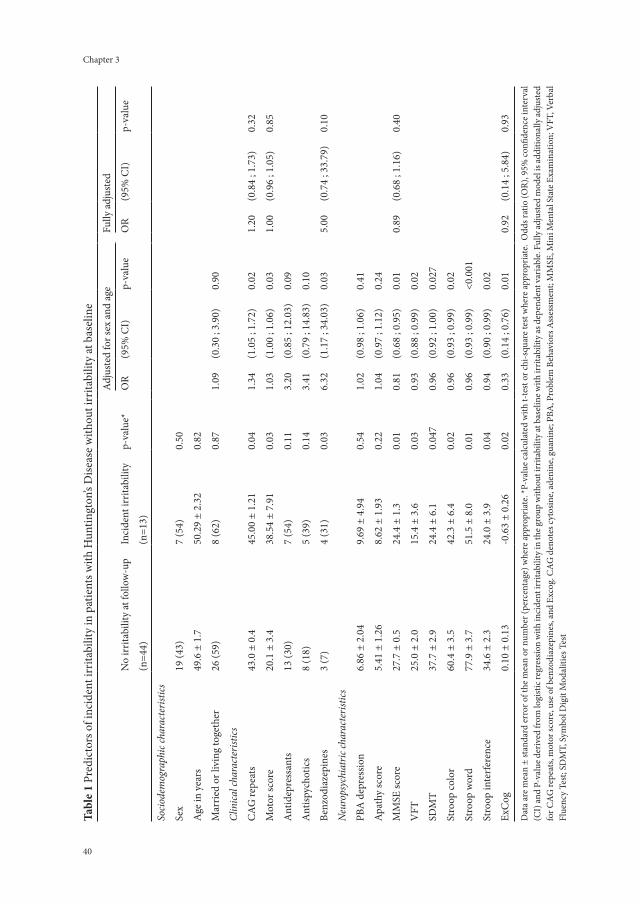
Chapter 3
40 41
Irritability in a prospective cohort of Huntington’s disease mutation carriers
tabl
e 1
Pred
icto
rs o
f inc
iden
t irr
itabi
lity
in p
atie
nts w
ith H
untin
gton
’s D
iseas
e w
ithou
t irr
itabi
lity
at b
asel
ine
Adj
uste
d fo
r sex
and
age
Fully
adj
uste
d
No
irrita
bilit
y at
follo
w-u
pIn
cide
nt ir
ritab
ility
p-va
lue*
OR
(95%
CI)
p-va
lue
OR
(95%
CI)
p-va
lue
(n
=44)
(n=1
3)
Socio
dem
ogra
phic
char
acte
risti
cs
Sex
19 (4
3)7
(54)
0.50
Age
in y
ears
49.6
± 1
.750
.29
± 2.
320.
82
Mar
ried
or li
ving
toge
ther
26 (5
9)8
(62)
0.87
1.09
(0.3
0 ; 3
.90)
0.90
Clin
ical c
hara
cter
istics
CA
G re
peat
s43
.0 ±
0.4
45.0
0 ±
1.21
0.04
1.34
(1.0
5 ; 1
.72)
0.02
1.20
(0.8
4 ; 1
.73)
0.32
Mot
or sc
ore
20.1
± 3
.438
.54
± 7.
910.
031.
03(1
.00
; 1.0
6)0.
031.
00(0
.96
; 1.0
5)0.
85
Ant
idep
ress
ants
13 (3
0)7
(54)
0.11
3.20
(0.8
5 ; 1
2.03
)0.
09
Ant
ispyc
hotic
s8
(18)
5 (3
9)0.
143.
41(0
.79
; 14.
83)
0.10
Benz
odia
zepi
nes
3 (7
)4
(31)
0.03
6.32
(1.1
7 ; 3
4.03
)0.
035.
00(0
.74
; 33.
79)
0.10
Neu
rops
ychi
atric
char
acte
risti
cs
PBA
dep
ress
ion
6.86
± 2
.04
9.69
± 4
.94
0.54
1.02
(0.9
8 ; 1
.06)
0.41
Apat
hy sc
ore
5.41
± 1
.26
8.62
± 1
.93
0.22
1.04
(0.9
7 ; 1
.12)
0.24
MM
SE sc
ore
27.7
± 0
.524
.4 ±
1.3
0.01
0.81
(0.6
8 ; 0
.95)
0.01
0.89
(0.6
8 ; 1
.16)
0.40
VFT
25.0
± 2
.015
.4 ±
3.6
0.03
0.93
(0.8
8 ; 0
.99)
0.02
SDM
T37
.7 ±
2.9
24.4
± 6
.10.
047
0.96
(0.9
2 ; 1
.00)
0.02
7
Stro
op co
lor
60.4
± 3
.542
.3 ±
6.4
0.02
0.96
(0.9
3 ; 0
.99)
0.02
Stro
op w
ord
77.9
± 3
.751
.5 ±
8.0
0.01
0.96
(0.9
3 ; 0
.99)
<0.0
01
Stro
op in
terf
eren
ce34
.6 ±
2.3
24.0
± 3
.90.
040.
94(0
.90
; 0.9
9)0.
02
ExC
og0.
10 ±
0.1
3-0
.63
± 0.
260.
020.
33(0
.14
; 0.7
6)0.
010.
92(0
.14
; 5.8
4)0.
93
Dat
a ar
e mea
n ±
stan
dard
erro
r of t
he m
ean
or n
umbe
r (pe
rcen
tage
) whe
re ap
prop
riate
. *P-
valu
e cal
cula
ted
with
t-te
st o
r chi
-squ
are t
est w
here
appr
opria
te.
Odd
s rat
io (O
R), 9
5% co
nfide
nce i
nter
val
(CI)
and
P-va
lue d
eriv
ed fr
om lo
gist
ic re
gres
sion
with
inci
dent
irrit
abili
ty in
the g
roup
with
out i
rrita
bilit
y at
bas
elin
e with
irrit
abili
ty as
dep
ende
nt v
aria
ble.
Fully
adju
sted
mod
el is
addi
tiona
lly ad
just
ed
for C
AG
repe
ats,
mot
or sc
ore,
use o
f ben
zodi
azep
ines
, and
Exc
og. C
AG
den
otes
cyto
sine,
aden
ine,
guan
ine;
PBA
, Pro
blem
Beh
avio
rs A
sses
smen
t; M
MSE
, Min
i Men
tal S
tate
Exa
min
atio
n; V
FT, V
erba
l Fl
uenc
y Te
st; S
DM
T, S
ymbo
l Dig
it M
odal
ities
Tes
t
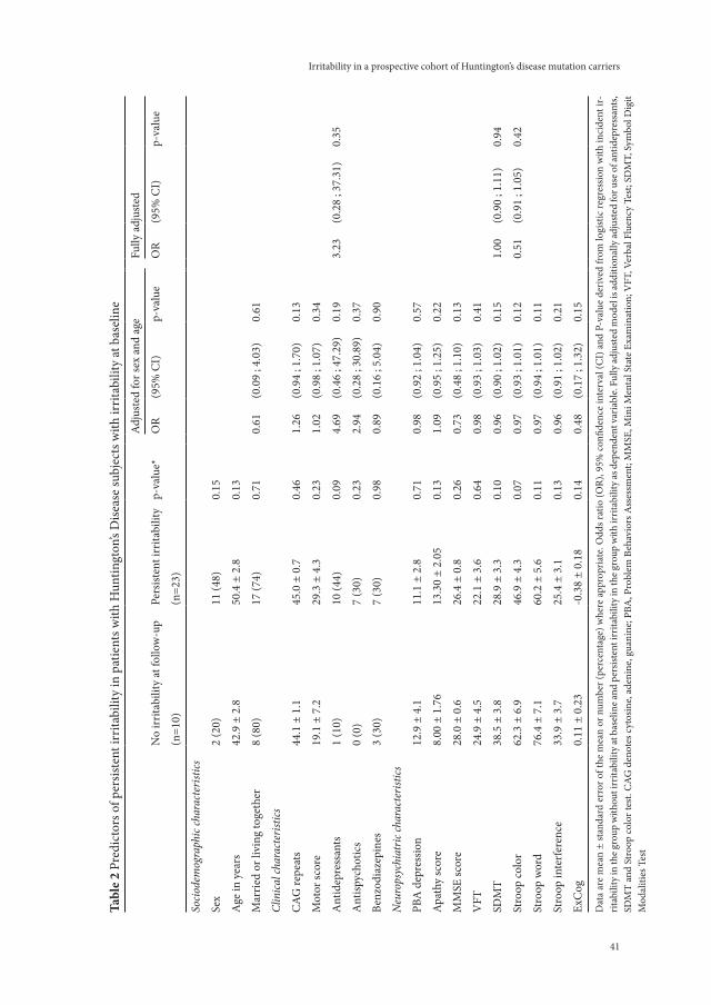
Chapter 3
40 41
Irritability in a prospective cohort of Huntington’s disease mutation carriers
tabl
e 2
Pred
icto
rs o
f per
siste
nt ir
ritab
ility
in p
atie
nts w
ith H
untin
gton
’s D
iseas
e su
bjec
ts w
ith ir
ritab
ility
at b
asel
ine
Adj
uste
d fo
r sex
and
age
Fully
adj
uste
d
No
irrita
bilit
y at
follo
w-u
pPe
rsist
ent i
rrita
bilit
yp-
valu
e*O
R(9
5% C
I)p-
valu
eO
R(9
5% C
I)p-
valu
e
(n
=10)
(n=2
3)
Socio
dem
ogra
phic
char
acte
risti
cs
Sex
2 (2
0)11
(48)
0.15
Age
in y
ears
42.9
± 2
.850
.4 ±
2.8
0.13
Mar
ried
or li
ving
toge
ther
8 (8
0)17
(74)
0.71
0.61
(0.0
9 ; 4
.03)
0.61
Clin
ical c
hara
cter
istics
CA
G re
peat
s44
.1 ±
1.1
45.0
± 0
.70.
461.
26(0
.94
; 1.7
0)0.
13
Mot
or sc
ore
19.1
± 7
.229
.3 ±
4.3
0.23
1.02
(0.9
8 ; 1
.07)
0.34
Ant
idep
ress
ants
1 (1
0)10
(44)
0.09
4.69
(0.4
6 ; 4
7.29
)0.
193.
23(0
.28
; 37.
31)
0.35
Ant
ispyc
hotic
s0
(0)
7 (3
0)0.
232.
94(0
.28
; 30.
89)
0.37
Benz
odia
zepi
nes
3 (3
0)7
(30)
0.98
0.89
(0.1
6 ; 5
.04)
0.90
Neu
rops
ychi
atric
char
acte
risti
cs
PBA
dep
ress
ion
12.9
± 4
.111
.1 ±
2.8
0.71
0.98
(0.9
2 ; 1
.04)
0.57
Apat
hy sc
ore
8.00
± 1
.76
13.3
0 ±
2.05
0.13
1.09
(0.9
5 ; 1
.25)
0.22
MM
SE sc
ore
28.0
± 0
.626
.4 ±
0.8
0.26
0.73
(0.4
8 ; 1
.10)
0.13
VFT
24.9
± 4
.522
.1 ±
3.6
0.64
0.98
(0.9
3 ; 1
.03)
0.41
SDM
T38
.5 ±
3.8
28.9
± 3
.30.
100.
96(0
.90
; 1.0
2)0.
151.
00(0
.90
; 1.1
1)0.
94
Stro
op co
lor
62.3
± 6
.946
.9 ±
4.3
0.07
0.97
(0.9
3 ; 1
.01)
0.12
0.51
(0.9
1 ; 1
.05)
0.42
Stro
op w
ord
76.4
± 7
.160
.2 ±
5.6
0.11
0.97
(0.9
4 ; 1
.01)
0.11
Stro
op in
terf
eren
ce33
.9 ±
3.7
25.4
± 3
.10.
130.
96(0
.91
; 1.0
2)0.
21
ExC
og0.
11 ±
0.2
3-0
.38
± 0.
180.
140.
48(0
.17
; 1.3
2)0.
15
Dat
a ar
e m
ean
± st
anda
rd e
rror
of t
he m
ean
or n
umbe
r (pe
rcen
tage
) whe
re a
ppro
pria
te. O
dds r
atio
(OR)
, 95%
con
fiden
ce in
terv
al (C
I) a
nd P
-val
ue d
eriv
ed fr
om lo
gist
ic re
gres
sion
with
inci
dent
ir-
ritab
ility
in th
e gro
up w
ithou
t irr
itabi
lity
at b
asel
ine a
nd p
ersis
tent
irrit
abili
ty in
the g
roup
with
irrit
abili
ty as
dep
ende
nt v
aria
ble.
Fully
adju
sted
mod
el is
addi
tiona
lly ad
just
ed fo
r use
of a
ntid
epre
ssan
ts,
SDM
T an
d St
roop
col
or te
st. C
AG
den
otes
cyt
osin
e, ad
enin
e, gu
anin
e; P
BA, P
robl
em B
ehav
iors
Ass
essm
ent;
MM
SE, M
ini M
enta
l Sta
te E
xam
inat
ion;
VFT
, Ver
bal F
luen
cy T
est;
SDM
T, S
ymbo
l Dig
it M
odal
ities
Tes
t

Chapter 3
42
We analyzed the association between changes in the clinical and neuropsychiatric char-acteristics with the change in Irritability Scale score. In univariate analyses of these changes, we found that an increase in UHDRS-m score, an increase in the Apathy Scale and the use of both antipsychotics and of benzodiazepines were associated with an increase in the Irritability Scale score over time. Next, we built a final multivariate regression model, comprising sex and age at baseline, increase in UHDRS-m score, Apathy Scale score, and the change in use of both antipsychotics and of benzodiazepines. We found that the association between change in Ir-ritability Scale score and change in Apathy Scale score persisted after adjustment for confounders (Figure 2). Furthermore the continuous use of antipsychotics was independently associated with an increase in Irritability Scale score.
-9
-6
-3
0
3
6
9
12
15
≤ –2 –2 to –1
-9
-6
-3
0
3
6
9
12
15
Chan
ge in
irrit
abili
ty (I
rrita
bilit
y Sc
ale)
Chan
ge in
irrit
abili
ty (I
rrita
bilit
y Sc
ale)
Change in apathy (Apathy Scale)Change in apathy (PBA apathy dimension)
b = 0.22P = 0.052
b = 0.41P < 0.001
–1 to 0 0 to 1 1 to 2 >2 ≤ –5 –4 to 0 1 to 5 6 to 10 11 to 15 >15
figure 2 Fully adjusted multivariate linear regression of changes in Irritability Scale score on changes in apathy as assessed with the PBA apathy factor (left) and the Apathy Scale (right)
table 3 Associations between changes in Irritability Scale score and changes in apathyChanges in Irritability Scale score
β (95% CI) p-value
Changes in Apathy Scale score
Crude 0.29 (0.08 ; 0.49) 0.01
Adjusted for sex and age 0.29 (0.09 ; 0.49) 0.01
Fully adjusted* 0.41 (0.19 ; 0.62) <0.001
Beta and 95% confidence interval (CI) derived from linear regression with change in irritability score between baseline and follow-up as dependent variable. *Fully adjusted model is also adjusted for increase in motor score, use of benzodiazepines and use of antipsychotics.
43
Irritability in a prospective cohort of Huntington’s disease mutation carriers
dIsCussIon
In this study, we investigated the course of irritability and its associations with clinical and neu-ropsychiatric symptoms in a cohort of 90 HD mutation carriers who were followed for 2 years. An increasing severity of apathy and the continuous use of antipsychotics were associated with increasing severity of irritability over 2 years.
A recent longitudinal study of neuropsychiatric symptoms followed 111 HD patients for a mean of 5 years {15}. At five moments of assessment during follow-up, a minimum of 49% of HD patients had one or more symptom of irritability and a maximum of 83% had one or more symp-tom of irritability. However, the proportions of any of these symptoms cannot be translated easily into a prevalence rate of the syndrome of irritability which we used in out analyses. Therefore, our incidence of 23% may be a good representation of new-onset irritability at 2-year follow-up in individuals with HD.
We found a link between apathy and irritability in HD, reflected by our finding that an in-crease in apathy coincided with an increase in irritability over a 2-year period. At first glance this may seem paradoxical. Apathy is a disorder of motivation that is mainly characterized by a lack of goal-directed behavior and cognition and by decreased emotional responsiveness. On the other hand, irritability has been characterized as a mood state predisposing toward negative or hostile appraisals and to the experience of anger. Although irritability may lead to the overt expression of anger, it may also only bother the patient himself without overt expression of irritability. In this paradigm, an individual with apathy may well experience irritability, a hypothesis which is supported by our study. An explanation for this comorbidity of apathy and irritability in HD may be that they have a related pathophysiology since both are caused by neural degeneration in subcortical and frontal circuits{28;29}, which is a key feature of HD, in particular the anterior cingulate cortex (ACC) and the orbitofrontal cortex (OFC). The ACC plays an important role in affect-regulation{30} as well as in decision making{31}. Lesions in the ACC, or in neuronal circuitry connected to it, have been associated with apathy{28} as well as behavioral inhibition, and aggression{28}. The orbitofrontal cortex is involved in impulse inhibition and lesions in the area of the OFC are associated with behavioral disinhibition and emotional lability {28}. In addition, hypoactivation of the OFC, as well as the ACC, is associated with impulsive aggres-sion {29}. Although emotional lability and impulsive aggression may be components of overt, observable irritability, these symptoms are absent in irritability that remains out of awareness and to our knowledge, the role of subcortical circuitry has not been investigated in this type of irritability. Therefore, the comorbidity of apathy and irritability may only be partly due to a related pathophysiology. Another factor to be considered is the possibility between construct overlap of irritability and apathy as measured by the Irritability and the Apathy Scales respec-tively. When looking at the items of both scales, there are no obvious similarities or overlapping items. Using Spearman’s correlation analyses, we found that the largest correlation coefficient was 0,44 between any of the 14 items of the Irritability scale and any of the 14 items of the Apathy
Chapter 3
44
Scale. This rather low correlation on any two single items suggest that phenomenological or scale reasons cannot account for the associations we found over time between irritability and apathy .
We found an association between the use of antipsychotics and increasing irritability. Al-though selective serotonin reuptake inhibitors are recommended as a first-step treatment, anti-psychotics are frequently prescribed for irritability and related behaviors {32}. Several underlying mechanisms may explain the association between the used of antipsychotics and irritability. On the one hand, antipsychotics may have been prescribed to treat (among others) irritability, likely resulting in confounding by indication as the treated group may on average still be more irritable than the untreated group. On the other hand, antipsychotics may have caused irritability or some other third factor may underlie the relationship. Furthermore, in some patients antipsychotics (both first-generation and second-generation) are known to cause akathisia, a state of psychomo-tor agitation{33}. Patients experiencing psychomotor agitation associated with akathisia, may either become irritable or may have reported positively on certain items on the Irritability Scale due to some overlap between the two constructs.
We assessed irritability using the Irritability Scale because this scale was used to assess ir-ritability in our earlier cross-sectional study{2}. On this scale, our previously validated cut-off score of 14 points for the presence of irritability enabled us to investigate the occurrence and correlates of incident and persistent irritability.
The strengths of this study are its prospective design, a relatively large cohort of HD mu-tation carriers, and the use of validated instruments for the assessments of determinants and outcomes. A few limitations should also be addressed. First, although we have validated the use of the Irritability Scale in our previous study, there is no gold standard for the assessment of irritability. Second, a relatively high number of mutation carriers dropped-out and, therefore, did not participate in the current analysis; irritability at follow-up may have been a reason not to participate. Selection bias may account for an underestimation of the incidence of irritability and for the underestimation of the predictive value of some predictors because of insufficient power. Third, we cannot exclude the possibility that cognitive dysfunction in our HD patients has distorted the reliability and sensitivity of scales that assessed of neuropsychiatric characteristics.
In conclusion, we found an unexpected association between irritability and apathy. HD mutation carriers that become more apathetic over time also seem to become more irritable. The association between irritability and apathy may imply a shared underlying pathophysiol-ogy between these neuropsychiatric symptoms in HD. Since apathy may mask irritability, we recommend asking mutation carriers with apathy and their caregivers about the symptoms of irritability in order to provide adequate support and treatment.

45
Irritability in a prospective cohort of Huntington’s disease mutation carriers
referenCes
1. Paulsen JS, Ready RE, Hamilton JM, et al: Neuropsychiatric aspects of Huntington’s disease. J Neurol Neurosurg
Psychiatry 2001;71: 310-314
2. Reedeker N, Bouwens JA, Giltay EJ, et al: Irritability in Huntington’s disease. Psychiatry Res 2012;200: 813-818
3. van Duijn E, Kingma EM, van der Mast RC: Psychopathology in verified Huntington’s disease gene carriers. J
Neuropsychiatry Clin Neurosci 2007;19: 441-448
4. Craufurd D, Thompson JC, Snowden JS: Behavioral changes in Huntington Disease. Neuropsychiatry Neuropsychol
Behav Neurol 2001;14: 219-226
5. Kingma EM, van Duijn E, Timman R, et al: Behavioural problems in Huntington’s disease using the Problem
Behaviours Assessment. Gen Hosp Psychiatry 2008;30: 155-161
6. Rickards H, De SJ, van WM, et al: Factor analysis of behavioural symptoms in Huntington’s disease. J Neurol
Neurosurg Psychiatry 2011;82: 411-412
7. Craig KJ, Hietanen H, Markova IS, et al: The Irritability Questionnaire: a new scale for the measurement of irritabil-
ity. Psychiatry Res 2008;159: 367-375
8. Snaith RP, Taylor CM: Irritability: definition, assessment and associated factors. Br J Psychiatry 1985;147: 127-136
9. Julien CL, Thompson JC, Wild S, et al: Psychiatric disorders in preclinical Huntington’s disease. J Neurol Neurosurg
Psychiatry 2007;78: 939-943
10. Hamilton JM, Salmon DP, Corey-Bloom J, et al: Behavioural abnormalities contribute to functional decline in
Huntington’s disease. J Neurol Neurosurg Psychiatry 2003;74: 120-122
11. Wheelock VL, Tempkin T, Marder K, et al: Predictors of nursing home placement in Huntington disease. Neurology
2003;60: 998-1001
12. van Duijn E, Kingma EM, van der Mast RC: Psychopathology in verified Huntington’s disease gene carriers. J
Neuropsychiatry Clin Neurosci 2007;19: 441-448
13. Orth M, Handley OJ, Schwenke C, et al: Observing Huntington’s Disease: the European Huntington’s Disease
Network’s REGISTRY. PLoS Curr 2010;2: RRN1184
14. Kirkwood SC, Siemers E, Viken R, et al: Longitudinal personality changes among presymptomatic Huntington
disease gene carriers. Neuropsychiatry Neuropsychol Behav Neurol 2002;15: 192-197
15. Thompson JC, Harris J, Sollom AC, et al: Longitudinal evaluation of neuropsychiatric symptoms in Huntington’s
disease. J Neuropsychiatry Clin Neurosci 2012;24: 53-60
16. Anderson KE, Gehl CR, Marder KS, et al: Comorbidities of obsessive and compulsive symptoms in Huntington’s
disease. J Nerv Ment Dis 2010;198: 334-338
17. Wetzel HH, Gehl CR, Dellefave-Castillo L, et al: Suicidal ideation in Huntington disease: the role of comorbidity.
Psychiatry Res 2011;188: 372-376
18. Chatterjee A, Anderson KE, Moskowitz CB, et al: A comparison of self-report and caregiver assessment of depres-
sion, apathy, and irritability in Huntington’s disease. J Neuropsychiatry Clin Neurosci 2005;17: 378-383
19. Kloppel S, Stonnington CM, Petrovic P, et al: Irritability in pre-clinical Huntington’s disease. Neuropsychologia
2010;48: 549-557
20. van Duijn E, Kingma EM, Timman R, et al: Cross-sectional study on prevalences of psychiatric disorders in mu-
tation carriers of Huntington’s disease compared with mutation-negative first-degree relatives. J Clin Psychiatry
2008;69: 1804-1810
21. Starkstein SE, Fedoroff JP, Price TR, et al: Apathy following cerebrovascular lesions. Stroke 1993;24: 1625-1630
22. Shoulson I, Fahn S: Huntington disease: clinical care and evaluation. Neurology 1979;29: 1-3
23. Huntington Study Group: Unified Huntington’s Disease Rating Scale: reliability and consistency. Huntington Study
Group. Mov Disord 1996;11: 136-142

Chapter 3
46
24. Folstein MF, Folstein SE, McHugh PR: “Mini-mental state”. A practical method for grading the cognitive state of
patients for the clinician. J Psychiatr Res 1975;12: 189-198
25. Smith A: The Symbol Digit Modalities Test: a neuropsychological test for economic screening of learning and other
cerebral disorders. Learn Disord 1968;3: 83-91
26. Hodges JR: Initiation: verbal fluency tests. Cognitive Assessment for Clinicians. Oxford University Press, Oxford,
United Kingdom.; 2003:118-119
27. Stroop JR: Studies of interference in serial verbal reactions. Journal of Experimental Psychology 1935;18: 643-662
28. Bonelli RM, Cummings JL: Frontal-subcortical circuitry and behavior. Dialogues Clin Neurosci 2007;9: 141-151
29. Miller LA, Collins RL, Kent TA: Language and the modulation of impulsive aggression. J Neuropsychiatry Clin
Neurosci 2008;20: 261-273
32. van Duijn E: Treatment of Irritability in Huntington’s Disease. Curr Treat Options Neurol 2010;12: 424-433
30. Stevens FL, Hurley RA, Taber KH: Anterior cingulate cortex: Unique role in cognition and emotion. J Neuropsy-
chiatry Clin Neurosci 2011;23:121-125
31. Rosenbloom MH, Schmahmann JD, Price BH: The functional neuroanatomy of decision-making. J Neuropsychia-
try Clin Neurosci 2012;24:266-277
33. Kane JM, Fleischhacker WW, Hansen L, et al: Akathisia: an updated review focusing on second-generation antipsy-
chotics. J Clin Psychiatry 2009;70: 627-643

Chapter 4
acute-phase proteins in relation to neuropsychiatric symptoms and use of psychotropic medication in huntington’s disease
J.A. Bouwens, A.A. Hubers, E. van Duijn, C.M. Cobbaert, R.A.C. Roos, R.C. van der Mast, E.J. Giltay
European Neuropsychopharmacology.2014 Aug;24(8):1248-56
Chapter 4
48
aBstraCt
Activation of the innate immune system has been postulated in the pathogenesis of Huntington’s disease (HD). We studied serum concentrations of C-reactive protein (CRP) and albumin as positive and negative acute-phase proteins in HD. Multivariate linear and logistic regression was used to study the association between acute-phase protein levels in relation to clinical, neuropsychiatric, cognitive, and psychotropic use characteristics in a cohort consisting of 122 HD mutation carriers and 42 controls at first biomarker measurement, and 85 HD mutation carriers and 32 controls at second biomarker measurement. Significant associations were found between acute-phase protein levels and Total Functioning Capacity (TFC) score, severity of apathy, cognitive impairment, and the use of antipsychotics. Interestingly, all significant results with neuropsychiatric symptoms disappeared after additional adjusting for antipsychotic use. High sensitivity CRP levels were highest and albumin levels were lowest in mutation carriers who continuously used antipsychotics during follow-up versus those who had never used anti-psychotics (mean difference for CRP 1.4 SE mg/L; P = 0.04; mean difference for albumin 3 SE g/L; P < 0.001). The associations found between acute-phase proteins and TFC score, apathy, and cognitive impairment could mainly be attributed to the use of antipsychotics. This study provides evidence that HD mutation carriers who use antipsychotics are prone to develop an acute-phase response.

49
Acute-phase proteins in relation to neuropsychiatric symptoms and use of psychotropic medication in Huntington’s disease
IntroduCtIon
Huntington’s disease (HD) is an autosomal dominant neurodegenerative disease, characterized by motor abnormalities, cognitive dysfunction, and neuropsychiatric symptoms1 including depressed mood,2 apathy,3;4 and irritability.5 Although the mean age of onset is 40 years, there is a wide range in age at onset, severity, and symptomatology of HD. HD is caused by an expanded trinucleotide cytosine-adenine-guanine (CAG) repeat on chromosome 4 coding for the mutant huntingtin protein.6 Although the genetic defect has been elucidated, its role in the pathogenesis of HD remains unclear.
There is increasing evidence that the immune system may be involved in the pathogenesis of HD. Increased activation of components of the innate and adaptive immune systems has been found in the central nervous system (CNS)7-9 and peripheral tissues of HD patients.10 In par-ticular, increased concentrations of tumor necrosis factor (TNF)-α and interleukin (IL)-6, have been found in plasma of HD mutation carriers when compared to controls.11 Physiologically, TNF-α and IL-6 are pro-inflammatory cytokines which are released in the bloodstream as part of the acute-phase response.12;13 C-reactive protein (CRP) synthesis is upregulated in the liver under regulation of IL-6,14 whereas the synthesis of other proteins such as albumin is down-regulated. Since in the acute-phase response CRP levels increase, CRP can be thought of as a positive acute-phase protein and since albumin levels decrease, albumin can be thought of as a negative acute-phase protein.
Previously, CRP levels have been reported to be significantly increased in HD mutation carriers in advanced disease stage when compared to controls.15 Levels of CRP were significantly higher in a group of 13 HD mutation carriers when compared to 10 healthy controls. Also, levels of CRP were higher in HD mutation carriers with more advanced disease.16 A large set of markers for inflammation and innate immunity was measured in a recent study that included two cohorts comprising 81 HD mutation carriers and 40 controls, but none of the more than 20 plasma constituents were significantly different between the groups.17 Nevertheless, post-hoc tests revealed significantly lower CRP levels in HD mutation carriers with early disease versus controls, whereas premanifest HD had similar levels compared with controls.
In various other populations, there are reports indicating an association between CRP18-20 and albumin21-24 levels on the one hand, and both cognitive dysfunction and neuropsychiatric symptoms on the other hand. In a systematic review assessing the association between CRP level and stroke, cognitive disorders, and depression,18 raised CRP concentrations were associated with cognitive decline and increased risk of dementia. Low albumin levels were associated with cognitive dysfunction in two large population-based studies of the non-demented elderly.21;22 In a large longitudinal study with 73,131 participants from the general population, a positive association was found between CRP levels and depressive symptoms.19 Also, a meta-analysis showed a positive correlation between CRP levels and depression (d = 0.15; 95% confidence interval [CI] 0.10 – 0.21).20 Low albumin levels were found in depressed patients compared to
Chapter 4
50
healthy controls.23;24 Cross-sectionally, a significant association was found between CRP levels and apathy in elderly individuals.25 Longitudinally, however, higher CRP levels were not associ-ated with an increased risk of apathy in individuals aged 85 years and over.26
Antipsychotics are frequently prescribed for symptomatic treatment of chorea and neu-ropsychiatric symptoms in HD.27 It has become apparent that the use of antipsychotics may adversely affect CRP levels.23;28;29 In addition, weight gain is a well-known side-effect of treatment with atypical antipsychotics,30 which is associated with an increase of IL-6 and other proteins associated with low-grade inflammation.31 The effects of antipsychotics on levels of albumin have not been thoroughly studied, although serum albumin was decreased in a group of schizophrenic patients when compared with healthy controls. However, no significant effect of the use of anti-psychotics was demonstrated.32
This study aims to investigate CRP levels as a positive acute-phase protein and albumin levels as a negative acute-phase protein in relation to clinical, neuropsychiatric, and cognitive characteristics in a cohort of HD mutation carriers. For this, we took into account factors that might have confounded or otherwise influenced these associations, in particular the use of antipsychotics. Our hypothesis was that, with disease progression, inflammatory activity would increase leading to higher CRP levels and lower albumin levels, associated with a higher preva-lence of cognitive impairment and neuropsychiatric symptoms.
experImental proCedures
Population
Between May 2004 and August 2006, 152 HD mutation carriers and 56 controls were recruited for participation. Sources for recruitment were the outpatient departments of Neurology and Clinical Genetics of the Leiden University Medical Center (LUMC) and a regional nursing home specialized in care for HD patients. Besides HD mutation carriers, verified first-degree non-carriers participated in the study as controls. The study design is described in detail elsewhere.33 A second measurement was conducted two years after the baseline visit (including 128 mutation carriers and 42 controls) and a third measurement two years thereafter (including 94 HD muta-tion carriers and 32 controls). Blood samples suitable for the determination of high sensitivity CRP (hsCRP) and albumin levels were available at the second and the third measurement. Since 6 participants did not give blood at the first biomarker measurement (defined as t1) they were ex-cluded from the analyses, which resulted in 122 mutation carriers at t1. At the second biomarker measurement (defined as t2), 9 participants did not give blood, which resulted in 85 mutation carriers at t2.

51
Acute-phase proteins in relation to neuropsychiatric symptoms and use of psychotropic medication in Huntington’s disease
Sample withdrawal and biochemical analyses
At t1 and t2 EDTA blood was withdrawn between the years 2008 and 2012 and subsequently stored at -80 ºC within 2 h. Blood samples were stored until measurement of hsCRP and albumin levels in the fall of 2012. hsCRP was determined in EDTA plasma with an IFCC standardized method (Cat. no. 04628918190) on a COBAS INTEGRA 800 analyzer from Roche Diagnostics, according to the instructions of the manufacturer. The expected reference range for hsCRP in adult males and females is < 5 mg/L. Between-run coefficients of variation (CVs) during the study period (12 runs) were 5.8% at 4.09 mg/L, and 5.7% at 12.81 mg/L. Albumin in EDTA plasma was determined with an IFCC standardized method (Cat. no. 11970909216) on Modular P systems from Roche Diagnostics, using a BCG-based colorimetric assay with endpoint detection. This method has been standardized against the CRM 470 reference preparation. The reference range for serum albumin in adult males and females is 34–48 g/L. CVs during the study period (7 runs) were 2.94% at 37.5 g/L and 1.01% at 55.0 g/L.
Sociodemographic and clinical characteristics
Information on sociodemographic and clinical characteristics, including the potential con-founders like current smoking, high alcohol consumption (defined as drinking more than 14 standardized units of alcohol a week), body weight and height, and medication use, was collected using a standardized interview. Use of antipsychotics was divided in different categories: typical antipsychotics, atypical antipsychotics, and tiapride. Tiapride was considered a separate category as it was used by many participants and cannot be classified unambiguously within one of the other categories. The estimated duration of disease was calculated by the current age minus the estimated age of onset according to the equation of Vassos.34 The Total Functional Capacity (TFC) scale of the Unified Huntington’s Disease Rating Scale (UHDRS)35 was used to assess global daily functioning and disease stage.36 Total scores range from 0 to 13 points, with higher scores indicating better global functioning.37
Motor functioning was assessed by a trained neurologist using the motor scale of the UH-DRS.35 Total scores range from 0 to 124, with higher scores indicating worse motor functioning. The diagnostic Confidence Level (CL) of the UHDRS motor scale was used to define mutation carriers as pre-motor symptomatic (CL 0 or 1 point) or motor symptomatic (CL 2 to 4 points).
Neuropsychiatric characteristics
Neuropsychiatric characteristics were assessed by the Dutch version of the Problem Behaviours Assessment (PBA),38 a semi-structured interview, assessing the frequency and severity of 36 potential behavioral problems in HD. The inter-rater reliability of the Dutch version of the PBA is 0.82 for severity scores and 0.73 for frequency scores.39 In this study we used symptom factors that were previously estimated by factor analysis of the PBA, which resulted in three factors: apathy (range 0–64), irritability (range 0–80), and depression (range 0–80).39 Additionally, sui-cidality was assessed with the PBA38 by multiplying the severity and frequency score of the item
Chapter 4
52
‘suicidal ideation’. As done before, a total score > 1 point on this item was used to characterize the presence of suicidality.40
Cognitive characteristics
The Mini-Mental State Examination (MMSE) was used to assess global cognitive functioning. Total scores range from 0 to 30 points, with higher scores indicating better global cognitive functioning.41 The cognitive scales of the UHDRS,35 including the Verbal Fluency Test (VFT),42 the Symbol Digit Modalities Test (SDMT),43 and the Stroop tests,44 were used to assess executive cognitive functioning. For all cognitive scales, higher scores indicate better executive function-ing. The ExCog, a composite variable obtained by averaging the standardized z-scores of the other executive cognitive subscales, was used as a measure for executive cognitive functioning.
Statistical analyses
Data are presented as n (%), mean (± standard deviation [SD]), mean (95% CI), or median (interquartile range [IQR]) when appropriate.
Because of their positively skewed distribution, hsCRP levels were log transformed before being used in statistical analyses. Characteristics of HD mutation carriers and controls at t1 were compared by chi-squared tests for categorical data, t-tests for independent samples with normal distributions, or non-parametric Whitney-U tests for continuous variables without normal distributions.
Associations between hsCRP and albumin levels and clinical, neuropsychiatric, and cogni-tive characteristics were determined by linear regression analysis for continuous outcomes and logistic regression analysis for dichotomous outcomes. Apart from the crude model, we adjusted in the first multivariate models for the potential confounders sex, age, body mass index (BMI), smoking, and alcohol consumption at t1. Since previous literature indicated that the use of antipsychotics may affect CRP levels,23;28;29 a second model was built, including the variables from the adjusted model and the use of antipsychotics. Furthermore, analysis of covariance (ANCOVA) was used to compare acute-phase protein levels between participants who did not use antipsychotics, participants who started using antipsychotics during the study period, and participants who continuously used antipsychotics, with adjustment for sex, age, BMI, smoking, and alcohol consumption at t1. In an additional model comparing acute-phase protein levels at t2 between the different groups, we additionally adjusted for acute-phase protein levels at t1. As a sensitivity analysis, this ANCOVA was repeated without participants whose hsCRP level was above 10 mg/L, due to their possible association with acute infectious diseases. Since almost all participants who used antipsychotics were in later disease stages and disease stage may also be related to the acute-phase response, we performed a sensitivity analysis which only included the participants with a disease stage > 2 (TFC score < 7) at t1. As previous research showed that the effect of antipsychotic use on CRP levels is different for different kinds of antipsychotics,28 we performed a mixed model analysis (i.e., multilevel regression analysis) with adjustment for age,
53
Acute-phase proteins in relation to neuropsychiatric symptoms and use of psychotropic medication in Huntington’s disease
sex, BMI, smoking, and alcohol consumption at t1, to evaluate associations between the different categories of antipsychotics and acute-phase protein levels.
A p-value < 0.05 was considered statistically significant. p-Values presented were not cor-rected for multiple comparisons, since we interpreted the overall pattern of diminishing effect sizes and p-values after additional adjustment for the use of antipsychotics in multivariable models. SPSS version 20.0 was used.
results
Characteristics of mutation carriers versus controls
At t1, mutation carriers more often used psychotropic medication, had higher scores on the apathy, irritability, and depression factors of the PBA and worse scores on all cognitive tests, compared with controls (Table 1). When comparing acute-phase proteins, the hsCRP level of the mutation carriers at t1 was non-significantly higher in mutation carriers compared with controls (Table 1). At t2, the hsCRP level of mutation carriers was significantly higher compared with controls (mean: 1.94; 95% CI: 1.50–2.51 versus 0.99; 95% CI: 0.64–1.52, respectively; p = 0.002) (data not shown). There was a significant trend of increasing hsCRP levels across increas-ing disease stages at t1 and t2 (Suppl. Table 1). The albumin level of the mutation carriers was significantly lower compared with controls, both at t1 (Table 1) and t2 (mean: 46.4; 95% CI: 45.8–47.1 versus 47.6; 95% CI: 46.5–48.7, respectively; p = 0.03) (data not shown). However, after correction for potential confounders (sex, age, BMI, smoking, and alcohol consumption) only the hsCRP level at t2 remained significantly different between mutation carriers and controls. There was a significant trend of decreasing albumin levels across increasing disease stages at t1 and t2 (Suppl. Table 1).
Associations between acute-phase proteins and clinical characteristics at t1
Serum hsCRP level was significantly associated with TFC score, apathy score (Table 2), benzodi-azepine use, and antipsychotic use (Table 3). Using multivariate linear regression analyses, with adjustment for potential confounders, the hsCRP level remained significantly associated with apathy score, Stroop interference test, and ExCog (Table 2). However, after additional adjustment for use of antipsychotics, all these associations lost their statistical significance (Tables 2 and 3).
Serum albumin level was independently associated with TFC score, apathy score, SDMT score, the Stroop test scores, ExCog (Table 2), MMSE score, use of benzodiazepines, and use of antipsychotics (Table 3). Likewise, after additional adjustment for use of antipsychotics, all these associations lost their statistical significance (Tables 2 and 3).

Chapter 4
54
table 1. Characteristics of HD mutation carriers and controls at first biomarker measurement (t1).Mutation carriers Controls p-valuea
n = 122 n = 42Sociodemographic characteristicsMale gender 54 (44%) 19 (45%) 0.91Age (years) 49.2 ± 11.5 41.0 ± 11.2 <0.001BMI (kg/m2) 25.6 ± 5.1 25.1 ± 4.3 0.62Smoking 30 (26%) 12 (29%) 0.73High alcohol consumption 11 (9%) 1 (2%) 0.30
Biological characteristicsAlbumin (mean in g/L, 95% CI) 47.1 (46.5 – 47.7) 48.5 (47.6 – 49.5) 0.01hsCRPb (mean in mg/L, 95% CI) 1.49 (1.20 – 1.86) 1.23 (0.85 – 1.79) 0.39
Clinical characteristicsCAG repeats (number) 44.1 ± 3.2 21.6 ± 4.2 <0.001Estimated disease duration (years) 3.8 ± 11.4 NA NATFC score 8 (3 – 13) 13 (13 – 13) <0.001UHDRS motor score 19 (5 – 48) NA NAPre-motor symptomatic 33 (28%) NA NAAntidepressant use 44 (36%) 1 (2%) <0.001Benzodiazepine use 25 (21%) 0 (0%) 0.001Antipsychotic use 35 (29%) 0 (0%) <0.001
Neuropsychiatric characteristicsPBA apathy factor score 2 (0 – 18) 0 (0 – 0) <0.001PBA irritability factor score 4 (0 – 15) 0 (0 – 2) <0.001PBA depression factor score 4 (0 – 12) 0 (0 – 6) 0.04PBA suicidality 14 (12%) 1 (2%) 0.08
Cognitive characteristics MMSE score 28 (24 – 29) 29 (29 – 30) <0.001SDMT score 35 (12 – 50) 53 (47 – 58) <0.001VFT score 20 (10 – 30) 29 (25 – 37) <0.001Stroop colour score 50 (33 – 71) 76 (69 – 87) <0.001Stroop word score 71 (41 – 98) 99 (99 – 100) <0.001Stroop interference score 29 (16 – 40) 44 (39 – 47) <0.001ExCog -0.20 ± 0.9 0.68 ± 0.3 <0.001
Data are presented as n (%), mean (± SD), or median (interquartile range [IQR]), unless otherwise specified.BMI denotes Body Mass Index; hsCRP, high sensitivity C-reactive protein; CI, confidence interval; NA, applicable; TFC, total functional capacity; UHDRS, Unified Huntington’s Disease Rating Scale; PBA, Problem Behaviour Assessment; MMSE, Mini Mental State Examination; SDMT, Symbol-Digit Modalities Test; VFT, Verbal Fluency Test.a p-Values by chi-squared test for categorical data, by t-test for independent samples with normal distributions, or non-para-metric Whitney-U tests for independent samples without normal distributions.b Because of its skewed distribution, hsCRP was log transformed before the analyses.

55
Acute-phase proteins in relation to neuropsychiatric symptoms and use of psychotropic medication in Huntington’s disease
Associations between acute-phase proteins and clinical characteristics at t2
When analyzing cross-sectional associations between hsCRP and albumin levels on the one hand and clinical, neuropsychiatric, and cognitive characteristics on the other hand at t2, similar as-sociations were found as at t1. Once more, significant associations disappeared after additionally adjusting for antipsychotic use (data not shown).
table 2. Cross-sectional relationships between acute-phase proteins and continuous clinical, neu-ropsychiatric, and cognitive characteristics in 122 HD mutation carriers at first biomarker mea-surement (t1).
Crude Model 1a Model 2a
hsCRPb Albumin hsCRPb Albumin hsCRPb Albumin
Clinical characteristics
CAG repeats 0.01 0.11 0.17 -0.04 0.15 0.00
Estimated disease duration 0.12 -0.27** 0.10 -0.04 0.09 -0.01
TFC scoreb -0.19* 0.38*** -0.15 0.28** -0.07 0.16
UHDRS motor scoreb 0.12 -0.24** 0.14 -0.14 0.09 -0.05
Neuropsychiatric characteristics
PBA apathy factor scoreb 0.21* -0.29*** 0.22* -0.24* 0.15 -0.12
PBA irritability factor scoreb 0.06 -0.05 0.02 -0.10 0.02 -0.11
PBA depression factor scoreb 0.09 0.04 0.08 0.02 0.07 0.04
Cognitive characteristics
SDMT scoreb -0.13 0.31*** -0.16 0.20* -0.08 0.06
VFT scoreb -0.12 0.24** -0.17 0.18 -0.11 0.07
Stroop Word Test scoreb -0.14 0.27** -0.19 0.18* -0.12 0.08
Stroop Color Test scoreb -0.13 0.32*** -0.18 0.24* -0.10 0.11
Stroop Interference Test scoreb -0.13 0.32*** -0.22* 0.24* -0.14 0.10
ExCog -0.14 0.30** -0.20* 0.22* -0.12 0.09
Data are standardized betas. Analyses performed by linear multivariate regression analyses. Model 1: adjusted for sex, age, BMI, smoking, and alcohol consumption at t1; Model 2: additional adjustment for use of antipsychotics at t1.hsCRP denotes high sensitivity C-reactive protein; TFC, total functional capacity; UHDRS, Unified Huntington’s Disease Rat-ing Scale; PBA, Problem Behaviour Assessment; SDMT, Symbol-Digit Modalities Test; VFT, Verbal Fluency Test; BMI, Body Mass Index.*p < 0.05.** p < 0.01. *** p < 0.001.a n=115 mutation carriers because of missing values.b Because of its skewed distribution, hsCRP, TFC score, UHDRS motor score, PBA factor scores, SDMT score, VFT score, and Stroop scores were log transformed before the analyses.

Chapter 4
56
Use of antipsychotics and acute-phase proteins
When comparing mutation carriers who started using antipsychotics during the study period with mutation carriers who did not use antipsychotics during the study period, the hsCRP level of starters at t1 was not significantly different from non-users. At t2, however, the hsCRP level of starters had increased and was higher compared with non-users, although statistical significance disappeared after adjustment for the hsCRP level at t1 (Figure 1 and Suppl. Table 2). Compared with non-users, mutation carriers who continuously used antipsychotics had significantly higher hsCRP levels at both measurements and also had a larger increase in hsCRP level between the two measurements (Figure 1 and Suppl. Table 2).
Likewise, serum albumin levels of antipsychotic starters tended to decline, but the com-parison with non-users was not significant at both measurements (Figure 1 and Suppl. Table 2). Albumin levels of continuous users were significantly lower compared with non-users at both t1 and t2, even after adjustment for the albumin level at t1 (Figure 1 and Suppl. Table 2).
After excluding participants with an hsCRP level above 10 mg/L, differences in hsCRP and albumin levels between non-users and starters and between non-users and continuous users at t1 persisted. At t2, continuous users had significantly more often hsCRP levels above 10 mg/L than non-users (21.1% versus 4.1%, respectively; p = 0.03). hsCRP levels remained higher and albumin levels lower in starters and continuous users compared with non-users, although the absolute differences substantially attenuated.
Seru
m h
sCR
P (m
g/L)
0
1
2
3
4
5
6
First biomarker measurement
Second biomarker measurement
P=0.04
P=0.03P=0.15
P=0.13
Seru
m A
lbum
in (
g/L)
42
44
46
48
50
52
First biomarker measurement
Second biomarker measurement
P=0.38
P=0.003
P=0.10
P<0.001
figure 1 High sensitivity C-reactive protein (hsCRP) and albumin levels in controls and mutation carriers. For hsCRP levels the adjusted, backtransformed geometric mean values and for albumin the adjusted mean values are presented. Error bars represent the 95% confidence intervals. Muta-tion carriers were categorized according to antipsychotic (AP) usage into those: (1) who did not use AP at either time points; (2) who started the use of AP; and (3) who continuously used AP at both time points. Data were analyzed using analyses of covariance (ANCOVA) adjusting for sex, age, body mass index, smoking, and alcohol use at first biomarker measurement.

57
Acute-phase proteins in relation to neuropsychiatric symptoms and use of psychotropic medication in Huntington’s disease
When repeating the analysis only in participants with a disease stage > 2, the differences that were found between continuous users and non-users at both t1 and t2 were comparable to those in the original analysis, while this was not the case for the differences between starters and non-users.
Using linear mixed models analyses, with adjustment for potential confounders, all three cat-egories of antipsychotics were positively associated with hsCRP levels. Effect estimates indicated a rather similar effect sizes for typical antipsychotics (n = 32; ß = 0.42), atypical antipsychotics (n = 26; ß = 0.25), and tiapride (n = 28; ß = 0.30). Likewise, inverse associations were found with albumin: typical antipsychotics (n = 32; ß = -0.47), atypical antipsychotic (n = 26; ß = -0.53), and tiapride (n = 28; ß = -0.36) (data not shown).
dIsCussIon
We found strong associations between the use of antipsychotics and an elevated acute-phase response in HD. Other associations between hsCRP and albumin levels on the one hand and
table 3. Cross-sectional relationships between acute-phase proteins and dichotomous clinical, neuropsychiatric, and cognitive characteristics in 122 HD mutation carriers at first biomarker mea-surement (t1).
Crude Model 1a Model 2a
hsCRPb Albumin hsCRPb Albumin hsCRPb Albumin
Clinical characteristics
Pre-motor symptomatic 1.20 0.92 1.31 0.97 1.18 1.05
Antidepressant use 1.15 0.88* 0.88 0.92 0.74 0.98
Benzodiazepine use 1.47* 0.75*** 1.21 0.79* 1.08 0.84
Antipsychotic use 1.64** 0.72*** 1.54 0.77** n/a n/a
Neuropsychiatric characteristics
PBA suicidality 1.05 0.83* 0.92 0.83 0.80 0.86
Cognitive characteristics
MMSE score 0.79 1.18** 0.71 1.19* 0.78 1.13
Data are odds ratios. Analyses performed by logistic multivariate regression analyses. Model 1: adjusted for sex, age, BMI, smoking, and alcohol consumption at t1; Model 2: additional adjustment for use of antipsychotics at t1.hsCRP denotes high sensitivity C-reactive protein; PBA, Problem Behaviour Assessment; MMSE, Mini Mental State Examina-tion; BMI, Body Mass Index.*p < 0.05.** p < 0.01. *** p < 0.001.a n=115 mutation carriers because of missing values.b Because of its positively skewed distribution, hsCRP was log transformed before the analyses.

Chapter 4
58
TFC score, apathy, and cognitive impairment on the other hand, disappeared when adjusting for the use of antipsychotics. Also, mutation carriers who continuously used antipsychotics had significantly higher hsCRP levels and lower albumin levels compared with mutation carriers who did not use antipsychotics during the study period.
Increases in CRP levels have previously been found in HD mutation carriers when compared to controls and one study also found a correlation between disease stage and CRP levels.15;16 In contrast, we only found a significant increasing trend of CRP across increasing disease stages but no differences in CRP levels between the whole group of HD mutation carriers and controls. The relatively low number of HD mutation carriers in more advanced disease stages in our cohort may have accounted for this finding. This idea is in line with the finding of a study reporting that premanifest and early-stage HD mutation carriers, who are in lower disease stages, had similar or lower CRP levels respectively, when compared with controls.17
Previous publications offer several mechanisms that could explain our finding of an elevated acute-phase reaction in mutation carriers who used antipsychotics. First, the use of antipsy-chotics may cause symptoms associated with the metabolic syndrome which in turn induces an acute-phase response. Atypical antipsychotics are strongly associated with the development of metabolic syndrome, which is characterized by abdominal obesity, dyslipidemia, hypertension, and insulin resistance. Obesity,45 dyslipidemia,31 and insulin resistance46 may induce low-grade inflammation reflected in increased CRP levels. In rats, it has been shown that treatment with olanzapine induces weight gain and increased adipose tissue. In addition, the adipose tissue of olanzapine-treated rats had become infiltrated with macrophages and there was a 2-fold increase in the expression of the TNF-α, both indicative of low-grade inflammation.47 Second, the use of antipsychotics may directly induce low-grade inflammation through their hepatic impact since antipsychotics are eliminated predominantly by hepatic metabolism. In the CATIE trial,28 increased CRP levels were indicative of having more signs of the metabolic syndrome but were also independently associated with the use of atypical antipsychotics, in particular olanzapine. In a smaller open-label study, with 111 patients randomized to haloperidol, olanzapine, and ris-peridone, patients on haloperidol showed the strongest increase in CRP levels after three months of treatment.29
Since the effects in our study were largely unaffected by adjustment for BMI, we assume that the use of antipsychotics directly induced an acute-phase response. Atypical antipsychotics are most often implicated in induction of inflammation but in our mixed-model analyses, we did not find a differential role for atypical antipsychotics, typical antipsychotics, and tiapride. Therefore these groups were combined.
Immune activation in HD is widespread48 and is known to positively correlate with disease stage. IL-6, a cytokine that plays a main role in the innate immune response, was shown to be up-regulated both centrally and peripherally in HD.11 One of its functions is to initiate the acute-phase response, thus subsequently increasing the synthesis of CRP while decreasing albumin levels. Neuroinflammation leads to neuronal degeneration through several mechanisms, and

59
Acute-phase proteins in relation to neuropsychiatric symptoms and use of psychotropic medication in Huntington’s disease
neuronal degeneration in the striatum is one of the key histopathological features of HD that is associated with movement abnormalities, cognitive and several neuropsychiatric symptoms. Our findings may therefore also be explained by reverse causation meaning that neuroinflammation has caused movement abnormalities and neuropsychiatric symptoms, which in turn has led to the prescription of antipsychotics.
Although influenced by the use of antipsychotics, we did find associations between levels of acute-phase proteins and several clinical variables. We found an association for TFC, apathy, and cognitive impairment. Previous studies in non-HD populations also found associations between levels of acute-phase proteins and both apathy25 and cognitive impairment.18;21;22 In contrast to our study, these studies did not investigate the role of antipsychotics in these associations. In our study, the elevated acute-phase reaction was strongly associated with the use of antipsychot-ics and all associations between acute-phase proteins and clinical variables disappeared when adjusting for antipsychotics. As previously argued, we hypothesize that the use of antipsychotics induced an acute-phase response which in turn was associated with several neuropsychiatric symptoms and cognitive impairment. But associations between acute-phase proteins on the one hand and TFC, apathy, and cognitive impairment on the other hand, may have been confounded by the use of antipsychotics. Alternatively, confounding by indication may explain our results.
To our knowledge, this is the first study investigating the role of the acute-phase response in the occurrence of neuropsychiatric symptoms and cognitive dysfunction in HD. Strengths of this study are the use of a comparison group (consisting of first-degree non-carriers), the use of validated measurement tools in a standardized interview, and the adjustment for potential confounders, including antipsychotic use, in the analyses. There are several limitations that war-rant discussion. Since inflammation is associated with the neurodegenerative process in HD, relationships found in this study might not be generalized to other disorders and the general population. Given the observational rather than experimental nature of our study, it is impos-sible to make causal inferences about our findings.
Future research on the role of antipsychotic use on the acute-phase reaction is necessary, both in HD and in other populations. We found strong evidence for a role of antipsychotics in inducing an acute-phase response in HD mutation carriers. Although we could not make infer-ences regarding the direction of causation, HD mutation carriers who receive antipsychotics may be prone to the development of low-grade inflammation, which may subsequently increase their risk of cardiovascular morbidity, lower functioning capacity, apathy, and cognitive dysfunction.
aCknowledgments
We thank N.A. Aziz, S.J.A. Bogaard, and Y.A.M. Grimbergen, neurologists for performing UH-DRS motor score.

Chapter 4
60
referenCes
(1) Walker FO. Huntington’s disease. Lancet 2007;369:218-228.
(2) van Duijn E, Kingma EM, van der Mast RC. Psychopathology in verified Huntington’s disease gene carriers. J
Neuropsychiatry Clin Neurosci 2007;19:441-448.
(3) van Duijn E, Reedeker N, Giltay EJ, Roos RA, van der Mast RC. Correlates of apathy in Huntington’s disease. J
Neuropsychiatry Clin Neurosci 2010;22:287-294.
(4) Reedeker N, Bouwens JA, van Duijn E, Giltay EJ, Roos RA, van der Mast RC. Incidence, course, and predictors of
apathy in Huntington’s disease: a two-year prospective study. J Neuropsychiatry Clin Neurosci 2011;23:434-441.
(5) Reedeker N, Bouwens JA, Giltay EJ et al. Irritability in Huntington’s disease. Psychiatry Res 2012;200:813-818.
(6) Hoogeveen AT, Willemsen R, Meyer N et al. Characterization and localization of the Huntington disease gene
product. Hum Mol Genet 1993;2:2069-2073.
(7) Hodges A, Strand AD, Aragaki AK et al. Regional and cellular gene expression changes in human Huntington’s
disease brain. Hum Mol Genet 2006;15:965-977.
(8) Silvestroni A, Faull RL, Strand AD, Moller T. Distinct neuroinflammatory profile in post-mortem human Hunting-
ton’s disease. Neuroreport 2009;20:1098-1103.
(9) Tai YF, Pavese N, Gerhard A et al. Microglial activation in presymptomatic Huntington’s disease gene carriers. Brain
2007;130:1759-1766.
(10) Leblhuber F, Walli J, Jellinger K et al. Activated immune system in patients with Huntington’s disease. Clin Chem
Lab Med 1998;36:747-750.
(11) Bjorkqvist M, Wild EJ, Thiele J et al. A novel pathogenic pathway of immune activation detectable before clinical
onset in Huntington’s disease. J Exp Med 2008;205:1869-1877.
(12) Dofferhoff AS, Vellenga E, Limburg PC, van Zanten A, Mulder PO, Weits J. Tumour necrosis factor (cachectin) and
other cytokines in septic shock: a review of the literature. Neth J Med 1991;39:45-62.
(13) Koj A, Magielska-Zero D, Bereta J, Kurdowska A, Rokita H, Gauldie J. The cascade of inflammatory cytokines
regulating synthesis of acute phase proteins. Tokai J Exp Clin Med 1988;13:255-264.
(14) Pepys MB, Hirschfield GM. C-reactive protein: a critical update. J Clin Invest 2003;111:1805-1812.
(15) Stoy N, Mackay GM, Forrest CM et al. Tryptophan metabolism and oxidative stress in patients with Huntington’s
disease. J Neurochem 2005;93:611-623.
(16) Sanchez-Lopez F, Tasset I, Aguera E et al. Oxidative stress and inflammation biomarkers in the blood of patients
with Huntington’s disease. Neurol Res 2012;34:721-724.
(17) Silajdzic E, Rezeli M, Vegvari A et al. A critical evaluation of inflammatory markers in Huntington’s Disease plasma.
J Huntingtons Dis 2013;2:125-134.
(18) Kuo HK, Yen CJ, Chang CH, Kuo CK, Chen JH, Sorond F. Relation of C-reactive protein to stroke, cognitive disor-
ders, and depression in the general population: systematic review and meta-analysis. Lancet Neurol 2005;4:371-380.
(19) Wium-Andersen MK, Orsted DD, Nielsen SF, Nordestgaard BG. Elevated C-reactive protein levels, psychological
distress, and depression in 73, 131 individuals. JAMA Psychiatry 2013;70:176-184.
(20) Howren MB, Lamkin DM, Suls J. Associations of depression with C-reactive protein, IL-1, and IL-6: a meta-analysis.
Psychosom Med 2009;71:171-186.
(21) Ng TP, Feng L, Niti M, Yap KB. Albumin, haemoglobin, BMI and cognitive performance in older adults. Age Ageing
2008;37:423-429.
(22) Llewellyn DJ, Langa KM, Friedland RP, Lang IA. Serum albumin concentration and cognitive impairment. Curr
Alzheimer Res 2010;7:91-96.
(23) Maes M, Wauters A, Neels H et al. Total serum protein and serum protein fractions in depression: relationships to
depressive symptoms and glucocorticoid activity. J Affect Disord 1995;34:61-69.

61
Acute-phase proteins in relation to neuropsychiatric symptoms and use of psychotropic medication in Huntington’s disease
(24) Van Hunsel F, Wauters A, Vandoolaeghe E, Neels H, Demedts P, Maes M. Lower total serum protein, albumin,
and beta- and gamma-globulin in major and treatment-resistant depression: effects of antidepressant treatments.
Psychiatry Res 1996;65:159-169.
(25) Ligthart SA, Richard E, Fransen NL et al. Association of vascular factors with apathy in community-dwelling elderly
individuals. Arch Gen Psychiatry 2012;69:636-642.
(26) Maas DW, van der Mast RC, de Craen AJ. Increased C-reactive protein is not associated with apathy: the Leiden
85-Plus Study. Int J Geriatr Psychiatry 2009;24:1177-1184.
(27) Venuto CS, McGarry A, Ma Q, Kieburtz K. Pharmacologic approaches to the treatment of Huntington’s disease.
Mov Disord 2012;27:31-41.
(28) Meyer JM, McEvoy JP, Davis VG et al. Inflammatory markers in schizophrenia: comparing antipsychotic effects in
phase 1 of the clinical antipsychotic trials of intervention effectiveness study. Biol Psychiatry 2009;66:1013-1022.
(29) Diaz FJ, Perez-Iglesias R, Mata I et al. Possible effects of some antipsychotic drugs on C-reactive protein in a drug-
naive psychotic sample. Schizophr Res 2010;121:207-212.
(30) Newcomer JW. Second-generation (atypical) antipsychotics and metabolic effects: a comprehensive literature
review. CNS Drugs 2005;19 Suppl 1:1-93.
(31) Calder PC, Ahluwalia N, Brouns F et al. Dietary factors and low-grade inflammation in relation to overweight and
obesity. Br J Nutr 2011;106 Suppl 3:S5-78.
(32) Yao JK, Reddy R, van Kammen DP. Abnormal age-related changes of plasma antioxidant proteins in schizophrenia.
Psychiatry Res 2000;97:137-151.
(33) van Duijn E, Kingma EM, Timman R et al. Cross-sectional study on prevalences of psychiatric disorders in muta-
tion carriers of Huntington’s disease compared with mutation-negative first-degree relatives. J Clin Psychiatry
2008;69:1804-1810.
(34) Vassos E, Panas M, Kladi A, Vassilopoulos D. Effect of CAG repeat length on psychiatric disorders in Huntington’s
disease. J Psychiatr Res 2008;42:544-549.
(35) Huntington Study Group. Unified Huntington’s Disease Rating Scale: reliability and consistency. Mov Disord
1996;11:136-142.
(36) Shoulson I. Huntington disease: functional capacities in patients treated with neuroleptic and antidepressant drugs.
Neurology 1981;31:1333-1335.
(37) Shoulson I, Fahn S. Huntington disease: clinical care and evaluation. Neurology 1979;29:1-3.
(38) Craufurd D, Thompson JC, Snowden JS. Behavioral changes in Huntington’s disease. Neuropsychiatry Neuropsy-
chol Behav Neurol 2001;14:219-226.
(39) Kingma EM, van Duijn E, Timman R, van der Mast RC, Roos RA. Behavioural problems in Huntington’s disease
using the Problem Behaviours Assessment. Gen Hosp Psychiatry 2008;30:155-161.
(40) Hubers AA, Reedeker N, Giltay EJ, Roos RA, van Duijn E, van der Mast RC. Suicidality in Huntington’s disease. J
Affect Disord 2012;136:550-557.
(41) Folstein MF, Folstein SE, McHugh PR. “Mini-mental state”. A practical method for grading the cognitive state of
patients for the clinician. J Psychiatr Res 1975;12:189-198.
(42) Hodges JR. Initiation: verbal fluency tests. In: Cognitive Assessment for Clinicians. Oxford, UK: Oxford University
Press; 2003;118-119.
(43) Smith A. The Symbol Digit Modalities Test: a neuropsychologic test for economic screening of learning and other
cerebral disorders. Learning Disorders 1968;3:83-91.
(44) Stroop JR. Studies of interference in serial verbal reactions. Journal of Experimental Psychology 1935;18:643-662.
(45) Raedler TJ. Cardiovascular aspects of antipsychotics. Curr Opin Psychiatry 2010;23:574-581.
(46) Kim SH, Reaven G, Lindley S. Relationship between insulin resistance and C-reactive protein in a patient popula-
tion treate d with second generation antipsychotic medications. Int Clin Psychopharmacol 2011;26:43-47.

Chapter 4
62
(47) Victoriano M, de Beaurepaire R, Naour N et al. Olanzapine-induced accumulation of adipose tissue is associated
with an inflammatory state. Brain Res 2010;1350:167-175.
(48) Ellrichmann G, Reick C, Saft C, Linker RA. The role of the immune system in Huntington’s disease. Clin Dev
Immunol 2013;2013:541259.

63
Acute-phase proteins in relation to neuropsychiatric symptoms and use of psychotropic medication in Huntington’s disease
supplementary table 1. Levels of hsCRP and albumin at different disease stages according to TFC score at t1 and t2
Disease stage according to TFC score*
Stage I Stage II Stage III Stage IV Stage V p-value for
trend
t1:
• No. of mutation carriers 52 19 25 10 16
• Albumin (g/L) 48.2 ± 2.8 47.0 ± 2.3 47.1 ± 2.9 46.0 ± 3.3 44.2 ± 4.2 <0.001
• hsCRP (mg/L) 2.3 ± 3.3 2.4 ± 2.9 4.2 ± 5.2 3.2 ± 1.8 5.5 ± 6.1 0.02
• Antipsychotic use 1 (2%) 4 (21%) 13 (52%) 7 (70%) 10 (63%) <0.001
t2:
• No. of mutation carriers 33 16 22 7 7
• Albumin (g/L) 47.5 ± 3.7 46.9 ± 2.5 46.5 ± 2.5 43.2 ± 0.8 43.2 ± 3.8 <0.001
• hsCRP (mg/L) 2.4 ± 2.7 5.4 ± 12.3 4.1 ± 4.9 3.3 ± 2.1 17.6 ± 25.8 0.01
• Antipsychotic use 0 (0%) 5 (31%) 15 (68%) 7 (100%) 7 (100%) <0.001
Data are numbers, means ± standard deviation (SD), or numbers (percentages) where appropriate.hsCRP denotes high sensitivity C-reactive protein; TFC, total functional capacity.*: Stage I: TFC>10, Stage II: 6<TFC<11, Stage III: 2<TFC<7, Stage IV: 0<TFC<3, Stage V: TFC=0. P-value for trend computed by linear regression analysis or chi-squared test, linear by linear term.

64
supplementary table 2. Levels of acute-phase proteins in mutation carriers according to antipsy-chotic use.
Mutation carriers not using antipsychotics(n = 51; reference)
Mutation carriers who started
antipsychotics(n = 15)
p-value Mutation carriers who continuously used
antipsychotics(n = 23)
p-value
high sensitivity C-reactive protein (mg/l)a:
First biomarker measurement
- Crude 1.10 (0.76 – 1.58) 1.67 (0.79 – 3.50) 0.28 1.93 (1.27 – 2.93) 0.04
- Model 1b 1.08 (0.77 – 1.50) 1.83 (1.01 – 3.32) 0.13 1.71 (1.03 – 2.82) 0.15
Second biomarker measurement
- Crudec 1.40 (0.99 – 1.98) 2.57 (1.29 – 5.12) 0.10 3.61 (2.02 – 6.46) 0.005
- Model 1d 1.37 (0.97 – 1.94) 2.88 (1.54 – 5.40) 0.04 3.00 (1.68 – 5.37) 0.03
- Model 2e 1.47 (1.07 – 2.01) 2.31 (1.29 – 4.15) 0.18 2.82 (1.67 – 4.77) 0.04
albumin (g/l):
First biomarker measurement
- Crude 48.1 (47.4 – 48.8) 47.2 (45.3 – 49.1) 0.27 45.3 (44.1 – 46.5) <0.001
- Model 1b 48.0 (47.2 – 48.8) 47.2 (45.8 – 48.7) 0.38 45.7 (44.5 – 46.9) 0.003
Second biomarker measurement
- Crudec 47.6 (46.9 – 48.3) 46.4 (45.0 – 47.9) 0.10 44.0 (42.6 – 45.4) <0.001
- Model 1d 47.7 (46.9 – 48.5) 46.4 (45.0 – 47.8) 0.10 44.1 (42.8 – 45.4) <0.001
- Model 2e 47.4 (46.8 – 48.0) 47.0 (45.8 – 48.2) 0.55 44.7 (43.6 – 45.7) <0.001
Data are presented as adjusted (geometric) mean values (with 95% confidence interval [CI]). P-values by t-tests for crude mod-els and by analyses of covariance (ANCOVA) for model 1 and model 2.Model 1: adjusted for sex, age, body mass index, smoking, and alcohol use at t1; Model 2: additional adjustment for acute-phase protein level (hsCRP/albumin) at t1.a Because of its positively skewed distribution, high sensitivity C-reactive protein was log transformed before the analyses.b 2 missings for non-users; c 2 missings for non-users, 4 for continuous users; d4 missings for non-users, 4 for continuous users; e 4 missings for non-users, 1 for starters and 4 for continuous users.

Chapter 5
disease stage and plasma levels of cyotkines in huntington’s disease: a 2-year follow-up study
J.A. Bouwens, E. van Duijn, C.M. Cobbaert, R.A.C. Roos, R.C. van der Mast, E.J. Giltay
Movement Disorders, 2017, 32: 1103–1104
Chapter 5
66
BaCkground
It has been shown that plasma levels of cytokines are elevated in Huntington’s Disease (HD) gene expansion carriers compared to controls, and are higher in advanced disease stages (1). To test the hypothesis that plasma cytokines may serve as a biomarker of disease progression (2), levels of pro-inflammatory and anti-inflammatory cytokines were measured in different disease stages at baseline and during the 2-year follow-up.
methods
In a cohort of 124 HD gene expansion carriers [described in (3)], of whom 92 could be reassessed at 2-year follow-up, plasma levels of tumor necrosis factor a (TNF-a), interleukin (IL)-1b, IL-1 receptor antagonist (ra), IL-5, IL-6, IL-8, and IL-10 were measured. High sensitivity (hs) ELISA assays were used to measure TNF-a, IL-1b, IL-6, and IL-10. Coefficients of variation ranged from 5.9-64%. The diagnostic confidence level (CL) of the Unified Huntington’s Disease Rating Scale (UHDRS) was used to define pre-motor symptomatic (CL 0-1) and motor symptomatic (CL 2-4) HD (5). The disease burden score (6), the Total Functional Capacity (TFC) scale (4), and the Total Motor Score (TMS) of the UHDRS were used as measures of disease progression. To as-sess longitudinal changes, paired-sample t-tests were used. Group comparisons were conducted using independent sample t-tests. Associations between disease stage and cytokine levels were analyzed using data from both assessments by multilevel analysis, accounting for covariance.
results
At baseline, the average age of participants was 49 years, 56% were male and 27% were pre-motor symptomatic. During the 2-year follow-up, increases were found in the levels of TNF-α [1.48 pg/mL standard error (SE) 1.05 pg/mL vs. 1.65 pg/mL SE 1.06 pg/mL, p=0.01] and IL-10 (0.38 pg/mL SE 1.03 pg/mL vs. 0.41 pg/mL SE 1.03 pg/mL, p=0.04). Motor symptomatic participants had higher levels of IL-6 than pre-motor symptomatic participants (0.98 pg/mL SE 1.06 pg/mL vs. 0.64 pg/mL SE 1.07 pg/mL, p=0.007). Levels of IL-5 were lower in motor symptomatic compared with pre-motor symptomatic participants (0.075 pg/mL SE 1.25 pg/mL vs. 0.151 pg/mL SE 1.25 pg/mL, p<0.001).
With regard to measures of disease progression, only the levels of IL-6 and IL-8 were inversely associated with the TFC score, indicating higher levels in more advanced disease stages (Table 1). After exclusion of participants who had an inflammatory disease, used anti-inflammatory agents, or had hsCRP levels >10 mg/mL (38 time points), effect sizes were not attenuated. No association was found between cytokine levels and disease burden score or TMS.
67
Disease stage and plasma levels of cyotkines in Huntington’s disease: a 2-year follow-up study
dIsCussIon
In the present study, in the total group of HD gene expansion carriers, an increase was found TNF-α and IL-10 levels during the 2-year follow-up. This may reflect a reactive elevation of anti-inflammatory cytokines after the initial pro-inflammatory response. IL-6 and IL-8 were inversely associated with the TFC score, whereas no association was found for the other cytokines and measures of disease progression. These findings are only partly in agreement with previous re-ports in which cytokine levels also correlated with estimated time until onset of disease (derived from the disease burden score) and with TMS (1). Therefore, currently, we cannot decisively conclude that cytokines are useful biomarkers of disease progression in HD.
A limitation of the present study is that we did not control for diurnal variation of cytokine levels. Also, the coefficients of variation were high for IL-1ra, IL-8 and IL-5, thereby increasing the probability of a type II error. Future studies should track cytokine levels in larger populations using multiple assessments in order to verify whether cytokine levels might be markers of disease progression in HD.

Chapter 5
68
referenCes
1. Bjorkqvist M, Wild EJ, Thiele J, Silvestroni A, Andre R, Lahiri N, et al. A novel pathogenic pathway of immune
activation detectable before clinical onset in Huntington’s disease. J Exp Med. 2008;205(8):1869-77.
2. Andre R, Scahill RI, Haider S, Tabrizi SJ. Biomarker development for Huntington’s disease. Drug Discov Today.
2014;19(7):972-9.
3. Kingma EM, van Duijn E, Timman R, van der Mast RC, Roos RA. Behavioural problems in Huntington’s disease
using the Problem Behaviours Assessment. Gen Hosp Psychiatry. 2008;30(2):155-61.
4. Shoulson I, Fahn S. Huntington disease: clinical care and evaluation. Neurology. 1979;29(1):1-3.
5. Group Huntington Study. Unified Huntington’s Disease Rating Scale: reliability and consistency. Huntington Study
Group. Mov Disord. 1996;11(2):136-42.
6. Penney JB, Jr., Vonsattel JP, MacDonald ME, Gusella JF, Myers RH. CAG repeat number governs the development
rate of pathology in Huntington’s disease. Ann Neurol. 1997;41(5):689-92.
Chapter 6
plasma cytokine levels in relation to neuropsychiatric symptoms and cognitive dysfunction in hungtington’s disease
J.A. Bouwens, E. van Duijn, C.M. Cobbaert, R.A.C. Roos, R.C. van der Mast, E.J. Giltay
Journal of Huntington’s Disease, vol. 5, no. 4, pp. 369-377, 2016
Chapter 6
70
aBstraCt
Background: In Huntington’s disease (HD) the innate immune system is activated, as reflected by increased plasma levels of different cytokines. Objective: To explore whether increased cytokine levels are associated with neuropsychiatric symptoms and cognitive dysfunction in HD mutation carriers. Method: Plasma cytokine levels of TNF-alpha, interleukin (IL)-1ra, IL-1β, IL-5, IL-6, IL-8 and Il-10 were assessed in 124 HD mutation carriers at two time points 2 years apart (total-ling 214 observations). Using multilevel regression analysis, cytokines were analysed in relation to neuropsychiatric symptoms and cognitive dysfunction. Depressed mood was assessed with the depression subscale of the Problem Behaviours Assessment (PBA), apathy with the Apathy Scale, and irritability with the Irritability Scale. Cognitive functioning was assessed using the Mini-Mental State Examination (MMSE) and a battery of executive cognitive functioning tests, aggregated into an executive cognitive functioning (ExCog) score. Results: Inverse associations were found in adjusted models between IL-6 and ExCog score (β =–0.114; p=0.01) and between IL-1ra and ExCog score (β=–0.110; p=0.02). No associations between cytokine levels and any of the other neuropsychiatric symptom scores remained statistically significant in adjusted models. Conclusion: Higher plasma levels of IL-6 and IL-1ra are weakly associated with cognitive dys-function in HD, but not with other neuropsychiatric symptoms.
71
Plasma cytokine levels in relation to neuropsychiatric symptoms and cognitive dysfunction in Hungtington’s disease
IntroduCtIon
Huntington’s disease (HD) is an autosomal dominant heritable disease, characterized by neu-ropsychiatric symptoms, cognitive dysfunction and motor disturbances [1]. The prevalence of neuropsychiatric symptoms ranged between 33%-69% for a depressed mood, 34%-76% for apathy and 33%-73% for irritability [2, 3]. Neuropsychiatric symptoms may occur at any disease stage; only the prevalence of apathy is clearly positively associated with progression of the disease [4]. Executive cognitive impairment is a prominent component of cognitive dysfunction in HD and is positively correlated with disease progression and with whole brain volume loss. Execu-tive cognitive impairment can become noticeable already an early stage in HD patients [1, 5]. HD is caused by an expanded cytosine-adenine-guanine (CAG) repeat in the huntingtin protein gene on the short arm of chromosome 4. This genetic defect leads to the expression of mutant huntingtin (mhtt). The exact pathophysiological mechanisms by which mhtt causes HD is not yet fully understood [6].
The role of immune activation in HD neurodegeneration is supported by several lines of evidence [6, 7]. Activated microglia – the immunocompetent cells in the central nervous system (CNS) – have been demonstrated in post-mortem samples [8] and on cerebral positron emission tomography (PET)-scans [9] of HD patients. Immune activation has also been demonstrated in plasma of HD patients and may reflect immune activation within the CNS [10]. Several cross-sectional studies have shown increased plasma levels of different cytokines in HD, such as interleukin [IL]-6, soluble (s) IL-2 receptor (R), s tumor necrosis factor (TNF)-αR, IL-4, IL-5, and IL-10. Particularly IL-6 levels were found to be consistently higher in HD mutation carriers than in matched controls [10-13]. Levels of IL-6 also correlated positively with the disease stage of HD [10].
In an earlier investigation, we studied the association between plasma acute-phase protein C-reactive protein (CRP) and neuropsychiatric symptoms including cognitive dysfunction. Since CRP is synthesized in the liver predominantly under the influence of several pro-inflammatory cytokines such as IL-6, the results of that study are relevant to our current investigation. Plasma levels of CRP were associated with disease progression, apathy, and cognitive dysfunction[14]. However, these results were likely mediated by the use of antipsychotics that may have augmented the hepatic production of CRP.
To date, plasma levels of cytokines have not been linked to the occurrence of neuropsychiatric symptoms and cognitive dysfunction in HD. In study populations other than HD, levels of plasma cytokines have been associated with neuropsychiatric symptoms like anger, hostility, irritability, apathy, depression and cognitive dysfunction [15-23]. In line with the findings in other popula-tions, we expect positive associations between pro-inflammatory cytokines and, more speculative, inverse associations between anti-inflammatory cytokines on the one hand and neuropsychiatric symptoms and cognitive dysfunction, on the other hand. Since the highest level of plasma cy-tokines have been found in late disease stages [10], we expect to find the strongest associations

Chapter 6
72
between cytokines and apathy and cognitive dysfunction in late disease stages. We investigated these associations using all data of up to two time points in HD mutation carriers, taking into account the covariance between both measurements that were two years apart from each other.
materIals and methods
Population
In total, 124 HD mutation carriers comprising both pre-motor symptomatic and motor symp-tomatic mutation carriers were recruited from the outpatient departments of Neurology and Clinical Genetics of the Leiden University Medical Center and a regional nursing home special-ized in care for advanced HD patients. The study design is described in detail elsewhere [24]. All participants were proven HD mutation carriers with a CAG repeat length ≥ 36 repeats. Baseline measurements were conducted between May 2004 and August 2006. Two years later a second measurement was conducted and a third measurement two years thereafter (Figure 1). Blood samples suitable for determination of cytokine levels were drawn only at the second and third measurement. Thus, for the present study, data from the second and third measurement were used, referred to as t1 and t2 hereafter. The study was approved by the medical ethical committee of the LUMC and informed consent was obtained from all participants.
152 mutation carriers
Subjects recruited from department of Neurology and Clinical Genetics and nursing home
124 mutation carriers
92 mutation carriers
32 drop-outs
baseline
Third wave (t2)
Second wave (t1)
figure 1

73
Plasma cytokine levels in relation to neuropsychiatric symptoms and cognitive dysfunction in Hungtington’s disease
Specimen collection and storage
EDTA samples were collected and centrifuged for 15 minutes at 3000 g within 30 minutes of collection. Blood samples were collected between 9 am and 3 pm, but time of withdrawal was not ascertained for each blood sampling. EDTA-plasmas were aliquoted in 500 µL fractions in cryo vials from Sarstedt and stored at -80 °C until analysis took place. EDTA-plasmas sampled at t1 had median (min-max) storage times of 328 (246-361) weeks before cytokine analysis; t2 samples were stored up to 170 (77-203) weeks before analysis. Plasma aliquots used for IL1ra, IL5 and IL8 were thawed twice before analysis, whereas plasma aliquots used for high sensitivity (hs) IL-1b, hs IL6, hs IL10, hs TNF-a were thawed only once. Samples were assessed in duplicates, taking the average of both measurements. To explore the possibility of IL-6 degradation due to prolonged storage in the freezer, we performed an analysis correlating levels of IL-6 with levels of C-reactive protein (CRP) which are known to be stable during storage. In this analysis, we found a correlation of 0.49 which is comparable to the correlation of 0.49 found in a large meta-analysis that comprised 19,053 participants [25]. In our sample of 90 HD patients with assessments at both time points, correlation coefficients varied between between 0.483 and 0.728 (P<0.001).
Cytokine Reagents
Cytokine analyses were done using traditional uniplex ELISA’s, the assay in principle being based on quantitative sandwich enzyme immunoassay technology. Standardization is done according to purified NIBSC/WHO recombinant standards. Analytical selectivity is guaranteed by means of pre-coated monoclonal antibodies, specific for each cytokine under investigation. Detection took place using a Reader SpectraMAX250 (Molecular Devices) and Softmax Pro calculation software (version 5.4). The cytokines IL6, IL10, TNFα and IL1β were measured with Quantikine® hs Human Immunoassay kits from R&D Systems. These hs ELISA’s for IL1b, IL6, IL10 and TNFa are optimized for measuring cytokines in the low plasma range with inter-assay coefficients of variation (CVs) below 10%. Minimum detectable levels, defined as two standard deviations above the mean of twenty zero standard replicates, are 0.057; 0.039; 0.09 and 0.106 pg/mL for IL1b, IL6, IL10 and TNFa respectively. The cytokines IL8, IL1-ra and IL5 were analysed with Quantikine® ELISA Human Immunoassay from R&D Systems, which is the non-high sensitivity version as the high sensitivity version is not available. These ELISA’s are optimized for cytokines in the pathophysiological range. Minimum detectable levels are 6.26; 0.29 and 3.5 pg/mL for IL1ra, IL5 and IL8.
Assessment of neuropsychiatric symptoms
Depressive mood was assessed using the depression subscale of the Dutch translation of the Problem Behaviours Assessment (NL-PBA) [24]. This scale has a range from 0 to 80. Apathy was assessed using the Apathy Scale[26], a 14-item questionnaire with a sum score range between 0 and 42. Caregivers’ information and the interviewers’ judgment were included in rating the Apathy Scale, since patients with apathy may lack adequate insight into their own symptoms.

Chapter 6
74
Irritability was assessed using the irritability scale[27], a 14-item questionnaire with a sum score range between 0 and 42.
Assessment of cognition
Global cognitive functioning was assessed using The Mini-Mental State Examination (MMSE) [28]. Executive cognitive functioning was assessed using the cognitive scales of the Unified Huntington’s Disease Rating Scale (UHDRS), including the Verbal Fluency Test (VFT)[29] the Symbol Digit Modalities Test (SDMT)[30], and the three Stroop tests [31]. For all cognitive scales, lower scores indicate worse executive functioning. Because of substantial co-linearity between the executive cognitive functioning scales (Pearson’s r > 0.80), executive cognitive functioning was summarised in the ExCog variable, a composite variable obtained by averaging the standardized z-scores of the five executive cognitive scales (i.e. VFT, SDMT and Stroop tests).
Assessment of clinical characteristics
Information on sociodemographic and clinical characteristics including alcohol consumption, use of psychotropic medication and information about the household was collected in a stan-dardized manner. Global daily functioning was assessed using the Total Functional Capacity (TFC) scale of the UHDRS[32]. Trained neurologists assessed motor symptoms using the motor section of the UHDRS (UHDRS-m). Mutation carriers with an UHDRS-m confidence level >1 were considered motor symptomatic. Disease stage was classified according to functional status (TFC) in five categories with scores on the TFC of 11 to 13, 7 to 10, 3 to 6, 1 to 2 and 0 cor-responding to disease stages I, II, III, IV and V, respectively. Stages I – III were defined as early disease and stages IV-V as late disease.
Statistical analysis
Data are presented as n (%), mean (± standard deviation [SD]), mean (95% CI), or median (inter-quartile range [IQR]), when appropriate. Because of skewed distributions of values and residuals in our regression analyses, scores on the irritability scale, apathy scale and PBA depres-sion subscale were naturally loge-transformed. After loge-transformation, residuals displayed approximately normal distributions. For the ExCog score, no such transformation was necessary. Multilevel regression models (i.e., Mixed Models) were used to analyse the association between cytokine levels (i.e. independent variable) and neuropsychiatric symptoms (i.e. dependent vari-able) using a two–level structure, with plasma cytokine levels at the two time points as the lower level and patients as the upper levels. To increase power, we used data from both time points, taking into account the covariance between both measurements within patients. In multivariable models, we adjusted for sex, age, body mass index (BMI), smoking and the use of antipsychotics. We used a compound symmetry covariance matrix to ensure that data from the same individual at two different time-points are not treated as independent observations. We chose not to adjust for disease stage as we hypothesized that inflammation (reflected in serum cytokine levels) is
75
Plasma cytokine levels in relation to neuropsychiatric symptoms and cognitive dysfunction in Hungtington’s disease
part of the causal pathway between disease progression and neuropsychiatric symptoms, and therefore adding disease stage as a confounding variable would lead to over-adjustment. To ascertain whether the associations between cytokine levels and outcome variables were stronger in late versus early disease stages (defined as Stages IV–V vs. I-III according to TFC), appropriate interaction terms were added to the statistical models. To adjust for the potential confound-ing effect of prevalent infections, a sensitivity analysis was carried out in which we excluded 18 observations with hsCRP levels > 10 mmol/mL. Also, a partial correlation coefficient was calculated for the similarity of levels at second measurement and third measurement and for the correlation with hsCRP as a measure of validity. The SPSS 21.0 software package was used for statistical analyses. A two-tailed p-value <0.05 was considered to denote statistical significance.
results
Population characteristics
Data from 124 observations at t1 and 90 observations from t2 were available and used in the pres-ent analysis. (Table 1) On average, mutation carriers were 50.7 years old, 57% was male, 71% was motor symptomatic and the average TFC score was 7.7. Twenty percent of the mutation carriers were in a late stage. Cytokine levels could not be obtained in 5 mutation carriers. In addition, data was missing for the apathy scale, irritability scale, MMSE and ExCog in respectively 1, 2, 2 and 4 mutation carriers out of the 214 observations.
Associations between cytokine levels and neuropsychiatric symptoms
No associations were found between cytokine levels and the PBA depression subscale score, neither in univariate nor in multivariate models. The apathy scale score was positively associated with Il-1β, IL-6 and IL-8, but after adjustment for sex, age, BMI, smoking status and the use of antipsychotics, these associations were no longer statistically significant. No associations were found between cytokine levels and irritability. The score on the MMSE was inversely associated with IL-1β and IL-1ra, but again only in crude but not in adjusted models.
The ExCog score was inversely associated with IL-6 and IL-1ra in both crude and adjusted models (β= -0.114 and β= -0.110, respectively; Table 2 and Figure 2). Interaction terms between disease stage and cytokine level for the relationship with ExCog were not statistically significant. When excluding a total of 18 observations with hsCRP levels > 10 mmol/mL, effect sizes for the adjusted association between ExCog on the one hand and both IL-6 and IL-1ra on the other hand remained significant (β= -0.192, p <0.001 and β= -0.125, p=0.02, respectively). Also, after adjustment for hsCRP levels, the effect sizes for the adjusted association remained similar and significant (β= -0.146, p=0.003 and β= -0.108, p=0.04, respectively).
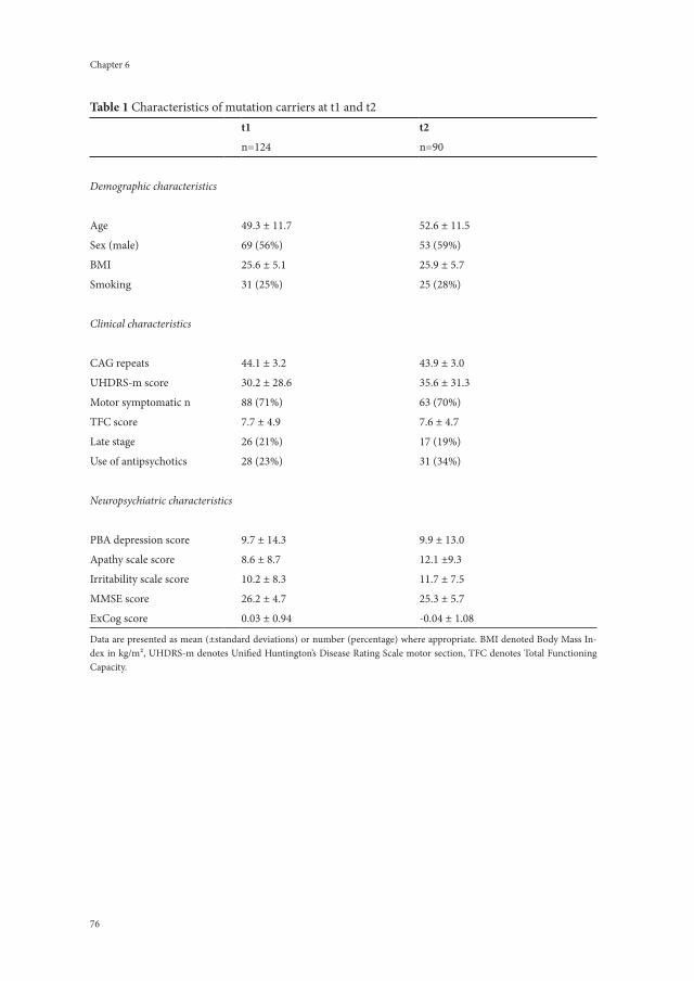
Chapter 6
76 77
Plasma cytokine levels in relation to neuropsychiatric symptoms and cognitive dysfunction in Hungtington’s disease
table 1 Characteristics of mutation carriers at t1 and t2t1 t2
n=124 n=90
Demographic characteristics
Age 49.3 ± 11.7 52.6 ± 11.5
Sex (male) 69 (56%) 53 (59%)
BMI 25.6 ± 5.1 25.9 ± 5.7
Smoking 31 (25%) 25 (28%)
Clinical characteristics
CAG repeats 44.1 ± 3.2 43.9 ± 3.0
UHDRS-m score 30.2 ± 28.6 35.6 ± 31.3
Motor symptomatic n 88 (71%) 63 (70%)
TFC score 7.7 ± 4.9 7.6 ± 4.7
Late stage 26 (21%) 17 (19%)
Use of antipsychotics 28 (23%) 31 (34%)
Neuropsychiatric characteristics
PBA depression score 9.7 ± 14.3 9.9 ± 13.0
Apathy scale score 8.6 ± 8.7 12.1 ±9.3
Irritability scale score 10.2 ± 8.3 11.7 ± 7.5
MMSE score 26.2 ± 4.7 25.3 ± 5.7
ExCog score 0.03 ± 0.94 -0.04 ± 1.08
Data are presented as mean (±standard deviations) or number (percentage) where appropriate. BMI denoted Body Mass In-dex in kg/m², UHDRS-m denotes Unified Huntington’s Disease Rating Scale motor section, TFC denotes Total Functioning Capacity.
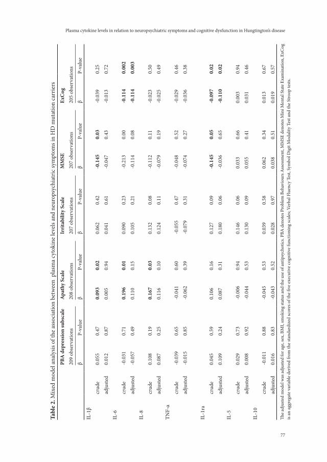
Chapter 6
76 77
Plasma cytokine levels in relation to neuropsychiatric symptoms and cognitive dysfunction in Hungtington’s disease
tabl
e 2.
Mix
ed m
odel
ana
lysis
of t
he a
ssoc
iatio
n be
twee
n p
lasm
a cy
toki
ne le
vels
and
neur
opsy
chia
tric
sym
ptom
s in
HD
mut
atio
n ca
rrie
rspB
a d
epre
ssio
n su
bsca
lea
path
y sc
ale
Irri
tabi
lity
scal
em
mse
exC
og20
9 ob
serv
atio
ns20
8 ob
serv
atio
ns20
7 ob
serv
atio
ns20
7 ob
serv
atio
ns20
5 ob
serv
atio
nsβ
P-va
lue
β P-
valu
eβ
P-va
lue
β P-
valu
eβ
P-va
lue
IL-1
βcr
ude
0.05
50.
470.
093
0.02
0.06
20.
42-0
.145
0.03
-0.0
390.
25ad
just
ed0.
012
0.87
0.00
50.
940.
041
0.61
-0.0
470.
43-0
.013
0.72
IL-6
crud
e-0
.031
0.71
0.19
60.
010.
090
0.23
-0.2
130.
00-0
.114
0.00
2ad
just
ed-0
.057
0.49
0.11
00.
150.
105
0.21
-0.1
140.
08-0
.114
0.00
3IL
-8cr
ude
0.10
80.
190.
167
0.03
0.13
20.
08-0
.112
0.11
-0.0
230.
50ad
just
ed0.
087
0.25
0.11
60.
100.
124
0.11
-0.0
790.
19-0
.025
0.49
TNF-
αcr
ude
-0.0
390.
65-0
.041
0.60
-0.0
550.
47-0
.048
0.52
-0.0
290.
46ad
just
ed-0
.015
0.85
-0.0
620.
39-0
.079
0.31
-0.0
740.
27-0
.036
0.38
IL-1
racr
ude
0.04
50.
590.
106
0.16
0.12
70.
09-0
.145
0.05
-0.0
970.
02ad
just
ed0.
109
0.24
0.08
70.
310.
180
0.06
-0.0
360.
65-0
.110
0.02
IL-5
crud
e0.
029
0.73
-0.0
060.
940.
146
0.06
0.03
30.
660.
003
0.94
adju
sted
0.00
80.
92-0
.044
0.53
0.13
00.
090.
055
0.41
0.03
10.
46IL
-10
crud
e-0
.011
0.88
-0.0
450.
530.
039
0.58
0.06
20.
340.
013
0.67
ad
just
ed
0.01
60.
83
-0.0
430.
52
0.02
80.
97
0.03
80.
51
0.01
90.
57
The a
djus
ted
mod
el w
as ad
just
ed fo
r age
, sex
, BM
I, sm
okin
g st
atus
and
the u
se o
f ant
ipsy
chot
ics.
PBA
den
otes
Pro
blem
Beh
avio
urs A
sses
smen
t, M
MSE
den
otes
Min
i Men
tal S
tate
Exa
min
atio
n, E
xCog
is
an a
ggre
gate
var
iabl
e de
rived
from
the
stan
dard
ized
scor
es o
f the
five
exe
cutiv
e co
gniti
ve fu
nctio
ning
scal
es; V
erba
l Flu
ency
Tes
t, Sy
mbo
l Dig
it M
odal
ity T
est a
nd th
e St
roop
test
s.
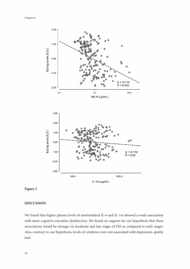
Chapter 6
78
dIsCussIon
We found that higher plasma levels of unstimulated IL-6 and IL-1ra showed a weak association with more cognitive executive dysfunction. We found no support for our hypothesis that these associations would be stronger in moderate and late stages of HD as compared to early stages. Also, contrary to our hypothesis, levels of cytokines were not associated with depression, apathy and.
figure 2
79
Plasma cytokine levels in relation to neuropsychiatric symptoms and cognitive dysfunction in Hungtington’s disease
Our findings that levels of IL-6 and IL-1ra in plasma were associated with cognitive dysfunction have not yet been reported in HD. In non-HD populations, levels of cytokines in relation to cognitive dysfunction have been studied most extensively in patients with Alzheimer’s disease (AD), showing that particularly IL-6 and TNF- levels were associated with cognitive dysfunction in some, but not all studies among patients with AD [20]. Both IL-6 and IL-1ra play an important role in the acute-phase response. Upon disturbance of the homeostasis by infection or tissue injury, cells of the innate immune system become activated and release pro-inflammatory cytokines [33]. One of the major pro-inflammatory cytokines is IL-6 [34]. Il-6 initiates the acute-phase response by induction of a systemic reaction comprising hepatic release of C-reactive protein (CRP) [35], the release of other pro-inflammatory cytokines and, activa-tion of the hypothalamic-pituitary-adrenal axis [36]. Another effect of IL-6 is the induction of IL-1ra [37]. IL-1ra is an inhibitor of the pro-inflammatory cytokine IL-1beta [38]. Thus, IL-1ra is regulator of the acute phase response by providing a feedback mechanism for the systemic effects of pro-inflammatory cytokines.
Given the cross-sectional nature of our investigation, the association between the cytokines IL-6 and IL-1ra with cognitive dysfunction may be explained by either causal effects, reverse cau-sation, or confounding by another process. For a causal effect to become likely, IL-6 and IL-1ra in the peripheral plasma should be able to influence the CNS in some way. Two direct pathways between the peripheral plasma and the CNS exist [39]. First, cytokines can be transported by active passage though the blood brain barrier [40]. Second, cytokines can diffuse passively into the CNS in the choroid plexus [41]. Furthermore, IL-6 can increase the permeability of the blood-brain barrier[42], thereby allowing toxins to enter the brain that can interfere with brain functions such as cognition. Indirectly, the CNS can be affected by stimulation of the vagal nerve by intra-peritoneal cytokines [43]. Also, under certain conditions, microglia and endothelial cells in the brain produce inflammatory factors [43]. Once in the brain, cytokines can activate other microglia which in turn secrete their own pro-inflammatory cytokines [44]. This activa-tion of microglia has indeed been demonstrated by several studies, in both PET scans [9] an in post-mortem brain tissue of HD patients [8]. Long-term exposure to cytokines, together with the presence of activated microglia may lead to neurodegeneration that causes cognitive dysfunction [39, 45]. In earlier studies, it has further been demonstrated that exposure to pro-inflammatory cytokines such as IL-6 impairs neurogenesis [39, 46, 47]. Also, due to its atherothrombotic potential, IL-6 may induce vascular pathology that contributes to neuronal dysfunction [48]. On the other hand, tissue damage due to neurodegeneration in HD caused by other pathophysiologi-cal mechanisms may be a common cause of both elevated plasma cytokine levels and cognitive dysfunction and may thus be a confounder for the relationship we found.
We were not able to demonstrate associations between cytokines and the neuropsychiatric symptoms depression, apathy and irritability. This finding contrasts with previous studies in non-HD populations demonstrating associations of cytokine levels with several neuropsychiatric symptoms [15-23]. Depressive symptoms in relation to plasma cytokines have been extensively

Chapter 6
80
investigated. In a meta-analyses of over 60 studies measuring depressive symptom severity, both IL-1 and IL-6 levels were increased in patients with a depressive disorder versus controls (Cohen’s d=0.35 and d=0.25, respectively) [49, 50]. Another meta-analysis of 24 studies among patients with major depressive disorder (MDD) showed that TNF-α and IL-6 levels were significantly higher in 492 patients compared to 400 controls [51]. No significant differences for IL-1β, IL-8, and IL-10 levels were found. Moreover, trials in cancer and hepatitis C patients also showed the increased risk of depression when treated with interferon (IFN)-gamma [15, 19]. Apathy and loss of interest have been studied less often in relation to inflammation; levels of IL-6 have been asso-ciated with these neuropsychiatric symptoms in 48 stroke patients [50]. The association between plasma cytokines and the hostility and anger seen in irritable subjects has been investigated both in healthy populations and patient groups [16-18, 21-23]. Observational studies showed that IL-1, IL-6, IL-8 and TNF-a were associated with higher levels of anger in populations of healthy couples [16], and healthy men [23] and women [22]. Intervention studies in hepatitis C patients showed increased anger and hostility upon treatment with IFN-gamma [17, 18]. However, these previous studied did not include HD patients, and our contrasting findings may be explained by disease specific factors of HD. Neurodegenerative changes in frontal-striatal circuits in HD may account for several neuropsychiatric symptoms, thereby obscuring the subtle associations with plasma cytokines, that tend to vary considerably over time.
The strengths of this study are the relatively large cohort of HD mutation carriers assessed at two time points, and the use of robust high sensitivity ELISA assays for the assessment of cytokine levels. Also some limitations warrant discussion. First, the average storage time of plasma samples was 3 years and it has been demonstrated that levels of particularly IL-6 may degrade significantly over longer storage times [52]. In addition, we did not ascertain time of withdrawal of our blood samples. As a consequence, this may have resulted in a measurement error, resulting in an under-estimation of the association with cognitive dysfunction. We found however, that cytokine plasma levels were rather stable and that measurement error was therefore likely within acceptable limits. As a limited number of HD mutation carriers completed both the second and third measurement (n=90), we could not perform longitudinal analyses because of the low statistical power as rela-tively few HD patients had new-onset neuropsychiatric symptoms during the 2 years of follow-up. Therefore, given the cross-sectional nature of our study, we cannot infer causal relationships.
In conclusion, levels of IL-6 and IL-1ra were positively associated with executive cognitive dysfunction in our cross-sectional study among HD mutation carriers. A causal relation may exist between pro-inflammatory cytokines and executive cognitive dysfunction; however the association may also be confounded by the neurodegenerative process or by environmental fac-tors. No associations were found between cytokine levels and other neuropsychiatric symptoms. Precise measurement of both cytokine levels as well as of neuropsychiatric symptoms is difficult and may introduce errors. Our results should therefore be interpreted with caution. Future stud-ies should have a longitudinal design to assess possible causal or alternative relations and should be designed to further reduce measurement errors.

81
Plasma cytokine levels in relation to neuropsychiatric symptoms and cognitive dysfunction in Hungtington’s disease
referenCes
[1] Walker FO. Huntington’s Disease. SeminNeurol. 2007;27(2):143-50.
[2] van Duijn E, Kingma EM, van der Mast RC. Psychopathology in verified Huntington’s disease gene carriers. JNeu-
ropsychiatry ClinNeurosci. 2007;19(4):441-8.
[3] Reedeker N, Bouwens JA, Giltay EJ, Le Mair SE, Roos RA, van der Mast RC, et al. Irritability in Huntington’s disease.
Psychiatry Res. 2012;200(2-3):813-8.
[4] Thompson JC, Harris J, Sollom AC, Stopford CL, Howard E, Snowden JS, et al. Longitudinal evaluation of neuro-
psychiatric symptoms in Huntington’s disease. 2014.
[5] Tabrizi SJ, Scahill RI, Durr A, Roos RAC, Leavitt BR, Jones R, et al. Biological and clinical changes in premanifest
and early stage Huntington’s disease in the TRACK-HD study: the 12-month longitudinal analysis. The Lancet
Neurology. 2011;10(1):31-42.
[6] Ellrichmann G, Reick C, Saft C, Linker RA. The Role of the Immune System in Huntington’s Disease. ClinDevIm-
munol. 2013;2013:541259.
[7] Borish LC, Steinke JW. 2. Cytokines and chemokines. Journal of Allergy and Clinical Immunology. 2003;111(2):S460-
S75.
[8] Sapp E, Kegel KB, Aronin N, Hashikawa T, Uchiyama Y, Tohyama K, et al. Early and progressive accumulation of
reactive microglia in the Huntington disease brain. JNeuropatholExpNeurol. 2001;60(2):161-72.
[9] Tai YF, Pavese N, Gerhard A, Tabrizi SJ, Barker RA, Brooks DJ, et al. Microglial activation in presymptomatic
Huntington’s disease gene carriers. Brain. 2007;130(Pt 7):1759-66.
[10] Bjorkqvist M, Wild EJ, Thiele J, Silvestroni A, Andre R, Lahiri N, et al. A novel pathogenic pathway of immune
activation detectable before clinical onset in Huntington’s disease. JExpMed. 2008;205(8):1869-77.
[11] Chang KH, Wu YR, Chen YC, Chen CM. Plasma inflammatory biomarkers for Huntington’s disease patients and
mouse model. Brain BehavImmun. 2014.
[12] Dalrymple A, Wild EJ, Joubert R, Sathasivam K, Bjorkqvist M, Petersen A, et al. Proteomic profiling of
plasma in Huntington’s disease reveals neuroinflammatory activation and biomarker candidates. JProteomeRes.
2007;6(7):2833-40.
[13] Sanchez-Lopez F, Tasset I, Aguera E, Feijoo M, Fernandez-Bolanos R, Sanchez FM, et al. Oxidative stress and
inflammation biomarkers in the blood of patients with Huntington’s disease. NeurolRes. 2012;34(7):721-4.
[14] Bouwens JA, Hubers AA, van Duijn E, Cobbaert CM, Roos RA, van der Mast RC, et al. Acute-phase proteins
in relation to neuropsychiatric symptoms and use of psychotropic medication in Huntington’s disease. European
neuropsychopharmacology : the journal of the European College of Neuropsychopharmacology. 2014;24(8):1248-
56.
[15] Capuron L, Ravaud A, Miller AH, Dantzer R. Baseline mood and psychosocial characteristics of patients devel-
oping depressive symptoms during interleukin-2 and/or interferon-alpha cancer therapy. Brain BehavImmun.
2004;18(3):205-13.
[16] Kiecolt-Glaser JK, Loving TJ, Stowell JR, Malarkey WB, Lemeshow S, Dickinson SL, et al. Hostile marital interac-
tions, proinflammatory cytokine production, and wound healing. ArchGenPsychiatry. 2005;62(12):1377-84.
[17] Kraus MR, Schafer A, Faller H, Csef H, Scheurlen M. Psychiatric symptoms in patients with chronic hepatitis C
receiving interferon alfa-2b therapy. JClinPsychiatry. 2003;64(6):708-14.
[18] Lotrich FE, Sears B, McNamara RK. Anger induced by interferon-alpha is moderated by ratio of arachidonic acid
to omega-3 fatty acids. JPsychosomRes. 2013;75(5):475-83.
[19] McHutchison JG, Gordon SC, Schiff ER, Shiffman ML, Lee WM, Rustgi VK, et al. Interferon alfa-2b alone or in
combination with ribavirin as initial treatment for chronic hepatitis C. Hepatitis Interventional Therapy Group.
NEnglJMed. 1998;339(21):1485-92.
Chapter 6
82
[20] Michaud M, Balardy L, Moulis G, Gaudin C, Peyrot C, Vellas B, et al. Proinflammatory cytokines, aging, and
age-related diseases. JAmMedDirAssoc. 2013;14(12):877-82.
[21] Preau M, Marcellin F, Spire B, Ravaux I, Dellamonica P, Blanc D, et al. Impaired anger control as an underap-
preciated side effect of treatments for chronic HCV infection in HIV-HCV coinfected patients. JClinGastroenterol.
2008;42(1):92-6.
[22] Suarez EC, Lewis JG, Krishnan RR, Young KH. Enhanced expression of cytokines and chemokines by blood mono-
cytes to in vitro lipopolysaccharide stimulation are associated with hostility and severity of depressive symptoms in
healthy women. Psychoneuroendocrinology. 2004;29(9):1119-28.
[23] Suarez EC, Lewis JG, Kuhn C. The relation of aggression, hostility, and anger to lipopolysaccharide-stimulated
tumor necrosis factor (TNF)-alpha by blood monocytes from normal men. Brain BehavImmun. 2002;16(6):675-84.
[24] Kingma EM, van Duijn E, Timman R, van der Mast RC, Roos RA. Behavioural problems in Huntington’s disease
using the Problem Behaviours Assessment. GenHospPsychiatry. 2008;30(2):155-61.
[25] Sarwar N, Butterworth AS, Freitag DF, Gregson J, Willeit P, Gorman DN, et al. Interleukin-6 receptor pathways in
coronary heart disease: a collaborative meta-analysis of 82 studies. Lancet. 2012;379(9822):1205-13.
[26] Starkstein SE, Fedoroff JP, Price TR, Leiguarda R, Robinson RG. Apathy following cerebrovascular lesions 19.
Stroke. 1993;24(11):1625-30.
[27] Chatterjee A, Anderson KE, Moskowitz CB, Hauser WA, Marder KS. A comparison of self-report and care-
giver assessment of depression, apathy, and irritability in Huntington’s disease. JNeuropsychiatry ClinNeurosci.
2005;17(3):378-83.
[28] Folstein MF, Folstein SE, McHugh PR. “Mini-mental state”. A practical method for grading the cognitive state of
patients for the clinician. JPsychiatrRes. 1975;12(3):189-98.
[29] Hodges JR. Initiation: verbal fluency tests. Cognitive Assessment for Clinicians: Oxford University Press, oxford,
united kingdom.; 2003. p. 118-9.
[30] Smith A. The Symbol Digit Modalities Test: a neuropsychological test for economic screening of learning and other
cerebral disorders. Learn Disord. 1968;3:83-91.
[31] Stroop JR. Studies of interference in serial verbal reactions. Journal of Experimental Psychology. 1935;18:643-62.
[32] Kremer HPH, Huntington Study G. Unified Huntington’s disease rating scale: reliability and consistency. 1996.
[33] Heinrich PC, Castell JV, Andus T. Interleukin-6 and the acute phase response. Biochemical Journal. 1990;265(3):621.
[34] Nishimoto N. Interleukin-6 as a therapeutic target in candidate inflammatory diseases. ClinPharmacolTher.
2010;87(4):483-7.
[35] Gabay C, Kushner I. Acute-phase proteins and other systemic responses to inflammation. NEnglJMed.
1999;340(6):448-54.
[36] Mastorakos G, Chrousos GP, Weber JS. Recombinant interleukin-6 activates the hypothalamic-pituitary-adrenal
axis in humans. The Journal of Clinical Endocrinology & Metabolism. 1993;77(6):1690-4.
[37] Tilg H, Trehu E, Atkins MB, Dinarello CA, Mier JW. Interleukin-6 (IL-6) as an anti-inflammatory cytokine: induc-
tion of circulating IL-1 receptor antagonist and soluble tumor necrosis factor receptor p55. Blood. 1994;83(1):113-
8.
[38] Dinarello CA, van der Meer JW. Treating inflammation by blocking interleukin-1 in humans. SeminImmunol.
2013;25(6):469-84.
[39] Rosano C, Marsland AL, Gianaros PJ. Maintaining brain health by monitoring inflammatory processes: a mecha-
nism to promote successful aging. Aging Dis. 2012;3(1):16-33.
[40] Banks WA. Blood-brain barrier transport of cytokines: a mechanism for neuropathology. CurrPharmDes.
2005;11(8):973-84.
[41] Konsman JP, Parnet P, Dantzer R. Cytokine-induced sickness behaviour: mechanisms and implications. Trends
Neurosci. 2002;25(3):154-9.

83
Plasma cytokine levels in relation to neuropsychiatric symptoms and cognitive dysfunction in Hungtington’s disease
[42] de Vries HE, Blom-Roosemalen MC, van Oosten M, de Boer AG, van Berkel TJ, Breimer DD, et al. The influence of
cytokines on the integrity of the blood-brain barrier in vitro. Journal of neuroimmunology. 1996;64(1):37-43.
[43] Licinio J, Wong ML. Pathways and mechanisms for cytokine signaling of the central nervous system. JClinInvest.
1997;100(12):2941-7.
[44] Hanisch UK. Microglia as a source and target of cytokines. Glia. 2002;40(2):140-55.
[45] Smith JA, Das A, Ray SK, Banik NL. Role of pro-inflammatory cytokines released from microglia in neurodegenera-
tive diseases. Brain ResBull. 2012;87(1):10-20.
[46] Godbout JP, Johnson RW. Interleukin-6 in the aging brain. JNeuroimmunol. 2004;147(1-2):141-4.
[47] Vallieres L, Campbell IL, Gage FH, Sawchenko PE. Reduced hippocampal neurogenesis in adult transgenic mice
with chronic astrocytic production of interleukin-6. JNeurosci. 2002;22(2):486-92.
[48] Ross R. Atherosclerosis--an inflammatory disease. NEnglJMed. 1999;340(2):115-26.
[49] Howren MB, Lamkin DM, Suls J. Associations of depression with C-reactive protein, IL-1, and IL-6: a meta-analysis.
PsychosomMed. 2009;71(2):171-86.
[50] Spalletta G, Cravello L, Imperiale F, Salani F, Bossu P, Picchetto L, et al. Neuropsychiatric symptoms and interleu-
kin-6 serum levels in acute stroke. JNeuropsychiatry ClinNeurosci. 2013;25(4):255-63.
[51] Dowlati Y, Herrmann N, Swardfager W, Liu H, Sham L, Reim EK, et al. A meta-analysis of cytokines in major
depression. BiolPsychiatry. 2010;67(5):446-57.
[52] de Jager W Bourcier K, Rijkers GT, Prakken BJ, Seyfert-Margolis V. Prerequisites for cytokine measurements in
clinical trials with multiplex immunoassays. BMCImmunol. 2009;10:52.


Chapter 7
general discussion and summary


87
General discussion and summary
general dIsCussIon
In this thesis, we investigated the activity of the innate immune system in HD as reflected in several inflammatory immune parameters. Also, we aimed to gain further insight into the neuropsychiatric symptoms in HD, in particular irritability. In addition, we investigated the as-sociation between inflammatory immune parameters on the one hand and measures of disease progression (including neuropsychiatric symptoms and cognitive decline) on the other. In aging, and in neurodegenerative disorders such as Parkinson’s disease and Alzheimer’s disease, (neuro)inflammation is associated with the loss of normal functioning. Investigation of inflammatory immune parameters of inflammation in relation to markers of disease progression may provide further insight into the development of symptoms of HD. Also, inflammatory immune param-eters that reflect disease severity in HD may be used as biomarkers in research and clinical care. In HD, such biomarkers are important because they may help to detect whether or not a potential therapeutic intervention is beneficial [1, 2]. We expected to find a correlation between increased levels of pro-inflammatory cytokines and positive acute-phase proteins on the one hand and markers of disease progression on the other; we also expected that, with progressing disease, levels of regulatory anti-inflammatory cytokines would decrease. Finally, we also expected to find an association between levels of immune parameters and neuropsychiatric symptoms.
IntegratIon of the study fIndIngs Into Current knowledge
Irritability
We assessed the psychometric properties of the Irritability Scale (IS) and established that a cut-off point of 14 provides a valid and reliable assessment of irritability in HD gene expansion carriers.
Using the IS, in our HD population the prevalence of irritability in the past two weeks was 35%, whereas in other studies the prevalence ranged from 33-73% [3]. These differences in reported prevalence can probably be attributed to the wide range of instruments used to assess irritability, the different time periods of the studies, as well as the sociodemographic and clinical differences in the HD populations investigated. Currently, the IS the only symptom-specific scale that is recommended for the assessment of irritability in HD [4]. Although several scales are available that assess irritability as one of the behavioral domains (such as the UHDRS behavioral subscale), these subscales have either not been extensively validated[5] or no data are available on their reliability or other psychometric properties in HD [6-8].
In our study, the incidence of irritability was found to be 23% over a two-year period. The rate of remittance (for which we were unable to find any predictors) was comparable to the in-cidence of irritability over time. Increasing irritability was associated with increased apathy and the continuous use of antipsychotics. However, we found no correlation between irritability and measures of disease progression, such as cognitive decline, motor abnormalities and functional

Chapter 7
88
incapacity. Also, at baseline, none of these parameters could predict the incidence of irritability over a two-year period. Earlier studies showed that irritability may be a characteristic neuro-psychiatric symptom of pre-manifest and early HD[9-11]. This is in line with findings of more recent imaging studies that demonstrated functional changes in the brain of gene expansion carriers, specifically for the early stages of HD, that correlated with irritability[12, 13]. Possible explanations for the development of irritability include overcompensation of damaged circuits early in the disease[13] and cognitive overload caused by early cognitive decline[12]. Also, the construct of irritability as assimilated in the IS may lose validity in later stages of HD because of the interference of severe motor dysfunction, cognitive decline and neuropsychiatric symptoms.
Immune parameters
We found that activation of the acute-phase response, as reflected in levels of both positive and negative acute-phase proteins, was associated with impaired daily functioning, apathy and cog-nitive impairment. However, these associations were confounded by the use of antipsychotics. Gene expansion carriers that continuously used antipsychotics had higher levels of hsCRP and lower levels of albumin than gene expansion carriers who did not use antipsychotics during the study period. In other HD cohorts, CRP levels were significantly increased in advanced disease stages when compared to healthy controls; to our knowledge, in these cohorts, no adjustment was made for the use of antipsychotics.
Levels of TNF-α and IL-10 increased over the two-year study period. Pre-motor symptom-atic gene expansion carriers had lower levels of IL-5 and IL-6 than symptomatic gene expansion carriers. IL-6 and IL-8 were inversely associated with daily functioning. Also, increased levels of both IL-1ra and IL-6 were independently associated with impaired cognitive functioning. Previ-ous cross-sectional studies with a limited number of mutation carriers in disease stages from premanifest to moderately advanced disease stage, presented evidence for increased immune activation in the plasma of HD mutation carriers[14-18]. Most consistently, levels of IL-6 were higher in motor symptomatic HD mutation carriers than in matched controls, whereas increased plasma levels of sIL-2R, sTNF-αR[18], IL-8, and TNF-α [14] were also found. The study that assessed IL-4, IL-5, and IL-10, found positive correlations with a more advanced disease stage, although levels of IL-4 and IL-5 were increased only in a moderately advanced disease stage, compared with controls[14].
The triggering of innate immune activation is also emerging in other neurodegenerative diseases[19]. However, the relationship between immune activation and disease progression is complex. In Alzheimer’s disease, inflammatory pathways are activated as reflected by increased levels of chemokines and cytokines in both the brain and cerebrospinal fluid of patients, and neuroinflammation has been demonstrated by PET scans[20]. On the other hand, inflamma-tory processes are also a risk factor for Alzheimer’s disease. Studies on patients with Alzheimer’s disease have demonstrated the negative effects of severe infection[21], sepsis[22] and periodon-titis[23] on cognition.

89
General discussion and summary
By chance, we found evidence for activation of the acute-phase response in gene-expansion carriers who used antipsychotics. This was found when adjusting for the use of antipsychotics in the relationship between acute-phase proteins on the one hand, and daily functioning, cognitive impairment and apathy on the other. After this adjustment, effect sizes for these relations were considerably reduced and no longer significant. Furthermore, the group of gene-expansion car-riers that continuously used antipsychotics had higher levels of CRP and lower levels of albumin than those who did not use antipsychotics. To our knowledge, this phenomenon has not earlier been described.
Antipsychotics are frequently prescribed in late-stage HD for the symptomatic treatment of movement disorder. Therefore, the idea that increased acute-phase response activity is at least partly due to the use of antipsychotics is in line with a study reporting that premanifest and early-HD gene-expansion carriers had similar lower CRP levels compared to controls[24]. In non-HD populations, two pathways for activation of the acute-phase response by antipsychotics have been described. First, low-grade inflammation may be the result of obesity[25], dyslip-idemia[26] and insulin resistance[27] which are elements of the metabolic syndrome, a well-known side-effect of second-generation antipsychotics. Second, both classical antipsychotics[28] and second-generation antipsychotics[29] may directly induce low-grade inflammation through their hepatic impact.
Strengths and limitations
The strengths of this study are the cohort study design and the well-characterized study popu-lation with HD. Furthermore, we included a control group consisting of first-degree relatives without the HD gene expansion which had been exposed to a similar environment as the gene expansion carriers. Also, the group of gene-expansion carriers was heterogeneous, comprising a range of pre-manifest individuals many years from clinical onset to institutionalized patients with end-stage disease. In addition, the same individuals were interviewed up to three times, with a two-year interval in between. Specifically, we used valid and reliable measurement tools in a standardized setting. Dropout rates between measurements were moderate and did not exceed 20%. Cytokine levels were also measured in a standardized and state-of-the-art manner. Where possible, we used high-sensitivity enzyme-linked-immuno-sorbent assays (ELISA); due to their conceptual design, ELISAs are less prone to altered levels of circulating proteins and inhibitors than multiplexed immunoassays[30]. Also, the number of freeze-thaw cycles was limited to one (for IL1-b, IL-6, IL-10 and TNF-α) or two (for IL-1ra, IL-5 and IL-8). Third, the methodological design eliminated any plate-to-plate and/or lot-to-lot variation and/or operator variability, as paired samples from HD gene expansion carriers were measured simultaneously on the same plate for the longitudinal analyses. Fourth, significant partial correlations were found for all cytokines between paired HD samples, ranging from 0.483 to 0.728 (p<0.001).
Some limitations also need to be addressed. An important drawback is the limited validity of the measurement of behavioral symptoms in HD. Validated systems, such as the Diagnostic

Chapter 7
90
and Statistical Manual of Mental Disorders (DSM), are used for the classification of psychiatric disorders. However, their usefulness in HD is limited because they were not designed to assess symptoms and do not assess the severity of symptoms in dimensional scales. Next, cytokine levels in the plasma may not accurately reflect the highly complex state of the immune system. Although blood and its subsequent handling was done carefully, we were not able to take into ac-count the effect of diurnal and seasonal changes in cytokine levels, which can be significant[31]. Another drawback is that we could not adjust for all possible confounding variables. Medication is frequently prescribed for movement disorder and behavioral symptoms; these medications include antipsychotics and benzodiazepines, which are known to have a wide range of biological adverse effects. Given the observational nature of our study, we could not determine whether the associations found were explained by causative mechanisms, confounding, or confounding by indication. Given the low incidence of HD, collecting a sufficiently large sample of HD gene expansion carriers remains a challenge. In particular, for analyses within subgroups with a siz-able number of covariates, sample sizes were too low to reach adequate statistical power; because this may have caused type-I and type-II errors, our findings need to be replicated in independent samples of HD gene-expansion carriers.
Implications for clinical practice
No specific treatment for irritability in HD is available. Current treatment guidelines are largely based on case series and expert opinion, and on findings from studies in patients with psychiatric or other neurodegenerative disorders[32-34]. Our study showed that, in a significant number of patients, irritability may remit over a two-year study period; however, we were unable to predict which patients had the highest chance to remit from irritability. Nonetheless, we found that the use of benzodiazepines and antipsychotics was associated with the prevalence and incidence of irritability, respectively. Both types of drugs can be used in the treatment of irritability; however, especially in patients who receive these drugs for other indications, clinicians should be aware that irritability might be a possible side-effect.
Although evidence for increased levels of (low-grade) systemic inflammation was found in our population, the cause of this systemic inflammation could not be identified; the inflamma-tory state most likely reflects a complex system of mechanisms. In other populations, the effects of systemic inflammation on mood[35-37] and cognition[38-40] are detrimental. Studies have also indicated negative effects on metabolic disease and cardiovascular health[41-43]. Several studies have investigated the positive effects of anti-inflammatory drugs in HD. However, until now, agents like minocycline[44, 45], cannabinoids[46, 47] and non-steroidal anti-inflammatory drugs[48, 49] have not proven effective in humans, although some studies may have been under-powered. Further studies are needed to assess whether a more specific tweaking of neuroinflam-mation may benefit the many ailments of HD gene expansion carriers. In HD, treatable causes of systemic inflammation, such as infectious processes, should be managed properly. Also, iat-rogenic damage, caused by the possible adverse effects of prescribed drugs, should be prevented.
91
General discussion and summary
Although our findings offer additional insight into the role of the innate immune system in HD, they also indicate that the rather crude immune parameters that we investigated were not of clinical use as biomarkers for the individual HD gene expansion carrier. It is important that a biomarker is measurable in a cost-effective manner, changes with the disease state over time, and is able to discriminate between gene expansion carriers in different disease states.
Directions for future research
The studies in this thesis confirm that inflammatory processes play an important role in HD. Immune parameters, indicating activation of the innate immune system, correlated with neuro-psychiatric and motor symptoms, as well as with functional decline. However, given the observa-tional nature of our investigation, we cannot infer any causal relations. Most likely, the immune system plays both a pathogenic and homeostatic role; however, more longitudinal studies are required to unravel its precise function in HD. Importantly, our findings also demonstrated that iatrogenic factors, such as the use of (psychotropic) medication, were related to both immune system parameters and outcome measures and, thus, need to be taken into account.
Although we have provided a thorough assessment of the psychometric properties of the IS, these findings still need to be replicated in larger studies. The validity and comparability of the scale, and its sum score in different stages of the disease, should be studied further using more so-phisticated analysis techniques that include confirmatory factor analysis to assess measurement invariance. This might help establish whether factor loadings, intercepts and residual variances are equivalent in a factor model that measures a latent variable irritability. If so, comparisons of irritability scores will be valid across disease stages and across time within a HD gene expansion carrier.
Our findings concerning the associations between immune system parameters on the one hand and neuropsychiatric symptoms and disease progression on the other, yielded far more modest effect sizes than other studies of a comparable size Therefore, larger studies are needed to replicate these findings. Moreover, these studies should be able to adequately assess cytokine levels, taking into account possible confounding factors such as diurnal and seasonal variations in the levels of cytokines and the use of medication.
In observational studies, no firm conclusions regarding causality can be drawn. In our study, the confounding effect of the use of antipsychotics on immune system parameters and neuropsychiatric symptoms, warrants investigating whether this is explained by confounding by indication or by a direct effect of these drugs. Therefore, the results of previous studies on anti-psychotics cannot be generalized. The effectiveness of antipsychotics should be assessed critically in well-designed and sufficiently powered studies and be weighed against the possible negative consequences, preferably in randomized controlled trials (RCTs) in a HD population. In addition to evaluation of the safety of antipsychotics, RCTs can shed more light on the treatment of vari-ous neuropsychiatric symptoms which, so far, relies mostly on case reports and expert opinion.

Chapter 7
92
summary
In a cohort of HD gene expansion carriers, we assessed neuropsychiatric symptoms and several inflammatory immune system parameters. The aim was to investigate neuropsychiatric symp-toms, in particular irritability, in more detail and gain further insight into the activation of the innate immune system in HD, as well as its role in disease progression.
The irritability scale (IS) was used to assess irritability. This scale proved to be a reliable and valid instrument for the assessment of irritability in HD. Using a cut-off point of 14, the prevalence of irritability was 35% in our population of HD gene expansion carriers. Gene ex-pansion carriers tended to underestimate their level of irritability when compared to reports of their caregivers or spouses. Being married/living together and a longer CAG expansion in the Htt-gene were independent correlates of self-reported irritability. The incidence of new-onset ir-ritability over a two-year study period was 23% and coincided with an increase of apathy; during this period, irritability remitted in 30% of previously irritable gene expansion carriers.
Levels of the pro-inflammatory cytokines TNF-α and IL-10 increased over the two-year study period. Also, pre-motor symptomatic gene expansion carriers had lower levels of IL-5 and IL-6 than motor symptomatic gene expansion carriers. Additionally, levels of IL-6 and IL-8 were inversely associated with the level of daily functioning.
Activation of the acute-phase response, as reflected by higher levels of CRP and lower levels of albumin, was associated with impaired daily functioning, apathy and cognitive impairment. However, these associations were confounded by the use of antipsychotics.
A weak association was found between increased levels of both IL-1ra and IL-6 on the one hand and cognitive decline on the other. No associations were found between cytokine levels and apathy, irritability and depressive mood.

93
General discussion and summary
referenCes
[1] Andre R, Scahill RI, Haider S, Tabrizi SJ. Biomarker development for Huntington’s disease. Drug DiscovToday.
2014;19(7):972-9.
[2] Ross CA, Aylward EH, Wild EJ, Langbehn DR, Long JD, Warner JH, et al. Huntington disease: natural history,
biomarkers and prospects for therapeutics. NatRevNeurol. 2014;10(4):204-16.
[3] van Duijn E, Kingma EM, van der Mast RC. Psychopathology in verified Huntington’s disease gene carriers. JNeu-
ropsychiatry ClinNeurosci. 2007;19(4):441-8.
[4] Mestre TA, van Duijn E, Davis AM, Bachoud‐Lévi AC, Busse M, Anderson KE, et al. Rating scales for behavioral
symptoms in Huntington’s disease: Critique and recommendations. Movement Disorders. 2016;31(10):1466-78.
[5] Snaith RP, Taylor CM. Irritability: definition, assessment and associated factors 1. BrJPsychiatry. 1985;147:127-36.
[6] Callaghan J, Stopford C, Arran N, Boisse MF, Coleman A, Santos RD, et al. Reliability and factor structure of the
Short Problem Behaviors Assessment for Huntington’s disease (PBA-s) in the TRACK-HD and REGISTRY studies.
The Journal of neuropsychiatry and clinical neurosciences. 2015;27(1):59-64.
[7] Craufurd D, Thompson JC, Snowden JS. Behavioral changes in Huntington Disease. Neuropsychiatry Neuropsy-
cholBehavNeurol. 2001;14(4):219-26.
[8] Group HS. Unified Huntington’s Disease Rating Scale: reliability and consistency. Huntington Study Group. Mov
Disord. 1996;11(2):136-42.
[9] Baudic S, Maison P, Dolbeau G, Boissé M-F, Bartolomeo P, Dalla Barba G, et al. Cognitive impairment related to
apathy in early Huntington’s disease. Dementia and geriatric cognitive disorders. 2006;21(5-6):316-21.
[10] Kingma EM, van DE, Timman R, van der Mast RC, Roos RA. Behavioural problems in Huntington’s disease using
the Problem Behaviours Assessment. GenHospPsychiatry. 2008;30(2):155-61.
[11] van Duijn E, Craufurd D, Hubers AA, Giltay EJ, Bonelli R, Rickards H, et al. Neuropsychiatric symptoms in a
European Huntington9s disease cohort (REGISTRY). J Neurol Neurosurg Psychiatry. 2014:jnnp-2013-307343.
[12] Gregory S, Scahill RI, Seunarine KK, Stopford C, Zhang H, Zhang J, et al. Neuropsychiatry and white matter
microstructure in Huntington’s disease. Journal of Huntington’s disease. 2015;4(3):239-49.
[13] Van den Stock J, De Winter FL, Ahmad R, Sunaert S, Van Laere K, Vandenberghe W, et al. Functional brain changes
underlying irritability in premanifest Huntington’s disease. Human brain mapping. 2015;36(7):2681-90.
[14] Bjorkqvist M, Wild EJ, Thiele J, Silvestroni A, Andre R, Lahiri N, et al. A novel pathogenic pathway of immune
activation detectable before clinical onset in Huntington’s disease. JExpMed. 2008;205(8):1869-77.
[15] Chang KH, Wu YR, Chen YC, Chen CM. Plasma inflammatory biomarkers for Huntington’s disease patients and
mouse model. Brain BehavImmun. 2014.
[16] Dalrymple A, Wild EJ, Joubert R, Sathasivam K, Bjorkqvist M, Petersen A, et al. Proteomic profiling of
plasma in Huntington’s disease reveals neuroinflammatory activation and biomarker candidates. JProteomeRes.
2007;6(7):2833-40.
[17] Leblhuber F, Walli J, Jellinger K, Tilz GP, Widner B, Laccone F, et al. Activated immune system in patients with
Huntington’s disease. ClinChemLab Med. 1998;36(10):747-50.
[18] Sanchez-Lopez F, Tasset I, Aguera E, Feijoo M, Fernandez-Bolanos R, Sanchez FM, et al. Oxidative stress and
inflammation biomarkers in the blood of patients with Huntington’s disease. NeurolRes. 2012;34(7):721-4.
[19] Heneka MT, Kummer MP, Latz E. Innate immune activation in neurodegenerative disease. Nat Rev Immunol.
2014;14(7):463-77.
[20] Cagnin A, Brooks DJ, Kennedy AM, Gunn RN, Myers R, Turkheimer FE, et al. In-vivo measurement of activated
microglia in dementia. The Lancet. 2001;358(9280):461-7.
[21] Holmes C, Cunningham C, Zotova E, Woolford J, Dean C, Kerr Su, et al. Systemic inflammation and disease
progression in Alzheimer disease. Neurology. 2009;73(10):768-74.
Chapter 7
94
[22] Iwashyna TJ, Ely EW, Smith DM, Langa KM. Long-term cognitive impairment and functional disability among
survivors of severe sepsis. Jama. 2010;304(16):1787-94.
[23] Kamer AR, Craig RG, Dasanayake AP, Brys M, Glodzik-Sobanska L, de Leon MJ. Inflammation and Alzheimer’s
disease: possible role of periodontal diseases. Alzheimer’s & Dementia. 2008;4(4):242-50.
[24] Silajdžić E, Rezeli M, Végvári Á, Lahiri N, Andre R, Magnusson-Lind A, et al. A critical evaluation of inflammatory
markers in Huntington’s disease plasma. Journal of Huntington’s disease. 2013;2(1):125-34.
[25] Raedler TJ. Cardiovascular aspects of antipsychotics. Current opinion in psychiatry. 2010;23(6):574-81.
[26] Calder PC, Ahluwalia N, Brouns F, Buetler T, Clement K, Cunningham K, et al. Dietary factors and low-grade
inflammation in relation to overweight and obesity. British Journal of Nutrition. 2011;106(S3):S1-S78.
[27] Kim SH, Reaven G, Lindley S. Relationship between insulin resistance and C-reactive protein in a patient
population treated with second generation antipsychotic medications. International clinical psychopharmacology.
2011;26(1):43.
[28] Diaz FJ, Perez-Iglesias R, Mata I, Martinez-Garcia O, Vazquez-Barquero JL, de Leon J, et al. Possible effects of some
antipsychotic drugs on C-reactive protein in a drug-naive psychotic sample. Schizophrenia research. 2010;121(1-
3):207-12.
[29] Meyer JM, McEvoy JP, Davis VG, Goff DC, Nasrallah HA, Davis SM, et al. Inflammatory markers in schizophrenia:
comparing antipsychotic effects in phase 1 of the clinical antipsychotic trials of intervention effectiveness study.
Biological psychiatry. 2009;66(11):1013-22.
[30] Leng SX, McElhaney JE, Walston JD, Xie D, Fedarko NS, Kuchel GA. ELISA and multiplex technologies for cyto-
kine measurement in inflammation and aging research. JGerontolA BiolSciMedSci. 2008;63(8):879-84.
[31] de Jager W Bourcier K, Rijkers GT, Prakken BJ, Seyfert-Margolis V. Prerequisites for cytokine measurements in
clinical trials with multiplex immunoassays. BMCImmunol. 2009;10:52.
[32] Groves M, van Duijn E, Anderson K, Craufurd D, Edmondson MC, Goodman N, et al. An international survey-
based algorithm for the pharmacologic treatment of irritability in Huntington’s disease. PLoS currents. 2011;3.
[33] Mestre T, Ferreira J, Coelho MM, Rosa M, Sampaio C. Therapeutic interventions for symptomatic treatment in
Huntington’s disease. The Cochrane Library. 2009.
[34] van Duijn E. Treatment of irritability in Huntington’s disease. Current treatment options in neurology.
2010;12(5):424-33.
[35] Beatriz Currier M, B Nemeroff C. Inflammation and mood disorders: proinflammatory cytokines and the patho-
genesis of depression. Anti-Inflammatory & Anti-Allergy Agents in Medicinal Chemistry (Formerly Current
Medicinal Chemistry-Anti-Inflammatory and Anti-Allergy Agents). 2010;9(3):212-20.
[36] Rosenblat JD, Cha DS, Mansur RB, McIntyre RS. Inflamed moods: a review of the interactions between inflamma-
tion and mood disorders. Progress in Neuro-Psychopharmacology and Biological Psychiatry. 2014;53:23-34.
[37] Swardfager W, Rosenblat JD, Benlamri M, McIntyre RS. Mapping inflammation onto mood: Inflammatory media-
tors of anhedonia. Neuroscience & Biobehavioral Reviews. 2016;64:148-66.
[38] Schram MT, Euser SM, De Craen AJ, Witteman JC, Frölich M, Hofman A, et al. Systemic markers of inflammation
and cognitive decline in old age. Journal of the American Geriatrics Society. 2007;55(5):708-16.
[39] Teunissen C, Van Boxtel M, Bosma H, Bosmans E, Delanghe J, De Bruijn C, et al. Inflammation markers in relation
to cognition in a healthy aging population. Journal of neuroimmunology. 2003;134(1):142-50.
[40] Yaffe K, Lindquist K, Penninx B, Simonsick E, Pahor M, Kritchevsky S, et al. Inflammatory markers and cognition
in well-functioning African-American and white elders. Neurology. 2003;61(1):76-80.
[41] Fearon WF, Fearon DT. Inflammation and Cardiovascular Disease. Am Heart Assoc; 2008.
[42] Pearson TA, Mensah GA, Alexander RW, Anderson JL, Cannon RO, Criqui M, et al. Markers of inflammation and
cardiovascular disease. Circulation. 2003;107(3):499-511.
[43] Ridker PM, Hennekens CH, Buring JE, Rifai N. C-reactive protein and other markers of inflammation in the
prediction of cardiovascular disease in women. New England Journal of Medicine. 2000;342(12):836-43.

95
General discussion and summary
[44] Huntington Study Group, Minocycline safety and tolerability in Huntington disease. Neurology. 2004;63(3):547-9.
[45] Li C, Yuan K, Schluesener H. Impact of minocycline on neurodegenerative diseases in rodents: a meta-analysis.
Reviews in the Neurosciences. 2013;24(5):553-62.
[46] Consroe P, Laguna J, Allender J, Snider S, Stern L, Sandyk R, et al. Controlled clinical trial of cannabidiol in Hun-
tington’s disease. Pharmacology Biochemistry and Behavior. 1991;40(3):701-8.
[47] Curtis A, Rickards H. Nabilone could treat chorea and irritability in Huntington’s disease. The Journal of neuropsy-
chiatry and clinical neurosciences. 2006;18(4):553-4.
[48] Kalonia H, Kumar A. Suppressing inflammatory cascade by cyclo-oxygenase inhibitors attenuates quinolinic acid
induced Huntington’s disease-like alterations in rats. Life sciences. 2011;88(17):784-91.
[49] Norflus F, Nanje A, Gutekunst C-A, Shi G, Cohen J, Bejarano M, et al. Anti-inflammatory treatment with acetyl-
salicylate or rofecoxib is not neuroprotective in Huntington’s disease transgenic mice. Neurobiology of disease.
2004;17(2):319-25.

97
nederlandse samenvatting
In dit proefschrift worden neuropsychiatrische symptomen bij de ziekte van Huntington (ZvH) beschreven. In het bijzonder wordt stilgestaan bij het vóórkomen en het beloop van prikkel-baarheid. Tevens wordt de rol van het immuunsysteem onderzocht. Hierbij wordt nagegaan of er toegenomen activiteit van met name het pro-inflammatoire gedeelte van het niet-adaptieve immuunsysteem optreedt en of deze activiteit van invloed is op het optreden van neuropsychia-trische symptomen.
Prikkelbaarheid is een tot nu toe niet-gevalideerd construct bij de ziekte van Huntington. In hoofdstuk 2 worden de psychometrische eigenschappen van de prikkelbaarheidsschaal (PS) beschreven en wordt aangetoond dat de PS een betrouwbaar en valide meetinstrument is voor het vaststellen van prikkelbaar. Een afkapwaarde van 14 punten op deze schaal blijkt hiervoor het meest betrouwbaar. Gebruik makend van die afkapwaarde wordt de prevalentie van irritabiliteit in ons cohort geschat op 35%. Genexpansiedragers onderschatten de mate van hun eigen prikkel-baarheid als deze wordt vergeleken met de rapportage van zorgverleners of partners. Getrouwd zijn en samenwonen, alsmede drager zijn van een langere genexpansie, zijn onafhankelijke cor-relaten van toegenomen prikkelbaarheid. In hoofdstuk 3 wordt vastgesteld dat de incidentie van prikkelbaarheid gedurende 2 jaar 23% is en samengaat met een toename van apathie. In diezelfde periode is er sprake van een remissie van 30% van bij baseline vastgestelde prikkelbaarheid.
In hoofdstuk 4 wordt de relatie onderzocht tussen de acuut-fase eiwitten C-reactief proteine (CRP) en albumine enerzijds, en het voorkomen van neuropsychiatrische symptomen anderzijds. Activatie van de acuut-fase respons blijkt geassocieerd met verminderd algemeen functioneren, toegenomen apathie en meer cognitieve achteruitgang. Ook blijkt dat het gebruik van antipsy-chotica dit verband geheel verklaart en dit gebruik dus mogelijk een oorzaak van beide is.
In hoofdstuk 5 worden de spiegels van zowel pro-inflammatoire als anti-inflammatoire cytokinen onderzocht in niet-motor symptomatische genexpansiedragers vergeleken met symptomatische genexpansiedragers. De hoogte van deze spiegels wordt gemeten op baseline, en bij follow-up 2 jaar later. Daarbij blijkt dat de cytokinen TNF-α en IL-10 gedurende deze periode toenemen. Niet-motor symptomatische genexpansiedragers hebben lagere spiegels van IL-5 en IL-6 dan symptomatische genexpansiedragers. Ook zijn hogere spiegels van IL-6 en IL-8 geassocieerd met verminderd algemeen functioneren. Hogere spiegels van IL-1ra en IL-6 zijn geassocieerd met cognitieve achteruitgang zoals beschreven in hoofdstuk 6.

99
dankwoord
Mijn dank gaat uit naar iedereen die deze promotie heeft mogelijk gemaakt en eraan heeft bijge-dragen. Allereerst wil ik mijn promotor prof. dr. Roos van der Mast en copromotoren dr. Erik Giltay en dr. Erik van Duijn bedanken voor hun ondersteuning, kritische bijdragen en geduld. Ook gaat mijn dank uit naar prof. dr. Raymund Roos. Tevens gaat mijn dank uit naar mijn medeauteurs dr. Marloes Hubers, dr. Nanda Reedeker en dr. Christa Cobbaert. In het bijzonder ben ik dank verschuldigd aan de deelnemers van dit onderzoek.
Rotterdam, mei 2018


101
Curriculum vitae
Jos Bouwens werd op 15 oktober 1985 in Rotterdam geboren. Zijn middelbare school volgde hij aan het R.K. Alfrink college in Zoetermeer. Na het behalen van het VWO diploma in 2003, begon hij met zijn studie geneeskunde in Leiden.
Tijdens zijn studie volbracht hij zijn semi-arts stage en zijn wetenschapsstage op de afdeling psychiatrie van het Leids Universitair Medisch Centrum (LUMC). Gedurende deze wetensc-hapsstage verzamelde hij gegevens waarop dit proefschrift is gebaseerd en schreef hij mee aan een destijds gepubliceerd artikel. Zijn artsenbul behaalde hij in december 2009.
In januari 2010 begon hij als arts-assistent op de high care van de GGZ Haagstreek te Leidschendam. In oktober van dat jaar begon hij met zijn opleiding tot psychiater in het LUMC. Gedurende zijn opleiding werkte hij in het LUMC, op verschillende afdelingen van GGZ Rivier-duinen en bij de Brijder verslavingszorg. Tijdens en naast zijn opleiding heeft hij ook de gegevens verzameld voor zijn proefschriftonderzoek en waarop de inmiddels gepubliceerde artikelen zijn gebaseerd.
Sinds 2015 is hij geregistreerd als psychiater. Hij werkte sindsdien in het Erasmus Medisch Centrum, binnen de verslavingszorg en op dit moment binnen de forensische psychiatrische zorg.


103
publicaties
Reedeker, N., Bouwens, J. A., van Duijn, E., Giltay, E. J., Roos, R. A., & van der Mast, R. C. (2011). Incidence, course, and predictors of apathy in Huntington’s disease: a two-year prospec-tive study. The Journal of neuropsychiatry and clinical neurosciences, 23(4), 434-441.
Wes, J. T., Bouwens, J. A., Van Fenema, E. M., Van Merkesteyn, J. P. R., & Gortzak, R. T. (2012). Automutilatie van het palatum bij een psychiatrische patiënt. Nederlands tijdschrift voor tand-heelkunde, 120(119).
Irritability in Huntington’s disease W. Reedeker, J.A. Bouwens, E.J. Giltay, S.E. Le Mair, R.A.C. Roos, R.C. van der Mast, E. van Duijn Psychiatry Research 2012 Dec 30;200(2-3):813-8
Acute-phase proteins in relation to neuropsychiatric symptoms and use of psychotropic medica-tion in Huntington’s disease J.A. Bouwens, A.A. Hubers, E. van Duijn, C.M. Cobbaert, R.A.C. Roos, R.C. van der Mast, E.J. Giltay European Neuropsychopharmacology.2014 Aug;24(8):1248-56
Irritability in a prospective cohort of Huntington’s disease mutation carriers J.A. Bouwens, E. van Duijn, R.C. van der Mast, R.A.C. Roos, E.J. Giltay Journal of Neuropsychiatry and Clinical Neurosciences 2015 Summer;27(3):206-12
Plasma cytokine levels in relation to neuropsychiatric symptoms and cognitive dysfunction in Hungtington’s disease J.A. Bouwens, E. van Duijn, C.M. Cobbaert, R.A.C. Roos, R.C. van der Mast, E.J. Giltay Journal of Huntington’s Disease, vol. 5, no. 4, pp. 369-377, 2016
Disease stage and plasma levels of cyotkines in Huntington’s disease: a 2-year follow-up study J.A. Bouwens, E. van Duijn, C.M. Cobbaert, R.A.C. Roos, R.C. van der Mast, E.J. Giltay Move-ment Disorders, 2017, 32: 1103–1104