Embed Size (px)
Citation preview
Proc. Natl. Acad. Sci. USAVol. 81, pp. 4741-4745, August 1984Biochemistry
Bovine leukemia virus: Unique structural features of its longterminal repeats and its evolutionary relationship tohuman T-cell leukemia virus
(integrated provirus/secondary structure/enhancer element/retrovirus evolution)
NORIYUKI SAGATA*, TERUO YASUNAGAt, YASUKI OGAWA*, JUNKO TSUZUKU-KAWAMURA*, AND YOJI IKAWA*
*Laboratory of Molecular Oncology and tComputation Center, The Institute of Physical and Chemical Research, Wako, Saitama 351, Japan; and tDepartmentof Epizootiology, Faculty of Veterinary Medicine, Hokkaido University, Sapporo, Hokkaido 060, Japan
Communicated by Motoo Kimura, April 25, 1984
ABSTRACT The nucleotide sequence of the long terminalrepeat (LTR) of bovine leukemia virus, a unique oncogenicretrovirus of cattle, was determined. The LTR consisted of 530base pairs (bp) with an inverted repeat of 6 bp at its 5' and 3'ends, flanked by a direct repeat of 6 bp of host ceil origin. AtRNAI)° binding site for minus-strand DNA synthesis followedthe 5' LTR. The U3 region contained putative transcriptionalpromoters, "CAT" box and "TATA" box, but they had pecu-liar sequences (C-C-A-A-C-T and G-A-T-A-A-A-T). The U3region also contained a potential enhancer element, whose se-quence partially resembled those of other viral and cellular,especially of immunoglobulin, enhancers. The most strikingstructural feature of the LTR was an exceptionally long R re-gion (228 bp), which separated a poly(A) addition signal (A-A-T-A-A-A) from a poly(A) site as far apart as 260 bp. The longR region was suggested to form a large stable hairpin structureon a nascent RNA chain, making the two transcription termi-nation signals close together and thus ensuring normal termi-nation of the chain. This structural feature of the bovine leuke-mia virus LTR was analogous to that of human T-cell leukemiavirus LTR and, in fact, slight sequence homology (at most50%) was observed between the R regions of these two retro-viruses, indicating their evolutionary relationship. The uniquestructural feature of bovine leukemia virus and human T-ceilleukemia virus LTRs may thus bear some relation to the bio-logical features commonly shared by these retroviruses.
An important structural feature of the retroviral genome isthe presence of two long terminal repeats (LTRs) at the 5'and 3' ends of the proviral genome (1). Each LTR containsthe sequences U3-R-U5, where U3 and U5 represent uniquesequences derived from the 3' and 5' ends, respectively, ofviral RNA, and R represents a short, terminally redundantsequence present at both termini of viral RNA, and it is al-ways terminated by short inverted repeats and is flanked bydirect repeats of host cell origin (2). The LTR plays a crucialrole in retrovirus replication and is thus known to containregulatory sequences necessary for transcription (1). TheLTR also contains an enhancer element that greatly in-creases the transcriptional efficiencies of both viral andnearby cellular genes (3). Furthermore, the LTR shows sig-nificant sequence homology between those of related retro-viruses, providing models for retrovirus evolution (4-7).Bovine leukemia virus (BLV), an exogenous type C retro-
virus, is a causative agent of enzootic bovine leukosis orlymphosarcoma in cattle (8). BLV differs in several biologi-cal and biochemical properties from other retroviruses. It ismorphologically atypical (9), and, unlike other mammaliantype C retroviruses, its reverse transcriptase has an absolute
requirement for Mg2+ (10). It shows no extensive nucleotideor structural protein homologies with other known retrovir-uses (11, 12). Curiously, its productively infected cell linehas rarely been established (13) and, in fact, it does not repli-cate efficiently in the natural target B lymphocytes (14). Fi-nally, BLV is not integrated into a common site in the tumorcell chromosome (15, 16) and does not activate a down-stream host cell gene (14). In the present study, we examinedthe nucleotide sequence of the cloned BLV LTR, which weexpected to show some unique structural features, becauseof its essential role in viral replication. We found that it hasseveral unique features with respect to its nucleotide se-quence and the locations of regulatory signals for transcrip-tion and also found that BLV is distantly related only to hu-man (or adult) T-cell leukemia virus (HTLV or ATLV).
MATERIALS AND METHODSMolecular Cloning of BLV DNA. The isolation of a DNA
clone of integrated BLV from tumor cells in X Charon 4Aand its subcloning in pBR322 have been described (17).DNA Sequence Analysis. The nucleotide sequence was de-
termined as described by Maxam and Gilbert (18). DNAfragments were obtained by use of various restriction endo-nucleases (Takara-Shuzo, Kyoto, Japan) and all of thesewere labeled at the 3' end by using cordycepin 5'-[a-32P]tri-phosphate (Amersham, 3000 Ci/mmol; 1 Ci = 37 GBq).Computer-Aided Analysis of the Sequence Determined. Se-
quence homology was examined by the DNA version of themethod developed for distantly related proteins (19). Sec-ondary structures were deduced, as the forms with minimalfree energies, according to the computer program developedfor large RNA sequences (20). Sequence alignment of dis-tantly related DNA sequences was performed by a modifica-tion of the method initially developed for proteins (21).
RESULTSLocalization of LTR Sequences Within Integrated BLV
DNA and Sequencing Strategy. The isolation and character-ization of a molecular clone (XBLV-1) of integrated BLVprovirus DNA has been described (17). Only restriction sitesof interest are shown in Fig. 1. The LTR sequences withinthe BLV genome were located on the basis of the identicalrestriction sites near either end ofBLV DNA and the hybrid-ization of the sequences to a BLV LTR-specific probe (17).We sequenced both the 5' and 3' LTRs and the host flankingsequences by the strategy shown in Fig. 1.
Structural Features Common to Other Retrovirus LTRs.The nucleotide sequences of the 5' and 3' LTRs and the cel-lular flanking regions are shown in Fig. 2A. The BLV LTR
Abbreviations: BLV, bovine leukemia virus; bp, base pair(s);HTLV (or ATLV), human (or adult) T-cell leukemia virus; kb, kilo-base(s); LTR, long terminal repeat.
4741
The publication costs of this article were defrayed in part by page chargepayment. This article must therefore be hereby marked "advertisement"in accordance with 18 U.S.C. §1734 solely to indicate this fact.
Proc. Natl. Acad Sci. USA 81 (1984)
consisted of 530 bp and contained an imperfect inverted re-peat of 6 bp with the sequence 5' T-G-T-A-T-G 3' at its 5'and 3' termini. There was a 6-bp host sequence (G-A-C-A-G-G), directly repeated at the site of virus integration. Immedi-ately following the 5' LTR, the BLV genome bore a stretchof 18 bp (at positions 533-550) that was complementary tothe 3' sequence of proline tRNA, suggesting that, as withother mammalian type C viruses (2), tRNAF"o may serve as aprimer for reverse transcription of BLV. Preceding the 3'LTR, on the other hand, there was a polypurine tract of 9 bp(G-A-G-G-G-G-G-A-G), which might serve as a primer forplus-strand DNA synthesis (1). Thus, as schematically rep-resented in Fig. 2B, the termini and boundaries of the BLVLTRs have the same structural organization as those of otherknown retrovirus LTRs.
Unique Structural Features of the BLV LTR Sequence. Be-sides the common features described above, we found sever-al unique features in the internal sequence organization ofthe BLV LTR. We first attempted to identify the R region, aportion of the LTR unit (U3-R-U5) that normally begins witha guanosine (G) residue (i.e., cap site) and terminates with adinucleotide C-A [i.e., poly(A) addition site] (1). BLVstrong-stop cDNA is 320 bp long (22), predicting that the Gresidue at the cap site should precede the tRNApro bindingsite by 320 bp. We found a G residue at exactly the predictedposition (position 212, Fig. 2A), suggesting that this G resi-due (or a closely located G residue at position 215) is the capsite or the beginning of the R region. On the other hand, asmany as 10 C-A dinucleotides were found as possible candi-dates for the poly(A) site downstream of the putative capsite. Careful examination of their flanking sequences, how-ever, revealed that only the dinucleotide C-A at positions438-439 formed a part of 11-bp stretch (T-C-T-G-G-C-T-T-G-C-A) that closely matched the transcription termi-nation signal (T-T-T-G-C-N-1-T-T-G-C-A) of other retro-viruses (23). Furthermore, this C-A was followed by T-T-G-T(at positions 447-450), a sequence that is frequently ob-served immediately after the poly(A) site (1). Thus, the C-Asequence at positions 438-439 is the most likely poly(A) site,or the end of the R region. We estimated from these observa-tions that the R region ofBLV LTR is 228 bp long and hencethat the U3 region is 211 bp long and the U5 region is 91 bplong (Fig. 2B). The R region thus constitutes 43% of the LTR
PBS ( t me"r)B TG
5'LTR
.-'~~~~~viral Genome-GACAGG ,3, ObpU,,3.;
ASkb
Gbox? box? signal sife SiI PPT115 169 177 212 439
AXBLV-1 -9kb
HeSC sO He Ps H' SC RI eCo ll
ll
5'LTR X.*,'3LTR "
Hc/ PPV SC SC PsRI PVPV Sc Sc Hc
.~~~ ~~~~~ f.,f.. fF , ._: . * .
.,-200bp,
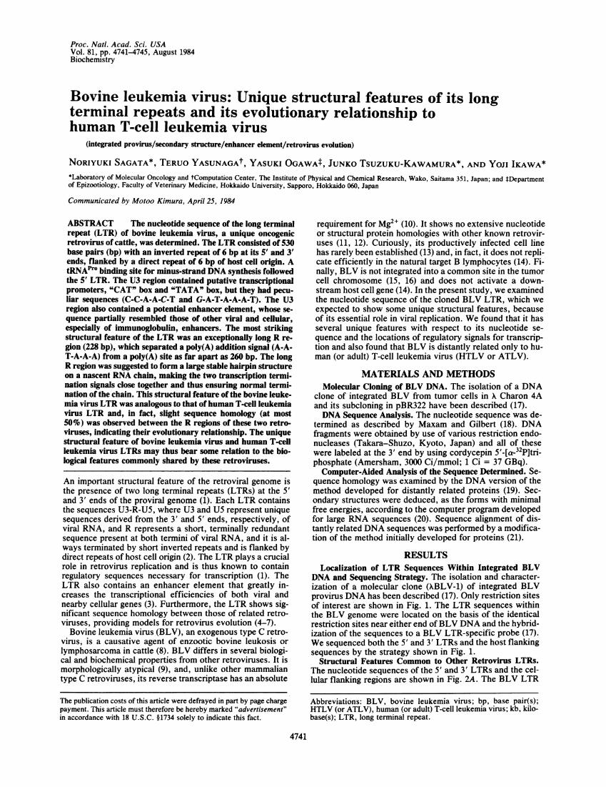
FIG. 1. Restriction enzyme map and strategy for sequencingBLV LTR. The isolation and characterization of integrated BLVprovirus DNA, XBLV-1, were described (17). The 3' end-labeled [a-32P]DNA fragments were sequenced by the procedure of Maxamand Gilbert (18). RI, EcoRI; Sc, Sac I; Hc, HincII; Ps, Pst I; Pv, PvuII. *, Labeled end of each fragment. The extent and direction ofsequencing are indicated by arrows. bp, Base pairs; kb, kilobases.
sequence, which is much more than in other retroviruses(16-79 bp, constituting 1.2-14% of the LTR; ref. 1). But sur-prisingly, it was identical to that of HTLV LTR (22). [Itshould be noted, however, that the U3 and U5 regions ofBLV LTR were both much shorter than those (351 and 175bp, respectively; ref. 22) of HTLV LTR.]We then attempted to identify the transcription promoter
signals, the CAT box (consensus; C-C-A-A-T) and TATAbox (T-A-T-A-T-A-T), which are usually located in the U3region, 70-90 and 20-30 bp, respectively, upstream from theG residue of the cap site (1). We could not find any typicalsignal sequences anywhere in the LTR, but closely relatedsequences were present 92 and 37 bp upstream from the capsite: the sequence at positions 115-120 had a single-base in-sertion (C-C-A-A-C-T) and that at positions 169-175 had asingle-base substitution (G-A-T-A-A-A-T). Although thesesequences are peculiar, they probably represent CAT andTATA boxes, judging from their locations at about the ex-pected positions.The canonical signal sequence (A-A-T-A-A-A) for polya-
denylylation is located 10-20 bp upstream from the poly(A)site in most retroviruses (1). In the BLV LTR, this hexanu-cleotide was not at the expected position but as far as 260 bpupstream (positions 177-182) from the poly(A) site. If this
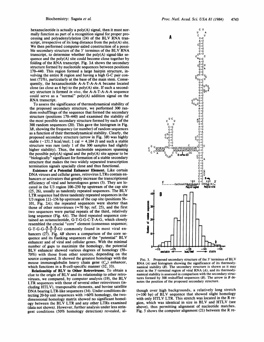
FIG. 2. Sequence (A) andsummary of the major features (B)of BLV LTR and its adjacent re-gions in the integrated proviralgenome. (A) The sequence ofLTR and its flanking regions pro-ceeding in the 5' to 3' directionwith the same polarity as that ofthe BLV genomic RNA areshown. (B) Important features ofthe BLV LTR sequence are illus-trated diagramatically. U3, se-quences unique to the 3' end of vi-ral RNA; R, terminally redundantsequences of viral RNA; U5, se-quences unique to the 5' end of vi-ral RNA; IR, inverted repeat; DR,direct repeat; PBS, primer bind-ing site for minus-strand DNAsynthesis; PPT, polypurine tractfor plus-strand DNA synthesis. C-C-A-A-C-T at positions 115-120and G-A-T-A-A-A-T at positions169-175 are the most likely candi-dates for the "CAT" box and"TATA" box. Sequences in brack-ets (positions 56-101) denote a
potential enhancer element.
Cell| R |U5 |6AC
228 bp 91 bp
A _4 . CtPb[O"5'ltR, U3 50A -40 Cellular.DNA r5LR35
- -ATGGACACGAGTTTGAGTGAACTCCGGGAGTTGGTGATGGACAG.GTGTATGAAAGATCATCCGACCTAGGACCCGCCACCCCCCCGTCCAGPvull Pvull DR 100 IV 150
ACAGAPACGiCAG'CTGCCAGkAACTGCGTGAdCCCCGTAC CTCCCCAACTTCCCCTTTC6CCGAAAAATCCACACCCTGAG200 U3.¶mRCATbox 250
CTGCTGACCTCACCTGCTGATAAATTAAT~AAATGCCGGCCCTGTC GAG3TTAGCGGCACCAGAAGCGTTCTTCTCCTGAGACCCTCGTGCTCAGCTCTC G
Sacl TATAbox Poly(A)signal 300 Cap Sito 350GTCCTGAGCACTCTTGCTCCCGAGACCTTCTGGTCGGCTATCCGGCAGCGGTCAGGTAAGGCAAGCACGGTTTGGAGGGTGGTTCTCGGCTGAGACCACC
SicI~400 R45540GCGAGCTCTATCTCCGGTCCTCTGACCGTCTCCAC GTGGACTCTCTCCTTTGCCTCCTGACCCCGCGCTCCAAGGGCGTCTGGCTTGCACCCGCGTTTGi
500 U5.nTR- PXly(A)site 550TTCCTGTCTTACTTTCTGTTTCTCGCGGCCCGCGCTCTCTCCTTCGGCGCCCTCTAGCGGCCAGGAGAGACCGGCAMCAATTGGGGGCTCGTCCGGGAT
Vl 4R POSTGATCACCCC ------------- --l-- --------------------------- AAATGAATGGCTCTCCCGCCTTTTTTGAGGGGGAGTCATT
10.3'LTR U3 R U5 3YLTR41 Cellular DNA PPTTGTA~kAMAT---------------------------GACCGGCAAACACACAGGGAGGCCTGGCGTGCTGCAATCATGGGGTTGCAAAAGT ------
lR DR
4742 Biochemistry: Sagata et aL
Biochemistry: Sagata et al. Proc. NatL. Acad. Sci. USA 81 (1984) 4743
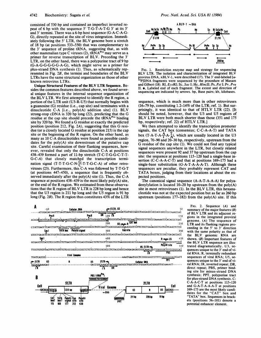
hexanucleotide is actually a poly(A) signal, then it must nor-mally function as part of a recognition signal for proper pro-cessing and polyadenylylation (24) of the BLV RNA tran-script, irrespective of its long distance from the poly(A) site.We then performed computer-aided construction of a possi-ble secondary structure of the 3' terminus of the BLV RNAtranscript, to determine whether the poly(A) signal-like se-quence and the poly(A) site could become close together byfolding of the RNA transcript. Fig. 3A shows the secondarystructure formed by nucleotide sequences between positions170-440. This region formed a large hairpin structure, in-volving the entire R region and having a high G-C pair con-tent (71%o), particularly at the base of the main stem. Conse-quently, the hexanucleotide A-A-T-A-A-A became locatedclose (as close as 6 bp) to the poly(A) site. If such a second-ary structure is formed in vivo, the A-A-T-A-A-A sequencecould serve as a "normal" poly(A) addition signal on theRNA transcript.To assess the significance of thermodynamical stability of
the proposed secondary structure, we performed 300 ran-dom reshufflings of the sequence that formed the secondarystructure (positions 170-440) and examined the stability ofthe most possible secondary structure formed by each of the300 random sequences (20). This gave the histogram in Fig.3B, showing the frequency (or number) ofrandom sequencesas a function of their thermodynamical stability. Clearly, theproposed secondary structure (arrow in Fig. 3B) was highlystable (-151.5 kcal/mol; 1 cal = 4.184 J) and such a stablestructure was rare (only 1 of the 300 samples had slightlyhigher stability). Thus, the nucleotide sequences spanningthe possible poly(A) signal and the poly(A) site appear to be"biologically" significant for formation of a stable secondarystructure that makes the two widely separated transcriptiontermination signals spacially close and thus functional.
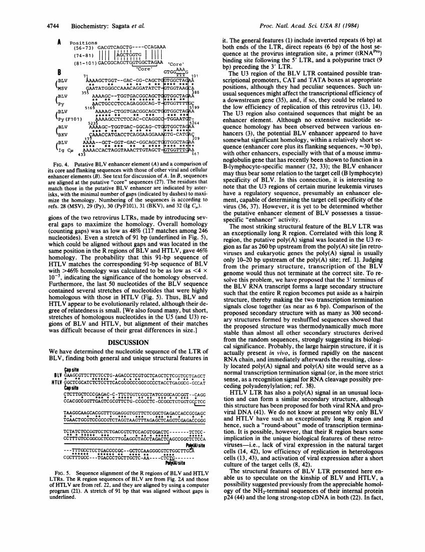
Existence of a Potential Enhancer Element. Like certainDNA viruses and cellular genes, retrovirus LTRs contain en-hancers or activators that greatly increase the transcriptionalefficiency of viral and heterologous genes (3). They are lo-cated in the U3 region 100-250 bp upstream of the cap site(25, 26), usually as tandemly repeated sequences. The BLVLTR sequence had three tandemly repeated sequences in theU3 region 111-156 bp upstream of the cap site (positions 56-101, Fig. 2A); the repeated sequences were shorter thanthose of other retroviruses (-70 bp; ref. 25), and the firsttwo sequences were partial repeats of the third, relativelylong sequence (Fig. 4A). The third repeated sequence con-tained an octanucleotide, G-T-G-G-C-T-A-G, which closelyresembled the crucial "core" element (consensus sequence;G-T-G-G-TA A- AG) commonly found in most viral en-hancers (27). Fig. 4B shows a comparison of the core se-quence and its flanking sequences of the "potential" BLVenhancer and of viral and cellular genes. With the minimalnumber of gaps to maximize the homology, the potentialBLV enhancer showed various degrees of homology (50-70%o) with those from other sources, depending on thesource compared. It showed the greatest homology with themouse immunoglobulin heavy chain gene (C,,) enhancer,which functions in a B-cell-specific manner (32, 33).
Relationship of BLV to Other Retroviruses. To obtain aclue to the origin of BLV and its relationship to other retro-viruses, we compared, by computer analysis (19), the BLVLTR sequences with those of several other retroviruses (in-cluding HTLV), transposable elements, and bovine satelliteDNA bearing LTR-like sequences (34). Under conditions de-tecting 20-bp unit sequences with >60% homology, the two-dimensional homology matrix showed no significant homol-ogy between the BCV LTR and any other _LTRs examined(data not shown). However, further analysis under less strin-gent conditions (50% homology detection) revealed, al-
C UU A
A-UCC *-CG-CC *GC *G
CA- CAG-CA-U
UCGcUG~c
G *C6UC-GUCUG-CUG *CUAAGUGG:C GG *CbcAG-CuA *U
GU UG CGCACG U
A .UC *GG *CG *C
U C AA-UC U UG-CU U G-CC-G A.UG-C C-GA.U U-A
CUG-C G-C
CGG C ACCG-CC-G G-C
C G GCUCAQC'UC-GACCqVU9UygGGCUAU6 GU ...d .0 UGGAAGACK ACCGUG C
U *AC *GC *GC *G
cG.C GUuC.G R-0
50, ~~~~1§0 G-C 4i0 3'------GAUAAUMI&WU UUGCA (A )n
Poly(A) signal hoIY(A Sit$
B
ea 30
220
Z10
FIG. 3. Proposed secondary structure of the 3' terminus of BLVRNA (A) and histogram showing the significance of its thermody-namical stability (B). The secondary structure is shown as it mayexist in the 3'-termninal region of viral RNA (A), and its thermody-namical stability is assessed in comparison with the secondary struc-tures formed by 300 reshuffled sequences (B). The arrow in B de-notes the position of the proposed secondary structure.
though over high backgrounds, a relatively long stretch(-100 bp) of BLV sequence that showed slight homologywith only HTLV LTR. This stretch was located in the R re-gion, which was identical in size in BLV and HTLV (seeabove), thus permitting alignment of nucleotide matches.Fig. 5 shows the computer alignment (21) between the R re-
5 _1 5 _1 S- -1
I
-5 -1unstable -P11/1 stablh
Proc. NatL Acad. Sci. USA 81 (1984)
A
BBLV
MSV
(BLVPyBLV
Py (IBLV
BKV
BLV
Ig (
Positions(56-73) GACGTCAGCTG----CCAGAAA
(74-81) | AGCTGGTGi1
(81-101) GACGGCAGCTGGTGGCTAGA 'Core'
'Core ' AAAGTGG~ G
71 TTT 101
AAAAGCTGGT--GAC-GG-CAGCT TGGCTA** ** ** ** * ** *** *** *
G.AATATGGGCCAAACAGGATATCT TGGTAA A355 388
AAAAGC--TGGTGACGGCAGCT TGGCTA A** ** * ** ***** * **** **
AACTGCCCTCCAGAGGGCAG-T-GTGGTTT G516 9 5199
AAAAG-CTGGTGACGGCAGCTC GTGGCTA A
***** ** ** *** *** **
F101) AAAAGCCTCTCCAC-CCAGGCC -TGGAAT -
5235 5264AAAAGC-TGGTGAC-GGCAG-CT TGGCTA A*** * ** * ** ** *** *****
C.AAACCATGACCTCAGGAAGG TG-CATCA209AAAA--GCT-GGT-GAC-GGCAGCT TGGCTAC AA**** ** *** ** * **** *** *****
CLuAAAACCACTAGGTAAACTTGTAGCT ATGGTTT433 467
FIG. 4. Putative BLV enhancer element (A) and a comparison ofits core and flanking sequences with those of other viral and cellularenhancer elements (B). See text for discussion of A. In B, sequencesare aligned at the putative "core" sequences (27). The residues thatmatch those in the putative BLV enhancer are indicated by aster-isks, with the minimal number of gaps (indicated by dashes) to maxi-mize the homology. Numbering of the sequences is according torefs. 28 (MSV), 29 (Py), 30 (PyF101), 31 (BKV), and 32 (Ig CJ).
gions of the two retrovirus LTRs, made by introducing sev-eral gaps to maximize the homology. Overall homology(counting gaps) was as low as 48% (117 matches among 246nucleotides). Even a stretch of 91 bp (underlined in Fig. 5),which could be aligned without gaps and was located in thesame position in the R regions of BLV and HTLV, gave 46%homology. The probability that this 91-bp sequence ofHTLV matches the corresponding 91-bp sequence of BLVwith >46% homology was calculated to be as low as <4 x
10-5, indicating the significance of the homology observed.Furthermore, the last 50 nucleotides of the BLV sequencecontained several stretches of nucleotides that were highlyhomologous with those in HTLV (Fig. 5). Thus, BLV andHTLV appear to be evolutionarily related, although their de-gree of relatedness is small. [We also found many, but short,stretches of homologous nucleotides in the U5 (and U3) re-gions of BLV and HTLV, but alignment of their matcheswas difficult because of their great differences in size.]
DISCUSSIONWe have determined the nucleotide sequence of the LTR ofBLV, finding both general and unique structural features in
;aP siteBLV GAAGCGTTCTTCTCCTG-AGACCCTCGTGCTCAGCTCTCGGTCCTGAGCT
* ** ****** * * * ** * ** * ** * *
HTLV GGCTCGCATCTCTCCTTCACGCGCCCGCCGCCCTACCTGAGGCG-GCCATlapsiteCTCTTGCTCCCGAGAC-C-TTCTGGTCGGCTATCCGGCAGCGGT--CAGG* * *** * * ***** ** ** *** * * *** *
CCACGCCGGTTGAGTCGCGTTCTG-CCGCCTC-CCGCCTGTGGTGCCTCC
TAAGGCAAGCACGGTTTGGAGGGTGGTTCTCGGCTGAGACCACCGCGAGC* * * ** * *** *** *** ** * * *
TGAACTGCGTCCGCCGTCTAGGTAAGTTTAGAGCTCAGGTCGAGACCGGG
TCTATCTCCGGTCCTCTGACCGTCTCCACGTGGACTC-------TCTCC-** * ***** *** * * ** * ***** ***
CCTTTGTCCGGCGCTCCCTTGGAGCCTACCTAGACTCAGCCGGCTCTCCAI
lP*t)site- - -TTTGCCTCCTGACCCCGC- -GCTCCAAGGGCGTCTGGCTTG M
****** ****** ** **** ** ****
CGCTTTGCC---TGACCCTGCTTGCTC-AA----CTCG-------IM site
FIG. 5. Sequence alignment of the R regions of BLV and HTLVLTRs. The R region sequences of BLV are from Fig. 2A and thoseof HTLV are from ref. 22, and they are aligned by using a computerprogram (21). A stretch of 91 bp that was aligned without gaps isunderlined.
it. The general features (1) include inverted repeats (6 bp) atboth ends of the LTR, direct repeats (6 bp) of the host se-quence at the provirus integration site, a primer (tRNAPIO)binding site following the 5' LTR, and a polypurine tract (9bp) preceding the 3' LTR.The U3 region of the BLV LTR contained possible tran-
scriptional promoters, CAT and TATA boxes at appropriatepositions, although they had peculiar sequences. Such un-usual sequences might affect the transcriptional efficiency ofa downstream gene (35), and, if so, they could be related tothe low efficiency of replication of this retrovirus (13, 14).The U3 region also contained sequences that might be anenhancer element. Although no extensive nucleotide se-quence homology has been observed between various en-hancers (3), the potential BLV enhancer appeared to havesomewhat significant homology, within a relatively short se-quence (enhancer core plus its flanking sequences, -30 bp),with other enhancers, especially with that of a mouse immu-noglobulin gene that has recently been shown to function in aB-lymphocyte-specific manner (32, 33); the BLV enhancermay thus bear some relation to the target cell (B lymphocyte)specificity of BLV. In this connection, it is interesting tonote that the U3 regions of certain murine leukemia viruseshave a regulatory sequence, presumably an enhancer ele-ment, capable of determining the target cell specificity of thevirus (36, 37). However, it is yet to be determined whetherthe putative enhancer element of BLV possesses a tissue-specific "enhancer" activity.The most striking structural feature of the BLV LTR was
an exceptionally long R region. Correlated with this long Rregion, the putative poly(A) signal was located in the U3 re-gion as far as 260 bp upstream from the poly(A) site [in retro-viruses and eukaryotic genes the poly(A) signal is usuallyonly 10-20 bp upstream of the poly(A) site; ref. 1]. Judgingfrom the primary structure, transcription of the BLVgenome would thus not terminate at the correct site. To re-solve this problem, we have proposed that the 3' terminus ofthe BLV RNA transcript forms a large secondary structuresuch that the entire R region becomes put aside as a hairpinstructure, thereby making the two transcription terminationsignals close together (as near as 6 bp). Comparison of theproposed secondary structure with as many as 300 second-ary structures formed by reshuffled sequences showed thatthe proposed structure was thermodynamically much morestable than almost all other secondary structures derivedfrom the random sequences, strongly suggesting its biologi-cal significance. Probably, the large hairpin structure, if it isactually present in vivo, is formed rapidly on the nascentRNA chain, and immediately afterwards the resulting, close-ly located poly(A) signal and poly(A) site would serve as anormal transcription termination signal (or, in the more strictsense, as a recognition signal for RNA cleavage possibly pre-ceding polyadenylylation; ref. 38).HTLV LTR has also a poly(A) signal in an unusual loca-
tion and can form a similar secondary structure, althoughthis structure has been proposed for both viral RNA and pro-viral DNA (41). We do not know at present why only BLVand HTLV have such an exceptionally long R region andhence, such a "round-about" mode of transcription termina-tion. It is possible, however, that their R region bears someimplication in the unique biological features of these retro-viruses-i.e., lack of viral expression in the natural targetcells (14, 42), low efficiency of replication in heterologouscells (13, 43), and activation of viral expression after a shortculture of the target cells (8, 42).The structural features of BLV LTR presented here en-
able us to speculate on the kinship of BLV and HTLV, apossibility suggested previously from the appreciable homol-ogy of the NH2-terminal sequences of their internal proteinp24 (44) and the long strong-stop cDNA in both (22). In fact,
4744 Biochemistry: Sagata et aL
Proc. NatL. Acad ScL USA 81 (1984) 4745
439 445XBLV-1 ---GCGTCTGGCTTGCACCCGCy;TT---
pLV 12 --- (GCGTCTGGCTTGCACCCGCA)654 660
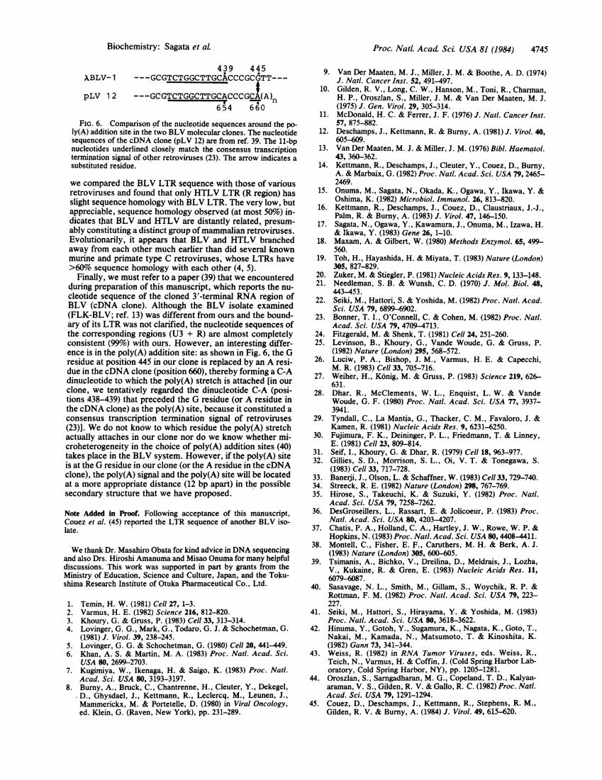
FIG. 6. Comparison of the nucleotide sequences around the po-ly(A) addition site in the two BLV molecular clones. The nucleotidesequences of the cDNA clone (pLV 12) are from ref. 39. The 11-bpnucleotides underlined closely match the consensus transcriptiontermination signal of other retroviruses (23). The arrow indicates asubstituted residue.
we compared the BLV LTR sequence with those of variousretroviruses and found that only HTLV LTR (R region) hasslight sequence homology with BLV LTR. The very low, butappreciable, sequence homology observed (at most 50%) in-dicates that BLV and HTLV are distantly related, presum-ably constituting a distinct group ofmammalian retroviruses.Evolutionarily, it appears that BLV and HTLV branchedaway from each other much earlier than did several knownmurine and primate type C retroviruses, whose LTRs have>60% sequence homology with each other (4, 5).
Finally, we must refer to a paper (39) that we encounteredduring preparation of this manuscript, which reports the nu-cleotide sequence of the cloned 3'-terminal RNA region ofBLV (cDNA clone). Although the BLV isolate examined(FLK-BLV; ref. 13) was different from ours and the bound-ary of its LTR was not clarified, the nucleotide sequences ofthe corresponding regions (U3 + R) are almost completelyconsistent (99%) with ours. However, an interesting differ-ence is in the poly(A) addition site: as shown in Fig. 6, the Gresidue at position 445 in our clone is replaced by an A resi-due in the cDNA clone (position 660), thereby forming a C-Adinucleotide to which the poly(A) stretch is attached [in ourclone, we tentatively regarded the dinucleotide C-A (posi-tions 438-439) that preceded the G residue (or A residue inthe cDNA clone) as the poly(A) site, because it constituted aconsensus transcription termination signal of retroviruses(23)]. We do not know to which residue the poly(A) stretchactually attaches in our clone nor do we know whether mi-croheterogeneity in the choice of poly(A) addition sites (40)takes place in the BLV system. However, if the poly(A) siteis at the G residue in our clone (or the A residue in the cDNAclone), the poly(A) signal and the poly(A) site will be locatedat a more appropriate distance (12 bp apart) in the possiblesecondary structure that we have proposed.
Note Added in Proof. Following acceptance of this manuscript,Couez et al. (45) reported the LTR sequence of another BLV iso-late.
We thank Dr. Masahiro Obata for kind advice in DNA sequencingand also Drs. Hiroshi Amanuma and Misao Onuma for many helpfuldiscussions. This work was supported in part by grants from theMinistry of Education, Science and Culture, Japan, and the Toku-shima Research Institute of Otuka Pharmaceutical Co., Ltd.
1. Temin, H. W. (1981) Cell 27, 1-3.2. Varmus, H. E. (1982) Science 216, 812-820.3. Khoury, G. & Gruss, P. (1983) Cell 33, 313-314.4. Lovinger, G. G., Mark, G., Todaro, G. J. & Schochetman, G.
(1981) J. Virol. 39, 238-245.5. Lovinger, G. G. & Schochetman, G. (1980) Cell 20, 441-449.6. Khan, A. S. & Martin, M. A. (1983) Proc. Natl. Acad. Sci.
USA 80, 2699-2703.7. Kugimiya, W., Ikenaga, H. & Saigo, K. (1983) Proc. Natl.
Acad. Sci. USA 80, 3193-3197.8. Burny, A., Bruck, C., Chantrenne, H., Cleuter, Y., Dekegel,
D., Ghysdael, J., Kettmann, R., Leclercq, M., Leunen, J.,Mammerickx, M. & Portetelle, D. (1980) in Viral Oncology,ed. Klein, G. (Raven, New York), pp. 231-289.
9. Van Der Maaten, M. J., Miller, J. M. & Boothe, A. D. (1974)J. Natl. Cancer Inst. 52, 491-497.
10. Gilden, R. V., Long, C. W., Hanson, M., Toni, R., Charluan,H. P., Oroszlan, S., Miller, J. M. & Van Der Maaten, M. J.(1975) J. Gen. Virol. 29, 305-314.
11. McDonald, H. C. & Ferrer, J. F. (1976) J. Natl. Cancer Inst.57, 875-882.
12. Deschamps, J., Kettmann, R. & Burny, A. (1981) J. Virol. 40,605-609.
13. Van Der Maaten, M. J. & Miller, J. M. (1976) Bibl. Haematol.43, 360-362.
14. Kettmann, R., Deschamps, J., Cleuter, Y., Couez, D., Burny,A. & Marbaix, G. (1982) Proc. Natl. Acad. Sci. USA 79, 2465-2469.
15. Onuma, M., Sagata, N., Okada, K., Ogawa, Y., Ikawa, Y. &Oshima, K. (1982) Microbiol. Immunol. 26, 813-820.
16. Kettmann, R., Deschamps, J., Couez, D., Claustriaux, J.-J.,Palm, R. & Burny, A. (1983) J. Virol. 47, 146-150.
17. Sagata, N., Ogawa, Y., Kawamura, J., Onuma, M., Izawa, H.& Ikawa, Y. (1983) Gene 26, 1-10.
18. Maxam, A. & Gilbert, W. (1980) Methods Enzymol. 65, 499-560.
19. Toh, H., Hayashida, H. & Miyata, T. (1983) Nature (London)305, 827-829.
20. Zuker, M. & Stiegler, P. (1981) Nucleic Acids Res. 9, 133-148.21. Needleman, S. B. & Wunsh, C. D. (1970) J. Mol. Biol. 48,
443-453.22. Seiki, M., Hattori, S. & Yoshida, M. (1982) Proc. Natl. Acad.
Sci. USA 79, 6899-6902.23. Bonner, T. I., O'Connell, C. & Cohen, M. (1982) Proc. Natl.
Acad. Sci. USA 79, 4709-4713.24. Fitzgerald, M. & Shenk, T. (1981) Cell 24, 251-260.25. Levinson, B., Khoury, G., Vande Woude, G. & Gruss, P.
(1982) Nature (London) 295, 568-572.26. Luciw, P. A., Bishop, J. M., Varmus, H. E. & Capecchi,
M. R. (1983) Cell 33, 705-716.27. Weiher, H., Konig, M. & Gruss, P. (1983) Science 219, 626-
631.28. Dhar, R., McClements, W. L., Enquist, L. W. & Vande
Woude, G. F. (1980) Proc. Natl. Acad. Sci. USA 77, 3937-3941.
29. Tyndall, C., La Mantia, G., Thacker, C. M., Favaloro, J. &Kamen, R. (1981) Nucleic Acids Res. 9, 6231-6250.
30. Fujimura, F. K., Deininger, P. L., Friedmann, T. & Linney,E. (1981) Cell 23, 809-814.
31. Seif, I., Khoury, G. & Dhar, R. (1979) Cell 18, 963-977.32. Gillies, S. D., Morrison, S. L., Oi, V. T. & Tonegawa, S.
(1983) Cell 33, 717-728.33. Banerji, J., Olson, L. & Schaffner, W. (1983) Cell 33, 729-740.34. Streeck, R. E. (1982) Nature (London) 298, 767-769.35. Hirose, S., Takeuchi, K. & Suzuki, Y. (1982) Proc. Natl.
Acad. Sci. USA 79, 7258-7262.36. DesGroseillers, L., Rassart, E. & Jolicoeur, P. (1983) Proc.
Natl. Acad. Sci. USA 80, 4203-4207.37. Chatis, P. A., Holland, C. A., Hartley, J. W., Rowe, W. P. &
Hopkins, N. (1983) Proc. Natl. Acad. Sci. USA 80,4408-4411.38. Montell, C., Fisher, E. F., Caruthers, M. H. & Berk, A. J.
(1983) Nature (London) 305, 600-605.39. Tsimanis, A., Bichko, V., Dreilina, D., Meldrais, J., Lozha,
V., Kukaine, R. & Gren, E. (1983) Nucleic Acids Res. 11,6079-6087.
40. Sasavage, N. L., Smith, M., Gillam, S., Woychik, R. P. &Rottman, F. M. (1982) Proc. Natl. Acad. Sci. USA 79, 223-227.
41. Seiki, M., Hattori, S., Hirayama, Y. & Yoshida, M. (1983)Proc. Natl. Acad. Sci. USA 80, 3618-3622.
42. Hinuma, Y., Gotoh, Y., Sugamura, K., Nagata, K., Goto, T.,Nakai, M., Kamada, N., Matsumoto, T. & Kinoshita, K.(1982) Gann 73, 341-344.
43. Weiss, R. (1982) in RNA Tumor Viruses, eds. Weiss, R.,Teich, N., Varmus, H. & Coffin, J. (Cold Spring Harbor Lab-oratory, Cold Spring Harbor, NY), pp. 1205-1281.
44. Oroszlan, S., Sarngadharan, M. Q., Copeland, T. D., Kalyan-araman, V. S., Gilden, R. V. & Gallo, R. C. (1982) Proc. Nail.Acad. Sci. USA 79, 1291-1294.
45. Couez, D., Deschamps, J., Kettmann, R., Stephens, R. M.,Gilden, R. V. & Burny, A. (1984) J. Virol. 49, 615-620.
Bioche istrIm y: Sagata et aL