Embed Size (px)
Citation preview
Caracterización de la disfunción vascular sanguínea y linfática en la cirrosis hepática:
Evaluación de la inhibición del factor decrecimiento placentario y del óxido nítrico
como estrategias terapéuticas
Jordi Ribera Sabaté
ADVERTIMENT. La consulta d’aquesta tesi queda condicionada a l’acceptació de les següents condicions d'ús: La difusió d’aquesta tesi per mitjà del servei TDX (www.tdx.cat) ha estat autoritzada pels titulars dels drets de propietat intel·lectual únicament per a usos privats emmarcats en activitats d’investigació i docència. No s’autoritza la seva reproducció amb finalitats de lucre ni la seva difusió i posada a disposició des d’un lloc aliè al servei TDX. No s’autoritza la presentació delseu contingut en una finestra o marc aliè a TDX (framing). Aquesta reserva de drets afecta tant al resum de presentació de la tesi com als seus continguts. En la utilització o cita de parts de la tesi és obligat indicar el nom de la persona autora.
ADVERTENCIA. La consulta de esta tesis queda condicionada a la aceptación de las siguientes condiciones de uso: La difusión de esta tesis por medio del servicio TDR (www.tdx.cat) ha sido autorizada por los titulares de los derechos de propiedad intelectual únicamente para usos privados enmarcados en actividades de investigación y docencia. No se autoriza su reproducción con finalidades de lucro ni su difusión y puesta a disposición desde un sitio ajeno al servicio TDR. No se autoriza la presentación de su contenido en una ventana o marco ajeno a TDR (framing). Esta reserva de derechos afecta tanto al resumen de presentación de la tesis como a sus contenidos. En la utilización o cita de partes de la tesis es obligado indicar el nombre de la persona autora.
WARNING. On having consulted this thesis you’re accepting the following use conditions: Spreading this thesis by the TDX (www.tdx.cat) service has been authorized by the titular of the intellectual property rights only for private uses placed in investigation and teaching activities. Reproduction with lucrative aims is not authorized neither its spreading and availability from a site foreign to the TDX service. Introducing its content in a window or frame foreign to the TDX service isnot authorized (framing). This rights affect to the presentation summary of the thesis as well as to its contents. In the usingor citation of parts of the thesis it’s obliged to indicate the name of the author.
CARACTERIZACIÓN DE LA DISFUNCIÓN VASCULAR SANGUÍNEA Y LINFÁTICA
EN LA CIRROSIS HEPÁTICA:
EVALUACIÓN DE LA INHIBICIÓN DEL FACTOR DE CRECIMIENTO PLACENTARIO
Y DEL ÓXIDO NÍTRICO COMO ESTRATEGIAS TERAPÉUTICAS
Memoria presentada por
JORDI RIBERA SABATÉ
para optar al título de Doctor en Bioquímica por la Universitat de Barcelona
Trabajo realizado bajo la dirección del Dr. Manuel Morales Ruiz
Servicio de Bioquímica y Genética Molecular
Hospital Clínic de Barcelona
Tesis inscrita en el programa de doctorado de Medicina
Departamento de Medicina, Facultad de Medicina


Aquesta tesi està dedicada
als meus pares i a tota la meva família

ABREVIATURAS
ACE Enzima convertidora de la angiotensina
ADH Hormona antidiurética
αSMA Actina de músculo liso α
Ang-II Angiotensina-2
AVP Vasopresina
BODIPY Boro-dipirrometeno
BrdU Bromodeoxiuridina
Cav-1 Caveolina-1
CCL21 Ligando de quimioquinas C-C 21
CCl4 Tetracloruro de carbono
CCR7 Receptor de quimioquinas C-C tipo 7
CH Cirrosis, cirrótico
CLEC-2 Familia 2 del dominio de lectina tipo C
CLEVER-1 Receptor endotelial vascular y endotelial linfático común-1
CT Control
CXCL12 Ligando de quimioquinas CxC 12
CXCR4 Receptor de quimioquinas CxC tipo 4
DETANONOate Dietilentriamina NONOate
eNOS Sintasa de óxido nítrico endotelial
ERK Quinasa regulada por señales extracelulares
ET-1 Endotelina-1
FDA Food and Drug Administration
FAK Quinasa de adhesiones focales
FITC Fluoresceina isotiocianato
GMPc Guanosín monofosfato cíclico
GTP Guanosín trifosfato
HSC Célula estrellada hepática
ICAM-1 Molécula de adhesión intercelular-1
IFN Interferón
IL Interleuquina
iNOS Sintasa de óxido nítrico inducible
JAM-2 Molécula de adhesión cruzada tipo B
KC Célula de Kupffer
LDL-R Receptor de las lipoproteínas de baja densidad
L-NAME Metil éster de NG-nitro-L-arginina
L-NIO N-iminoetil-L-ornitina
L-NMMA NG-monometil-L-arginina
L-NOArg NG-nitro-L-arginina
LPS Lipopolisacárico
LSEC Célula endotelial sinusoidal
LyEC Célula endotelial linfática
LYVE-1 Receptor endotelial de vasos linfáticos-1
MAPK Proteínas quinasa activadas por mitógenos
MEK Proteína quinasa quinasa
MMP Metaloproteinasa
NADPH Nicotinamida adenina dinucleótido fosfato
nNOS Sintasa de óxido nítrico neuronal
NO Óxido nítrico
NOS Sintasa de óxido nítrico
PI3K Fosfatidil inositol 3-quinasa
PlGF Factor de crecimiento placentario
PDGF Factor de crecimiento derivado de plaquetas
PDGFR Receptor del factor de crecimiento derivado de plaquetas
PKC Proteína quinasa C
PKG Proteína quinasa dependiente de GMPc
RNAm Ácido ribonucleico mensajero
ROS Especies reactivas de oxígeno
RT Retrotranscripción
RTK Receptor tirosina quinasa
SLP-76 Proteína leucocitaria de 76 KDa
SMC Célula muscular lisa
SNS Sistema nervioso simpático
SRAA Sistema renina-angiotensina-aldosterona
SYK Tirosina quinasa de bazo
TGF Factor de crecimiento transformante
TIMP Inhibidor de las metaloproteinasas
TNF Factor de necrosis tumoral
VCAM-1 Molécula de adhesión vascular-1
VEGF Factor de crecimiento endotelial vascular
VEGFR Receptor del factor de crecimiento endotelial vascular
vWF Factor de Von Willebrand


ÍNDICE

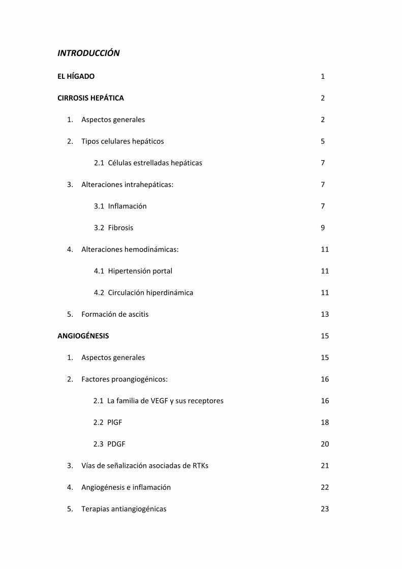
INTRODUCCIÓN
EL HÍGADO 1
CIRROSIS HEPÁTICA 2
1. Aspectos generales 2
2. Tipos celulares hepáticos 5
2.1 Células estrelladas hepáticas 7
3. Alteraciones intrahepáticas: 7
3.1 Inflamación 7
3.2 Fibrosis 9
4. Alteraciones hemodinámicas: 11
4.1 Hipertensión portal 11
4.2 Circulación hiperdinámica 11
5. Formación de ascitis 13
ANGIOGÉNESIS 15
1. Aspectos generales 15
2. Factores proangiogénicos: 16
2.1 La familia de VEGF y sus receptores 16
2.2 PlGF 18
2.3 PDGF 20
3. Vías de señalización asociadas de RTKs 21
4. Angiogénesis e inflamación 22
5. Terapias antiangiogénicas 23
EL ÓXIDO NÍTRICO 24
1. Características y funciones generales 24
2. Sintasas del óxido nítrico 26
3. Inhibidores de las NOS 27
4. Papel fisiopatológico del óxido nítrico en la cirrosis 28
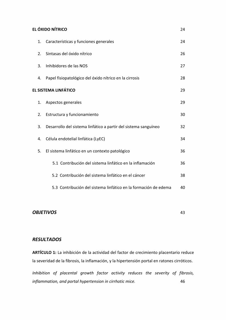
EL SISTEMA LINFÁTICO 29
1. Aspectos generales 29
2. Estructura y funcionamiento 30
3. Desarrollo del sistema linfático a partir del sistema sanguíneo 32
4. Célula endotelial linfática (LyEC) 34
5. El sistema linfático en un contexto patológico 36
5.1 Contribución del sistema linfático en la inflamación 36
5.2 Contribución del sistema linfático en el cáncer 38
5.3 Contribución del sistema linfático en la formación de edema 40
OBJETIVOS 43
RESULTADOS
ARTÍCULO 1: La inhibición de la actividad del factor de crecimiento placentario reduce
la severidad de la fibrosis, la inflamación, y la hipertensión portal en ratones cirróticos.
Inhibition of placental growth factor activity reduces the severity of fibrosis,
inflammation, and portal hypertension in cirrhotic mice. 46
ARTÍCULO 2: La sobreproducción de óxido nítrico en células endoteliales linfáticas
provoca una disfunción en el drenaje linfático en ratas cirróticas.
Increase nitric oxide production in lymphatic endothelial cells causes impairment of
lymphatic drainage in cirrhotic rats. 52
DISCUSIÓN 56
CONCLUSIONES 64
BIBLIOGRAFÍA 67
AGRADECIMIENTOS 86
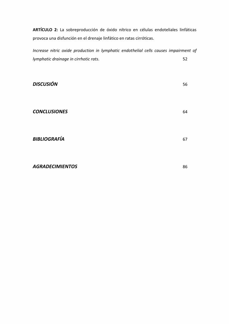
ÍNDICE DE FIGURAS
Figura 1. Estructura del hígado 1
Figura 2. Tipos celulares hepáticos 6
Figura 3. Respuesta inflamatoria tras un daño hepático 8
Figura 4. Proceso de fibrosis en el territorio hepático 10
Figura 5. Fisiopatología de la formación de ascitis 14
Figura 6. Mecanismos básicos de formación de vasos 15
Figura 7. Esquema de la familia de VEGF y sus receptores 18
Figura 8. Vías de señalización activadas por receptores tirosina quinasa 21
Figura 9. Generación de NO y efecto sobre las células musculares lisas 25
Figura 10. Funciones de la vasculatura linfática 30
Figura 11. Estructura y funcionamiento del sistema linfático 32
Figura 12. Desarrollo embrionario del sistema linfático 33
Figura 13. Función de los vasos linfáticos en los procesos inflamatorios 37
Figura 14. Metástasis linfática del cáncer 39
Figura 15. Vasculatura linfática en el linfedema 41
ÍNDICE DE TABLAS
Tabla 1. Alteraciones hemodinámicas en la cirrosis 12
Tabla 2. Clasificación de los diferentes tipos de sintasas de NO 26
Tabla 3. Inhibidores de las NOS 28

INTRODUCCIÓN

INTRODUCCIÓN _______________________________________________________________________
1
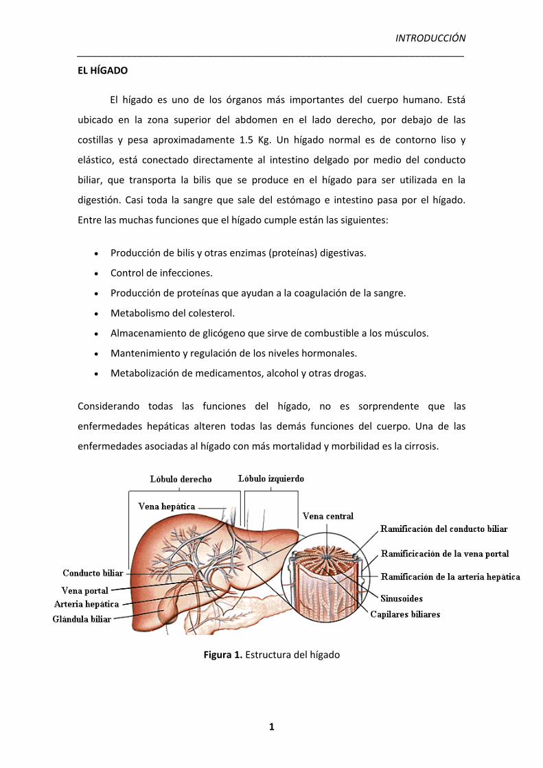
EL HÍGADO
El hígado es uno de los órganos más importantes del cuerpo humano. Está
ubicado en la zona superior del abdomen en el lado derecho, por debajo de las
costillas y pesa aproximadamente 1.5 Kg. Un hígado normal es de contorno liso y
elástico, está conectado directamente al intestino delgado por medio del conducto
biliar, que transporta la bilis que se produce en el hígado para ser utilizada en la
digestión. Casi toda la sangre que sale del estómago e intestino pasa por el hígado.
Entre las muchas funciones que el hígado cumple están las siguientes:
� Producción de bilis y otras enzimas (proteínas) digestivas.
� Control de infecciones.
� Producción de proteínas que ayudan a la coagulación de la sangre.
� Metabolismo del colesterol.
� Almacenamiento de glicógeno que sirve de combustible a los músculos.
� Mantenimiento y regulación de los niveles hormonales.
� Metabolización de medicamentos, alcohol y otras drogas.
Considerando todas las funciones del hígado, no es sorprendente que las
enfermedades hepáticas alteren todas las demás funciones del cuerpo. Una de las
enfermedades asociadas al hígado con más mortalidad y morbilidad es la cirrosis.
Figura 1. Estructura del hígado
INTRODUCCIÓN _______________________________________________________________________
2
CIRROSIS HEPÁTICA
1. ASPECTOS GENERALES
La cirrosis es la consecuencia final de un daño acumulado en el hígado,
habitualmente durante varios años, que se caracteriza por la acumulación de fibra en
el tejido hepático y por una disminución del tejido hepático funcional. Estos cambios
del hígado interfieren con la estructura y el funcionamiento normal del hígado,
ocasionando serias complicaciones en la circulación de la sangre a través de dicho
órgano y en sus funciones. Según la Organización Mundial de la Salud (OMS),
anualmente mueren más de 200.000 personas por esta enfermedad en los países
desarrollados, siendo la sexta causa de muerte más frecuente en el mundo (Anderson
et al., 2003).
De forma similar a lo que acontece en otros tejidos, la inflamación hepática es
un proceso necesario en la reparación tisular. En condiciones normales, este tipo de
respuesta restaura la estructura y las funciones originales del hígado, y mantiene la
homeostasis tisular. Sin embargo, hay ocasiones en que la lesión es demasiado intensa
o persistente, y el propio proceso inflamatorio compromete la integridad estructural
del órgano a través de mecanismos como una excesiva formación de matriz
extracelular (fibrosis), con posterior esclerosis (Schuppan, 1990). El exceso de matriz se
acumula en la zona perisinusoidal, formándose una barrera que impide la correcta
difusión de oxígeno y nutrientes a través del hígado, provocando la muerte celular de
los hepatocitos. Esto conduce a la distorsión de la arquitectura lobular normal del
parénquima hepático y de la red vascular, derivando en la formación de nódulos
hepáticos separados por fibras (nódulos de regeneración). Esta fase final del proceso
es la que se conoce como cirrosis (Erlinger y Benhamou, 1999).
INTRODUCCIÓN _______________________________________________________________________
3
En la evolución de la enfermedad, podemos distinguir dos fases: cirrosis
compensada y descompensada. Las complicaciones que definen la cirrosis
descompensada son (Conn and Atterbury, 1987; Friedman, 2003):
� Ascitis: acúmulo de líquido libre intraabdominal con características de
transudado (filtrado de plasma con bajo contenido en proteínas). Este
transudado además puede infectarse (peritonitis bacteriana espontánea),
habitualmente a causa de la traslocación bacteriana. Es la complicación más
común y más temprana de la cirrosis.
� Síndrome hepatorrenal: insuficiencia renal funcional y reversible sin que exista
alteración de la estructura renal. Se produce debido a una intensa
vasoconstricción renal que conduce a una insuficiencia que se establece en el
plazo de días o semanas. Suele aparecer de forma muy agresiva y tiene un
pronóstico fatal a corto plazo, en la mayoría de los casos en semanas (síndrome
hepatorrenal tipo I) o de forma más insidiosa, con un pronóstico ligeramente
mejor y una supervivencia media de alrededor de seis meses (síndrome
hepatorrenal tipo II).
� Encefalopatía hepática: síndrome neuropsiquiátrico secundario producido por
el deterioro de la función neurológica, habitualmente episódico y reversible.
Aparece cuando determinados metabolitos, como por ejemplo amonio, pasan
directamente de la circulación portal a la circulación sistémica, y acaban
afectando al sistema nervioso.
� Hemorragia digestiva por varices gastroesofágicas: dilataciones de las venas
gástricas y del esófago producidas por aumento de la presión en la vena porta,
como consecuencia de la cirrosis hepática. Al aumentar la presión dentro de las
varices, se rompen produciendo hemorragias.
� Ictericia: tinte amarillento de la piel y las mucosas a consecuencia del acúmulo
de bilirrubina.
La cirrosis descompensada predice habitualmente una importante disminución de la
supervivencia, y tiene un mal pronóstico a corto plazo. Además de las complicaciones
INTRODUCCIÓN _______________________________________________________________________
4
descritas los pacientes con cirrosis hepática tienen un riesgo aumentado de desarrollar
hepatocarcinoma (Gines et al, 2004; Bataller and Brenner, 2005).
Existen numerosas causas que pueden desencadenar la cirrosis hepática, entre
las principales están (Anthony et al., 1978; Fattovich et al., 1997):
� Virus de la hepatitis B, hepatitis C, y hepatitis D (los virus de la hepatitis son
la causa de cirrosis más habitual en la actualidad).
� Consumo excesivo de alcohol (sigue siendo todavía la causa del 40% de los
casos de cirrosis).
� Enfermedad del hígado graso no alcohólico, también llamada
esteatohepatitis no alcohólica. Es una condición frecuente en la población
general debido al cambio de hábitos alimentarios y al sedentarismo. Está
asociada a diabetes y obesidad.
� Enfermedades autoinmunes, como la hepatitis autoinmune o la cirrosis
biliar primaria.
� Enfermedades hereditarias o congénitas como la hemocromatosis, en la
cual se acumula hierro dentro del hígado dañando el tejido. Otro ejemplo
sería la enfermedad de Wilson, que es causada por una alteración en el
transporte de cobre, acumulándose en el hígado y otros tejidos.
� Obstrucción prolongada del conducto biliar, como pasa en la colangitis
esclerosante.
� Ausencia de proteínas específicas o enzimas para metabolizar diferentes
substancias en el hígado, como la deficiencia de la alfa 1-antitripsina.
� Ciertas enfermedades del corazón, como la insuficiencia cardíaca.
� Reacción severa a drogas o medicamentos.
� Exposición prolongada a agentes tóxicos en el medio ambiente.
La cirrosis como tal carece de tratamiento médico específico. El tratamiento
definitivo de la cirrosis es el trasplante hepático, pero el rechazo crónico del injerto y el
desequilibrio existente entre la demanda y la disponibilidad de órganos comprometen
esta estrategia.
INTRODUCCIÓN _______________________________________________________________________
5
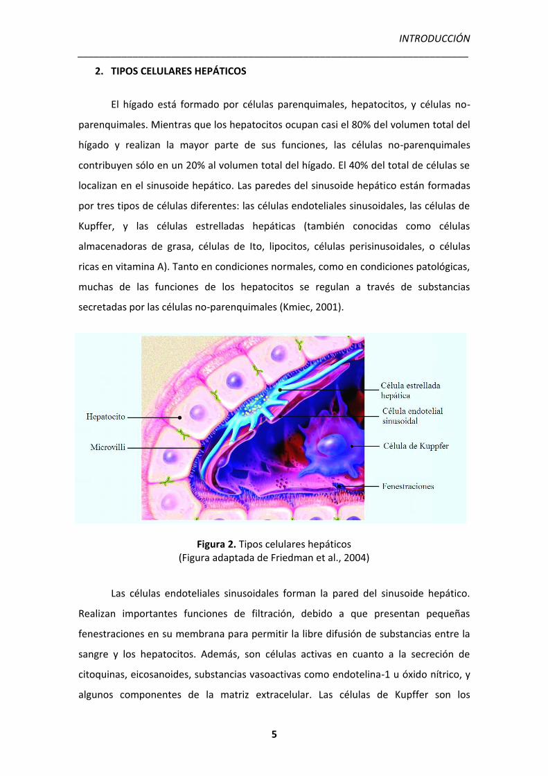
2. TIPOS CELULARES HEPÁTICOS
El hígado está formado por células parenquimales, hepatocitos, y células no-
parenquimales. Mientras que los hepatocitos ocupan casi el 80% del volumen total del
hígado y realizan la mayor parte de sus funciones, las células no-parenquimales
contribuyen sólo en un 20% al volumen total del hígado. El 40% del total de células se
localizan en el sinusoide hepático. Las paredes del sinusoide hepático están formadas
por tres tipos de células diferentes: las células endoteliales sinusoidales, las células de
Kupffer, y las células estrelladas hepáticas (también conocidas como células
almacenadoras de grasa, células de Ito, lipocitos, células perisinusoidales, o células
ricas en vitamina A). Tanto en condiciones normales, como en condiciones patológicas,
muchas de las funciones de los hepatocitos se regulan a través de substancias
secretadas por las células no-parenquimales (Kmiec, 2001).
Figura 2. Tipos celulares hepáticos (Figura adaptada de Friedman et al., 2004)
Las células endoteliales sinusoidales forman la pared del sinusoide hepático.
Realizan importantes funciones de filtración, debido a que presentan pequeñas
fenestraciones en su membrana para permitir la libre difusión de substancias entre la
sangre y los hepatocitos. Además, son células activas en cuanto a la secreción de
citoquinas, eicosanoides, substancias vasoactivas como endotelina-1 u óxido nítrico, y
algunos componentes de la matriz extracelular. Las células de Kupffer son los
INTRODUCCIÓN _______________________________________________________________________
6
macrófagos residentes del hígado, tienen capacidad endocítica y fagocítica, y producen
potentes mediadores de la respuesta inflamatoria (especies reactivas de oxígeno,
eicosanoides, óxido nítrico, monóxido de carbono, TNF-alfa, y otras citoquinas) (Luckey
and Petersen, 2001; Melgert et al., 2001). Por ello, controlan las primeras fases de la
inflamación hepática (Titos et al., 2003; Claria and Titos, 2004). Durante la enfermedad
hepática, las células de Kupffer secretan enzimas y citoquinas que pueden dañar a los
hepatocitos, y son activas en el remodelado de la matriz extracelular (Bouwens et al.,
1992; Wisse et al., 1996).
2.1 CÉLULAS ESTRELLADAS HEPÁTICAS
Las células estrelladas hepáticas están ubicadas en el espacio perisinusoidal de
Disse, que es la zona limitada por los hepatocitos y la pared sinusoidal formada por las
células endoteliales sinusoidales (Friedman, 2003). En el hígado sano las células
hepáticas estrelladas almacenan vitamina A, controlan la degradación y formación de
matriz extracelular, y regulan la contractibilidad del sinusoide (Flisiak, 1997). En este
estado expresan marcadores característicos de adipocito (PPARγ, SREBP-1c y leptina).
Sin embargo, el daño hepático activa estas células, transformándolas de un estado
quiescente a un fenotipo miofibroblástico, que tiene un papel muy importante en el
desarrollo de inflamación y fibrosis. En este estado expresan marcadores miogénicos
(α-SMA, c-myb, miosina o desmina), algunos marcadores neuroendocrinos (reelina,
nestina, neurotrofinas, sinaptofisina y GFAP) y receptores de neurotransmisores. Estos
nuevos miofibroblastos constituyen una nueva fuente de múltiples tipos de proteínas
colágenas y no colágenas de la matriz extracelular, como el colágeno tipo I, tipo III, tipo
IV, laminina, elastina, fibronectina y diversos proteoglucanos como el
condroitinsulfato, el dermatansulfato y el heparansulfato. Además, la velocidad de
proliferación de las células estrelladas hepáticas se incrementa después de su
activación, amplificando así el número de células fibrogénicas presentes en el hígado
(Sarem et al., 2006; Pinzani et al., 2005; Alcolado et al., 1997; Bataller and Brenner,
2005).
INTRODUCCIÓN _______________________________________________________________________
7
3. ALTERACIONES INTRAHEPÁTICAS
3.1 INFLAMACIÓN
La inflamación es un proceso clave en la progresión de diversas enfermedades
como el cáncer, el asma, la enfermedad inflamatoria intestinal o las enfermedades
hepáticas crónicas, y tiene como finalidad la reparación de una lesión tisular
eliminando las células y tejidos necróticos (Vinay et al., 2005).
Debido a su función y situación anatómica, el hígado está constantemente
expuesto a agresiones que pueden inducir inflamación. El proceso inflamatorio en el
hígado es dinámico y si no se resuelve adecuadamente se cronifica en el tiempo y se
acaba llegando al estado de fibrosis (Iredale, 2007). El primer paso es la activación de
los hepatocitos cuando éstos son dañados por metabolitos tóxicos. Los hepatocitos
activados liberan citoquinas que, junto con las propias moléculas causantes del daño,
inducen la activación del resto de células hepáticas y el reclutamiento de leucocitos del
torrente sanguíneo (Ramadori et al., 2008). Hay un incremento en la expresión de
moléculas de adhesión, como ICAM-1, y el reclutamiento se realiza gracias a la
interacción entre las moléculas de adhesión existentes en la superficie de las células
inflamatorias y las células endoteliales hepáticas. La composición de este infiltrado
inflamatorio incluye monocitos, linfocitos T, linfocitos B, células dendríticas,
eosinófilos, neutrófilos, células NK y mastocitos. La primera función del infiltrado es
eliminar el daño hepático provocando la muerte de los hepatocitos no viables.
Adicionalmente, otras subpoblaciones de células hepáticas también pueden ser dianas
para la respuesta inflamatoria, como las células endoteliales o las estrelladas. Esto
provoca una disfunción en estas células y aumentan la síntesis de substancias
proinflamatorias. (Maher, 2001; Dasarathy, 2008).
Los leucocitos, junto con las células inflamatorias residentes en el hígado
(células de Kupffer), inician la respuesta inflamatoria en el tejido y empiezan a producir
una serie de moléculas que acaban alterando el funcionamiento de los demás tipos
celulares hepáticos. Se producen grandes cantidades de NO, especies reactivas de
oxígeno, factores de crecimiento y citoquinas proinflamatorias, como el TNFα, TGF-β,
INTRODUCCIÓN _______________________________________________________________________
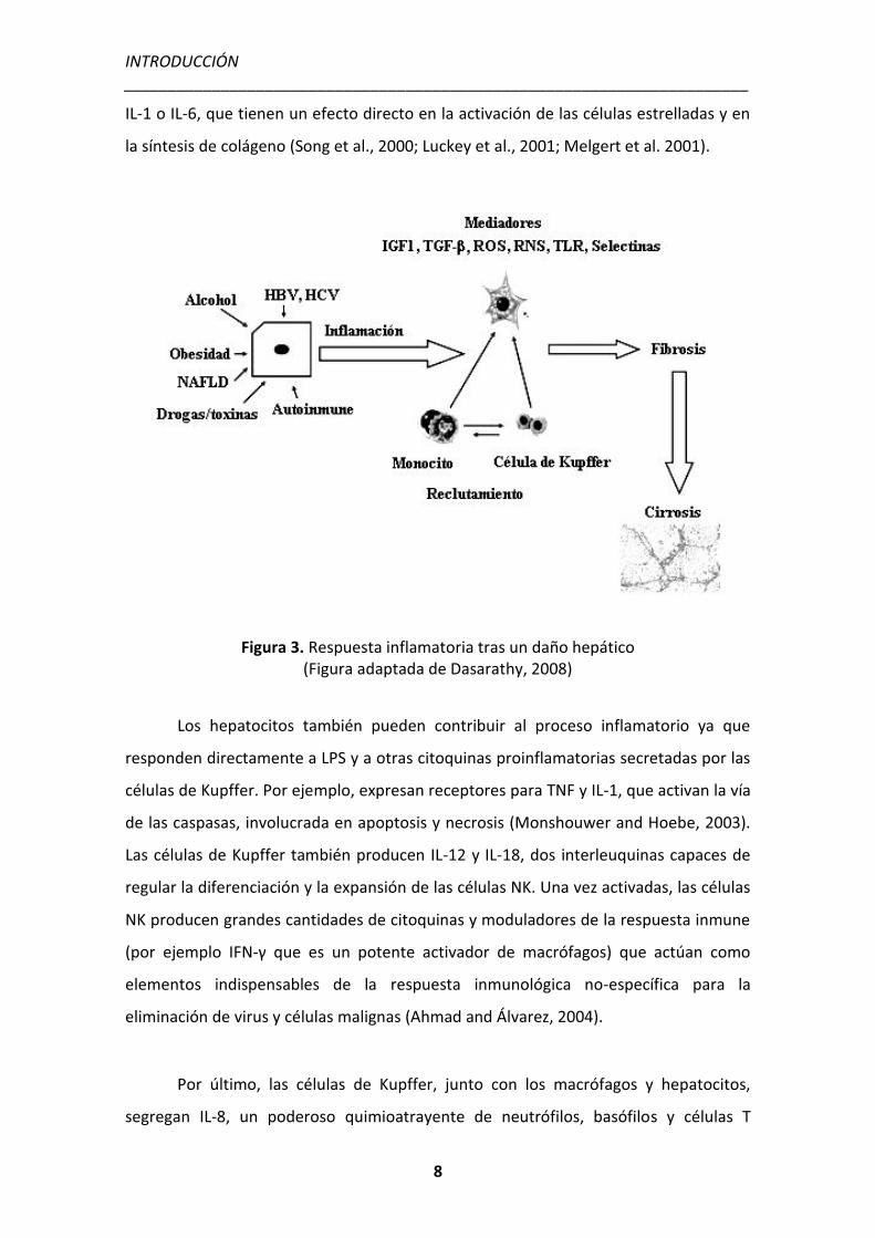
8
IL-1 o IL-6, que tienen un efecto directo en la activación de las células estrelladas y en
la síntesis de colágeno (Song et al., 2000; Luckey et al., 2001; Melgert et al. 2001).
Figura 3. Respuesta inflamatoria tras un daño hepático (Figura adaptada de Dasarathy, 2008)
Los hepatocitos también pueden contribuir al proceso inflamatorio ya que
responden directamente a LPS y a otras citoquinas proinflamatorias secretadas por las
células de Kupffer. Por ejemplo, expresan receptores para TNF y IL-1, que activan la vía
de las caspasas, involucrada en apoptosis y necrosis (Monshouwer and Hoebe, 2003).
Las células de Kupffer también producen IL-12 y IL-18, dos interleuquinas capaces de
regular la diferenciación y la expansión de las células NK. Una vez activadas, las células
NK producen grandes cantidades de citoquinas y moduladores de la respuesta inmune
(por ejemplo IFN-γ que es un potente activador de macrófagos) que actúan como
elementos indispensables de la respuesta inmunológica no-específica para la
eliminación de virus y células malignas (Ahmad and Álvarez, 2004).
Por último, las células de Kupffer, junto con los macrófagos y hepatocitos,
segregan IL-8, un poderoso quimioatrayente de neutrófilos, basófilos y células T
INTRODUCCIÓN _______________________________________________________________________
9
(Kershenobich and Bonder, 2003). La función de los linfocitos B y T en el proceso de
inflamación hepática no está bien descrita, aunque hay evidencias que sugieren que
los linfocitos T podrían activar las células de Kuppfer y aumentar su producción de
TNFα (Iredale, 2007).
3.2 FIBROSIS
La fibrosis hepática es un proceso dinámico de reparación tisular en respuesta a
un daño hepático crónico y está precedida siempre por una fase inflamatoria. Este
proceso provoca cambios en la formación, composición y degradación de la matriz
extracelular en el hígado. Si el daño perdura y se vuelve crónico, el proceso de
degradación de matriz extracelular no puede contrarrestar al de síntesis. Como
consecuencia de esto, se secreta un exceso de matriz extracelular que se acumula en la
zona perisinusoidal e impide la correcta difusión de oxígeno y nutrientes. Al final de
este proceso, la estructura del parénquima hepático queda totalmente alterada y el
hígado deja de llevar a cabo sus funciones; es la fase conocida como cirrosis (Iredale,
2003; Gutiérrez-Ruiz and Gómez-Quiroz, 2007).
Durante el proceso de fibrosis, existe una interacción compleja entre todos los
tipos celulares hepáticos. Los hepatocitos son la primera diana de los agentes
hepatotóxicos, una vez dañados liberan ROS y mediadores fibrogénicos e inducen el
reclutamiento de más infiltrado inflamatorio. De todas las citoquinas y factores de
crecimiento producidos durante el proceso inflamatorio, IL-6, TGF-β y PDGF son las
principales citoquinas profibrogénicas (Friedman, 2000). PDGF está producido
principalmente por las células de Kupffer y por las células endoteliales, y actúa como
factor mitogénico y quimiotáctico sobre las células estrelladas. Es la activación celular
la que induce la expresión del receptor de PDGF en las HSC, ya que en estado
quiescente no presentan receptores para este factor de crecimiento (Friedman et al.,
1989; Wong et al., 1994). TGF-β también es un potente activador de las células
estrelladas hepáticas, aumenta la transcripción de genes de colágeno e induce la
expresión de TIMP-1, un inhibidor de las MMPs involucradas en la degradación del
colágeno (Benyon et al., 2001). Además, TGF-β induce la trascripción de su propio
RNAm, de manera que se crea una retroalimentación circular que mantiene los niveles
INTRODUCCIÓN _______________________________________________________________________
10
de esta citoquina elevados continuamente en los hígados fibróticos, estableciéndose
un círculo vicioso donde células inflamatorias y fibrogénicas se estimulan entre ellas
perpetuando la patología (Bissell, 1998; Kershenobich and Bonder, 2003; Iredale,
2003).
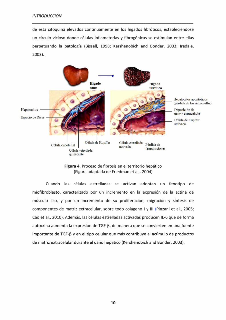
Figura 4. Proceso de fibrosis en el territorio hepático (Figura adaptada de Friedman et al., 2004)
Cuando las células estrelladas se activan adoptan un fenotipo de
miofibroblasto, caracterizado por un incremento en la expresión de la actina de
músculo liso, y por un incremento de su proliferación, migración y síntesis de
componentes de matriz extracelular, sobre todo colágeno I y III (Pinzani et al., 2005;
Cao et al., 2010). Además, las células estrelladas activadas producen IL-6 que de forma
autocrina aumenta la expresión de TGF-β, de manera que se convierten en una fuente
importante de TGF-β y en el tipo celular que más contribuye al acúmulo de productos
de matriz extracelular durante el daño hepático (Kershenobich and Bonder, 2003).
INTRODUCCIÓN _______________________________________________________________________
11
4. ALTERACIONES INTRAHEPÁTICAS
4.1 HIPERTENSIÓN PORTAL
La hipertensión portal es la manifestación clínica más frecuente en la cirrosis
hepática. Se caracteriza por un aumento patológico de la presión hidrostática en el
territorio venoso portal por encima de su intervalo de normalidad (1-5 mmHg). Esto da
lugar a la formación de una extensa red de vasos colaterales que derivan parte del
flujo sanguíneo portal hacia la circulación sistémica e impiden su paso a través del
hígado. Como consecuencia de este síndrome pueden aparecer algunas de las
complicaciones más importantes de la cirrosis como las varices gastroesofágicas o la
ascitis (Abraldes and Bosch, 2002).
Durante el proceso de cirrosis existen dos componentes que afectan a la
resistencia vascular intrahepática: 1) un componente mecánico caracterizado por
anomalías anatómicas con la presencia de nódulos regenerativos que dificultan el paso
de la sangre a través del hígado, y 2) un componente dinámico proporcionado por
factores vasoactivos y el control del tono vascular. En condiciones normales existe un
equilibrio entre la producción de sustancias vasodilatadoras (NO, prostaciclina, ...) y
sustancias vasoconstrictoras (ET-1, Ang-II, norepinefrina, AVP, ...) Sin embargo, en
condiciones patológicas este equilibrio se rompe y se decanta hacia la producción de
vasoconstrictores, sobretodo ET-1, a la vez que disminuye la síntesis de
vasodilatadores, sobretodo NO. Al efecto producido por el desequilibrio de agentes
vasoactivos intrahepáticos hay que sumar el efecto derivado del incremento en el flujo
portal que es el resultado de una marcada vasodilatación arterial en los órganos
esplácnicos que drenan hacia el sistema venoso portal (Rockey, 2003).
4.2 CIRCULACIÓN HIPERDINÁMICA
La vasodilatación esplácnica causa una disminución en las resistencias
vasculares sistémicas y por consiguiente, una disminución en el volumen arterial
efectivo, el cual estimula a nivel renal la reabsorción tubular de sodio y agua,
resultando en la expansión del volumen plasmático. A nivel venoso, existe una
INTRODUCCIÓN _______________________________________________________________________
12
dilatación y un aumento de la capacidad vascular total como adaptación al aumento de
la volemia. En esta fase inicial de la cirrosis, la homeostasis circulatoria se mantiene
por el desarrollo de una circulación hiperdinámica, que se caracterizada por un
aumento en el gasto cardíaco y la frecuencia cardíaca, además de una disminución en
las resistencias vasculares periféricas. Con la progresión de la enfermedad hepática y
de la vasodilatación esplácnica, disminuye la presión arterial y el volumen sanguíneo
central, y como consecuencia la circulación hiperdinámica es insuficiente para
mantener la homeostasis circulatoria. La hipovolemia, mediante un reflejo de los
baroreceptores, activa los sistemas vasoactivos hormonales (sistema renina-
angiotensina-aldosterona, sistema nervioso simpático e hipersecreción no osmótica de
hormona antidiurética) con carácter homeostático para mantener la presión arterial
(Schrier et al., 1988).
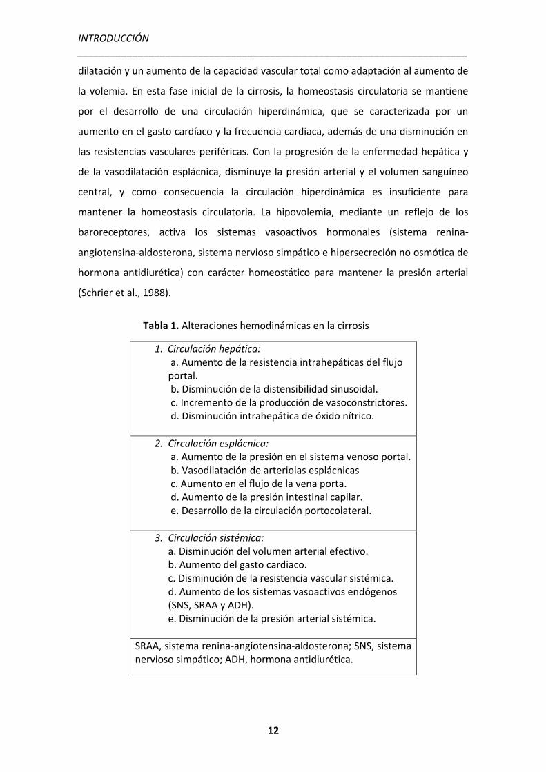
Tabla 1. Alteraciones hemodinámicas en la cirrosis
1. Circulación hepática: a. Aumento de la resistencia intrahepáticas del flujo portal. b. Disminución de la distensibilidad sinusoidal. c. Incremento de la producción de vasoconstrictores. d. Disminución intrahepática de óxido nítrico.
2. Circulación esplácnica: a. Aumento de la presión en el sistema venoso portal. b. Vasodilatación de arteriolas esplácnicas c. Aumento en el flujo de la vena porta. d. Aumento de la presión intestinal capilar. e. Desarrollo de la circulación portocolateral.
3. Circulación sistémica: a. Disminución del volumen arterial efectivo. b. Aumento del gasto cardiaco. c. Disminución de la resistencia vascular sistémica. d. Aumento de los sistemas vasoactivos endógenos (SNS, SRAA y ADH). e. Disminución de la presión arterial sistémica.
SRAA, sistema renina-angiotensina-aldosterona; SNS, sistema nervioso simpático; ADH, hormona antidiurética.
INTRODUCCIÓN _______________________________________________________________________
13
En la cirrosis avanzada, la disfunción circulatoria y la progresiva activación de
los sistemas vasoactivos endógenos produce una alteración en diferentes territorios
vasculares, afectando sobretodo la perfusión renal y pulmonar. Esto provoca una
alteración en la filtración glomerular y en el intercambio de gases, respectivamente.
Dichos fenómenos se conocen como síndrome hepatorrenal y síndrome
hepatopulmonar, y son dos de las principales causas de muerte en pacientes cirróticos
(Arroyo and Jiménez, 2000).
5. FORMACIÓN DE ASCITIS
La ascitis se define como un exceso de líquido en la cavidad peritoneal, que en
situaciones fisiológicas se cifra en sólo unos pocos centímetros cúbicos. La ascitis es,
dentro de las descompensaciones mayores de una hepatopatía crónica evolucionada,
la más frecuente. Aproximadamente el 50% de los pacientes con cirrosis compensada
desarrollarán ascitis en un intervalo prospectivo de 10 años y la mitad de los pacientes
con ascitis fallecerán en un plazo de 2 años.
El primer paso en la formación de ascitis consiste en el desarrollo del síndrome
de hipertensión portal. En pacientes con cirrosis compensada, el grado de hipertensión
portal y de vasodilatación esplácnica es moderado. En esta situación, el hecho de no
poder llenar la vasculatura arterial se compensa con un incremento del volumen
plasmático y del gasto cardíaco (Arroyo et al., 1988). Una vez que este estado
empeora, el índice cardíaco y el volumen plasmático ya no pueden compensar las
consecuencias de la vasodilatación esplácnica. Los mecanismos fisiológicos
compensatorios se activan a través de los receptores de presión y volumen corporales
posibilitando la retención renal de sodio y agua. Esta retención se produce mediante la
actuación de distintos sistemas neuro-hormonales como la secreción de la hormona
antidiurética, la activación del sistema nervioso simpático y del sistema renina-
angiotensina-aldosterona. Dada la vasodilatación periférica, el fluido retenido es
insuficiente para llenar el árbol vascular arterial, y el mecanismo se perpetúa y agrava
(Arroyo and Jiménez, 2000). El exceso de líquido se escapa del compartimento
INTRODUCCIÓN _______________________________________________________________________
14
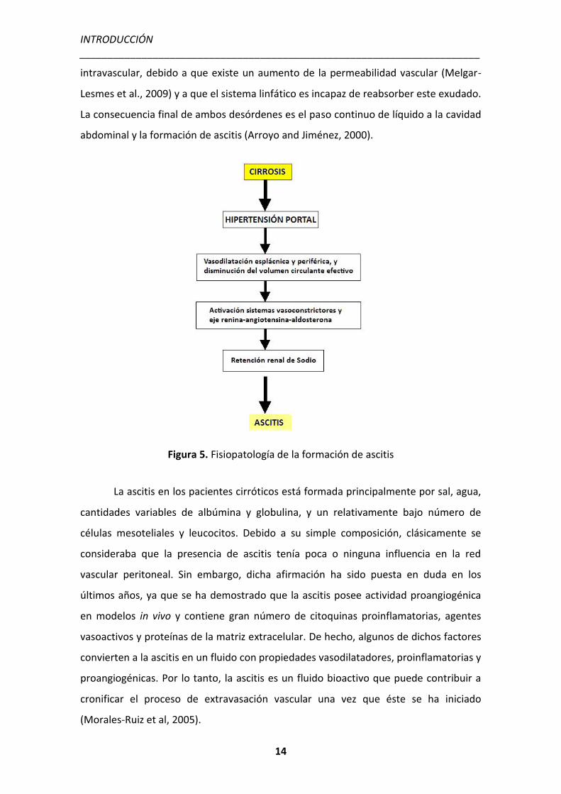
intravascular, debido a que existe un aumento de la permeabilidad vascular (Melgar-
Lesmes et al., 2009) y a que el sistema linfático es incapaz de reabsorber este exudado.
La consecuencia final de ambos desórdenes es el paso continuo de líquido a la cavidad
abdominal y la formación de ascitis (Arroyo and Jiménez, 2000).
Figura 5. Fisiopatología de la formación de ascitis
La ascitis en los pacientes cirróticos está formada principalmente por sal, agua,
cantidades variables de albúmina y globulina, y un relativamente bajo número de
células mesoteliales y leucocitos. Debido a su simple composición, clásicamente se
consideraba que la presencia de ascitis tenía poca o ninguna influencia en la red
vascular peritoneal. Sin embargo, dicha afirmación ha sido puesta en duda en los
últimos años, ya que se ha demostrado que la ascitis posee actividad proangiogénica
en modelos in vivo y contiene gran número de citoquinas proinflamatorias, agentes
vasoactivos y proteínas de la matriz extracelular. De hecho, algunos de dichos factores
convierten a la ascitis en un fluido con propiedades vasodilatadores, proinflamatorias y
proangiogénicas. Por lo tanto, la ascitis es un fluido bioactivo que puede contribuir a
cronificar el proceso de extravasación vascular una vez que éste se ha iniciado
(Morales-Ruiz et al, 2005).
INTRODUCCIÓN _______________________________________________________________________
15
ANGIOGÉNESIS
1. ASPECTOS GENERALES
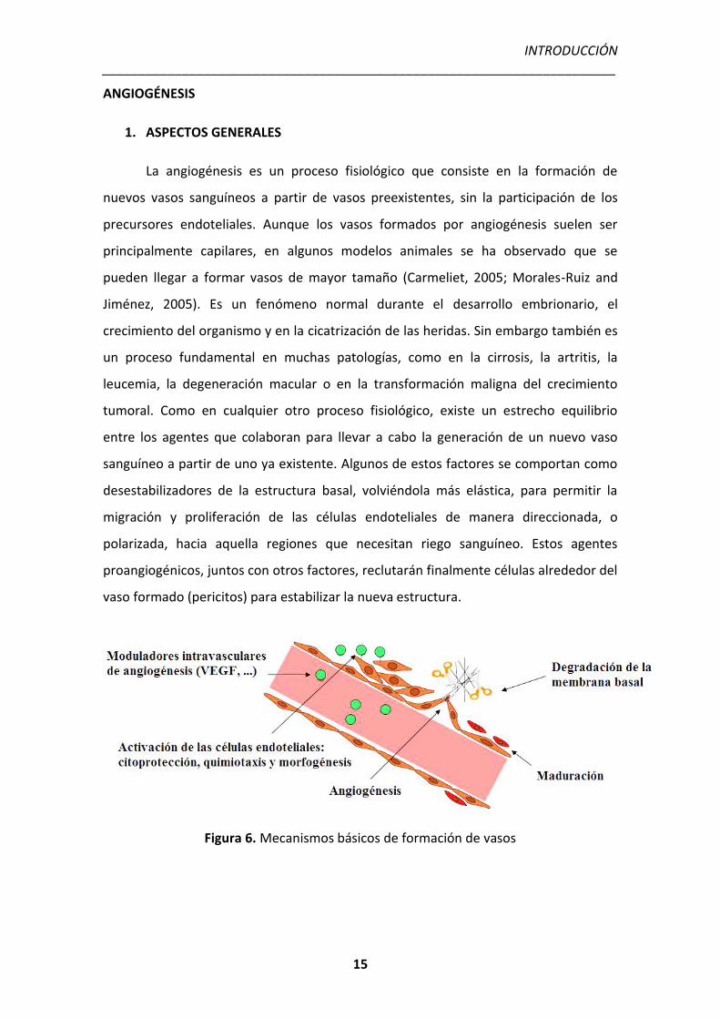
La angiogénesis es un proceso fisiológico que consiste en la formación de
nuevos vasos sanguíneos a partir de vasos preexistentes, sin la participación de los
precursores endoteliales. Aunque los vasos formados por angiogénesis suelen ser
principalmente capilares, en algunos modelos animales se ha observado que se
pueden llegar a formar vasos de mayor tamaño (Carmeliet, 2005; Morales-Ruiz and
Jiménez, 2005). Es un fenómeno normal durante el desarrollo embrionario, el
crecimiento del organismo y en la cicatrización de las heridas. Sin embargo también es
un proceso fundamental en muchas patologías, como en la cirrosis, la artritis, la
leucemia, la degeneración macular o en la transformación maligna del crecimiento
tumoral. Como en cualquier otro proceso fisiológico, existe un estrecho equilibrio
entre los agentes que colaboran para llevar a cabo la generación de un nuevo vaso
sanguíneo a partir de uno ya existente. Algunos de estos factores se comportan como
desestabilizadores de la estructura basal, volviéndola más elástica, para permitir la
migración y proliferación de las células endoteliales de manera direccionada, o
polarizada, hacia aquella regiones que necesitan riego sanguíneo. Estos agentes
proangiogénicos, juntos con otros factores, reclutarán finalmente células alrededor del
vaso formado (pericitos) para estabilizar la nueva estructura.
Figura 6. Mecanismos básicos de formación de vasos
INTRODUCCIÓN _______________________________________________________________________
16
El hígado cirrótico experimenta un intenso proceso de angiogénesis en
respuesta a la hipoxia tisular y al aumento de la resistencia intrahepática. Esto lleva a
una disrupción de la arquitectura vascular hepática que acelera la progresión de la
cirrosis, ya que aumenta la hipertensión portal y disminuye la perfusión de los
hepatocitos. Por otra parte, la angiogénesis regula la formación de vasos colaterales e
incrementa el flujo sanguíneo esplácnico. Complicaciones comunes en el progreso de
la cirrosis (Morales-Ruiz and Jiménez, 2005; Tugues et al., 2007; Fernández et al, 2008).
2. FACTORES PROANGIOGÉNICOS
2.1 LA FAMILIA DE VEGF Y SUS RECEPTORES
El factor de crecimiento endotelial vascular (VEGF) es una proteína señalizadora
con un gran potencial inductor de formación de vasos sanguíneos, tanto durante el
desarrollo embrionario (vasculogénesis) como en el crecimiento de vasos nuevos en el
adulto (angiogénesis). Además, promueve angiogénesis en los procesos de inflamación
crónica, cicatrización y en tumores. VEGF es secretado por muchas células del
mesénquima y del estroma, induce la migración de células precursoras endoteliales a
partir de la médula ósea, y estimula la proliferación y diferenciación de estas células en
los sitios de angiogénesis. Cuando la angiogénesis se origina a partir de vasos
preexistentes (y no a partir de células precursoras), VEGF estimula la supervivencia de
las células endoteliales, su proliferación y su motilidad, iniciando la gemación de
nuevos capilares. La familia de VEGF es esencial para los procesos de diferenciación,
proliferación y quimiotaxis en la vasculogénesis, angiogénesis fisiológica y patológica, y
en la linfangiogénesis. La importancia de la función de este sistema en el organismo se
ejemplifica en que la pérdida de un solo alelo del gen VEGF o de alguno de sus
receptores causa defectos vasculares embrionarios letales (Carmeliet et al., 1996;
Ferrara et al., 1996; Ferrara et al., 2003; Holmes et al., 2007).
La familia de VEGF incluye a diferentes miembros: VEGF-A (existen 5 isoformas
diferentes), VEGF-B, VEGF-C, VEGF-D, VEGF-E (del virus huérfano parapox), VEGF-F (del
veneno de serpiente) y el factor de crecimiento placentario (PlGF), los cuales se unen a
INTRODUCCIÓN _______________________________________________________________________
17
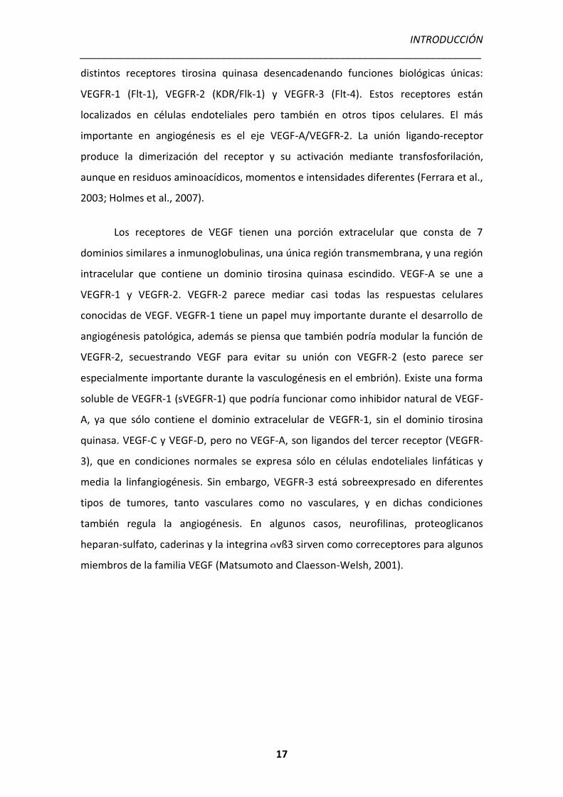
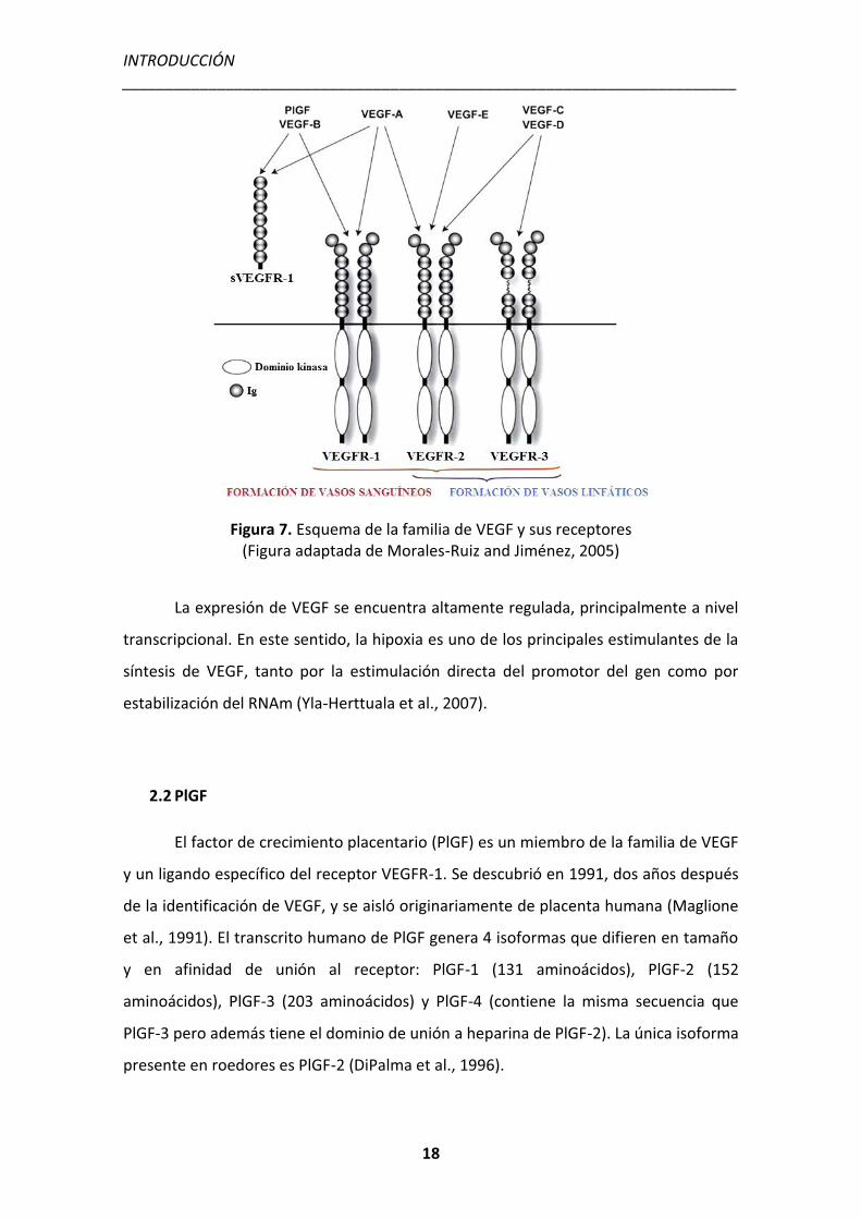
distintos receptores tirosina quinasa desencadenando funciones biológicas únicas:
VEGFR-1 (Flt-1), VEGFR-2 (KDR/Flk-1) y VEGFR-3 (Flt-4). Estos receptores están
localizados en células endoteliales pero también en otros tipos celulares. El más
importante en angiogénesis es el eje VEGF-A/VEGFR-2. La unión ligando-receptor
produce la dimerización del receptor y su activación mediante transfosforilación,
aunque en residuos aminoacídicos, momentos e intensidades diferentes (Ferrara et al.,
2003; Holmes et al., 2007).
Los receptores de VEGF tienen una porción extracelular que consta de 7
dominios similares a inmunoglobulinas, una única región transmembrana, y una región
intracelular que contiene un dominio tirosina quinasa escindido. VEGF-A se une a
VEGFR-1 y VEGFR-2. VEGFR-2 parece mediar casi todas las respuestas celulares
conocidas de VEGF. VEGFR-1 tiene un papel muy importante durante el desarrollo de
angiogénesis patológica, además se piensa que también podría modular la función de
VEGFR-2, secuestrando VEGF para evitar su unión con VEGFR-2 (esto parece ser
especialmente importante durante la vasculogénesis en el embrión). Existe una forma
soluble de VEGFR-1 (sVEGFR-1) que podría funcionar como inhibidor natural de VEGF-
A, ya que sólo contiene el dominio extracelular de VEGFR-1, sin el dominio tirosina
quinasa. VEGF-C y VEGF-D, pero no VEGF-A, son ligandos del tercer receptor (VEGFR-
3), que en condiciones normales se expresa sólo en células endoteliales linfáticas y
media la linfangiogénesis. Sin embargo, VEGFR-3 está sobreexpresado en diferentes
tipos de tumores, tanto vasculares como no vasculares, y en dichas condiciones
también regula la angiogénesis. En algunos casos, neurofilinas, proteoglicanos
heparan-sulfato, caderinas y la integrina vß3 sirven como correceptores para algunos
miembros de la familia VEGF (Matsumoto and Claesson-Welsh, 2001).
INTRODUCCIÓN _______________________________________________________________________
18
Figura 7. Esquema de la familia de VEGF y sus receptores (Figura adaptada de Morales-Ruiz and Jiménez, 2005)
La expresión de VEGF se encuentra altamente regulada, principalmente a nivel
transcripcional. En este sentido, la hipoxia es uno de los principales estimulantes de la
síntesis de VEGF, tanto por la estimulación directa del promotor del gen como por
estabilización del RNAm (Yla-Herttuala et al., 2007).
2.2 PlGF
El factor de crecimiento placentario (PlGF) es un miembro de la familia de VEGF
y un ligando específico del receptor VEGFR-1. Se descubrió en 1991, dos años después
de la identificación de VEGF, y se aisló originariamente de placenta humana (Maglione
et al., 1991). El transcrito humano de PlGF genera 4 isoformas que difieren en tamaño
y en afinidad de unión al receptor: PlGF-1 (131 aminoácidos), PlGF-2 (152
aminoácidos), PlGF-3 (203 aminoácidos) y PlGF-4 (contiene la misma secuencia que
PlGF-3 pero además tiene el dominio de unión a heparina de PlGF-2). La única isoforma
presente en roedores es PlGF-2 (DiPalma et al., 1996).
INTRODUCCIÓN _______________________________________________________________________
19
PlGF tiene acción sobre varios tipos celulares y está implicado en multitud de
respuestas biológicas. Esta citoquina actúa principalmente sobre las células
endoteliales estimulando el crecimiento y la maduración de los vasos (Ziche et al.,
1997; Yonekura et al., 1999; Carmeliet et al., 2001), pero también estimula la
proliferación de los fibroblastos, regula la capacidad contráctil de las células
musculares lisas que recubren el endotelio (Bellik et al., 2005) y está implicada en el
reclutamiento de macrófagos (Selvaraj et al., 2003). Sin embargo, la capacidad
proangiogénica de PlGF en condiciones fisiológicas es menor que la de VEGF y la
presencia de PlGF no es necesario para el mantenimiento de la quiescencia vascular en
órganos sanos. De hecho, la expresión de PlGF es prácticamente indetectable en la
mayoría de tejidos sanos, por el contrario, la sobreexpresión de PlGF se ha asociado a
diferentes situaciones de angiogénesis patológica (Autiero et al., 2003; Van Steenkiste
et al., 2009). Los estímulos que inducen la sobreexpresión de PlGF en condiciones
patológicas son múltiples, incluyendo la hipoxia, factores de crecimiento, hormonas u
oncogenes. Estudios recientes en ratones transgénicos deficientes en PlGF
demostraron que PlGF ejerce su función únicamente en condiciones patológicas. Los
ratones deficientes para PlGF no mostraron ningún fenotipo anormal aparente,
indicando que PlGF no es indispensable para el desarrollo y la homeostasis vascular.
Sin embargo, estos ratones presentaron una lenta recuperación después de inducirles
infarto de miocardio y defectos en la formación de vasos colaterales en respuesta a
una isquemia inducida en las extremidades (Carmeliet et al., 2001).
Debido a su efecto pleiotrópico, existen patologías caracterizadas por una
deficiencia de PlGF y en las que la terapia con PlGF consigue mejorar la enfermedad,
como por ejemplo en el infarto de miocardio (Luttun et al., 2002; Roncal et al. 2008) o
en la isquemia cerebral (Liu et al., 2006). En otras patologías en cambio, es el bloqueo
de la actividad de PlGF el que consigue mejorar la enfermedad, como pasa en el edema
ocular (Van de Veire et al., 2010), el enfisema pulmonar (Cheng et al., 2009), la cirrosis
hepática (Van Steenkiste et al., 2011) o en distintos tipos de cáncer (Van de Veire et al.,
2010; Rolny et al., 2011). En este contexto, se ha demostrado que en contraposición
con los inhibidores clásicos de VEGF, los anticuerpos monoclonales contra PlGF
reducen la angiogénesis patológica en varios tipos de cáncer y en otros modelos de
INTRODUCCIÓN _______________________________________________________________________
20
enfermedades, sin afectar a la vasculatura sana y sin mayores efectos secundarios ni
en ratones ni en humanos (Fischer et al., 2007).
2.3 PDGF
El factor de crecimiento derivado de plaquetas (PDGF) es uno de los numerosos
factores de crecimiento que regulan el crecimiento y la división celular, sobre todo en
células mesenquimales. Existen cinco isoformas diferentes de PDGF (A, B, C, D y el
heterodímero AB) que activan la respuesta celular a través de dos receptores: el
receptor alfa (PDGFRA) y el beta (PDGFRB), que se expresan principalmente en células
endoteliales y células musculares lisas. El PDGF desempeña un rol significativo en el
desarrollo embriogénico, proliferación celular, migración celular y en la angiogénesis
(Heiradan et al., 1991).
Los receptores PDGFR pertenecen a la clase de receptores con actividad
tirosina quinasa intrínseca, dentro del grupo de receptores transmembrana. El PDGFR
tipo alfa se une al PDGF-AA, PDGF-BB y PDGF-AB mientras el PDGFR tipo beta se une
con alta afinidad al PDGF-BB y PDGF-AB. La vía de PDGF más implicada en angiogénesis
es la activada por PDGF-BB y su receptor PDGFRB. Las células endoteliales liberan
PDGF-BB y con ello estimulan el crecimiento, migración, supervivencia y diferenciación
de las células mesenquimales que expresan PDGFRB, y las reclutan alrededor de los
nuevos vasos estabilizándolos (Lindahl et al., 1997). PDGF-C i PDGF-D también
promueven angiogénesis, pero su función en este proceso aun no está bien
caracterizada.
INTRODUCCIÓN _______________________________________________________________________
21
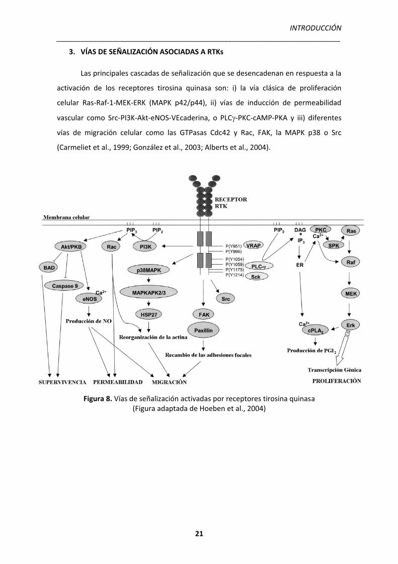
3. VÍAS DE SEÑALIZACIÓN ASOCIADAS A RTKs
Las principales cascadas de señalización que se desencadenan en respuesta a la
activación de los receptores tirosina quinasa son: i) la vía clásica de proliferación
celular Ras-Raf-1-MEK-ERK (MAPK p42/p44), ii) vías de inducción de permeabilidad
vascular como Src-PI3K-Akt-eNOS-VEcaderina, o PLC�-PKC-cAMP-PKA y iii) diferentes
vías de migración celular como las GTPasas Cdc42 y Rac, FAK, la MAPK p38 o Src
(Carmeliet et al., 1999; González et al., 2003; Alberts et al., 2004).
Figura 8. Vías de señalización activadas por receptores tirosina quinasa (Figura adaptada de Hoeben et al., 2004)
INTRODUCCIÓN _______________________________________________________________________
22
4. ANGIOGÉNESIS E INFLAMACIÓN
Aunque la inflamación es un proceso crucial para defender nuestro organismo
contra patógenos, cuando la respuesta inflamatoria perdura y se cronifica, aparecen
efectos adversos que se intensifican con la activación de la angiogénesis. Hay muchas
evidencias que demuestran que la inflamación crónica y la angiogénesis se pueden
inducir por los mismos estímulos y que ambos procesos dependen uno del otro, ya que
se han encontrado mecanismos moleculares compartidos por los dos (Costa et al.,
2007). La inflamación crónica está relacionada con proliferación y migración celular, y
con el reclutamiento de células inflamatorias que pueden llegar a ser extremadamente
dañinas para el tejido. Muchos tipos celulares importantes durante los procesos
inflamatorios liberan diferentes factores de crecimiento como TNF-α, TGF-β, IL-1, IL-6,
IL-8 o IL-18, que estimulan la angiogénesis de manera directa sobre las células
endoteliales (Ezaki et al., 2001).
Muchas enfermedades hepáticas crónicas se caracterizan por tener procesos de
inflamación y fibrosis. El tejido inflamado/fibrótico ofrece resistencia al flujo sanguíneo
y a la distribución de oxígeno, cosa que provoca que el tejido se vuelva hipóxico. Esto
estimula la expresión de factores proangiogénicos inducibles por hipoxia y el
crecimiento de nuevos vasos. Los macrófagos y las células T, por ejemplo, producen
gran cantidad de factores angiogénicos bajo condiciones de hipoxia, como VEGF.
Además las células T activadas expresan VEGFR-2, de manera que ellas mismas pueden
responder a VEGF aumentando la síntesis de IFN-� (activador de macrófagos) e
inhibiendo la secreción de IL-10 (citoquina antiinflamatoria) (Mor et al., 2004).
Los mediadores de inflamación pueden promover, directa o indirectamente,
angiogénesis. Y la angiogénesis, a su vez, contribuye a la inflamación. Los nuevos vasos
mantienen el estado crónico de inflamación transportando células inflamatorias al
tejido inflamado y aportándole nutrientes y oxígeno. Además, el incremento de la
superficie endotelial conlleva un gran aumento de la producción de citoquinas,
moléculas de adhesión y otros estímulos proinflamatorios (Jackson et al., 1997). Estas
observaciones han llevado al desarrollo de estrategias terapéuticas dirigidas contra la
angiogénesis y la inflamación crónica.
INTRODUCCIÓN _______________________________________________________________________
23
5. TERAPIAS ANTIANGIOGÉNICAS
Las terapias antiangiogénicas están dirigidas en su mayoría al tratamiento del
cáncer. En este caso, la angiogénesis no inicia la enfermedad, pero si promueve la
progresión tumoral y la metástasis. Por ello se propuso que inhibir la angiogénesis
podría ser un tratamiento antitumoral efectivo. Además, las células endoteliales no
son como las células tumorales, son genómicamente estables y se consideran ideales
como dianas terapéuticas no resistentes a la terapia antiangiogénica (Ferrara and
Alitalo, 1999).
VEGF y sus receptores son la principal diana de las terapias antiangiogénicas
clásicas. En el año 2004 fue aprobado por la FDA el anticuerpo monoclonal humano
anti-VEGFA, llamado Bevacizumab, que se une de manera específica a VEGF y ha
demostrado tener importantes beneficios clínicos en pacientes con diferentes tipos de
cáncer. Otros anticuerpos dirigidos contra los receptores de VEGF también han
demostrado tener efectos antiangiogénicos, como el anticuerpo específico de VEGFR-2
DC101 (Prewett et al., 1999). Una manera de incrementar el efecto consiste en actuar
no sólo sobre la vía de VEGF, sino también inhibiendo el reclutamiento de las células
murales bloqueando los receptores de PDGF (Erber et al., 2004). En los últimos años se
han desarrollado gran cantidad de pequeñas moléculas sintéticas con capacidad
antiangiogénica dirigidas a bloquear de manera múltiple los receptores tirosina
quinasa. Las más importantes son: Vatalanib, Sunitinib, Sorafenib y Pazopanib.
Sin embargo, estos tratamientos antiangiogénicos clásicos se han asociado a
graves efectos adversos, ya que VEGF no sólo actúa en la formación de nuevos vasos,
también es importante en la hematopoyesis, mielopoyesis y en la supervivencia de las
células endoteliales. VEGF es necesario en la coagulación, en la cicatrización de
heridas, activa a la célula endotelial para que produzca NO, regula parte de la función
renal, está involucrado en el mantenimiento de la homeostasis de la piel, en la
maduración de las células dendríticas del sistema inmunitario, etc. De manera que el
bloqueo de la actividad de VEGF bloquea múltiples vías de una gran importancia
biológica. De entre los diferentes efectos secundarios asociados a las terapias
antiangiogénicas clásicas destacan: sangrado, disminución de la densidad vascular en
INTRODUCCIÓN _______________________________________________________________________
24
órganos sanos, trombosis, hipertensión, hipotiroidismo, fatiga, proteinuria, edema,
toxicidad en la piel, linfopenia y leucopenia (Wu et al., 2008).
EL ÓXIDO NÍTRICO
1. CARACTERÍSTICAS Y FUNCIONES GENERALES
El óxido nítrico es un gas biológicamente activo, incoloro y poco soluble en
agua presente en pequeñas cantidades en los mamíferos. Es una molécula altamente
inestable en el aire, ya que presenta un electrón desapareado en su configuración
electrónica, con lo se oxida rápidamente en presencia de oxígeno convirtiéndose en
dióxido de nitrógeno. Por esta razón se la considera también un radical libre. En el
cuerpo, tiene una vida media de sólo unos pocos segundos, en gran parte porque el
anión superóxido (O2-) tiene gran afinidad por él y reduce su biodisponibilidad.
También se une al grupo hemo de la hemoglobina (en los glóbulos rojos) y al grupo
hemo del enzima guanilato ciclasa, que se encuentro en muchos tipos celulares, como
las células musculares lisas vasculares (Moncada et al., 1991).
El NO es producido por una amplia variedad de tipos celulares que incluyen
células epiteliales, nerviosas, endoteliales e inflamatorias. La magnitud y la duración de
la síntesis de NO por las células determina que su acción sea fisiológica o patológica.
Las acciones fisiológicas son mediadas por pulsos de pequeñas cantidades de NO. Las
actividades patológicas son el resultado de la producción sostenida de altos niveles de
NO. En las neuronas puede funcionar como neurotransmisor, atravesando fácilmente
las membranas celulares por su carácter lipófilo. Los macrófagos sintetizan NO para
destruir los microorganismos que han sido fagocitados. Cuando es producido en las
células endoteliales de los vasos sanguíneos induce vasodilatación. La activación del
endotelio por el estrés de cizalla o por la activación de receptores por parte de
agonistas, induce un aumento del Ca2+ intracelular. Este incremento estimula la
producción de NO, que funciona como regulador paracrino; difunde desde la célula de
origen hasta el interior de las células musculares lisas adyacentes, donde induce la
producción de GMPc (guanosin monofosfato cíclico) por su unión al grupo hemo de la
INTRODUCCIÓN _______________________________________________________________________
25
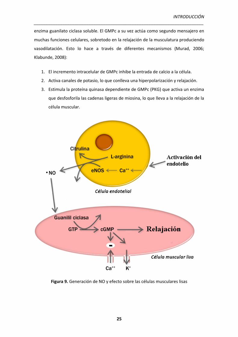
enzima guanilato ciclasa soluble. El GMPc a su vez actúa como segundo mensajero en
muchas funciones celulares, sobretodo en la relajación de la musculatura produciendo
vasodilatación. Esto lo hace a través de diferentes mecanismos (Murad, 2006;
Klabunde, 2008):
1. El incremento intracelular de GMPc inhibe la entrada de calcio a la célula.
2. Activa canales de potasio, lo que conlleva una hiperpolarización y relajación.
3. Estimula la proteína quinasa dependiente de GMPc (PKG) que activa un enzima
que desfosforila las cadenas ligeras de miosina, lo que lleva a la relajación de la
célula muscular.
Figura 9. Generación de NO y efecto sobre las células musculares lisas
INTRODUCCIÓN _______________________________________________________________________
26
2. SINTASAS DEL ÓXIDO NÍTRICO
La síntesis de NO se realiza por acción de una enzima, la óxido nítrico sintasa
(NOS), a partir del aminoácido L-arginina y requiriendo la presencia de un cofactor
(específicamente, una coenzima, la nicotinamida adenín-dinucleótido fosfato reducido
o nad-fosfato reducido (NADPH)) y de oxígeno. Los productos de la acción de NOS son
NO y L-citrulina. Existen tres isoformas distintas de NOS, dos constitutivas y
dependientes de calcio (cNOS): la endotelial (eNOS, NOS3) y la neuronal (nNOS, NOS1),
que sintetizan NO en condiciones normales. Y una isoforma inducible e independiente
de calcio (iNOS, NOS2), que no se expresa, o lo hace muy débilmente, en condiciones
fisiológicas (Knowles and Moncada, 1994; Lamas et al., 1992).
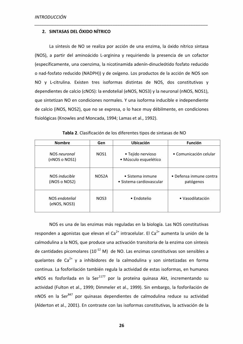
Tabla 2. Clasificación de los diferentes tipos de sintasas de NO
Nombre Gen Ubicación Función
NOS neuronal (nNOS o NOS1)
NOS1
• Tejido nervioso
• Músculo esquelético
• Comunicación celular
NOS inducible (iNOS o NOS2)
NOS2A
• Sistema inmune
• Sistema cardiovascular
• Defensa inmune contra
patógenos
NOS endotelial (eNOS, NOS3)
NOS3
• Endotelio
• Vasodilatación
NOS es una de las enzimas más reguladas en la biología. Las NOS constitutivas
responden a agonistas que elevan el Ca2+ intracelular. El Ca2+ aumenta la unión de la
calmodulina a la NOS, que produce una activación transitoria de la enzima con síntesis
de cantidades picomolares (10-12 M) de NO. Las enzimas constitutivas son sensibles a
quelantes de Ca2+ y a inhibidores de la calmodulina y son sintetizadas en forma
continua. La fosforilación también regula la actividad de estas isoformas, en humanos
eNOS es fosforilada en la Ser1177 por la proteína quinasa Akt, incrementando su
actividad (Fulton et al., 1999; Dimmeler et al., 1999). Sin embargo, la fosforilación de
nNOS en la Ser847 por quinasas dependientes de calmodulina reduce su actividad
(Alderton et al., 2001). En contraste con las isoformas constitutivas, la activación de la
INTRODUCCIÓN _______________________________________________________________________
27
NOS inducible es a nivel transcripcional. Los macrófagos, células prototípicas de la NOS
inducible, comienzan a producir NO varias horas después de la estimulación con
citoquinas. Después de la transcripción del gen y la expresión de la proteína, los
macrófagos activados producen cantidades nanomolares (10-9 M) de NO durante días,
hasta que la enzima es degradada por proteólisis. Los dos tipos de NOS pueden
coexistir en la misma célula. Las células endoteliales, por ejemplo, liberan NO por un
tiempo prolongado en respuesta a TNF-alfa y sólo brevemente cuando son estimuladas
con un agonista de Ca2+ (Busconi et al., 1993).
3. INHIBIDORES DE LAS NOS
Los inhibidores de las NOS han sido esenciales para evaluar el rol del NO en los
procesos fisiológicos y fisiopatológicos. La mayoría de ellos son análogos de la L-
arginina o contienen un grupo guanidínico (como la aminoguanidina). Los más
utilizados son la NG-monometil-L-arginina (L-NMMA), la NG-nitro-L-arginina (L-NOArg) y
su metil éster (L-NAME) y la N-iminoetil-L-ornitina (L-NIO). Estos compuestos inhiben
por competición con el substrato, la L-arginina, uniéndose ellos al centro activo de la
enzima inactivándola (Alderton et al., 2001). Cabe destacar que la potencia de estos
inhibidores no es la misma frente a todas las isoenzimas (Rees et al., 1990).
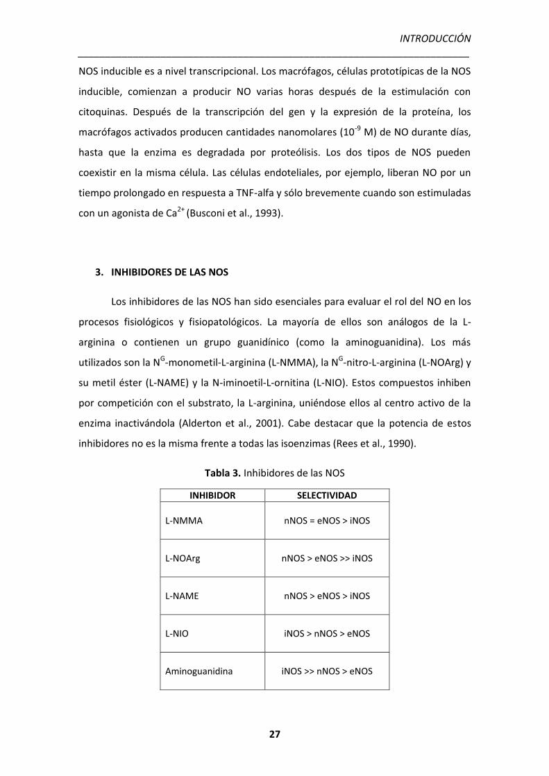
Tabla 3. Inhibidores de las NOS
INHIBIDOR SELECTIVIDAD L-NMMA
nNOS = eNOS > iNOS
L-NOArg
nNOS > eNOS >> iNOS
L-NAME
nNOS > eNOS > iNOS
L-NIO
iNOS > nNOS > eNOS
Aminoguanidina
iNOS >> nNOS > eNOS
INTRODUCCIÓN _______________________________________________________________________
28
4. PAPEL FISIOPATOLÓGICO DEL NO EN LA CIRROSIS
eNOS es una enzima extremadamente importante en el hígado y durante todo
el proceso cirrótico. En condiciones normales, las células endoteliales hepáticas
aumentan su producción de NO en respuesta a un incremento del flujo para disminuir
la presión de perfusión intrahepática (Shah et al., 1999). Sin embargo, la producción de
NO en el hígado cirrótico es muy reducida. La actividad de eNOS disminuye un 75%
(Rockey et al., 1998), dando lugar a una vasoconstricción sinusoidal y a un aumento de
la presión de perfusión en respuesta al incremento progresivo de flujo y al estrés
celular. Al contrario de lo que se podría pensar, las células endoteliales de ratas
cirróticas tienen igual cantidad de RNAm y de proteína eNOS que las de animales
controles, sugiriendo un control postraduccional de la actividad de la enzima (Gupta et
al., 1998). Por ejemplo, las células endoteliales de hígados cirróticos presentan un
aumento en la expresión y en la interacción de Cav-1 con eNOS, cosa que impide que
la enzima se una a la calmodulina (Hendrickson et al., 2003). También se ha
demostrado que los hígados de animales cirróticos tienen una activación deficiente de
la proteína quinasa Akt, lo que le impide fosforilar a eNOS y, como consecuencia,
disminuye su actividad (Morales-Ruiz et al., 2003).
Contrariamente a lo que ocurre en la microcirculación hepática, los niveles de
NO en la circulación esplácnica o sistémica están aumentados. Diversos estudios han
demostrado que la actividad de eNOS está incrementada en los vasos sanguíneos de
ratas cirróticas (Ros et al., 1995). Este exceso en la actividad de eNOS provoca una
vasodilatación arterial y un remodelado vascular (Fernández-Varo et al., 2003), además
del desarrollo del síndrome de circulación hiperdinámica que lleva a las graves
complicaciones asociadas a la cirrosis y a otras enfermedades hepáticas crónicas
(Langer and Shah, 2006). Una manifestación clásica de esta vasodilatación arterial es la
hiporeactividad arterial a los vasoconstrictores (Sieber et al., 1993). Además, la
inhibición de la síntesis de NO corrige, en parte, la hemodinámica esplácnica y
sistémica en diferentes modelos experimentales de cirrosis (Clària et al., 1992).
INTRODUCCIÓN _______________________________________________________________________
29
EL SISTEMA LINFÁTICO
1. ASPECTOS GENERALES
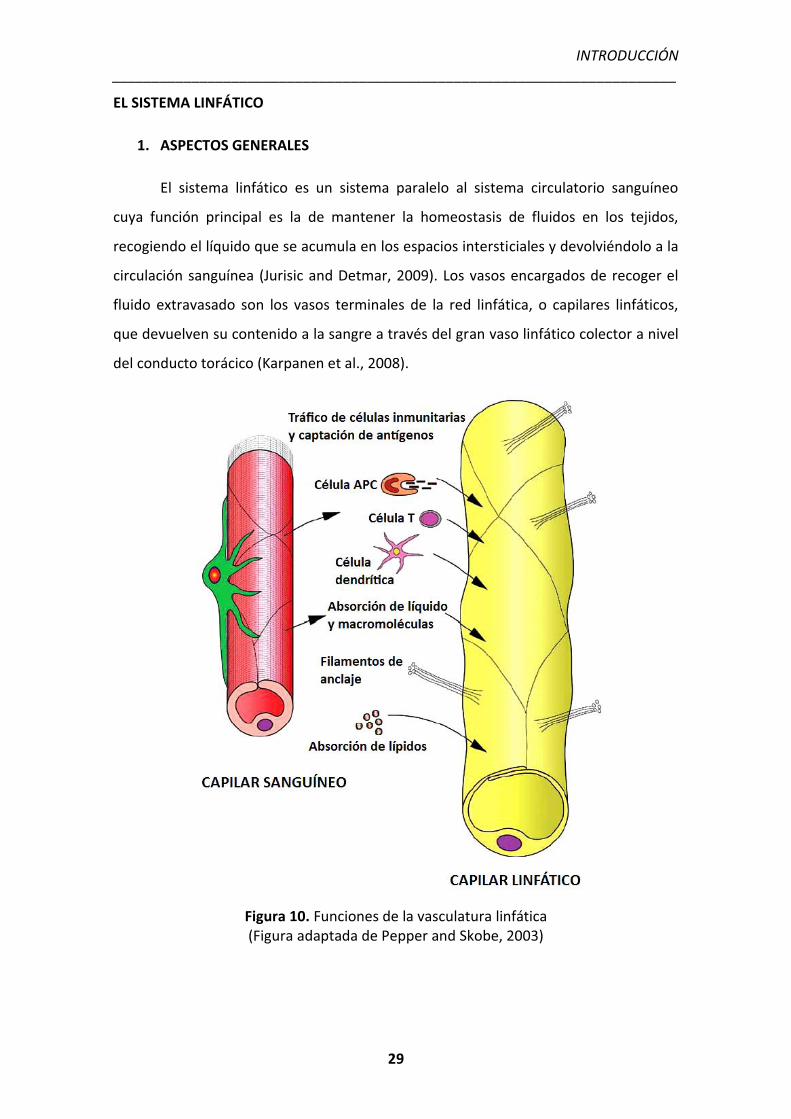
El sistema linfático es un sistema paralelo al sistema circulatorio sanguíneo
cuya función principal es la de mantener la homeostasis de fluidos en los tejidos,
recogiendo el líquido que se acumula en los espacios intersticiales y devolviéndolo a la
circulación sanguínea (Jurisic and Detmar, 2009). Los vasos encargados de recoger el
fluido extravasado son los vasos terminales de la red linfática, o capilares linfáticos,
que devuelven su contenido a la sangre a través del gran vaso linfático colector a nivel
del conducto torácico (Karpanen et al., 2008).
Figura 10. Funciones de la vasculatura linfática (Figura adaptada de Pepper and Skobe, 2003)
INTRODUCCIÓN _______________________________________________________________________
30
Además de su función principal como sistema de drenaje, el sistema linfático
también forma parte del sistema inmunitario del cuerpo. Los vasos linfáticos
transportan células dendríticas, células T, macrófagos, células presentadoras de
antígeno (células APC) y antígenos, desde los espacios periféricos hasta los nódulos
linfáticos donde se inicia la respuesta inmune (Olszewski, 2005). Por último, los vasos
linfáticos también son esenciales para la absorción de los ácidos grasos de cadena
larga ingeridos en la dieta, como las vitaminas solubles A, D, E y K, así como de los
compuestos lipofílicos liberados en el intestino en forma de quilomicrones (Bruyère
and Noël, 2010).
2. ESTRUCTURA Y FUNCIONAMIENTO
La red linfática está compuesta por capilares linfáticos y por vasos linfáticos
colectores. Los capilares linfáticos están formados por una fina capa de células
endoteliales linfáticas, sin membrana basal y rodeados por muy pocos pericitos, que se
anclan a la matriz extracelular a través de filamentos de fibrilina (Gerli et al., 2000).
Este anclaje a la matriz permite que los capilares se abran cuando la presión aumenta
debido al fluido intersticial, permitiendo el paso del líquido hacia el vaso (Schmid-
Schönbein, 1990). El flujo linfático es unidireccional, desde los capilares hasta el gran
vaso linfático colector del conducto torácico, que vacía su contenido en la vena cava
inferior (Cueni and Detmar, 2006). Los vasos linfáticos colectores difieren de los
capilares en diversos aspectos. Tienen un diámetro mayor, están recubiertos por una
capa de células musculares lisas y, a intervalos regulares, tienen un sistema de válvulas
internas que impiden el retroceso del contenido linfático, asegurando así un flujo
unidireccional (Schmid-Schönbein, 1990). Los segmentos de vaso comprendidos entre
dos válvulas se llaman linfangiones y se contraen secuencialmente. A lo largo de los
vasos linfáticos colectores se encuentran los nódulos linfáticos, unas estructuras
especializadas donde los sistemas circulatorio y linfático se comunican. En los nódulos
linfáticos se almacenan los glóbulos blancos, más concretamente los linfocitos, que
interaccionarán con los antígenos capturados por el sistema linfático en los espacios
intersticiales de los tejidos para iniciar la respuesta inmune (Fu and Chaplin, 1999).
INTRODUCCIÓN _______________________________________________________________________
31
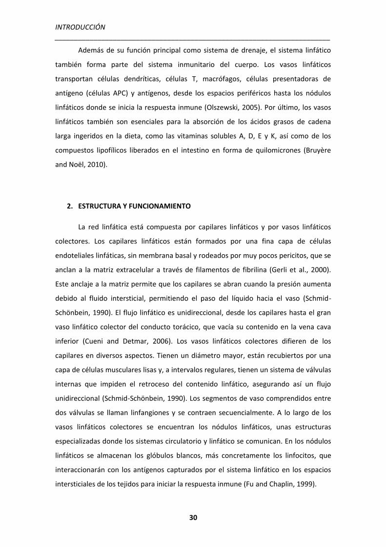
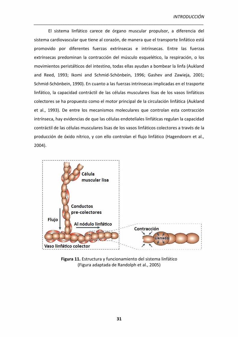
El sistema linfático carece de órgano muscular propulsor, a diferencia del
sistema cardiovascular que tiene al corazón, de manera que el transporte linfático está
promovido por diferentes fuerzas extrínsecas e intrínsecas. Entre las fuerzas
extrínsecas predominan la contracción del músculo esquelético, la respiración, o los
movimientos peristálticos del intestino, todas ellas ayudan a bombear la linfa (Aukland
and Reed, 1993; Ikomi and Schmid-Schönbein, 1996; Gashev and Zawieja, 2001;
Schmid-Schönbein, 1990). En cuanto a las fuerzas intrínsecas implicadas en el trasporte
linfático, la capacidad contráctil de las células musculares lisas de los vasos linfáticos
colectores se ha propuesto como el motor principal de la circulación linfática (Aukland
et al., 1993). De entre los mecanismos moleculares que controlan esta contracción
intrínseca, hay evidencias de que las células endoteliales linfáticas regulan la capacidad
contráctil de las células musculares lisas de los vasos linfáticos colectores a través de la
producción de óxido nítrico, y con ello controlan el flujo linfático (Hagendoorn et al.,
2004).
Figura 11. Estructura y funcionamiento del sistema linfático (Figura adaptada de Randolph et al., 2005)
INTRODUCCIÓN _______________________________________________________________________
32
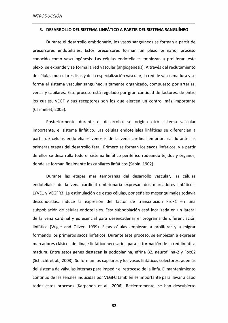
3. DESARROLLO DEL SISTEMA LINFÁTICO A PARTIR DEL SISTEMA SANGUÍNEO
Durante el desarrollo embrionario, los vasos sanguíneos se forman a partir de
precursores endoteliales. Estos precursores forman un plexo primario, proceso
conocido como vasculogénesis. Las células endoteliales empiezan a proliferar, este
plexo se expande y se forma la red vascular (angiogénesis). A través del reclutamiento
de células musculares lisas y de la especialización vascular, la red de vasos madura y se
forma el sistema vascular sanguíneo, altamente organizado, compuesto por arterias,
venas y capilares. Este proceso está regulado por gran cantidad de factores, de entre
los cuales, VEGF y sus receptores son los que ejercen un control más importante
(Carmeliet, 2005).
Posteriormente durante el desarrollo, se origina otro sistema vascular
importante, el sistema linfático. Las células endoteliales linfáticas se diferencian a
partir de células endoteliales venosas de la vena cardinal embrionaria durante las
primeras etapas del desarrollo fetal. Primero se forman los sacos linfáticos, y a partir
de ellos se desarrolla todo el sistema linfático periférico rodeando tejidos y órganos,
donde se forman finalmente los capilares linfáticos (Sabin, 1902).
Durante las etapas más tempranas del desarrollo vascular, las células
endoteliales de la vena cardinal embrionaria expresan dos marcadores linfáticos:
LYVE1 y VEGFR3. La estimulación de estas células, por señales mesenquimales todavía
desconocidas, induce la expresión del factor de transcripción Prox1 en una
subpoblación de células endoteliales. Esta subpoblación está localizada en un lateral
de la vena cardinal y es esencial para desencadenar el programa de diferenciación
linfática (Wigle and Oliver, 1999). Estas células empiezan a proliferar y a migrar
formando los primeros sacos linfáticos. Durante este proceso, se empiezan a expresar
marcadores clásicos del linaje linfático necesarios para la formación de la red linfática
madura. Entre estos genes destacan la podoplanina, efrina B2, neurofilina-2 y FoxC2
(Schacht et al., 2003). Se forman los capilares y los vasos linfáticos colectores, además
del sistema de válvulas internas para impedir el retroceso de la linfa. El mantenimiento
continuo de las señales inducidas por VEGFC también es importante para llevar a cabo
todos estos procesos (Karpanen et al., 2006). Recientemente, se han descubierto
INTRODUCCIÓN _______________________________________________________________________
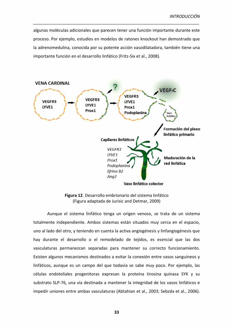
33
algunas moléculas adicionales que parecen tener una función importante durante este
proceso. Por ejemplo, estudios en modelos de ratones knockout han demostrado que
la adrenomedulina, conocida por su potente acción vasodilatadora, también tiene una
importante función en el desarrollo linfático (Fritz-Six et al., 2008).
Figura 12. Desarrollo embrionario del sistema linfático (Figura adaptada de Jurisic and Detmar, 2009)
Aunque el sistema linfático tenga un origen venoso, se trata de un sistema
totalmente independiente. Ambos sistemas están situados muy cerca en el espacio,
uno al lado del otro, y teniendo en cuenta la activa angiogénesis y linfangiogénesis que
hay durante el desarrollo o el remodelado de tejidos, es esencial que las dos
vasculaturas permanezcan separadas para mantener su correcto funcionamiento.
Existen algunos mecanismos destinados a evitar la conexión entre vasos sanguíneos y
linfáticos, aunque es un campo del que todavía se sabe muy poco. Por ejemplo, las
células endoteliales progenitoras expresan la proteína tirosina quinasa SYK y su
substrato SLP-76, una vía destinada a mantener la integridad de los vasos linfáticos e
impedir uniones entre ambas vasculaturas (Abtahian et al., 2003; Sebzda et al., 2006).
INTRODUCCIÓN _______________________________________________________________________
34
En este mecanismo también están implicados la podoplanina y el receptor CLEC-2 de
las plaquetas. La podoplanina activa CLEC-2 y provoca la agregación plaquetaria, y este
efecto lleva a la activación de SYK y SLP-76 (Christou et al., 2008; Suzuki-Inoue et al.,
2007). Otra molécula que parece estar involucrada en la correcta separación de ambas
vasculaturas es un factor adiposo llamado Fiaf. Estudios en ratones deficientes para
este factor han demostrado que presentan una expresión muy reducida del marcador
linfático Prox1 y unos vasos linfáticos llenos de sangre a nivel intestinal después del
nacimiento (Backhed et al., 2007). También está demostrado que numerosas
glicoproteínas regulan este proceso de separación entre los sistemas sanguíneo y
linfático (Bischoff, 1997; Julenius et al., 2005).
4. LA CÉLULA ENDOTELIAL LINFÁTICA
Las células endoteliales linfáticas, a diferencia de las sanguíneas, son células sin
fenestraciones y sin membrana basal. Aun así, comparten gran cantidad de
marcadores, como por ejemplo el factor VIII o factor de Von Willebrand, la
antitrombina 3, MHC 1, el receptor LDL-R, CD31 o la enzima convertidora de la
angiotensina (ACE) (Sleeman et al., 2001). Sin embargo, con el paso de los años, se han
ido descubriendo nuevos marcadores de superficie específicos de las células
endoteliales linfáticas, los más estudiados son LYVE-1, VEGFR-3, podoplanina y el
factor de transcripción Prox-1.
LYVE-1 es un homólogo del receptor de ácido hialurónico CD44 y está
involucrado en el metabolismo de este ácido en el sistema linfático (Banerji et al.,
1999). Es el primer marcador linfático que aparece durante el desarrollo. Se expresa en
los capilares linfáticos y en células endoteliales linfáticas aisladas, sin embargo está
ausente en los vasos linfáticos colectores (Banerji et al., 1999). En adultos, LYVE-1 no
sólo se expresa en el endotelio linfático, también lo hace en los sinusoides hepáticos,
en algunos vasos sanguíneos del pulmón y en tejidos con inflamación (Jackson, 2003).
INTRODUCCIÓN _______________________________________________________________________
35
VEGFR-3, también conocido como Flt4, es un receptor específico de los factores
de crecimiento vascular linfático VEGF-C y VEGF-D (Tammela et al., 2005). Las señales
de este receptor son esenciales para la proliferación, migración y supervivencia de las
células endoteliales linfáticas. Aunque su expresión es alta en la vasculatura sanguínea
durante el desarrollo embrionario, con el avance de la gestación la expresión se va
restringiendo gradualmente a las células endoteliales linfáticas (Kaipainen et al., 1995).
La podoplanina, también conocida como OTS-8, T1-alfa, gp36 o antígeno E11,
es una glicoproteína de superficie esencial para establecer la diferenciación entre el
sistema linfático y el sanguíneo. Está involucrada en la regulación de la permeabilidad
de los vasos linfáticos, en el mantenimiento de su tamaño y también podría controlar
la estructura de las válvulas internas. En adultos también se expresa en pulmón y en
riñón, donde controla la permeabilidad glomerular (Podgrabinska et al., 2002).
Prox-1, a diferencia de los demás marcadores linfáticos, es un factor de
transcripción que desencadena el programa de diferenciación linfática. Estudios en
ratones deficientes en este gen han demostrado que la falta de Prox-1 no afecta al
desarrollo de la vasculatura sanguínea, pero se inhibe totalmente la formación del
sistema linfático. La expresión de este factor en adultos se reduce únicamente a la
vasculatura linfática, de manera que comparado con otros marcadores, sería el
marcador más específico y exclusivo de las células endoteliales linfáticas (Wigle and
Oliver, 1999).
INTRODUCCIÓN _______________________________________________________________________
36
5. EL SISTEMA LINFÁTICO EN UN CONTEXTO PATOLÓGICO
La contribución de los vasos linfáticos en diferentes situaciones patológicas es
un campo que ha permanecido inexplorado durante muchos años debido a la falta de
marcadores específicos que permitieran distinguir el endotelio linfático del sanguíneo.
Con el descubrimiento de los marcadores linfáticos, en los últimos años se han hecho
gran cantidad de estudios y se han podido desarrollar modelos experimentales que
han elucidado la contribución del endotelio linfático en muchas enfermedades.
Actualmente se sabe que, en un contexto fisiopatológico, la vasculatura linfática está
muy involucrada en enfermedades inflamatorias, en la metástasis de las células
tumorales en el cáncer y en el crecimiento tumoral (Cassella et al., 2002; Detmar et al.,
2002) y en la formación de edema (linfedema) (Alitalo et al., 2005).
5.1 CONTRIBUCIÓN DEL SISTEMA LINFÁTICO EN LA INFLAMACIÓN
Los vasos linfáticos son esenciales para la migración de las células inmunitarias
hacia los nódulos linfáticos donde se desencadena la respuesta inmune adaptativa
(Halin and Detmar, 2006). Las señales inflamatorias activan la expresión de CCR7 en las
células dendríticas, a la vez que la vasculatura linfática expresa de forma constitutiva
su ligando CCL21, lo que provoca un proceso quimioatrayente de las células hacia los
vasos (Bromley et al., 2005; Debes et al., 2005). CCR7 también media la migración de
las células T CD4+ de los tejidos a los vasos linfáticos (Yuan et al., 2002). La inflamación
también induce la expresión de las moléculas de adhesión ICAM-1, VCAM-1 y E-
selectina en el endotelio linfático, que también facilitan la entrada de linfocitos a los
vasos (Johnson et al., 2006). Además, los estímulos inflamatorios hacen aumentar el
flujo de linfa, lo que promueve la migración de todas estas células hasta los nódulos
linfáticos (Gunn et al., 1998).
Estudios recientes han demostrado que durante los procesos inflamatorios hay
una activa linfangiogénesis (formación de nuevos vasos linfáticos) (Baluk et al., 2005).
Esto se debe, en parte, a que las células inflamatorias expresan VEGF-C y VEGF-A, dos
potentes factores linfangiogénicos (Ristimaki et al., 1998). La presencia de nuevos
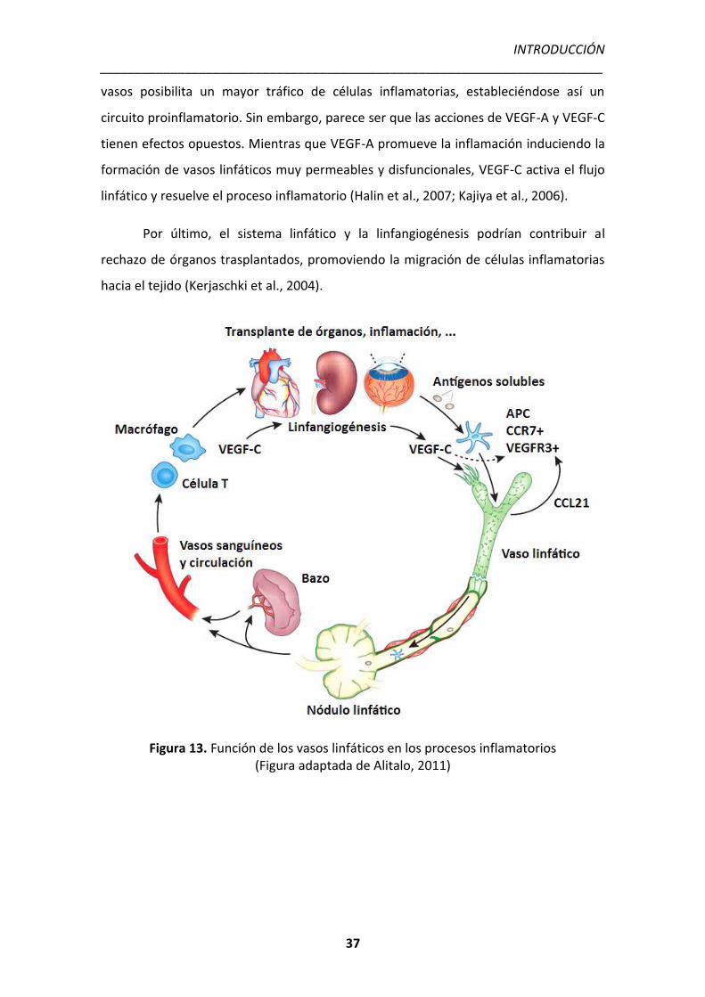
INTRODUCCIÓN _______________________________________________________________________
37
vasos posibilita un mayor tráfico de células inflamatorias, estableciéndose así un
circuito proinflamatorio. Sin embargo, parece ser que las acciones de VEGF-A y VEGF-C
tienen efectos opuestos. Mientras que VEGF-A promueve la inflamación induciendo la
formación de vasos linfáticos muy permeables y disfuncionales, VEGF-C activa el flujo
linfático y resuelve el proceso inflamatorio (Halin et al., 2007; Kajiya et al., 2006).
Por último, el sistema linfático y la linfangiogénesis podrían contribuir al
rechazo de órganos trasplantados, promoviendo la migración de células inflamatorias
hacia el tejido (Kerjaschki et al., 2004).
Figura 13. Función de los vasos linfáticos en los procesos inflamatorios (Figura adaptada de Alitalo, 2011)
INTRODUCCIÓN _______________________________________________________________________
38
5.2 CONTRIBUCIÓN DEL SISTEMA LINFÁTICO EN EL CÁNCER
El papel que tiene el sistema linfático durante los procesos tumorales no está
claro. Parece ser que las células cancerosas pueden inducir linfangiogénesis, e incluso
entrar pasivamente en los vasos linfáticos junto con el fluido intersticial y proteínas, y
usarlos como vías de metástasis (Sleeman et al., 2009; Achen et al., 2008; Mumprecht
et al., 2009). La metástasis de la mayoría de cánceres ocurre inicialmente a través de
los vasos linfáticos. Existe una compleja relación entre la vasculatura linfática y las
células tumorales. Las células endoteliales linfáticas atraen a las tumorales expresando
la molécula de laminina-421 (Saito et al., 2009), de igual forma, las células tumorales
pueden atraer a las linfáticas a través de la expresión de diferentes factores, como la
angiopoyetina-1 (Holopainen et al., 2009). La formación de nuevos vasos linfáticos
alrededor del tumor incrementa las probabilidades de que haya metástasis. Existen
numerosos estudios en humanos que han demostrado que hay una correlación entre
la expresión de VEGF-C en el tumor y la metástasis a los nódulos linfático (Stacker et
al., 2002; Pepper et al., 2003).
Aunque un incremento de la densidad de la red linfática puede promover la
diseminación tumoral incrementando las posibilidades pasivas de metástasis, los vasos
linfáticos también tienen una implicación más activa en la metástasis. Por ejemplo, las
células endoteliales linfáticas expresan constitutivamente factores solubles que
facilitan la invasión de los vasos por parte de las células tumorales. La activación de las
células linfáticas con VEGF-C u otros factores producidos por el tumor puede promover
la expresión de quimioquinas, que podrían atraer a las células tumorales al interior de
los vasos linfáticos. En este contexto, se ha encontrado que el receptor CXCR4, un
receptor de quimioquinas, tiene una importancia crucial en la metástasis del tumor a
los nódulos linfáticos. CXCR4 se ha encontrado sobreexpresado en el melanoma
maligno y en los tumores cerebrales, mientras que su ligando CXCL12 está altamente
expresado en los nódulos linfáticos (Muller et al., 2001).
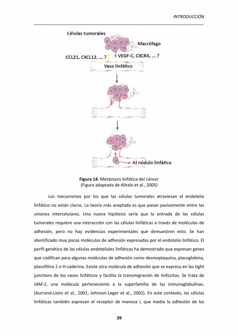
INTRODUCCIÓN _______________________________________________________________________
39
Figura 14. Metástasis linfática del cáncer (Figura adaptada de Alitalo et al., 2005)
Los mecanismos por los que las células tumorales atraviesan el endotelio
linfático no están claros. La teoría más aceptada es que pasan pasivamente entre las
uniones intercelulares. Una nueva hipótesis sería que la entrada de las células
tumorales requiere una interacción con las células linfáticas a través de moléculas de
adhesión, pero no hay evidencias experimentales que demuestren esto. Se han
identificado muy pocas moléculas de adhesión expresadas por el endotelio linfático. El
perfil genético de las células endoteliales linfáticas ha demostrado que expresan genes
que codifican para algunas moléculas de adhesión como desmoplaquina, placoglobina,
placofilina 2 o H-caderina. Existe otra molécula de adhesión que se expresa en las tight
junctions de los vasos linfáticos y facilita la transmigración de linfocitos. Se trata de
JAM-2, una molécula perteneciente a la superfamilia de las inmunoglobulinas.
(Aurrand-Lions et al., 2001; Johnson-Leger et al., 2002). En este contexto, las células
linfáticas también expresan el receptor de manosa I, que media la adhesión de los
INTRODUCCIÓN _______________________________________________________________________
40
linfocitos a los vasos linfáticos en los nódulos linfáticos (Irjala et al., 2001), y se ha
propuesto que esto podría controlar la salida estas células de los vasos a los nódulos o
a otros tejidos periféricos (Petrova et al., 2002; Podgrabinska et al., 2002).
Recientemente, se ha identificado otra molécula de adhesión implicada en la unión de
los linfocitos a las células linfáticas en los nódulos linfáticos, se trata del receptor
endotelial vascular 1, CLEVER-1 (Irjala et al., 2003b). Tanto la expresión del receptor de
manosa como de CLEVER-1, se ha detectado en los vasos linfáticos intratumorales en
diversos carcinomas humanos, y su expresión se ha asociado con un incremento de
metástasis a los nódulos linfáticos (Irjala et al., 2003a).
5.3 CONTRIBUCIÓN DEL SISTEMA LINFÁTICO EN LA FORMACIÓN DE EDEMA
El edema es la acumulación de líquido en el espacio tisular intersticial, que
suele suceder cuando hay un proceso inflamatorio. Cuando la acumulación de líquido
se debe a un fallo en el drenaje linfático, ya sea por malformación de los vasos, por un
transporte linfático deficiente o por una obstrucción del flujo linfático, se denomina
linfedema (Szuba and Rockson, 1997; Shin et al., 2008; Radhakrishnan and Rockson,
2008). Los linfedemas primarios son los que provienen de defectos genéticos, mientras
que los secundarios están causados principalmente por una patología, como una
inflamación crónica. Actualmente no existe ninguna cura para tratar el linfedema.
En cuanto a los linfedemas primarios, se han identificado algunas mutaciones
en genes humanos que comprometen el desarrollo y la función de los vasos linfáticos y
llevan a la formación de edema. Por ejemplo, la enfermedad de Milroy se caracteriza
por la presencia de mutaciones en el gen de VEGFR-3 que inactivan su dominio quinasa
e impiden su fosforilación (Brice et al., 2005; Butler et al., 2007; Karkkainen et al.,
2000). Las mutaciones de VEGFR-3 se han encontrado en muchos pacientes con
historia familiar de linfedema, aunque en los últimos años también se han reportado
mutaciones de novo (Ghalamkarpour et al., 2006). Mutaciones en el gen FOXC2 son
responsables del síndrome de distiquiasis-linfedema (Fang et al., 2000; Mangion et al.,
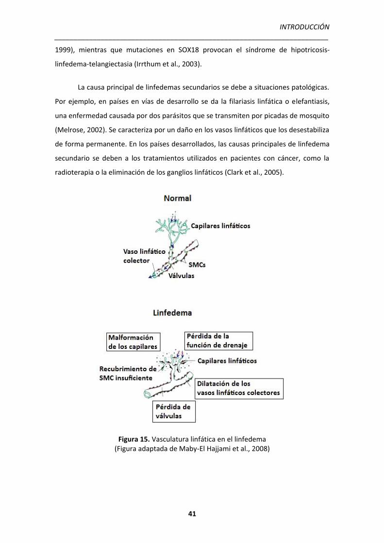
INTRODUCCIÓN _______________________________________________________________________
41
1999), mientras que mutaciones en SOX18 provocan el síndrome de hipotricosis-
linfedema-telangiectasia (Irrthum et al., 2003).
La causa principal de linfedemas secundarios se debe a situaciones patológicas.
Por ejemplo, en países en vías de desarrollo se da la filariasis linfática o elefantiasis,
una enfermedad causada por dos parásitos que se transmiten por picadas de mosquito
(Melrose, 2002). Se caracteriza por un daño en los vasos linfáticos que los desestabiliza
de forma permanente. En los países desarrollados, las causas principales de linfedema
secundario se deben a los tratamientos utilizados en pacientes con cáncer, como la
radioterapia o la eliminación de los ganglios linfáticos (Clark et al., 2005).
Figura 15. Vasculatura linfática en el linfedema (Figura adaptada de Maby-El Hajjami et al., 2008)
INTRODUCCIÓN _______________________________________________________________________
42
El descubrimiento de los factores de crecimiento linfáticos VEGF-C y VEGF-D,
capaces de inducir el crecimiento de nuevos vasos linfáticos, abrió un nuevo abanico
de posibilidades para tratar el linfedema (Rockson, 2005). La administración de VEGF-C
en un modelo de conejo con linfedema en la oreja, consiguió mejorar la enfermedad
considerablemente (Szuba et al., 2002). La terapia con VEGF-C y VEGF-D se relaciona
con una regeneración de la vasculatura linfática madura y mejora el drenaje linfático,
indicando que podría ser una estrategia potencial para tratar en el futuro el linfedema
secundario (Tammela et al., 2007).
En la cirrosis también puede aparecer edema debido a la disrupción u
obstrucción de la circulación linfática. En este contexto, la linfa intestinal (quilo) se
acumula en la cavidad peritoneal y da lugar a la aparición de ascitis quilosa. De hecho,
el 60% de los casos de ascitis quilosa en el mundo occidental se deben a una cirrosis
hepática. La ascitis quilosa se presenta en el 0.5-1% de los pacientes cirróticos y
ascíticos; también se puede manifestar como un síntoma u ocurrir de manera tardía en
el curso de un carcinoma hepatocelular. Fisiopatológicamente, este tipo de ascitis se
da por la ruptura de los vasos linfáticos al dilatarse debido al aumento de la presión
venosa portal, con un flujo excesivo de linfa (alrededor de 20 litros por día) (Rector,
1984; Cheng et al., 1989; Almakdisi et al., 2005).

OBJETIVOS

OBJETIVOS _______________________________________________________________________
43
A lo largo de la evolución, en los organismos complejos se han desarrollado dos
grandes redes vasculares complementarias para poder llevar a cabo todas las
funciones de transporte de fluidos, gases, nutrientes, moléculas de señalización, y
células a través de los tejidos, y hacer posible así la vida: la vasculatura sanguínea y la
vasculatura linfática. La fisiología y la fisiopatología del sistema vascular sanguíneo y la
implicación de la angiogénesis (la formación de nuevos vasos sanguíneos a partir de
otros preexistentes) en múltiples enfermedades, ha sido muy estudiada en las últimas
décadas (Carmeliet, 2003). Sin embargo, hace muy pocos años que se empezó a
estudiar el desarrollo y los mecanismos moleculares que controlan el sistema linfático,
identificado por primera vez en el siglo XVII (Asellius, 1627). El avance en el
conocimiento del sistema linfático se ha producido gracias al reciente descubrimiento
de marcadores específicos de las células endoteliales linfáticas. Aunque las células
endoteliales sanguíneas y linfáticas comparten algunas de sus características, ambas
tienen propiedades bien diferenciadas. Gracias a esta especialización endotelial, estas
dos redes vasculares mantienen su diferenciación funcional.
La cirrosis hepática constituye el estadío final de una fibrosis que se produce
como respuesta a un daño hepático prolongado y que tiene como consecuencia la
acumulación de tejido conectivo en el hígado, debido a un desequilibrio importante
entre la producción y la degradación de la matriz extracelular. Esta enfermedad sigue
estando vinculada de manera inevitable a una significativa disminución y a un
deterioro progresivo de la calidad y la esperanza de vida. Gracias a las investigaciones
realizadas durante las últimas dos décadas, se ha podido apreciar la importancia de la
vasculatura como regulador multifuncional de la fisiopatología de muchas de las
complicaciones de la cirrosis, como los procesos fibróticos e inflamatorios. Hay muchas
evidencias que sugieren que el incremento incontrolado y desorganizado de la
vasculatura sanguínea (angiogénesis) podría ser la causa y el mantenimiento de
algunas de las complicaciones asociadas a la cirrosis hepática. El hígado cirrótico
experimenta a lo largo de todo el desarrollo de la enfermedad una serie de cambios
estructurales, debidos a la acumulación de matriz extracelular y a la inflamación
crónica, que vuelven al tejido hipóxico. La hipoxia tisular provoca la liberación de
agentes proangiogénicos, como VEGF-A, que aumentan la irrigación del tejido para
OBJETIVOS _______________________________________________________________________
44
compensar la falta de oxígeno. Además, muchas citoquinas sintetizadas por las células
inflamatorias o por las células hepáticas estrelladas activadas, también tienen un alto
poder angiogénico. Estos nuevos vasos son una puerta de entrada de más infiltrado
inflamatorio, estableciéndose así un circuito que se retroalimenta entre angiogénesis e
inflamación que lleva a perpetuar la enfermedad. Además, esta expansión de la red
vascular hepática conlleva una serie de cambios morfológicos y funcionales, entre los
cuales destaca la capilarización sinusoidal, caracterizada por una membrana basal
capilar que cubre el sinusoide hepático y dificulta el intercambio de nutrientes y
oxígeno entre los hepatocitos y la sangre. Esto lleva a un incremento de la resistencia
intravascular hepática y produce isquemia y apoptosis de los hepatocitos. En este
contexto, las terapias antiangiogénicas clásicas dirigidas a bloquear VEGF o las vías de
los receptores tirosina quinasa, han demostrado ser eficaces en disminuir el grado de
angiogénesis, de fibrosis y de inflamación hepática, además de disminuir también la
presión portal (Mejias et al., 2009; Tugues et al., 2007). Sin embargo, este tipo de
terapias se han visto relacionadas con multitud de efectos adversos, como trombosis o
hipertensión, ya que no sólo inhiben la angiogénesis patológica sino que también
afectan a la vasculatura sana (Fischer et al., 2007; Wu et al., 2008). De manera que la
necesidad de encontrar nuevas dianas terapéuticas más seguras destinadas a modular
la angiogénesis en la cirrosis hepática sigue siendo uno de los objetivos de la
investigación científica actual.
Cuando la cirrosis es severa, se produce una desestructuración completa del
parénquima hepático que da lugar a la aparición de insuficiencia hepática y
complicaciones hemodinámicas graves. Estas complicaciones aparecen asociadas a un
síndrome de circulación hiperdinámica que se caracteriza por hipotensión arterial,
hipertensión portal, un aumento del gasto cardíaco y una disminución de las
resistencias periféricas totales. Cuando la cirrosis pasa a ser descompensada, los
pacientes cirróticos desarrollan importantes alteraciones en la homeostasis de los
líquidos corporales que dan lugar a la acumulación de ascitis. La formación de ascitis es
el trastorno más importante que presentan los pacientes cirróticos y constituye un
fenómeno extraordinariamente complejo en el que se han descrito la implicación de
diversos sistemas neurohormonales y sustancias endógenas capaces de regular el
OBJETIVOS _______________________________________________________________________
45
metabolismo renal de sodio y agua o la reactividad vascular (Arroyo and Jiménez,
2000). La probabilidad de fallecimiento a los 2 años en pacientes cirróticos, una vez
que desarrollan ascitis, es del 40%. A pesar de la enorme relevancia clínica de este
suceso, actualmente aun no se conocen con precisión todos los mecanismos
moleculares implicados en la formación y mantenimiento de la ascitis en la cirrosis. Se
sabe que en diferentes situaciones patológicas existen cuatro trastornos que
contribuyen a la formación de edema: un aumento de la presión hidrostática
intravascular, un descenso de la presión oncótica en el plasma, una retención renal de
sal y agua, y un fallo en el drenaje linfático (Gines et al., 2004; Van Steenkiste et al.,
2009; Henriksen et al., 2005). Si bien los tres primeros mecanismos están bien
estudiados y caracterizados en la cirrosis, la contribución de la funcionalidad linfática a
la acumulación de ascitis sigue siendo un campo poco estudiado.
Teniendo en cuenta todos los antecedentes descritos, nuestra hipótesis
formula que el estudio de las anomalías vasculares, tanto funcionales como
estructurales, posibilita el desarrollo de nuevas estrategias terapéuticas con
potencialidad traslacional en el contexto clínico del tratamiento de las enfermedades
hepáticas crónicas.
El objetivo global de esta tesis doctoral fue el de investigar la implicación de la
vasculatura, tanto sanguínea como linfática, en la patogenicidad de la cirrosis hepática.
De manera específica, la tesis está dividida en dos estudios dirigidos a:
I. Investigar el papel fisiopatológico del factor de crecimiento placentario
(PlGF) en la angiogénesis, la inflamación y la fibrosis hepática, así como
su posible papel profibrogénico, analizando su efecto biológico sobre las
células hepáticas estrelladas.
II. Caracterizar la funcionalidad y la estructura de la vasculatura linfática en
la cirrosis e investigar su contribución a la formación de ascitis, así
como identificar posibles mecanismos moleculares que puedan ser
responsables de una disfunción linfática durante la enfermedad.


RESULTADOS

RESULTADOS _______________________________________________________________________
46
Artículo 1: La inhibición de la actividad del factor de crecimiento placentario reduce
la severidad de la fibrosis, la inflamación, y la hipertensión portal en ratones
cirróticos.
Durante la progresión de gran cantidad de enfermedades hepáticas crónicas,
tales como las hepatitis B/C, la esteatosis no alcohólica, la cirrosis biliar y la cirrosis
alcohólica, tiene lugar un proceso de neoangiogénesis hepática que agrava
profundamente dichas patologías. Esta nueva vasculatura se localiza principalmente en
las zonas fibróticas y altera toda la angioarquitectura hepática, dando lugar a
anastomosis arterio-portal y porto-venosa (Fernández et al., 2009). Además, se ha
demostrado que diversos factores angiogénicos también son capaces de activar
diferentes subpoblaciones de células hepáticas estrelladas, estableciendo así un
vínculo entre inflamación, angiogénesis y fibrosis hepáticas (Thabut et al., 2010;
Semela et al., 2008). Son muchos los estudios que han puesto de manifiesto que las
terapias dirigidas, tanto a la inhibición de la angiogénesis como a la inhibición múltiple
de angiogénesis e inflamación, son capaces de frenar la progresión de la fibrosis a
cirrosis. En este contexto, se ha demostrado que la administración de sunitinib y
sorafenib a ratas cirróticas, dos inhibidores multidiana de receptores tirosina quinasa
que actúan en las vías de señalización de PDGF y VEGF, conseguía disminuir el grado de
angiogénesis, de fibrosis y de inflamación hepáticas, además de disminuir también la
presión portal (Mejias et al., 2009; Tugues et al., 2007). Sin embargo, este tipo de
terapias dirigidas a inhibir VEGF están asociadas a graves efectos adversos, como
trombosis, hipertensión o una disminución de la densidad vascular en órganos sanos
(Fischer et al., 2007), ya que VEGF también tiene importantes funciones en la
angiogénesis fisiológica. Esto limita su uso para el tratamiento de pacientes humanos.
Estudios recientes sugieren que las terapias enfocadas a inhibir el PlGF podrían tener
un perfil de bioseguridad mucho más óptimo que las terapias antiangiogénicas clásicas
(Fischer et al., 2007; Van de Veire et al., 2010). Esto es así porque la acción angiogénica
de PlGF se reduce únicamente a situaciones patológicas (Autiero et al., 2003; Van
Steenkiste et al., 2009), y se ha demostrado que su bloqueo reduce la angiogénesis en
varios modelos de cáncer y en otras enfermedades sin afectar a la vasculatura sana y
sin presentar mayores efectos secundarios (Lassen et al., 2009; Riisbro et al., 2009; Van
RESULTADOS _______________________________________________________________________
47
de Veire et al., 2010). Por ello, en este estudio se investigó el PlGF como potencial
candidato de futuras terapias contra la cirrosis, estudiando su función en la
angiogénesis, la inflamación y la fibrosis hepáticas. Los resultados más importantes
derivados de este estudio se enumeran a continuación:
1. Los niveles de PlGF están aumentados en ratones tratados con CCl4 y en pacientes
cirróticos.
En primer lugar, se analizaron los cambios de expresión de PlGF en modelos
experimentales de cirrosis en rata y ratón por ELISA y Western Blot. Después de 4
semanas de tratamiento con CCl4, los ratones incrementaron sus niveles hepáticos de
PlGF y éstos se mantuvieron elevados durante las 16 semanas que duró el tratamiento.
Este aumento también se detectó en el hígado de ratas cirróticas, donde la expresión
de PlGF estuvo aumentada del orden de 4 veces respecto a hígados control (Figura 1A
y 1B).
Para determinar si había un paralelismo en humanos, se analizaron los niveles
de PlGF en pacientes cirróticos, tanto en suero por ELISA como la expresión génica por
RT-PCR. Los niveles hepáticos de RNAm estuvieron significativamente elevados en
pacientes cirróticos comparados con pacientes control. En concordancia con estos
resultados, los niveles de PlGF en suero fueron entre 2 y 3 veces mayores en pacientes
cirróticos respecto a sujetos sanos. Además, se encontró una correlación significativa
entre los niveles de PlGF y el gradiente de presión portal en pacientes con hepatitis
alcohólica. (Figura 1C, 1D y 1E).
2. La deficiencia génica de PlGF y el tratamiento con anticuerpos anti-PlGF mejora la
hemodinámica y la hipertensión portal en ratones cirróticos.
El bloqueo de PlGF, tanto genéticamente como con una terapia con
anticuerpos, redujo el flujo arterial mesentérico un 36.8% y el gasto cardíaco un 17%.
Estos cambios hemodinámicos llevaron a una reducción del 27% de la presión portal
(Tabla 1).
RESULTADOS _______________________________________________________________________
48
3. El bloqueo de PlGF reduce el grado de inflamación hepática provocada por el
tratamiento con CCl4.
Estudios anteriores de nuestro laboratorio habían demostrado que los
inhibidores de angiogénesis, como sunitinib, reducían la inflamación de los hígados
cirróticos (Tugues et al., 2007). Es por ello que analizamos si el bloqueo de la actividad
de PlGF también era capaz de reducir la inflamación hepática, a través de un marcaje
de macrófagos (PAS-diastasa). El incremento del infiltrado de macrófagos típico de la
cirrosis se vio significativamente reducido en los ratones donde se había bloqueado la
actividad de PlGF (Figura 2). Además, también se observó una reducción de la
expresión de VCAM-1, una molécula de adhesión proinflamatoria, en la vasculatura de
los ratones cirróticos (Supplemental results Figura 2).
4. La inhibición de PlGF reduce la neoangiogénesis y la arteriogénesis intrahepáticas y
esplácnicas en los animales cirróticos.
Para investigar como PlGF estimulaba la angiogénesis durante la progresión de
la cirrosis realizamos tinciones inmuhistoquímicas con el marcador de vasos CD31.
Comparados con los ratones cirróticos, los ratones donde se bloqueó la actividad de
PlGF mostraron una reducción significativa de vasos en el hígado, en el mesenterio y
en el colon (Figura 3).
Para estudiar más el papel de PlGF en la angiogénesis patológica, se obtuvieron
moldes vasculares del mesenterio y del hígado de ratones cirróticos utilizando la
técnica de corrosión. Tanto el análisis con la lupa estereoscópica como por microscopía
electrónica de barrido, mostraron que los ratones cirróticos salvajes tenían una mayor
arborización vascular comparados con los ratones tratados contra PlGF. Además,
mientras que los ratones cirróticos mostraban un sinusoide hepático irregular, con
vasos tortuosos e inmaduros, el bloqueo de la actividad de PlGF conseguía normalizar
la estructura del sinusoide (Supplemental results Figura 4).
RESULTADOS _______________________________________________________________________
49
5. El bloqueo de la actividad de PlGF reduce el grado de fibrosis de los animales
cirróticos.
Para evaluar los efectos in vivo del bloqueo de la actividad de PlGF,
cuantificamos el grado de fibrosis por Sirius Red. Después de la administración crónica
de CCl4 (25 semanas) los ratones mostraron un elevado grado de fibrosis, con la
presencia de septos fibrosos centro-portales. Sin embargo, el bloqueo de PlGF redujo
el grado de fibrosis en un 36%. Además, la evaluación macroscópica y estereoscópica
de los hígados reveló una pérdida de nodularidad después del bloqueo de PlGF
comparado con los animales cirróticos salvajes (Figura 4).
6. Las células estrelladas hepáticas son la principal fuente de PlGF en el hígado
cirrótico.
Para entender porque una reducción de la actividad de PlGF se asocia con una
reducción en el grado de fibrosis, quisimos ver donde se localizaba la expresión de
PlGF en el hígado por inmunofluorescencia. En los hígados de los animales control la
señal de PlGF fue muy débil, sin embargo aumentó en los animales tratados con CCl4,
localizándose básicamente alrededor de los septos fibrosos (Figura 5A).
Teniendo en cuenta que el tipo celular que más participa en la formación de
fibra son las HSC, pensamos que quizá estas células podrían ser la fuente principal de
PlGF en el hígado. Esto lo confirmamos por PCR Real Time, donde se vio que las HSC
tienen una gran sobreexpresión de PlGF, mucho mayor que la de los otros tipos
celulares hepáticos (Supplemental results Figura 7C).
7. PlGF activa vías de señalización relacionadas con proliferación y migración celular en
las HSC.
Las HSC juegan un papel muy importante en la fisiopatología de la fibrosis, así
que quisimos estudiar el efecto de PlGF sobre este tipo celular activado, tanto en rata
como en humanos. Por RT-PCR detectamos expresión del receptor VEGFR1, el receptor
RESULTADOS _______________________________________________________________________
50
específico de PlGF, tanto en HSC aisladas de ratas cirróticas como en la línea LX-2, una
línea humana de HSC. Teniendo esto en cuenta, quisimos determinar que vías de
señalización estaban activadas en las HSC en respuesta a un estímulo de PlGF. Por
Western Blot vimos que el tratamiento de estas células con PlGF inducía la activación
sostenida de la vía de ERK 1/2 durante más de 60 minutos (Figura 5B y 5C).
Ya es conocido que la activación de la vía de ERK 1/2 se relaciona con
quimiotaxis y proliferación celular. Para corroborar si existía un paralelismo en las HSC,
cuantificamos la quimiotaxis celular en células LX-2 sin tratar o tratadas con PlGF, a
través de microfotografías tomadas a diferentes tiempos. Observamos que alrededor
de un 35% de las células migraron en respuesta a un tratamiento de PlGF durante 10
minutos. La migración celular se asocia con una reestructuración del citoesqueleto de
actina, por ello quisimos estudiar este efecto de PlGF en las HSC usando una tinción
con faloidina, que se une específicamente a las fibras de actina. PlGF indujo la
formación de filopodios alrededor de toda la células, indicando que sí es capaz de
promover la reorganización del citoesqueleto de actina (Figura 6A y 6C).
Para estudiar si aparte de inducir migración, PlGF era capaz de estimular la
proliferación de las HSC, hicimos un ensayo de proliferación con BrdU. Vimos que la
cantidad de BrdU incorporado en presencia de PlGF era del 21%, significativamente
mayor en comparación con el porcentaje de BrdU incorporado en células sin tratar.
Este % de incorporación de BrdU representa el % de células que están proliferando, y
esto indica que PlGF también es capaz de promover la proliferación de las HSC (Figura
6D).
Para profundizar más en los mecanismos de señalización a través de los cuales
PlGF induce una activación sostenida de ERK 1/2 y migración y proliferación celular,
analizamos la fosforilación de proteínas implicadas en la transducción de estas señales.
Para ello utilizamos unos arrays de anticuerpos que detectan los estados de
fosforilación de proteínas de señalización, y este estado de fosforilación es sinónimo
de actividad. Encontramos un total de 33 proteínas fosforiladas en Tyr después del
tratamiento con PlGF, y de acuerdo con los resultados de proliferación y migración
obtenidos anteriormente, el análisis bioinformático de los resultados mostró que las
RESULTADOS _______________________________________________________________________
51
proteínas fosforiladas se agrupaban principalmente en dos grupos funcionales: 1)
Desarrollo celular, desarrollo del sistema hematológico y viabilidad celular, y 2) Cáncer,
morfología tumoral y movimiento celular (Supplemental results Figura 9). Además, la
exposición de las HSC a PlGF también activó los receptores PDGFRA y EGFR, esto es
importante porque podría representar una asociación directa entre estos receptores
TK y el receptor de PlGF. Esto lo confirmamos con un ensayo de ligación por
proximidad (PLA), donde encontramos una interacción directa entre VEGFR1 y PDGFRA
(Supplemental results Figura 10).
8. El bloqueo de la actividad de PlGF presenta un perfil de bioseguridad óptimo
Uno de los problemas de los inhibidores clásicos de angiogénesis es que llevan
asociados múltiples efectos secundarios adversos, como trombosis, hipertensión o una
disminución de la densidad vascular en órganos sanos (Fischer et al., 2007). Por ello,
quisimos estudiar como afectaba el bloqueo de PlGF a la vasculatura sana en un
órgano sin angiogénesis patológica, como es la glándula tiroidea, por
inmunohistoquímica de CD31. En este contexto, ni los ratones tratados con
anticuerpos anti-PlGF ni los genéticamente deficientes para PlGF, mostraron cambios
significativos en la densidad vascular de este órgano. Otro punto crítico en la aplicación
clínica de antiangiogénicos es el desarrollo de resistencia al bloqueo de la angiogénesis
a través de la sobreexpresión de otros agentes proangiogénicos, como VEGF. Por esta
razón, estudiamos por ELISA si el bloqueo de PlGF estaba asociado a la inducción de
VEGF. El tratamiento contra PlGF tampoco produjo cambios significativos en los niveles
de expresión de VEGF en el mesenterio de ratones cirróticos (Supplemental results
Figura 11).
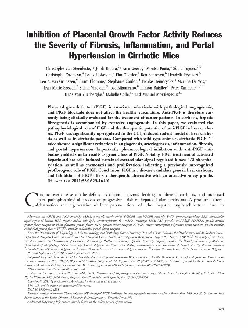
Inhibition of Placental Growth Factor Activity Reducesthe Severity of Fibrosis, Inflammation, and Portal
Hypertension in Cirrhotic MiceChristophe Van Steenkiste,1* Jordi Ribera,2* Anja Geerts,1 Montse Pauta,2 Sonia Tugues,2,3
Christophe Casteleyn,4 Louis Libbrecht,5 Kim Olievier,1 Ben Schroyen,6 Hendrik Reynaert,6
Leo A. van Grunsven,6 Bram Blomme,1 Stephanie Coulon,1 Femke Heindryckx,1 Martine De Vos,1
Jean Marie Stassen,7 Stefan Vinckier,9 Jose Altamirano,8 Ramon Bataller,8 Peter Carmeliet,9,10
Hans Van Vlierberghe,1 Isabelle Colle,1* and Manuel Morales-Ruiz2*
Placental growth factor (PlGF) is associated selectively with pathological angiogenesis,and PlGF blockade does not affect the healthy vasculature. Anti-PlGF is therefore cur-rently being clinically evaluated for the treatment of cancer patients. In cirrhosis, hepaticfibrogenesis is accompanied by extensive angiogenesis. In this paper, we evaluated thepathophysiological role of PlGF and the therapeutic potential of anti-PlGF in liver cirrho-sis. PlGF was significantly up-regulated in the CCl4-induced rodent model of liver cirrho-sis as well as in cirrhotic patients. Compared with wild-type animals, cirrhotic PlGF2/2
mice showed a significant reduction in angiogenesis, arteriogenesis, inflammation, fibrosis,and portal hypertension. Importantly, pharmacological inhibition with anti-PlGF anti-bodies yielded similar results as genetic loss of PlGF. Notably, PlGF treatment of activatedhepatic stellate cells induced sustained extracellular signal-regulated kinase 1/2 phospho-rylation, as well as chemotaxis and proliferation, indicating a previously unrecognizedprofibrogenic role of PlGF. Conclusion: PlGF is a disease-candidate gene in liver cirrhosis,and inhibition of PlGF offers a therapeutic alternative with an attractive safety profile.(HEPATOLOGY 2011;53:1629-1640)
Chronic liver disease can be defined as a com-plex pathophysiological process of progressivedestruction and regeneration of liver paren-
chyma, leading to fibrosis, cirrhosis, and increasedrisk of hepatocellular carcinoma. A profound altera-tion of the hepatic angioarchitecture due to
Abbreviations: aPlGF, anti-PlGF antibody; aSMA, a-smooth muscle actin; aVEGFR, anti-VEGFR antibody; BrdU, bromodeoxyuridine; ERK, extracellularsignal-regulated kinase; HSC, hepatic stellate cell; IgG1, immunoglobulin G1; mRNA, messenger RNA; PAS, periodic acid-Schiff; PDGFRA, platelet-derivedgrowth factor receptor-a; PlGF, placental growth factor; RTK, tyrosine kinase receptor; RT-PCR, reverse-transcription polymerase chain reaction; VEGF, vascularendothelial growth factor; VEGFR, vascular endothelial growth factor receptor.From the Departments of 1Hepatology and Gastroenterology and 5Pathology, Ghent University Hospital, Ghent, Belgium; the 2Biochemistry and Molecular Genetics
Department, Hospital Clinic, and the 8Liver Unit Hospital Clinic, Institut d’Investigacions Biomediques August Pi i Sunyer, CIBERehd, University of Barcelona,Barcelona, Spain; the 3Department of Genetics and Pathology, Rudbeck Laboratory, Uppsala University, Uppsala, Sweden; the 4Faculty of Veterinary Medicine,Department of Morphology, Ghent University, Ghent, Belgium; the 6Liver Cell Biology Laboratorium, Free University of Brussels (VUB), Brussels, Belgium;7ThromboGenics NV, Leuven, Belgium; the 9Vesalius Research Center, VIB, Leuven, Belgium; and the 10Vesalius Research Center, K. U. Leuven, Leuven, Belgium.Received September 16, 2010; accepted January 25, 2011.Supported by grants from the Fund for Scientific Research (Aspirant mandaat-FWO Vlaanderen, 1.1.466.09.N.0 to C. V. S.) and from the Ministerio de
Ciencia e Innovacion (SAF 2007-63069 and SAF 2010-19025 to M. M. R.) and AGAUR (2009 SGR 1496). CIBERehd is funded by the Instituto de SaludCarlos III-Ministerio de Ciencia e Innovacion. M. P. was supported by MICINN (contract number BES-2007-16909).*These authors contributed equally to this work.Address reprint requests to: Isabelle Colle, MD, Ph.D., Department of Hepatology and Gastroenterology, Ghent University Hospital, Building K12, First Floor
IE, De Pintelaan 185, 9000 Ghent, Belgium. E-mail: [email protected]; Fax: (32)-9-3324984.CopyrightVC 2011 by the American Association for the Study of Liver Diseases.View this article online at wileyonlinelibrary.com.DOI 10.1002/hep.24238Potential conflict of interest: ThromboGenics NV developed PlGF inhibitors for antiangiogenic treatment under a license from VIB and K. U. Leuven. Jean
Marie Stassen is the Senior Director of Research & Development at ThromboGenics NV.Additional Supporting Information may be found in the online version of this article.
1629
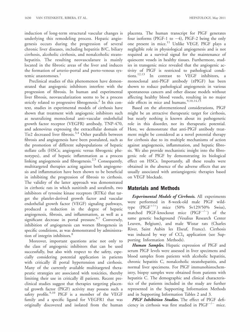
induction of long-term structural vascular changes isunderlying this remodeling process. Hepatic angio-genesis occurs during the progression of severalchronic liver diseases, including hepatitis B/C, biliarycirrhosis, alcoholic cirrhosis, and nonalcoholic steato-hepatitis. The resulting neovasculature is mainlylocated in the fibrotic areas of the liver and inducesthe formation of arterio-portal and porto-venous sys-temic anastomoses.1
Preclinical studies of this phenomenon have demon-strated that angiogenic inhibitors interfere with theprogression of fibrosis. In human and experimentalliver fibrosis, neovascularization seems to be a processstrictly related to progressive fibrogenesis.2 In this con-text, studies in experimental models of cirrhosis haveshown that treatment with angiogenic inhibitors suchas neutralizing monoclonal anti–vascular endothelialgrowth factor receptor (VEGFR) antibody, TNP-470,and adenovirus expressing the extracellular domain ofTie2 decreased liver fibrosis.3,4 Other parallels betweenfibrosis and angiogenesis have been postulated, such asthe promotion of different subpopulations of hepaticstellate cells (HSCs; angiogenic versus fibrogenic phe-notypes), and of hepatic inflammation as a processlinking angiogenesis and fibrogenesis.2,5 Consequently,multitargeted therapies acting against both angiogene-sis and inflammation have been shown to be beneficialin inhibiting the progression of fibrosis to cirrhosis.The validity of the latter approach was demonstratedin cirrhotic rats in which sunitinib and sorafenib, twoinhibitors of tyrosine kinase receptors (RTKs) that tar-get the platelet-derived growth factor and vascularendothelial growth factor (VEGF) signaling pathways,produced a reduction in the degree of hepaticangiogenesis, fibrosis, and inflammation, as well as asignificant decrease in portal pressure.6,7 Conversely,inhibition of angiogenesis can worsen fibrogenesis inspecific conditions, as was demonstrated by administra-tion of integrin inhibitors.8
Moreover, important questions arise not only tothe class of angiogenic inhibitors that can be usedsuccessfully, but also with respect to the safety, espe-cially considering potential application in patientswith critically ill portal hypertension and cirrhosis.Many of the currently available multitargeted thera-peutic strategies are associated with toxicities, therebylimiting their use in critically ill patients. Recent pre-clinical studies suggest that therapies targeting placen-tal growth factor (PlGF) activity may possess such asafety profile.9,10 PlGF is a member of the VEGFfamily and a specific ligand for VEGFR1 that wasoriginally discovered and isolated from the human
placenta. The human transcript for PlGF generatesfour isoforms (PlGF-1 to �4), PlGF-2 being the onlyone present in mice.11 Unlike VEGF, PlGF plays anegligible role in physiological angiogenesis and is notrequired as a survival signal for the maintenance ofquiescent vessels in healthy tissues. Furthermore, stud-ies in transgenic mice revealed that the angiogenic ac-tivity of PlGF is restricted to pathological condi-tions.12,13 In contrast to VEGF inhibitors, amonoclonal anti-PlGF antibody (aPlGF) has beenshown to reduce pathological angiogenesis in variousspontaneous cancers and other disease models withoutaffecting healthy blood vessels, resulting in no majorside effects in mice and humans.9,10,14,15
Based on the aforementioned considerations, PlGFmight be an attractive therapeutic target for cirrhosis,but nearly nothing is known about its pathogeneticrole in this disorder, nor its therapeutic potential.Here, we demonstrate that anti-PlGF antibody treat-ment might be considered as a novel potential therapyfor cirrhosis due to its multiple mechanisms of actionagainst angiogenesis, inflammation, and hepatic fibro-sis. We also provide mechanistic insight into the fibro-genic role of PlGF by demonstrating its biologicaleffect on HSCs. Importantly, all these results wereobtained in the absence of the adverse effects that areusually associated with antiangiogenic therapies basedon VEGF blockade.
Materials and Methods
Experimental Models of Cirrhosis. All experimentswere performed in 8-week-old male PlGF wild-type (PlGFþ/þ) mice (50% Sv129/50% Swiss),matched PlGF-knockout mice (PlGF�/�) of thesame genetic background (Vesalius Research CenterLeuven, Belgium), and male Wistar rats (CharlesRiver, Saint Aubin les Elseuf, France). Cirrhosiswas induced by way of CCl4 application (see Sup-porting Information Methods).Human Samples. Hepatic expression of PlGF and
serum PlGF levels were assessed in liver specimens andblood samples from patients with alcoholic hepatitis,chronic hepatitis C, nonalcoholic steatohepatitis, andnormal liver specimens. For PlGF immunohistochem-istry, biopsy samples were obtained from patients withhepatitis C. The demographic and clinical characteris-tics of the patients included in the study are furtherrepresented in the Supporting Information Methodsand in Supporting Information Tables 2 and 3.PlGF Inhibition Studies. The effect of PlGF defi-
ciency in cirrhosis was first studied in PlGF�/� mice.
1630 VAN STEENKISTE, RIBERA, ET AL. HEPATOLOGY, May 2011
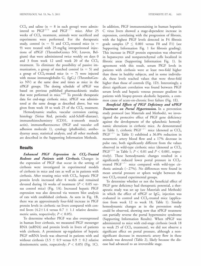
CCl4 and saline (n ¼ 8 in each group) were admin-istered to PlGFþ/þ and PlGF�/� mice. After 25weeks of CCl4 treatment, animals were sacrificed andexperiments were performed. For the therapeuticstudy, control (n ¼ 5) and CCl4-treated mice (n ¼9) were treated with 25-mg/kg intraperitoneal injec-tions of aPlGF (ThromboGenics NV, Leuven, Bel-gium) that were administered twice weekly on days 0and 3 from week 12 until week 20 of the CCl4treatment. To eliminate the possibility of passive im-munization, a group of matched control (n ¼ 5) anda group of CCl4-treated mice (n ¼ 7) were injectedwith mouse immunoglobulin G1 (IgG1) (ThromboGen-ics NV) at the same dose and times as mice in theaPlGF groups. The dosing schedule of aPlGF wasbased on previous published pharmacokinetic studiesthat were performed in mice.9,10 To provide therapeuticdata for end-stage cirrhotic mice, aPlGF was adminis-tered at the same dosage as described above, but wasgiven from week 18 to week 25 of the CCl4 treatment.Hemodynamic studies, vascular corrosion casting,
histology (Sirius Red, periodic acid-Schiff–diastase),immunohistochemistry (CD31, a-smooth muscleactin), immunofluorescence (PlGF and vascular celladhesion molecule 1), cytology (phalloidin), antibo-dyarray assay, statistical analysis, and all other methodsare described in the Supporting Information Methods.
Results
Enhanced PlGF Expression in CCl4-TreatedRodents and Patients with Cirrhosis. Changes inthe expression of PlGF that occur in the setting ofcirrhosis were investigated in experimental modelsof cirrhosis in mice and rats as well as in patients withcirrhosis. After treating mice with CCl4, hepatic PlGFprotein levels increased after 4 weeks and remainedelevated during 16 weeks of treatment (P < 0.05 ver-sus control mice) (Fig. 1A). Increased hepatic PlGFexpression was also detected via western blot analysisof rats with established cirrhosis. As seen in Fig. 1B,there was an approximately four-fold increase in PlGFprotein levels in cirrhotic rat livers compared with con-trol livers (4.261.4 versus 0.7 6 1.1 relative densito-metric units, respectively; P < 0.05).To determine whether PlGF was also overexpressed
in human liver cirrhosis, we measured PlGF messengerRNA (mRNA) and protein levels in livers of patientswith cirrhosis. A prominent up-regulation of hepaticPlGF mRNA levels was observed in patients with andwithout cirrhosis (3.5 6 0.9 versus 0.9 6 0.2 relativedensitometric units, respectively; P < 0.05) (Fig. 1C).
In addition, PlGF immunostaining in human hepatitisC virus livers showed a stage-dependent increase inexpression, correlating with the progression of fibrosis,with the highest PlGF levels detected in F4 fibrosisgrade samples (P � 0.001 versus F0 and F1) (seeSupporting Information Fig. 1 for fibrosis grading).This increase in PlGF protein expression was observedin hepatocytes and nonparenchymal cells localized infibrotic areas (Supporting Information Fig. 1). Inagreement with this result, serum PlGF levels inpatients with cirrhosis were at least two-fold higherthan those in healthy subjects, and in some individu-als, these levels reached values that were three-foldhigher than those of controls (Fig. 1D). Interestingly, adirect significant correlation was found between PlGFserum levels and hepatic venous pressure gradient inpatients with biopsy-proven alcoholic hepatitis, a com-mon cause of acute-on-chronic liver failure (Fig. 1E).Beneficial Effects of PlGF Deficiency and aPlGF
Treatment on Portal Hypertension. In a preventionstudy protocol (see Materials and Methods), we inves-tigated the protective effect of PlGF gene deficiencyagainst the development of the splanchnic hemody-namic alterations in cirrhotic mice. As demonstratedin Table 1, cirrhotic PlGF�/� mice (denoted as CCl4PlGF�/� in Table 1) exhibited a 36.8% reduction inmesenteric artery blood flow and a 17% decrease inpulse rate, both significantly different from the valuesobserved in wild-type cirrhotic mice (denoted as CCl4PlGFþ/þ in Table 1; P < 0.01 and P < 0.001, respec-tively). These hemodynamic changes resulted in asignificantly reduced lower portal pressure in CCl4-treated PlGF�/� mice compared with wild-type cir-rhotic animals (�27%). No differences were found inmean arterial pressure or spleen weight between thetwo CCl4-treated experimental groups.To determine whether or not the beneficial effect of
PlGF gene deficiency had therapeutic potential, a ther-apeutic study was set up (see Materials and Methods)in which the effect of aPlGF or IgG1 injection wasevaluated in control and CCl4-treated mice (applica-tion from week 12 to week 18, Table 1). Similarhemodynamic changes as in the prevention studycould be observed, showing now that aPlGF treatmentcan partially reverse the portal hypertensive syndrome(Supporting Information Results). When aPlGF wasadministered to mice with end-stage cirrhosis (week 18to week 25 of CCl4 treatment), we did not observe asignificant effect on portal pressure, although a non-significant decrease in mesenteric artery flow in theseanimals was detected (Table 2), likely because the dis-ease had advanced to an irreversible stage.
HEPATOLOGY, Vol. 53, No. 5, 2011 STEENKISTE ET AL. 1631
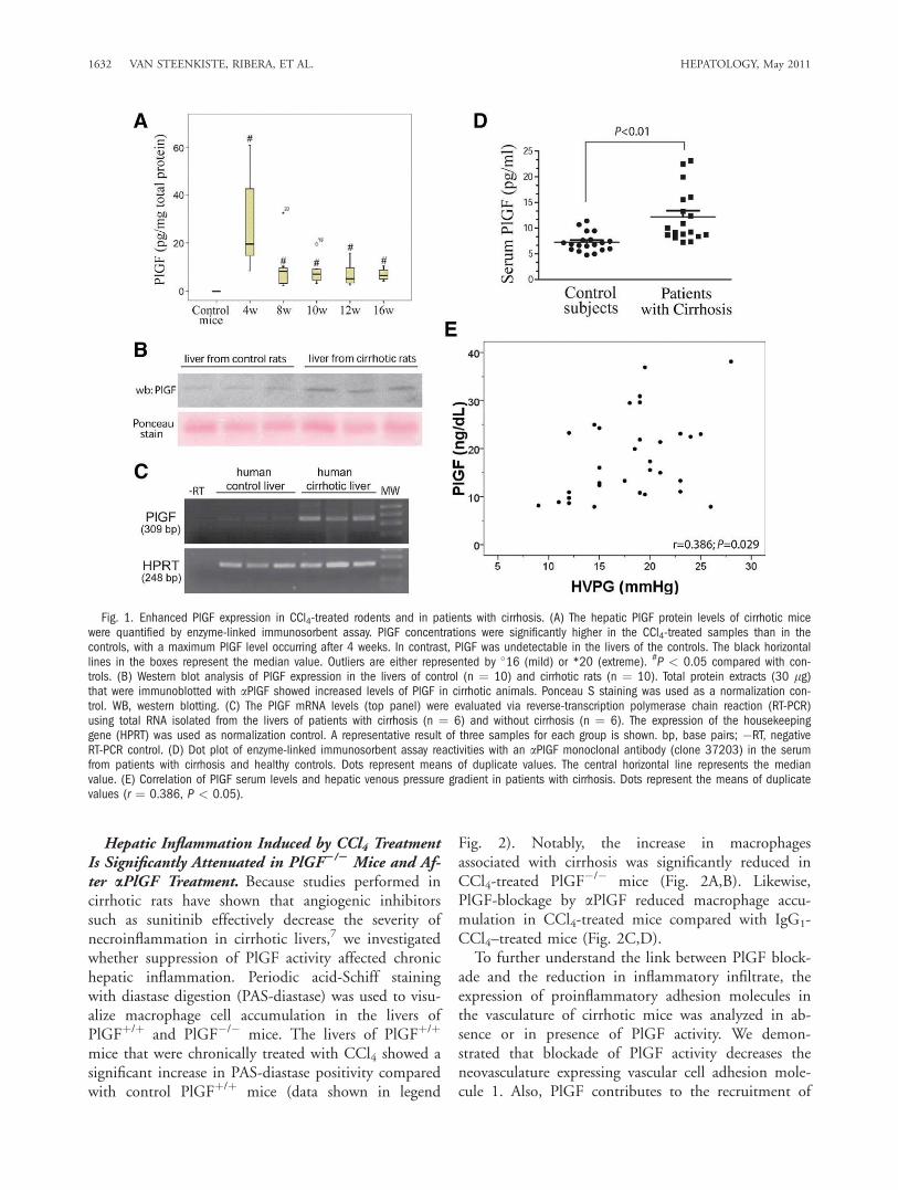
Hepatic Inflammation Induced by CCl4 TreatmentIs Significantly Attenuated in PlGF2/2 Mice and Af-ter aPlGF Treatment. Because studies performed incirrhotic rats have shown that angiogenic inhibitorssuch as sunitinib effectively decrease the severity ofnecroinflammation in cirrhotic livers,7 we investigatedwhether suppression of PlGF activity affected chronichepatic inflammation. Periodic acid-Schiff stainingwith diastase digestion (PAS-diastase) was used to visu-alize macrophage cell accumulation in the livers ofPlGFþ/þ and PlGF�/� mice. The livers of PlGFþ/þ
mice that were chronically treated with CCl4 showed asignificant increase in PAS-diastase positivity comparedwith control PlGFþ/þ mice (data shown in legend
Fig. 2). Notably, the increase in macrophagesassociated with cirrhosis was significantly reduced inCCl4-treated PlGF�/� mice (Fig. 2A,B). Likewise,PlGF-blockage by aPlGF reduced macrophage accu-mulation in CCl4-treated mice compared with IgG1-CCl4–treated mice (Fig. 2C,D).To further understand the link between PlGF block-
ade and the reduction in inflammatory infiltrate, theexpression of proinflammatory adhesion molecules inthe vasculature of cirrhotic mice was analyzed in ab-sence or in presence of PlGF activity. We demon-strated that blockade of PlGF activity decreases theneovasculature expressing vascular cell adhesion mole-cule 1. Also, PlGF contributes to the recruitment of
Fig. 1. Enhanced PlGF expression in CCl4-treated rodents and in patients with cirrhosis. (A) The hepatic PlGF protein levels of cirrhotic micewere quantified by enzyme-linked immunosorbent assay. PlGF concentrations were significantly higher in the CCl4-treated samples than in thecontrols, with a maximum PlGF level occurring after 4 weeks. In contrast, PlGF was undetectable in the livers of the controls. The black horizontallines in the boxes represent the median value. Outliers are either represented by �16 (mild) or *20 (extreme). #P < 0.05 compared with con-trols. (B) Western blot analysis of PlGF expression in the livers of control (n ¼ 10) and cirrhotic rats (n ¼ 10). Total protein extracts (30 lg)that were immunoblotted with aPlGF showed increased levels of PlGF in cirrhotic animals. Ponceau S staining was used as a normalization con-trol. WB, western blotting. (C) The PlGF mRNA levels (top panel) were evaluated via reverse-transcription polymerase chain reaction (RT-PCR)using total RNA isolated from the livers of patients with cirrhosis (n ¼ 6) and without cirrhosis (n ¼ 6). The expression of the housekeepinggene (HPRT) was used as normalization control. A representative result of three samples for each group is shown. bp, base pairs; �RT, negativeRT-PCR control. (D) Dot plot of enzyme-linked immunosorbent assay reactivities with an aPlGF monoclonal antibody (clone 37203) in the serumfrom patients with cirrhosis and healthy controls. Dots represent means of duplicate values. The central horizontal line represents the medianvalue. (E) Correlation of PlGF serum levels and hepatic venous pressure gradient in patients with cirrhosis. Dots represent the means of duplicatevalues (r ¼ 0.386, P < 0.05).
1632 VAN STEENKISTE, RIBERA, ET AL. HEPATOLOGY, May 2011
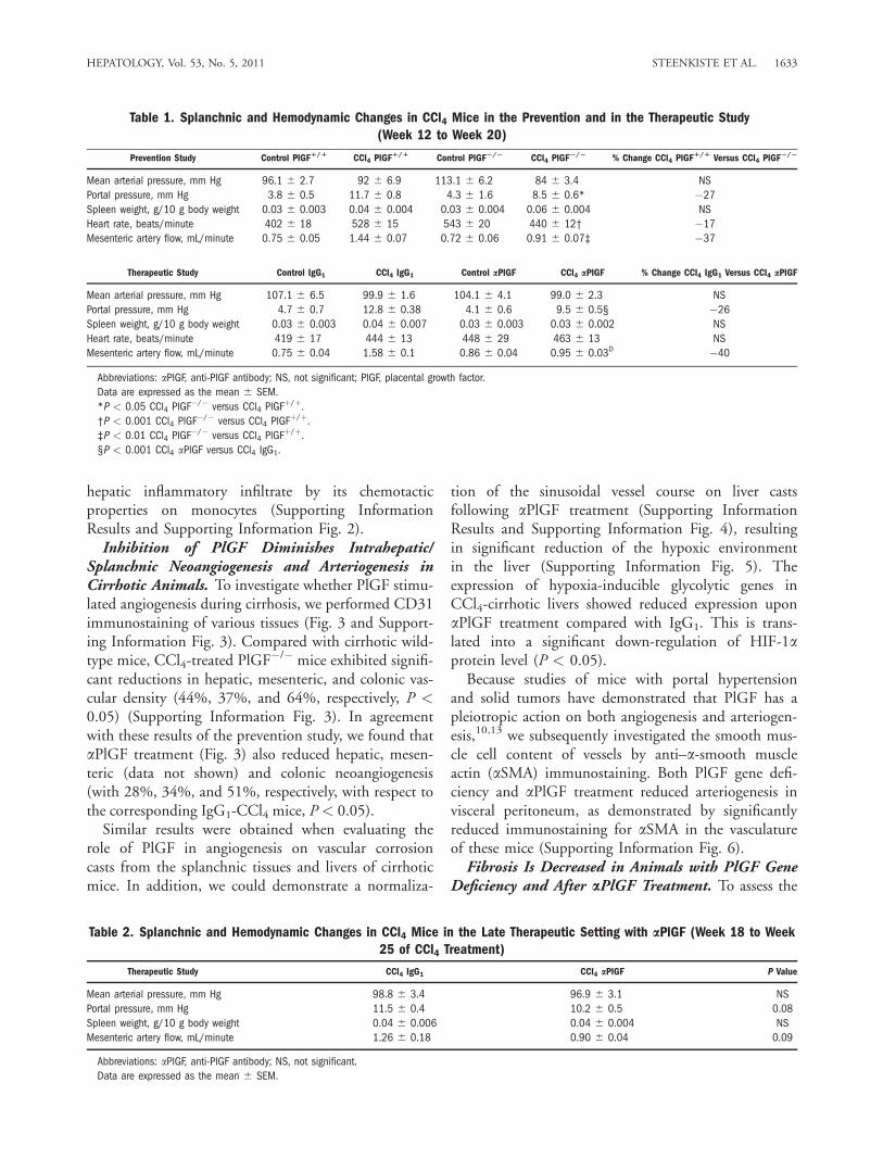
hepatic inflammatory infiltrate by its chemotacticproperties on monocytes (Supporting InformationResults and Supporting Information Fig. 2).Inhibition of PlGF Diminishes Intrahepatic/
Splanchnic Neoangiogenesis and Arteriogenesis inCirrhotic Animals. To investigate whether PlGF stimu-lated angiogenesis during cirrhosis, we performed CD31immunostaining of various tissues (Fig. 3 and Support-ing Information Fig. 3). Compared with cirrhotic wild-type mice, CCl4-treated PlGF
�/� mice exhibited signifi-cant reductions in hepatic, mesenteric, and colonic vas-cular density (44%, 37%, and 64%, respectively, P <0.05) (Supporting Information Fig. 3). In agreementwith these results of the prevention study, we found thataPlGF treatment (Fig. 3) also reduced hepatic, mesen-teric (data not shown) and colonic neoangiogenesis(with 28%, 34%, and 51%, respectively, with respect tothe corresponding IgG1-CCl4 mice, P < 0.05).Similar results were obtained when evaluating the
role of PlGF in angiogenesis on vascular corrosioncasts from the splanchnic tissues and livers of cirrhoticmice. In addition, we could demonstrate a normaliza-
tion of the sinusoidal vessel course on liver castsfollowing aPlGF treatment (Supporting InformationResults and Supporting Information Fig. 4), resultingin significant reduction of the hypoxic environmentin the liver (Supporting Information Fig. 5). Theexpression of hypoxia-inducible glycolytic genes inCCl4-cirrhotic livers showed reduced expression uponaPlGF treatment compared with IgG1. This is trans-lated into a significant down-regulation of HIF-1aprotein level (P < 0.05).Because studies of mice with portal hypertension
and solid tumors have demonstrated that PlGF has apleiotropic action on both angiogenesis and arteriogen-esis,10,13 we subsequently investigated the smooth mus-cle cell content of vessels by anti–a-smooth muscleactin (aSMA) immunostaining. Both PlGF gene defi-ciency and aPlGF treatment reduced arteriogenesis invisceral peritoneum, as demonstrated by significantlyreduced immunostaining for aSMA in the vasculatureof these mice (Supporting Information Fig. 6).Fibrosis Is Decreased in Animals with PlGF Gene
Deficiency and After aPlGF Treatment. To assess the
Table 1. Splanchnic and Hemodynamic Changes in CCl4 Mice in the Prevention and in the Therapeutic Study(Week 12 to Week 20)
Prevention Study Control PlGF1/1 CCl4 PlGF1/1 Control PlGF2/2 CCl4 PlGF
2/2 % Change CCl4 PlGF1/1 Versus CCl4 PlGF
2/2
Mean arterial pressure, mm Hg 96.1 6 2.7 92 6 6.9 113.1 6 6.2 84 6 3.4 NS
Portal pressure, mm Hg 3.8 6 0.5 11.7 6 0.8 4.3 6 1.6 8.5 6 0.6* �27
Spleen weight, g/10 g body weight 0.03 6 0.003 0.04 6 0.004 0.03 6 0.004 0.06 6 0.004 NS
Heart rate, beats/minute 402 6 18 528 6 15 543 6 20 440 6 12† �17
Mesenteric artery flow, mL/minute 0.75 6 0.05 1.44 6 0.07 0.72 6 0.06 0.91 6 0.07‡ �37
Therapeutic Study Control IgG1 CCl4 IgG1 Control aPlGF CCl4 aPlGF % Change CCl4 IgG1 Versus CCl4 aPlGF
Mean arterial pressure, mm Hg 107.1 6 6.5 99.9 6 1.6 104.1 6 4.1 99.0 6 2.3 NS
Portal pressure, mm Hg 4.7 6 0.7 12.8 6 0.38 4.1 6 0.6 9.5 6 0.5§ �26
Spleen weight, g/10 g body weight 0.03 6 0.003 0.04 6 0.007 0.03 6 0.003 0.03 6 0.002 NS
Heart rate, beats/minute 419 6 17 444 6 13 448 6 29 463 6 13 NS
Mesenteric artery flow, mL/minute 0.75 6 0.04 1.58 6 0.1 0.86 6 0.04 0.95 6 0.03D �40
Abbreviations: aPlGF, anti-PlGF antibody; NS, not significant; PlGF, placental growth factor.
Data are expressed as the mean 6 SEM.
*P < 0.05 CCl4 PlGF�/� versus CCl4 PlGFþ/þ.†P < 0.001 CCl4 PlGF�/� versus CCl4 PlGFþ/þ.‡P < 0.01 CCl4 PlGF�/� versus CCl4 PlGFþ/þ.§P < 0.001 CCl4 aPlGF versus CCl4 IgG1.
Table 2. Splanchnic and Hemodynamic Changes in CCl4 Mice in the Late Therapeutic Setting with aPlGF (Week 18 to Week25 of CCl4 Treatment)
Therapeutic Study CCl4 IgG1 CCl4 aPlGF P Value
Mean arterial pressure, mm Hg 98.8 6 3.4 96.9 6 3.1 NS
Portal pressure, mm Hg 11.5 6 0.4 10.2 6 0.5 0.08
Spleen weight, g/10 g body weight 0.04 6 0.006 0.04 6 0.004 NS
Mesenteric artery flow, mL/minute 1.26 6 0.18 0.90 6 0.04 0.09
Abbreviations: aPlGF, anti-PlGF antibody; NS, not significant.
Data are expressed as the mean 6 SEM.
HEPATOLOGY, Vol. 53, No. 5, 2011 STEENKISTE ET AL. 1633
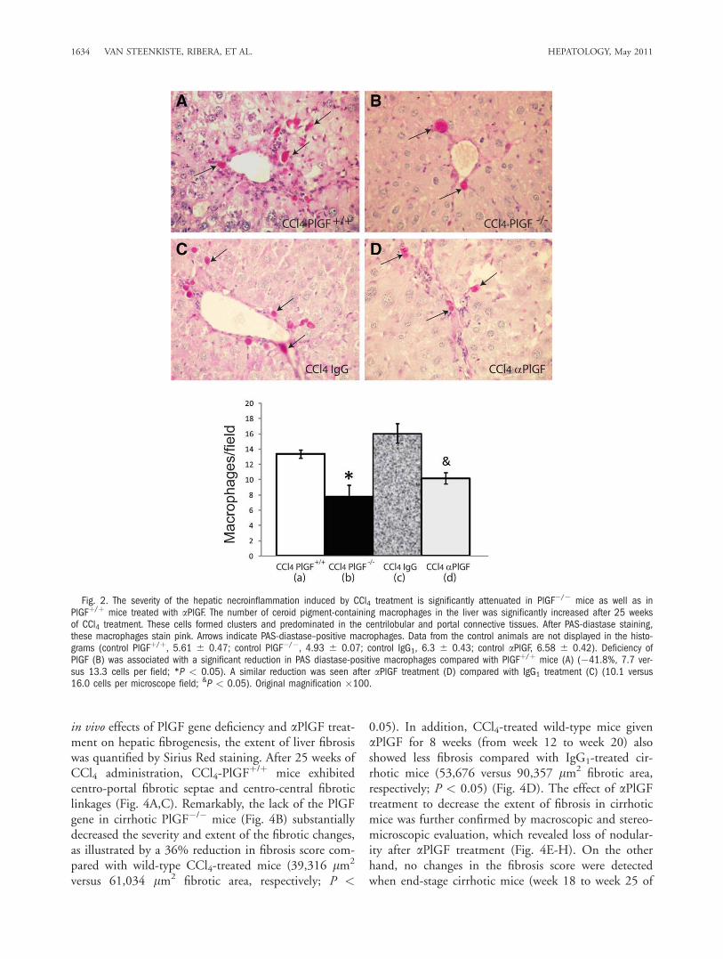
in vivo effects of PlGF gene deficiency and aPlGF treat-ment on hepatic fibrogenesis, the extent of liver fibrosiswas quantified by Sirius Red staining. After 25 weeks ofCCl4 administration, CCl4-PlGF
þ/þ mice exhibitedcentro-portal fibrotic septae and centro-central fibroticlinkages (Fig. 4A,C). Remarkably, the lack of the PlGFgene in cirrhotic PlGF�/� mice (Fig. 4B) substantiallydecreased the severity and extent of the fibrotic changes,as illustrated by a 36% reduction in fibrosis score com-pared with wild-type CCl4-treated mice (39,316 lm2
versus 61,034 lm2 fibrotic area, respectively; P <
0.05). In addition, CCl4-treated wild-type mice givenaPlGF for 8 weeks (from week 12 to week 20) alsoshowed less fibrosis compared with IgG1-treated cir-rhotic mice (53,676 versus 90,357 lm2 fibrotic area,respectively; P < 0.05) (Fig. 4D). The effect of aPlGFtreatment to decrease the extent of fibrosis in cirrhoticmice was further confirmed by macroscopic and stereo-microscopic evaluation, which revealed loss of nodular-ity after aPlGF treatment (Fig. 4E-H). On the otherhand, no changes in the fibrosis score were detectedwhen end-stage cirrhotic mice (week 18 to week 25 of
Fig. 2. The severity of the hepatic necroinflammation induced by CCl4 treatment is significantly attenuated in PlGF�/� mice as well as inPlGFþ/þ mice treated with aPlGF. The number of ceroid pigment-containing macrophages in the liver was significantly increased after 25 weeksof CCl4 treatment. These cells formed clusters and predominated in the centrilobular and portal connective tissues. After PAS-diastase staining,these macrophages stain pink. Arrows indicate PAS-diastase–positive macrophages. Data from the control animals are not displayed in the histo-grams (control PlGFþ/þ, 5.61 6 0.47; control PlGF�/�, 4.93 6 0.07; control IgG1, 6.3 6 0.43; control aPlGF, 6.58 6 0.42). Deficiency ofPlGF (B) was associated with a significant reduction in PAS diastase-positive macrophages compared with PlGFþ/þ mice (A) (�41.8%, 7.7 ver-sus 13.3 cells per field; *P < 0.05). A similar reduction was seen after aPlGF treatment (D) compared with IgG1 treatment (C) (10.1 versus16.0 cells per microscope field; &P < 0.05). Original magnification �100.
1634 VAN STEENKISTE, RIBERA, ET AL. HEPATOLOGY, May 2011
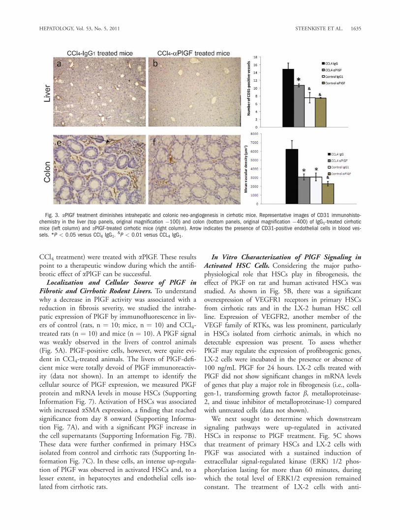
CCl4 treatment) were treated with aPlGF. These resultspoint to a therapeutic window during which the antifi-brotic effect of aPlGF can be successful.Localization and Cellular Source of PlGF in
Fibrotic and Cirrhotic Rodent Livers. To understandwhy a decrease in PlGF activity was associated with areduction in fibrosis severity, we studied the intrahe-patic expression of PlGF by immunofluorescence in liv-ers of control (rats, n ¼ 10; mice, n ¼ 10) and CCl4-treated rats (n ¼ 10) and mice (n ¼ 10). A PlGF signalwas weakly observed in the livers of control animals(Fig. 5A). PlGF-positive cells, however, were quite evi-dent in CCl4-treated animals. The livers of PlGF-defi-cient mice were totally devoid of PlGF immunoreactiv-ity (data not shown). In an attempt to identify thecellular source of PlGF expression, we measured PlGFprotein and mRNA levels in mouse HSCs (SupportingInformation Fig. 7). Activation of HSCs was associatedwith increased aSMA expression, a finding that reachedsignificance from day 8 onward (Supporting Informa-tion Fig. 7A), and with a significant PlGF increase inthe cell supernatants (Supporting Information Fig. 7B).These data were further confirmed in primary HSCsisolated from control and cirrhotic rats (Supporting In-formation Fig. 7C). In these cells, an intense up-regula-tion of PlGF was observed in activated HSCs and, to alesser extent, in hepatocytes and endothelial cells iso-lated from cirrhotic rats.
In Vitro Characterization of PlGF Signaling inActivated HSC Cells. Considering the major patho-physiological role that HSCs play in fibrogenesis, theeffect of PlGF on rat and human activated HSCs wasstudied. As shown in Fig. 5B, there was a significantoverexpression of VEGFR1 receptors in primary HSCsfrom cirrhotic rats and in the LX-2 human HSC cellline. Expression of VEGFR2, another member of theVEGF family of RTKs, was less prominent, particularlyin HSCs isolated from cirrhotic animals, in which nodetectable expression was present. To assess whetherPlGF may regulate the expression of profibrogenic genes,LX-2 cells were incubated in the presence or absence of100 ng/mL PlGF for 24 hours. LX-2 cells treated withPlGF did not show significant changes in mRNA levelsof genes that play a major role in fibrogenesis (i.e., colla-gen-1, transforming growth factor b, metalloproteinase-2, and tissue inhibitor of metalloproteinase-1) comparedwith untreated cells (data not shown).We next sought to determine which downstream
signaling pathways were up-regulated in activatedHSCs in response to PlGF treatment. Fig. 5C showsthat treatment of primary HSCs and LX-2 cells withPlGF was associated with a sustained induction ofextracellular signal-regulated kinase (ERK) 1/2 phos-phorylation lasting for more than 60 minutes, duringwhich the total level of ERK1/2 expression remainedconstant. The treatment of LX-2 cells with anti-
Fig. 3. aPlGF treatment diminishes intrahepatic and colonic neo-angiogenesis in cirrhotic mice. Representative images of CD31 immunohisto-chemistry in the liver (top panels, original magnification �100) and colon (bottom panels, original magnification �400) of IgG1-treated cirrhoticmice (left column) and aPlGF-treated cirrhotic mice (right column). Arrow indicates the presence of CD31-positive endothelial cells in blood ves-sels. *P < 0.05 versus CCl4 IgG1.
&P < 0.01 versus CCL4 IgG1.
HEPATOLOGY, Vol. 53, No. 5, 2011 STEENKISTE ET AL. 1635
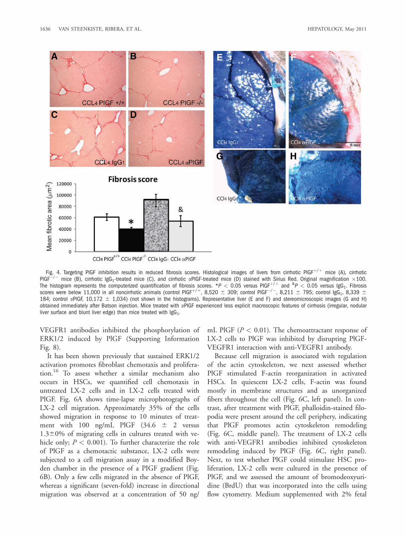
VEGFR1 antibodies inhibited the phosphorylation ofERK1/2 induced by PlGF (Supporting InformationFig. 8).It has been shown previously that sustained ERK1/2
activation promotes fibroblast chemotaxis and prolifera-tion.16 To assess whether a similar mechanism alsooccurs in HSCs, we quantified cell chemotaxis inuntreated LX-2 cells and in LX-2 cells treated withPlGF. Fig. 6A shows time-lapse microphotographs ofLX-2 cell migration. Approximately 35% of the cellsshowed migration in response to 10 minutes of treat-ment with 100 ng/mL PlGF (34.6 6 2 versus1.360% of migrating cells in cultures treated with ve-hicle only; P < 0.001). To further characterize the roleof PlGF as a chemotactic substance, LX-2 cells weresubjected to a cell migration assay in a modified Boy-den chamber in the presence of a PIGF gradient (Fig.6B). Only a few cells migrated in the absence of PlGF,whereas a significant (seven-fold) increase in directionalmigration was observed at a concentration of 50 ng/
mL PlGF (P < 0.01). The chemoattractant response ofLX-2 cells to PlGF was inhibited by disrupting PlGF-VEGFR1 interaction with anti-VEGFR1 antibody.Because cell migration is associated with regulation
of the actin cytoskeleton, we next assessed whetherPlGF stimulated F-actin reorganization in activatedHSCs. In quiescent LX-2 cells, F-actin was foundmostly in membrane structures and as unorganizedfibers throughout the cell (Fig. 6C, left panel). In con-trast, after treatment with PlGF, phalloidin-stained filo-podia were present around the cell periphery, indicatingthat PlGF promotes actin cytoskeleton remodeling(Fig. 6C, middle panel). The treatment of LX-2 cellswith anti-VEGFR1 antibodies inhibited cytoskeletonremodeling induced by PlGF (Fig. 6C, right panel).Next, to test whether PlGF could stimulate HSC pro-liferation, LX-2 cells were cultured in the presence ofPlGF, and we assessed the amount of bromodeoxyuri-dine (BrdU) that was incorporated into the cells usingflow cytometry. Medium supplemented with 2% fetal
Fig. 4. Targeting PlGF inhibition results in reduced fibrosis scores. Histological images of livers from cirrhotic PlGFþ/þ mice (A), cirrhoticPlGF�/� mice (B), cirrhotic IgG1-treated mice (C), and cirrhotic aPlGF-treated mice (D) stained with Sirius Red. Original magnification �100.The histogram represents the computerized quantification of fibrosis scores. *P < 0.05 versus PlGFþ/þ and &P < 0.05 versus IgG1. Fibrosisscores were below 11,000 in all noncirrhotic animals (control PlGFþ/þ, 8,520 6 309; control PlGF�/�, 8,211 6 795; control IgG1, 8,339 6184; control aPlGF, 10,172 6 1,034) (not shown in the histograms). Representative liver (E and F) and stereomicroscopic images (G and H)obtained immediately after Batson injection. Mice treated with aPlGF experienced less explicit macroscopic features of cirrhosis (irregular, nodularliver surface and blunt liver edge) than mice treated with IgG1.
1636 VAN STEENKISTE, RIBERA, ET AL. HEPATOLOGY, May 2011
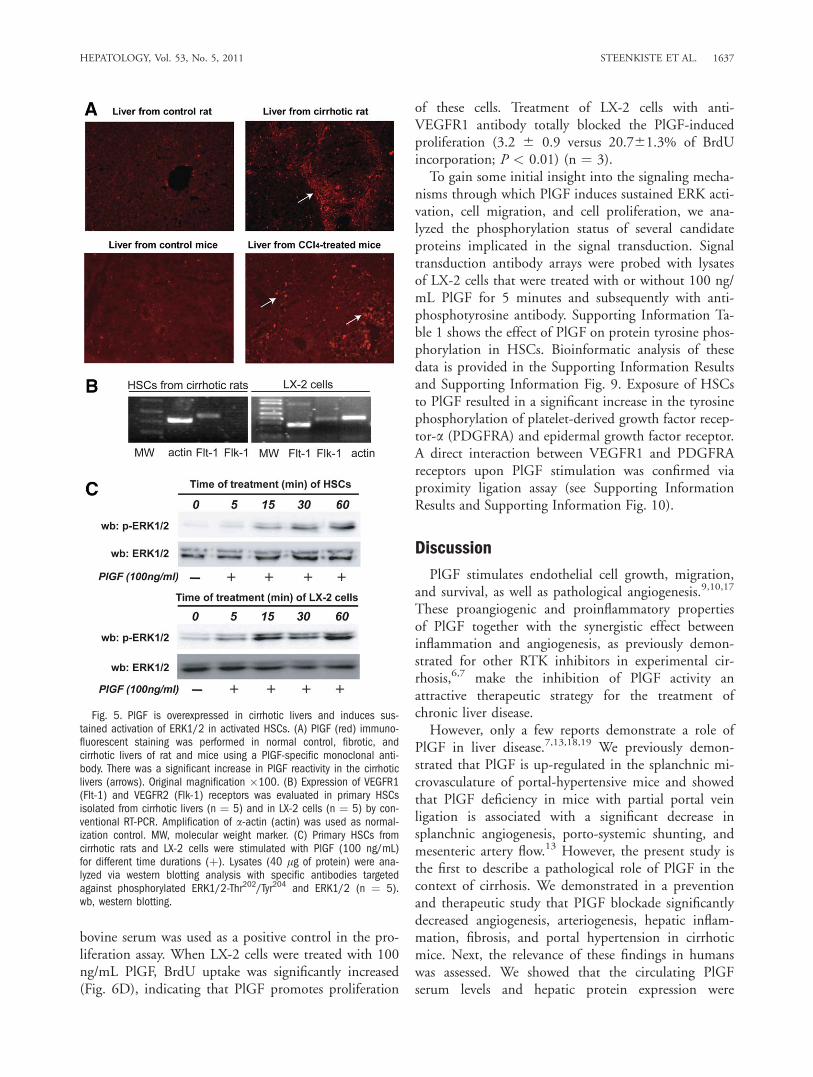
bovine serum was used as a positive control in the pro-liferation assay. When LX-2 cells were treated with 100ng/mL PlGF, BrdU uptake was significantly increased(Fig. 6D), indicating that PlGF promotes proliferation
of these cells. Treatment of LX-2 cells with anti-VEGFR1 antibody totally blocked the PlGF-inducedproliferation (3.2 6 0.9 versus 20.761.3% of BrdUincorporation; P < 0.01) (n ¼ 3).To gain some initial insight into the signaling mecha-
nisms through which PlGF induces sustained ERK acti-vation, cell migration, and cell proliferation, we ana-lyzed the phosphorylation status of several candidateproteins implicated in the signal transduction. Signaltransduction antibody arrays were probed with lysatesof LX-2 cells that were treated with or without 100 ng/mL PlGF for 5 minutes and subsequently with anti-phosphotyrosine antibody. Supporting Information Ta-ble 1 shows the effect of PlGF on protein tyrosine phos-phorylation in HSCs. Bioinformatic analysis of thesedata is provided in the Supporting Information Resultsand Supporting Information Fig. 9. Exposure of HSCsto PlGF resulted in a significant increase in the tyrosinephosphorylation of platelet-derived growth factor recep-tor-a (PDGFRA) and epidermal growth factor receptor.A direct interaction between VEGFR1 and PDGFRAreceptors upon PlGF stimulation was confirmed viaproximity ligation assay (see Supporting InformationResults and Supporting Information Fig. 10).
Discussion
PlGF stimulates endothelial cell growth, migration,and survival, as well as pathological angiogenesis.9,10,17
These proangiogenic and proinflammatory propertiesof PlGF together with the synergistic effect betweeninflammation and angiogenesis, as previously demon-strated for other RTK inhibitors in experimental cir-rhosis,6,7 make the inhibition of PlGF activity anattractive therapeutic strategy for the treatment ofchronic liver disease.However, only a few reports demonstrate a role of
PlGF in liver disease.7,13,18,19 We previously demon-strated that PlGF is up-regulated in the splanchnic mi-crovasculature of portal-hypertensive mice and showedthat PlGF deficiency in mice with partial portal veinligation is associated with a significant decrease insplanchnic angiogenesis, porto-systemic shunting, andmesenteric artery flow.13 However, the present study isthe first to describe a pathological role of PlGF in thecontext of cirrhosis. We demonstrated in a preventionand therapeutic study that PIGF blockade significantlydecreased angiogenesis, arteriogenesis, hepatic inflam-mation, fibrosis, and portal hypertension in cirrhoticmice. Next, the relevance of these findings in humanswas assessed. We showed that the circulating PlGFserum levels and hepatic protein expression were
Fig. 5. PlGF is overexpressed in cirrhotic livers and induces sus-tained activation of ERK1/2 in activated HSCs. (A) PlGF (red) immuno-fluorescent staining was performed in normal control, fibrotic, andcirrhotic livers of rat and mice using a PlGF-specific monoclonal anti-body. There was a significant increase in PlGF reactivity in the cirrhoticlivers (arrows). Original magnification �100. (B) Expression of VEGFR1(Flt-1) and VEGFR2 (Flk-1) receptors was evaluated in primary HSCsisolated from cirrhotic livers (n ¼ 5) and in LX-2 cells (n ¼ 5) by con-ventional RT-PCR. Amplification of a-actin (actin) was used as normal-ization control. MW, molecular weight marker. (C) Primary HSCs fromcirrhotic rats and LX-2 cells were stimulated with PlGF (100 ng/mL)for different time durations (þ). Lysates (40 lg of protein) were ana-lyzed via western blotting analysis with specific antibodies targetedagainst phosphorylated ERK1/2-Thr202/Tyr204 and ERK1/2 (n ¼ 5).wb, western blotting.
HEPATOLOGY, Vol. 53, No. 5, 2011 STEENKISTE ET AL. 1637
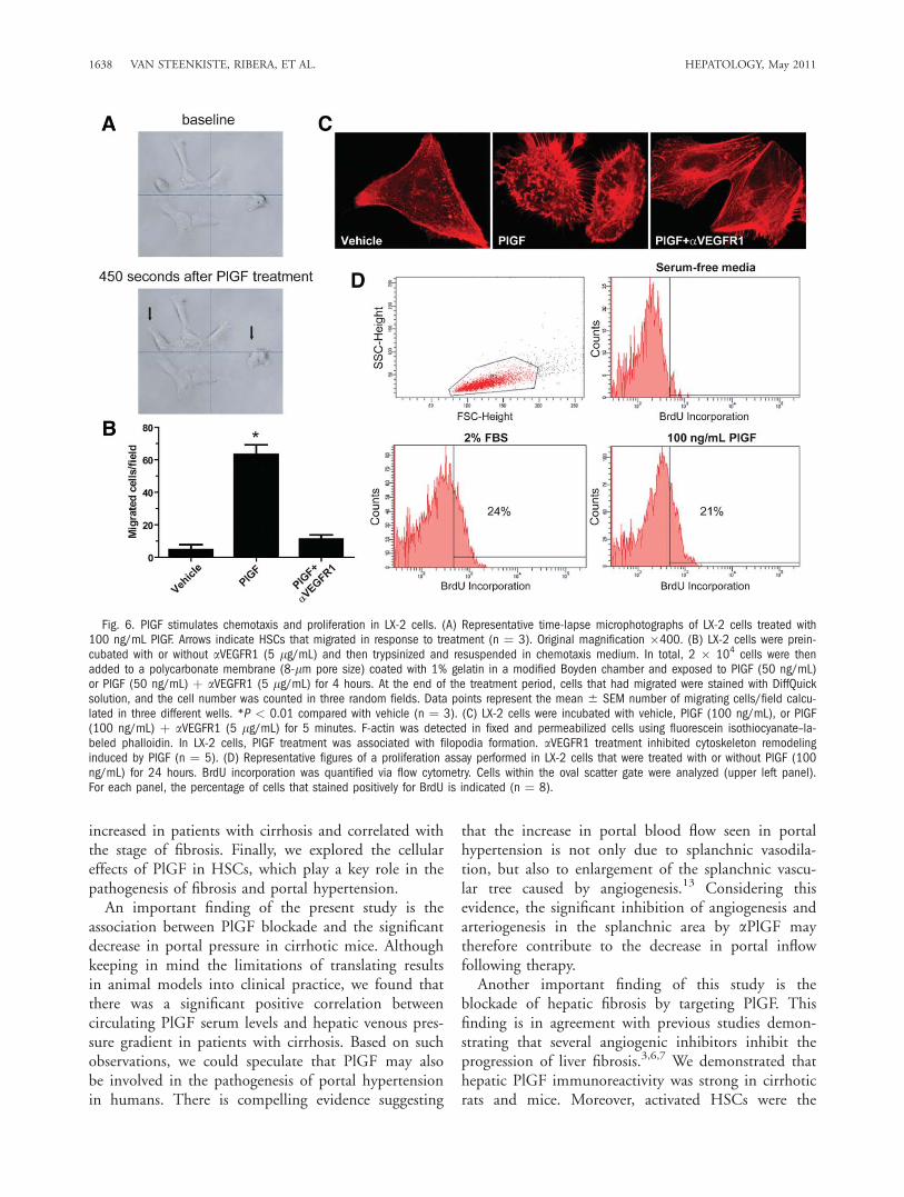
increased in patients with cirrhosis and correlated withthe stage of fibrosis. Finally, we explored the cellulareffects of PlGF in HSCs, which play a key role in thepathogenesis of fibrosis and portal hypertension.An important finding of the present study is the
association between PlGF blockade and the significantdecrease in portal pressure in cirrhotic mice. Althoughkeeping in mind the limitations of translating resultsin animal models into clinical practice, we found thatthere was a significant positive correlation betweencirculating PlGF serum levels and hepatic venous pres-sure gradient in patients with cirrhosis. Based on suchobservations, we could speculate that PlGF may alsobe involved in the pathogenesis of portal hypertensionin humans. There is compelling evidence suggesting
that the increase in portal blood flow seen in portalhypertension is not only due to splanchnic vasodila-tion, but also to enlargement of the splanchnic vascu-lar tree caused by angiogenesis.13 Considering thisevidence, the significant inhibition of angiogenesis andarteriogenesis in the splanchnic area by aPlGF maytherefore contribute to the decrease in portal inflowfollowing therapy.Another important finding of this study is the
blockade of hepatic fibrosis by targeting PlGF. Thisfinding is in agreement with previous studies demon-strating that several angiogenic inhibitors inhibit theprogression of liver fibrosis.3,6,7 We demonstrated thathepatic PlGF immunoreactivity was strong in cirrhoticrats and mice. Moreover, activated HSCs were the
Fig. 6. PlGF stimulates chemotaxis and proliferation in LX-2 cells. (A) Representative time-lapse microphotographs of LX-2 cells treated with100 ng/mL PlGF. Arrows indicate HSCs that migrated in response to treatment (n ¼ 3). Original magnification �400. (B) LX-2 cells were prein-cubated with or without aVEGFR1 (5 lg/mL) and then trypsinized and resuspended in chemotaxis medium. In total, 2 � 104 cells were thenadded to a polycarbonate membrane (8-lm pore size) coated with 1% gelatin in a modified Boyden chamber and exposed to PlGF (50 ng/mL)or PlGF (50 ng/mL) þ aVEGFR1 (5 lg/mL) for 4 hours. At the end of the treatment period, cells that had migrated were stained with DiffQuicksolution, and the cell number was counted in three random fields. Data points represent the mean 6 SEM number of migrating cells/field calcu-lated in three different wells. *P < 0.01 compared with vehicle (n ¼ 3). (C) LX-2 cells were incubated with vehicle, PlGF (100 ng/mL), or PlGF(100 ng/mL) þ aVEGFR1 (5 lg/mL) for 5 minutes. F-actin was detected in fixed and permeabilized cells using fluorescein isothiocyanate–la-beled phalloidin. In LX-2 cells, PlGF treatment was associated with filopodia formation. aVEGFR1 treatment inhibited cytoskeleton remodelinginduced by PlGF (n ¼ 5). (D) Representative figures of a proliferation assay performed in LX-2 cells that were treated with or without PlGF (100ng/mL) for 24 hours. BrdU incorporation was quantified via flow cytometry. Cells within the oval scatter gate were analyzed (upper left panel).For each panel, the percentage of cells that stained positively for BrdU is indicated (n ¼ 8).
1638 VAN STEENKISTE, RIBERA, ET AL. HEPATOLOGY, May 2011
major source of PlGF production in these rodents,and they exhibited substantial VEGFR1 expression.However, it is intriguing that although the blockade ofPlGF in vivo is antifibrogenic, we were unable to findsignificant changes in the expression of profibrogenicgenes when human activated HSCs were treated withPlGF. This discrepancy may be explained consideringthat PlGF promotes an angiogenic phenotype in HSCscharacterized by a sustained ERK1/2 phosphorylationas well as chemotaxis and proliferation. The acquisi-tion of an angiogenic phenotype by HSCs has beendescribed by others in response to PlGF and connectedto the enhanced HSCs coverage of sinousoid character-istic of cirrhotic livers.5 All of these changes result inabnormalities in hepatic blood vessels that compromisethe regulation of intrahepatic pressure and tissue perfu-sion. The sacculated and chaotically disorganizedappearance of the microvessels in the cirrhotic livers ofcontrol mice, as analyzed by the vascular corrosioncasts, is consistent with such vessel abnormalization.20
Interestingly, aPlGF treatment resulted in a partialnormalization of the three-dimensional architecture ofthe hepatic blood vessel network and induces a signifi-cant decrease of proinflammatory vasculature, which ischaracterized by the expression of vascular cell adhe-sion molecule 1. A similar mechanism of vessel nor-malization induced by aPlGF treatment was recentlydescribed in hepatocellular carcinoma nodules.10 Inter-estingly, a reduction in fibrosis was only demonstratedwhen mice were treated with aPlGF in the early phaseof cirrhosis induced by CCl4 treatment (from week 12to week 20). No significant beneficial effect wasobserved following aPlGF therapy in mice with end-stage cirrhosis induced by CCl4 (week 18 to week 25).This observation supports the idea that the acquisitionof an angiogenic phenotype by HSCs, in response toPlGF, causes an increase in the HSC population inearly phase of cirrhosis that correlates with the degreeof fibrosis. However, when the HSC populationreaches a critical mass, the therapeutic efficiency ofPlGF blockade is limited, because PlGF does not haveany effect on the regulation of profibrogenic genes. Inagreement with this hypothesis, it has been shown thatthe expression of angiogenic factors in fibrotic/cirrhoticlivers occurs mainly in areas of active fibrogenesis andnot in larger bridging septae or in end-stage cirrhotictissue.21 Therefore, this evidence points to a therapeu-tic window during which aPlGF treatment is effectiveat inhibiting and reducing fibrosis.The sustained ERK activation in response to PlGF
in HSCs prompted us to investigate the underlyingmechanisms, because VEGFR1 has a relatively weak
tyrosine kinase activity. Some authors also have sug-gested that VEGFR1 could function as a decoy recep-tor for VEGF-A, thereby amplifying the activity ofVEGF.12 However, HSCs did not express detectablelevels of VEGFR2, suggesting that VEGFR1’s roleextends beyond a mere decoy activity. Comparison ofthe protein tyrosine phosphorylation profile of acti-vated HSCs showed that PlGF induced the phospho-rylation of other tyrosine kinase receptors, includingPDGFRA and epidermal growth factor receptor. Thesefindings raise the intriguing possibility that upon PlGFactivation, VEGFR1 may amplify its own signaling byhighjacking other RTKs via a molecular association. Inour initial analysis, we identified PDGFRA as a candi-date of such molecular cross-talk that may furtherpotentiate sustained ERK activation. A similar cross-talk between VEGFR1 and VEGFR2, whereby PlGFamplifies VEGF-driven angiogenesis, has been docu-mented in endothelial cells.22 VEGFR1 also interactswith low-density lipoprotein receptor, that results inligand-independent activation of VEGFR1 by LDL.23
However, a molecular cross-talk between VEGFR1 andother types of RTKs, resulting in sustained signaling,has never been documented yet.Although antiangiogenic agents are frequently used
in the treatment of angiogenesis-related diseases, theirclinical use has been associated with adverse effects,such as hypertension, proteinuria, thrombosis, andreduced wound healing capacity. These adverse effectswarrant some caution to select angiogenic inhibitorsfor the treatment of patients with cirrhosis who arecritically ill. Studies in transgenic mice have shownthat loss of PlGF does not affect development, repro-duction, or normal postnatal health, but impairs path-ological angiogenesis in implanted and spontaneouslyarising cancer models.10 Moreover, administration ofaPlGF is not associated with vascular pruning inhealthy organs in mice,9 and is well tolerated inhumans, where phase I trials in healthy volunteers andpatients with solid tumors have thus far not revealedany major adverse effects.14,15 The present study con-firms the safety profile of aPlGF (Supporting Informa-tion Results and Supporting Information Fig. 11). Fur-thermore, aPlGF did not compensatorily up-regulatethe expression of VEGF; such up-regulation has beensuggested to represent a possible cause of resistance toantiangiogenic treatment (Supporting InformationResults and Supporting Information Fig. 11).In conclusion, this experimental study characterized
the pathophysiological mechanisms and moleculareffects that PlGF exerts on murine and human cir-rhotic livers and on HSCs. Blockade of the PlGF
HEPATOLOGY, Vol. 53, No. 5, 2011 STEENKISTE ET AL. 1639
pathway in cirrhotic mice by monoclonal antibodies orby genetic deficiency of PlGF decreased hepatic andmesenteric angiogenesis, mesenteric arterial blood flow,fibrosis, and inflammation, as well as portal pressure.Also because of its safety profile, aPlGF may be con-sidered as an attractive candidate for treating patientswith chronic liver disease.
Acknowledgment: We thank Julien Dupont andHuberte Moreau for technical assistance, Kin JipCheung for compiling the demographic data of thepatients, and Susana Kalko for technical assistancewith bioinformatic analysis. LX-2 cells were generouslysupplied by Scott L. Friedman; aPlGF was kindly pro-vided by ThromboGenics NV.
References1. Fernandez M, Semela D, Bruix J, Colle I, Pinzani M, Bosch J. Angio-genesis in liver disease. J Hepatol 2009;50:604-620.
2. Thabut D, Shah V. Intrahepatic angiogenesis and sinusoidal remodelingin chronic liver disease: new targets for the treatment of portal hyper-tension? J Hepatol 2010;53:976-980.
3. Taura K, De Minicis S, Seki E, Hatano E, Iwaisako K, OsterreicherCH, et al. Hepatic stellate cells secrete angiopoietin 1 that inducesangiogenesis in liver fibrosis. Gastroenterology 2008;135:1729-1738.
4. Wang YQ, Ikeda K, Ikebe T, Hirakawa K, Sowa M, Nakatani K, et al.Inhibition of hepatic stellate cell proliferation and activation by thesemisynthetic analogue of fumagillin TNP-470 in rats. HEPATOLOGY
2000;32:980-989.5. Semela D, Das A, Langer D, Kang N, Leof E, Shah V. Platelet-derivedgrowth factor signaling through ephrin-b2 regulates hepatic vascularstructure and function. Gastroenterology 2008;135:671-679.
6. Mejias M, Garcia-Pras E, Tiani C, Miquel R, Bosch J, Fernandez M.Beneficial effects of sorafenib on splanchnic, intrahepatic, and portocol-lateral circulations in portal hypertensive and cirrhotic rats. HEPATOLOGY
2009;49:1245-1256.7. Tugues S, Fernandez-Varo G, Munoz-Luque J, Ros J, Arroyo V, RodesJ, et al. Antiangiogenic treatment with sunitinib ameliorates inflamma-tory infiltrate, fibrosis, and portal pressure in cirrhotic rats. HEPATOLOGY
2007;46:1919-1926.8. Patsenker E, Popov Y, Stickel F, Schneider V, Ledermann M, SagesserH, et al. Pharmacological inhibition of integrin alphavbeta3 aggravatesexperimental liver fibrosis and suppresses hepatic angiogenesis. HEPATO-
LOGY 2009;50:1501-1511.9. Fischer C, Jonckx B, Mazzone M, Zacchigna S, Loges S, Pattarini L,et al. Anti-PlGF inhibits growth of VEGF(R)-inhibitor-resistant tumorswithout affecting healthy vessels. Cell 2007;131:463-475.
10. Van de Veire S, Stalmans I, Heindryckx F, Oura H, Tijeras-Raballand A,Schmidt T, et al. Further pharmacological and genetic evidence for the effi-cacy of PlGF inhibition in cancer and eye disease. Cell 2010;141:178-190.
11. DiPalma T, Tucci M, Russo G, Maglione D, Lago CT, Romano A,et al. The placenta growth factor gene of the mouse. Mamm Genome1996;7:6-12.
12. Autiero M, Luttun A, Tjwa M, Carmeliet P. Placental growth factorand its receptor, vascular endothelial growth factor receptor-1: noveltargets for stimulation of ischemic tissue revascularization and inhibitionof angiogenic and inflammatory disorders. J Thromb Haemost 2003;1:1356-1370.
13. Van Steenkiste C, Geerts A, Vanheule E, Van Vlierberghe H, De Vos F,Olievier K, et al. Role of placental growth factor in mesenteric neoan-giogenesis in a mouse model of portal hypertension. Gastroenterology2009;137:2112-2124.
14. Lassen U, Nielsen D, Sorensen M, Ronnengart E, Eldrup K, BentzonK, et al. A phase I, dose escalation study of TB-403, a monoclonalantibody directed against PlGF, in patients with solid tumors[Abstract]. Mol Cancer Ther 2009;8:A111.
15. Riisbro R, Larsson L, Winsted L, Niskanen T, Pakola S, Stassen JM,et al. A first-in-man phase I dose escalation study of TB403, a mono-clonal antibody directed against PlGF in healthy male subjects[Abstract]. Mol Cancer Ther 2009;8:A3.
16. Eliceiri BP, Klemke R, Stromblad S, Cheresh DA. Integrin alphavbeta3requirement for sustained mitogen-activated protein kinase activity dur-ing angiogenesis. J Cell Biol 1998;140:1255-1263.
17. Khurana R, Moons L, Shafi S, Luttun A, Collen D, Martin JF, et al.Placental growth factor promotes atherosclerotic intimal thickening andmacrophage accumulation. Circulation 2005;111:2828-2836.
18. Huang XX, McCaughan GW, Shackel NA, Gorrell MD. Up-regulationof proproliferative genes and the ligand/receptor pair placental growthfactor and vascular endothelial growth factor receptor 1 in hepatitis Ccirrhosis. Liver Int 2007;27:960-968.
19. Salcedo Mora X, Sanz-Cameno P, Medina J, Martin-Vilchez S, Garcia-Buey L, Borque MJ, et al. Association between angiogenesis soluble fac-tors and disease progression markers in chronic hepatitis C patients.Rev Esp Enferm Dig 2005;97:699-706.
20. Van Steenkiste C, Trachet B, Casteleyn D, van Loo L, Van HoorebekeL, Segers P, et al. Vascular corrosion casting: analyzing wall shear stressin the portal vein and vascular abnormalities in portal hypertensive andcirrhotic rodents. Lab Invest 2010;90:1558-1572.
21. Aleffi S, Petrai I, Bertolani C, Parola M, Colombatto S, Novo E, et al.Upregulation of proinflammatory and proangiogenic cytokinesby leptin in human hepatic stellate cells. HEPATOLOGY 2005;42:1339-1348.
22. Autiero M, Waltenberger J, Communi D, Kranz A, Moons L, Lam-brechts D, et al. Role of PlGF in the intra- and intermolecular crosstalk between the VEGF receptors Flt1 and Flk1. Nat Med 2003;9:936-943.
23. Usui R, Shibuya M, Ishibashi S, Maru Y. Ligand-independent activa-tion of vascular endothelial growth factor receptor 1 by low-density lip-oprotein. EMBO Rep 2007;8:1155-1161.
1640 VAN STEENKISTE, RIBERA, ET AL. HEPATOLOGY, May 2011
Supplemental information
1. Supplemental methods
2. Supplemental results
3. Supplemental figure legends
4. Supplemental tables
1. Supplemental methods
Experimental models of cirrhosis. All animals were kept under constant temperature
and humidity in a 12-h controlled dark/light cycle. Mice and rats were fed ad libitum
on a standard pellet diet. Cirrhosis in mice was induced by subcutaneous injection of
CCl4 (1 mL/kg, twice a week) during the entire period of the study. Additionally, 5%
ethanol was added to the animals’ drinking water, as previously described (1). After 12
weeks of CCl4 treatment, mice developed micronodular cirrhosis (METAVIR score
F3/F4). Controls received subcutaneous injections of 1 mL/kg body weight of saline
(0.9%) over a corresponding period and no ethanol was added to their drinking water.
Cirrhosis in rats was induced by CCl4 inhalation, which was administered twice weekly.
Control and cirrhotic rats were supplied with a standard diet and drinking water
containing 0.3 g/L phenobarbital (Kern Pharma; Barcelona, Spain) (2). All experiments
were approved by the Ethics Committees of the Ghent University and the Hospital
Clínic-University of Barcelona
Human samples. Hepatic expression of PlGF was assessed in liver specimens that were
obtained by a transjugular approach from patients with alcoholic hepatitis (n=3) and by
a percutaneous approach in patients with chronic hepatitis C (n=23). Normal liver
specimens (n=6) were obtained from tissue fragments obtained following resections of
colon cancer metastases from the liver (localized at a considerable distance from the
metastases) prior to vascular clamping as previously described (3). Fibrosis (F0=none;
F1=mild; F2=moderate; F3=advanced; F4=cirrhosis) and inflammatory activity
(A0=none; A1=mild; A2=moderate; A3=severe) were evaluated on liver biopsy
according to the METAVIR scoring system (1). Co-infected patients were excluded from
the study. For analysis of serum PlGF levels, blood samples were obtained from
patients with chronic hepatitis C (n=5), non-alcoholic steatohepatitis (n=5) and
alcoholic hepatitis (n=32). Samples were also collected from healthy controls (n=19)
who were matched with the patients with respect to age, sex, and body mass index.
For PlGF immunohistochemistry (IHC), biopsy samples were obtained from patients
with hepatitis C (n = 20). Patients showing signs of decompensation, co-infection or
hepatocellular carcinoma (HCC) were excluded from the IHC study group. The study
protocol was conform to the ethical guidelines of the 1975 Declaration of Helsinki and
was approved by the Ethics Committees of the Ghent University Hospital and the
Hospital Clínic of Barcelona. All patients gave informed consent prior to participation in
the study.
Hemodynamic studies. The animals were anaesthetized with a mixture of ketamine
(100 mg/kg body weight, Ketalar®, Pfizer, Brussels, Belgium) and xylazine (10mg/kg
body weight, Rompun®, Bayer, Brussels, Belgium) and prepared for measurement of
hemodynamic parameters (arterial blood pressure (mmHg, MAP), portal pressure
(mmHg), superior mesenteric artery flow (ml/min)). All studies were performed in
overnight-fasted mice. Both the jugular vein and the carotid artery were cannulated
for administration of fluid and monitoring of the mean arterial blood pressure (mmHg)
(MAP), respectively. To evaluate portal hypertension, the portal pressure (mmHg) was
measured via cannulation of the ileocolic vein with a 24-gauge catheter (Becton
Dickinson, Erembodegem-Aalst, Belgium). All catheters were connected to highly
sensitive pressure transducers of a multichannel computer-based recorder (Powerlab,
ADInstruments, Spechbach, Germany) and analyzed with Chart5 (ADInstruments). An
ultrasonic blood flow probe (Transonic Systems Inc., Ithaca, NY, USA) was placed
around the superior mesenteric artery, allowing measurements of the superior
mesenteric artery blood flow (ml/min).
Vascular corrosion casting. Each cast was prepared as previously described (4) by the
injection of Batson’s #17 casting medium (Brunschwig chemie, Amsterdam, The
Netherlands) and analyzed with a stereomicroscope (Olympus SZX7, Olympus Belgium,
Aartselaar, Belgium) and a Jeol JSM 5600 LV scanning electron microscope (Jeol Ltd.,
Tokyo, Japan).
Histology (Sirius Red, PAS-diastase), immunohistochemistry (CD31, ,
immunofluorescence (PlGF and VCAM-1), cytology (phalloidin) and (computerized)
image analysis. For our histology and immunohistochemistry experiments, liver
samples were fixed in 10% buffered formaldehyde solution, dehydrated and
embedded in paraffin. Immunolabeling was performed using the following antibodies:
anti–mouse CD31 (Santa Cruz Biotechnology, Santa Cruz, USA), anti-PlGF (Abcam,
Cambridge, UK) and anti–mouse
cells were detected with the LSAB2 System HRP (Dako; Hamburg, Germany). Collagen
deposition was detected by Sirius Red staining. Visualization of macrophages with
active phagocytic activity was achieved with PAS-diastase staining. Sections were pre-
digested with or without diastase, incubated with periodic acid for 5 min and stained
with Schiff's reagent for 10 min. Quantification of PAS-positive cells was performed in
seven randomly selected fields for each mouse, and the mean value of the cell counts
in these seven fields was calculated. Immunoreactivity and liver histology were
visualized by light microscopy (Nikon Eclipse E600, Kawasaki, Kanagawa, Japan). The
vascular density of specimens stained for CD31 was measured quantitatively using light
microscopy and computerized image analysis (automatic color detection by Olympus
Cell software, Olympus Corporation, Tokyo, Japan) using an adapted international
consensus method for quantification of angiogenesis (5). Likewise, the amount of Sirius
Red–stained collagen fibers were analyzed and quantified by automatic color
detection. The double layer of visceral peritoneum suspending the jejunum and ileum
(referred to as “mesenteric tissues”) was also analyzed by immunohistochemistry.
Because vascular density can vary between different parts of the mesentery, the first
six loops proximal to the cecum were chosen for analysis. For the immunofluorescence
assays, tissues were fixed in 4% paraformaldehyde, cryoprotected overnight in a 20%
sucrose solution and embedded in optimal cutting temperature medium. Next, 8-μm
frozen sections were rehydrated, blocked with 5% normal goat serum and incubated
with anti-PlGF antibody (sc-1882; Santa Cruz Biotechnology, Santa Cruz, CA, USA) or rat
anti-mouse CD31 (BD Pharmingen, San Diego, CA, USA) or goat anti-mouse VCAM-1
(R&D Systems, Minneapolis, USA). Tissues for which immunostaining was performed
without primary antibodies was used as the negative control. The binding sites of the
primary antibodies were revealed with Alexa Fluor 555–conjugated donkey anti–goat
IgG (Molecular Probes, Invitrogen, San Diego, CA, USA) and Alexa Fluor 488-conjugated
donkey anti-rat IgG (Molecular Probes, Invitrogen, San Diego, CA, USA). Samples were
visualized with a fluorescence microscope (Nikon Eclipse E600, Kawasaki, Kanagawa,
Japan). For assessment of filopodia and cytoskeleton remodeling, LX-2 cells were
stimulated with 100 ng/mL PlGF and then fixed with 4% paraformaldehyde at 4°C for
10 min. Cells were then permeabilized with Triton 0.1% for 4 min at 4°C and incubated
with phalloidin (Sigma-Aldrich, St Louis, MO, USA) for 40 min at room temperature.
The cytoskeleton was visualized by confocal microscopy (Leica Microsystems, Wetzlar,
Germany). More than 25 cells per well were evaluated under the microscope.
AntibodyArray assay and identification of the protein phoshorylation. To profile the
protein phosphorylation under different conditions, LX-2 cells that were grown to
subconfluence were serum-starved overnight and incubated with or without 100ng/mL
PlGF (Acris Antibodies, Herford, Germany) for 5 min. Cell extracts were incubated for
2h at room temperature with the Signal Transduction AntibodyArrayTM filter
(Hypromatrix, Worcester, USA). Further procedures were carried out according to the
manufacturer’s instructions. The reactions were visualized using enhanced
chemiluminescence (ECL) (Amersham, GE Healthcare, Buckinghamshire, UK). Post-
scanning raw signal intensity data for the antibody arrays were obtained, background
corrections were processed with the gel analysis function of Image J software, and the
spot intensities were log2-transformed. The between-array variation was normalized
using the phosphotyrosine signals of proteins not affected by PlGF. For each protein, the
average expression across the arrayed samples (n=3 for vehicle and n=3 for PlGF
treatment) was calculated. Results are expressed as the n-fold change in the difference
in the mean normalized expression after PlGF treatment and the mean normalized
expression of basal tyrosine phosphorylation. Only changes that were larger than 2-
fold were considered for inclusion in the final analysis. Intracellular signaling pathways
activated by PlGF treatment were modeled using Ingenuity Pathway Software
(Ingenuity®Systems, Inc., Redwood City, USA). The resulting P-values obtained by the
Ingenuity Pathways Knowledge Base were adjusted for multiple comparisons using
Benjamini and Hochberg’s method.
Proximity ligation assay (PLA). LX-2 cells that were plated at a density of 2x10⁴
cells/well were seeded into 8-well chamber slides. After incubation for 24 h, cells were
serum-starved overnight in 0.1% bovine serum albumin. The following day, they were
stimulated with 100 ng/mL PlGF for 5 min at 37°C. Cultures were fixed in 4%
paraformaldehyde and subjected to PLA (Olink Bioscience, Uppsala, Sweden). Briefly,
slides were blocked and incubated with primary antibodies [goat anti-human VEGFR1
(AF321; R&D Systems, Minneapolis, USA) and rabbit anti-PDGFRA (sc-338; Santa Cruz)].
Secondary antibodies (anti-goat and anti-rabbit) that were conjugated to unique DNA
probes (Olink bioscience, Uppsala, Sweden) were then added. Ligation and
circularization of the DNA was followed by a rolling circle amplification step. These
reactions were detected by a complementary Cy3-labeled DNA linker. The cells were
counterstained with Alexa Fluor 488 conjugated to phalloidin (Molecular Probes-
Invitrogen; San Diego, CA, USA). Slides were mounted using Vectashield (Vector
Laboratories, Burlingame, CA, USA) and examines using an LSM 510 META confocal
microscope (Carl Zeiss, Oberkochen, Germany).
Quantitative RT-PCR (qPCR). Total RNA was extracted from frozen rat livers with Trizol
reagent (Life Technologies, Rockville, USA). One microgram of total RNA was reverse-
transcribed using the First Strand cDNA Synthesis Kit (Roche, Mannheim, Germany).
Then, cDNA samples were amplified for 30–35 cycles (94°C for 30 s, 55–60 °C for 30 s
and 72 °C for 1 min.) The specific primers used for cDNA amplification were:
1) PlGF-2 (human): 5’-GGGGGCCCTGCTACCTGTTCTTG-3’ (forward) and 5’-
CCCGGGGCCCACTCTGTATGT-3’ (reverse);
2) PlGF (mouse): 5’-ATGACATTTTCTCAGGATGTGCT-3’ (forward) and 5’-
GGTTCCTCAGTCTGTGAGTTTC-3’ (reverse);
3) PFKM (mouse): 5’-GCCGGCTCAGTGAGACAAG-3’ (forward) and 5’-
TGGCACCTTCAGCAACAATG-3’ (reverse)
4) GLUT1 (mouse): 5’-GCTTATGGGCTTCTCCAAACT-3’ (forward) and 5‘-
GGTGACACCTCTCCCACATAC-3’ (reverse)
5) VEGFR1 (rat): 5’-CCCAAGGCCTCAATGAAAATAGAC-3’ (forward) and 5’-
GAGCCGAGCCGTTGGGACTTAGAA-3’ (reverse);
6) VEGFR1 (human): 5’-CACGCTGAGCTGGAAAGGAAAATC-3’ (forward) and 5’-
CGGGGGTTGGAGCAGGGAAGTCAT-3’ (reverse);
7) VEGFR2 (rat): 5’-TCCCGTCCTCAAAGCATCAGCATA-3’ (forward) and 5’-
GCAGGGGAGGGTTGGCATAGA-3’ (reverse primer);
8) VEGFR2 (human): 5’-AGAGCCGGCCTGTGAGTGTAAAAA-3’ (forward) and 5’-
TCAGTGTGGTCCCCGAGTCAGG-3’ (reverse);
9 -actin (rat): 5’-TAAGGCCAACCGTGAAAAGATGAC-3’ (forward) and 5’-
ATTGCCGATATGGATGACCTG-3’ (reverse);
10 -actin (human): 5'-GTGACGTGGACATCCGCAAAGAC-3' (forward) and 5'-
AAGAAAGGGTGTAACGCAACTAAG-3' (reverse);
11) α-SMA (mouse): 5’-CCAGCACCATGAAGATCAAG-3’ (forward) and 5’-
TGGAAGGTAGACAGCGAAGC-3’ (reverse) and
12) HPRT (human): 5’-GGGGGCTATAAGTTCTTTGCTGAC-3’ (forward) and 5’-
CCTCCCATCTCCTTCATCACATCT-3’ (reverse)
For qPCR analysis,
examined in a 96-well plate using an Opticon real-time PCR machine (MJ Research,
Waltham, USA). Changes in human CollagenA1, MMP2, TGF-β and TIMP1 expression
after LX-2 treatment with PlGF (100 ng/mL) for 24 h, were examined in a 384-well
plate using a LightCycler®480 Real-Time PCR System (Roche Applied Science,
Indianapolis, USA). All qPCR reactions were performed in the presence of specific
primers and oligonucleotide probes (TaqMan probe, Applied Biosystems, Foster City,
CA, USA). The number of PCR cycles required for the fluorescence intensity to exceed a
pre-determined threshold was measured during qPCR. Quantification of the initial
amount of template molecules relied on this number of PCR cycles, which is termed
the cycle of threshold (Ct). For each sample, the amount of the target gene that was
present was determined using the appropriate standard curve. The n-fold changes in
the amount of the target gene were calculated by dividing target values from cirrhotic
samples by those from the averaged control samples. The expression of the
housekeeping gene hypoxanthine phosphoribosyltransferase (HPRT) or 18s was used
as an internal control for normalization of RNA quantity. For PlGF, GLUT1 and PFKM
real-time detection and qPCR quantification, we used SYBR Green (Roche, Mannheim,
Germany) using the stably expressed reference gene hydroxymethylbilane synthase
(HMBS). For relative quantification, the ��Ct method was used. Transcript abundance
was estimated taking into account the amplification efficiency of each primer set.
Primer efficiencies were determined by subjecting a dilution series of mouse reference
cDNA to qPCR analysis and using the formula (10(-1/slope) -1) x 100.
Cell culture. Hepatic stellate cells (HSC), hepatocytes and liver sinusoidal endothelial
cells from control and cirrhotic rats were isolated and cultured as previously described
(6, 7). The LX-2 cell line, which is derived from normal primary human HSCs that have
been immortalized and that are able to grow under reduced serum conditions, was
maintained as described previously (8). LX-2 cells express markers of activated HSCs
and have a similar phenotype to that of activated HSCs in vivo. The THP-1 cells, a
human acute monocytic leukemia cell line, were cultured in Roswell Park Memorial
Institute Medium (RPMI-1640) (Invitrogen, Grand Island, USA), supplemented with
10% fetal calf serum, 50 U/ml penicillin and 50 μg/ml streptomycin.
Enzyme-linked immunosorbent assay (ELISA) of PlGF, VEGF and Protein lysates
from liver tissues were obtained as previously described (9). Quantification of PlGF and
VEGF levels in serum and hepatic tissue was performed using specific ELISAs (R&D
Systems, Minneapolis, USA) according to the manufacturer’s instructions. To
determine the concentration of αPlGF in serum samples, ELISA plates (Sigma-Aldrich,
Saint Louis, USA) were coated with PlGF (0.5 μg/mL, R&D Systems), blocked with
bovine serum albumin (BSA), and incubated with serial dilutions of serum. Bound
αPlGF was detected using goat anti–mouse IgG1 conjugated to horseradish peroxidase
(HRP) (Sigma-Aldrich).
Western blot experiments. Cell and tissue lysates were prepared in lysis buffer, and
western blot analysis was performed as previously described (6) using anti–Erk1/2
mitogen-activated protein kinase (MAPK), anti–phospho-Erk1/2-Thr202/Tyr204 MAPK
(Cell Signaling Technology, Beverly, MA), anti-PlGF (sc-1882; Santa Cruz Biotechnology,
Santa Cruz, USA) antibodies and anti-HIF-1α (Calbiochem, Nottingham, United
Kingdom). LX-2 cells were preincubated for 1h with anti-human VEGFR1 (R&D Systems,
Minneapolis, USA) (25 μg/mL) in order to block PlGF signal, and stimulated with PlGF
(100 ng/mL) or PlGF (100 ng/mL) plus anti-human VEGFR1 (25 μg/mL) prior to the lysis.
Cell proliferation assay. The BrdU cell proliferation assay kit (BrdU Flow kit; BD
Pharmigen, San Jose, USA) was used to measure the incorporation of BrdU during DNA
synthesis following the manufacturer’s protocols. Briefly, LX-2 cells were fasted in
serum-free medium for 24 h. Following treatment with PlGF (100 ng/mL) for 24 h,
BrdU (10 �M) was added to the culture medium for 5 h. The BrdU-labeled cells were
then fixed and the DNA was denatured in fixative solution for 1 h at 37 °C. The cells
were then incubated with fluorescein isothiocyanate (FITC)–conjugated anti-BrdU
antibody for 20 minutes at room temperature. Immunofluorescence was detected by
flow cytometry (FACSCanto II; BD Bioscience, Bedford, MA, USA).
Cell migration assays. Cell migration was assessed by time-lapse recording of activated
HSC cell behaviour in response to PlGF treatment (100 ng/mL) at 37 °C, 5% CO2 and
21% O2. The time-lapse recording was done using a digital camera (Leica TCS SL,
Wetzlar, Germany) and an inverted Leica DMIRE2 microscope (Leica Microsystems,
Wetzlar, Germany). In addition, directional migration was also evaluated by Boyden
chamber assay (NeuroProbe, Gaithersburg, USA). Briefly, LX-2 cells were fasted in
serum-free Dulbecco’s Modified Eagle Medium (DMEM) (Invitrogen, Grand Island,
USA) and THP-1 cells in RPMI-1640 (Invitrogen, Grand Island, USA) for 24 h prior to the
migration experiment. LX-2 cells were preincubated for 1h with anti-human VEGFR1 (5
μg/mL). Cells were suspended and counted, subsequently 50 �l of cell suspension (4 ×
105 cells/mL for LX-2 or 1 × 10⁵ cells/ml for THP-1) was placed in the upper
compartment with a gelatin-coated polycarbonate membrane separating the two
chambers. PlGF (50 ng/mL), PlGF (50 ng/mL) plus anti-VEGFR1 (5 μg/mL) or the
chemotactic tripeptide fMet-Leu-Phe (10-7 M) were placed in the lower compartment
of the chamber. After 4 h of incubation at 37 °C, cells that were on the upper side of
the membrane were scraped off using a cotton swab and cells that had migrated to the
lower side of the filter were fixed with methanol. After staining with DiffQuick®
solution (Dade Behring Inc., Newark, USA), cells that had migrated into the lower side
were counted manually. The cells’ migration ability was expressed as the average cell
number in four random microscopic fields (Olympus BX45, Olympus Corporation,
Tokyo, Japan).
Statistical analysis: Data analysis was performed using SPSS version 17.0 (SPSS Inc,
Chicago, USA). See Supplemental Methods. In the case of normally distributed data,
groups were compared using Student's t-test or ANOVA (analysis of variance) for
independent samples. For other types of data, the Mann–Whitney U test or the
Kruskal–Wallis test was used. Data are presented as means ± standard error of the
mean (SEMs) or as medians ± ranges when indicated. Differences were considered to
be statistically significant when the P-value < 0.05 (two-tailed). The statistical
significance of the correlation between circulating levels of PlGF and the hepatic vein
pressure gradient (HVPG) was calculated using the Pearson Correlation Coefficient.
2. Supplemental results
Beneficial effects of anti-PlGF antibody (αPlGF) treatment on splanchnic hyperemia
and portal hypertension in cirrhotic mice. The serum concentration of αPlGF (trough
level) was in the therapeutic range (774 ± 94 μg/ml). Compared with IgG1 treatment
(CCl4 IgG1), 8 weeks of αPlGF treatment (CCl4
portal pressure (P<0.001) and a 40% reduction in mesenteric artery blood flow
(P<0.001) (Table 1). Treatment with αPlGF did not affect MAP, splenic weight or heart
rate. These results demonstrate that αPlGF treatment can partially reverse the portal
hypertensive syndrome.
Linking PlGF to inflammation. To further understand the link between PlGF blockade
and the reduction in inflammatory infiltrate, two different experimental approaches
were used. First, the expression of pro-inflammatory adhesion molecules in the
vasculature of cirrhotic mice was analyzed in absence or in presence of PlGF activity. It
has been reported previously that VCAM-1 is overexpressed in areas of
neovascularization and inflammatory infíltrate and, in addition, plays a major role in
leukocyte recruitment and vascular transmigration. As shown in Supplemental Figure
2A, VCAM-1 (red) was weakly expressed in the sinusoidal lining cells of control livers
and did not colocalize with blood vessels (green). In contrast, VCAM-1 immunostaining
in cirrhotic livers was stronger and colocalized well with the neovasculature (yellow
color in the merge panels). CCl4-treated PlGF-/- mice exhibited a significant reduction in
hepatic VCAM-1 expression, compared with CCl4-treated PlGF+/+ mice (4.9±2.5 % of
VCAM-1 positive area vs 13.1±1.9 %, respectively; P<0.05). Interestingly, a significant
number of hepatic blood vessels did not express VCAM-1 (10) in CCl4-treated PlGF-/- mice
(67±5.1%), compared with CCl4-treated PlGF+/+ mice (19±3.4%; P<0.05). In the second
approach, we investigate the chemoattractant response of monocytes to PlGF. Few
THP-1 cells, a human monocytic cell line, migrate in the absence of PlGF. However, a
significant 7-fold increase in directional migration of THP-1 cells was observed when
PlGF was added to the modified Boyden chamber (supplemental Figure 2B). The degree
of chemotaxis in response to PlGF was similar to the chemotaxis induced by
formylated chemotactic tripeptide fMet-Leu-Phe (fMLP), which was used as a positive
control.
Evaluating the role of PlGF in angiogenesis on vascular corrosion casting.
Stereomicroscopy and scanning electron-microscopic evaluation showed that control
IgG1-CCl4 mice displayed increased splanchnic angiogenesis, which was reflected by
increased vascular arborization forming honeycomb-like vascular networks
(Supplemental Figure 4, panels a, c and e). Although only sporadically present,
intussusceptive angiogenesis was noticed in IgG1-CCl4 mice (detail from panel e). In
contrast, vascular pruning and reduced vascular density were seen in αPlGF-treated
CCl4 mice (panels b, d and f; see also detail from panel f). Blood vessels were organized
in a nodular pattern in the livers of both IgG1- and αPlGF-treated CCl4 mice. However, a
striking difference was observed in the three-dimensional organization of the
sinusoids. Whereas the sinusoids of IgG1 cirrhotic mice had an irregular, disrupted,
bulging and saccular appearance (panels g and i), those of CCl4 mice treated with αPlGF
had evidence of more quiescent, highly organized trabecular sinusoidal perfusion in
which the course of the vessels was less disrupted (panels h and j). This hepatic
angioarchitecture was comparable to the “Christmas-tree” appearance that has been
described in livers of control mice (4). However, this sinusoidal vessel normalization
following αPlGF treatment was not uniformly distributed throughout the entire liver,
as scattered areas with disordered architecture were still present in other areas.
Analysis of Hypoxia-Inducible Genes and HIF-1α levels in IgG1 and αPlGF-treated CCl4-
exposed livers. Quantitative PCR of the expression of hypoxia-inducible glycolytic
genes in CCL4-cirrhotic livers showed reduced expression upon αPlGF treatment
compared to IgG1 (Supplemental Figure 5). These findings suggest that αPlGF-treated
livers may be less glycolytic because of improved oxygenation. In agreement with this
metabolic reprogramming, PlGF-blockage lowered GLUT-1 expression (P=0.05) and
phosphofructokinase-1 expression (P<0.05), the most important regulatory enzyme of
the glycolysis, suggesting that PlGF-blocked cirrhotic livers have a more oxidative
metabolism due to higher oxygenation. This is translated into a significant
downregulation of HIF-1α protein level (p < 0.05) (Supplemental Figure 5). The balance
between oxygen supply and demand in the liver seems to be restored.
Further characterization of PlGF signaling pathways. In accordance to our findings in
the proliferation and cell migration experiments (shown in Figure 6), bioinformatic
analysis of these data showed clustering of the phosphorylated proteins into two
functional networks: 1) cellular development, hematological system development and
cell viability; and 2) cancer, tumor morphology and cellular movement (Supplemental
Figure 9). Interestingly, we also found that exposure of HSCs to PlGF resulted in a
significant increase in the tyrosine phosphorylation of platelet-derived growth factor
receptor- epidermal growth factor receptor (EGFR). Supplemental
Figure 10A shows representative areas of the arrays that show reactivity for PDGFRA
and p-EGFR, but not for PDGFRB or for the unphosphorylated form of EGFR. Increased
tyrosine phosphorylation of EGFR and PDGFRA might result from a direct association
between these RTKs and VEGFR1. To further explore this possibility and to validate the
array results, the interaction of VEGFR1 and PDGFRA receptors was examined by
proximity ligation assay (PLA). As shown in Supplemental Figure 10B, at time zero of
VEGFR1 activation, VEGFR1 and PDGFRA were not close enough to produce a
significant PLA signal in LX-2 cells. However, after 5 min of PlGF treatment, a
substantial increase in the PLA signal was present. These results demonstrate that PlGF
induced an interaction between VEGFR1 and PDGFRA, enabling proximity ligation and
subsequent detection by hybridization of labeled oligonucleotide probes.
Safety profile and adaptive resistance to PlGF inhibition. Treatment with classical
angiogenesis inhibitors can be associated with hematological side effects characterized by
thrombosis, hypertension and microvascular pruning in healthy organs (11). In this
context, neither cirrhotic mice treated with αPlGF nor cirrhotic PlGF–/– mice exhibited
significant changes in the vascular density of quiescent vessels in organs in which
pathological angiogenesis was not present, such as the thyroid gland (Supplemental
Figure 11A). Another critical point in the clinical application of anti-angiogenic
medication is the potential development of resistance through compensatory
overexpression of other pro-angiogenic factors. To evaluate whether or not αPlGF

therapy was associated with the induction of alternative pro-angiogenic mechanisms,
mesenteric VEGF expression was evaluated in the context of PlGF blockade. As seen in
Supplemental Figure 11B, αPlGF treatment did not produce significant changes in the
expression of this key regulator of angiogenesis.
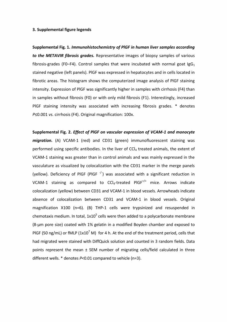
3. Supplemental figure legends
Supplemental Fig. 1. Immunohistochemistry of PlGF in human liver samples according
to the METAVIR fibrosis grades. Representative images of biopsy samples of various
fibrosis-grades (F0–F4). Control samples that were incubated with normal goat IgG1
stained negative (left panels). PlGF was expressed in hepatocytes and in cells located in
fibrotic areas. The histogram shows the computerized image analysis of PlGF staining
intensity. Expression of PlGF was significantly higher in samples with cirrhosis (F4) than
in samples without fibrosis (F0) or with only mild fibrosis (F1). Interestingly, increased
PlGF staining intensity was associated with increasing fibrosis grades. * denotes
P≤0.001 vs. cirrhosis (F4). Original magnification: 100x.
Supplemental Fig. 2. Effect of PlGF on vascular expression of VCAM-1 and monocyte
migration. (A) VCAM-1 (red) and CD31 (green) immunofluorescent staining was
performed using specific antibodies. In the liver of CCl4 treated animals, the extent of
VCAM-1 staining was greater than in control animals and was mainly expressed in the
vasculature as visualized by colocalization with the CD31 marker in the merge panels
(yellow). Deficiency of PlGF (PlGF -/-) was associated with a significant reduction in
VCAM-1 staining as compared to CCl4-treated PlGF+/+ mice. Arrows indicate
colocalization (yellow) between CD31 and VCAM-1 in blood vessels. Arrowheads indicate
absence of colocalization between CD31 and VCAM-1 in blood vessels. Original
magnification X100 (n=6). (B) THP-1 cells were trypsinized and resuspended in
chemotaxis medium. In total, 1x10⁵ cells were then added to a polycarbonate membrane
(8-μm pore size) coated with 1% gelatin in a modified Boyden chamber and exposed to
PlGF (50 ng/mL) or fMLP (1x10⁷ M) for 4 h. At the end of the treatment period, cells that
had migrated were stained with DiffQuick solution and counted in 3 random fields. Data
points represent the mean ± SEM number of migrating cells/field calculated in three
different wells. * denotes P<0.01 compared to vehicle (n=3).
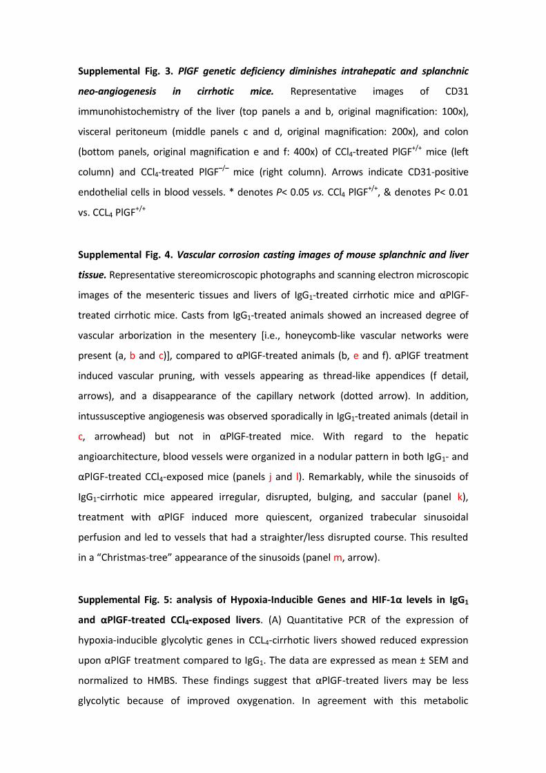
Supplemental Fig. 3. PlGF genetic deficiency diminishes intrahepatic and splanchnic
neo-angiogenesis in cirrhotic mice. Representative images of CD31
immunohistochemistry of the liver (top panels a and b, original magnification: 100x),
visceral peritoneum (middle panels c and d, original magnification: 200x), and colon
(bottom panels, original magnification e and f: 400x) of CCl4-treated PlGF+/+ mice (left
column) and CCl4-treated PlGF–/– mice (right column). Arrows indicate CD31-positive
endothelial cells in blood vessels. * denotes P< 0.05 vs. CCl4 PlGF+/+, & denotes P< 0.01
vs. CCL4 PlGF+/+
Supplemental Fig. 4. Vascular corrosion casting images of mouse splanchnic and liver
tissue. Representative stereomicroscopic photographs and scanning electron microscopic
images of the mesenteric tissues and livers of IgG1-treated cirrhotic mice and αPlGF-
treated cirrhotic mice. Casts from IgG1-treated animals showed an increased degree of
vascular arborization in the mesentery [i.e., honeycomb-like vascular networks were
present (a, b and c)], compared to αPlGF-treated animals (b, e and f). αPlGF treatment
induced vascular pruning, with vessels appearing as thread-like appendices (f detail,
arrows), and a disappearance of the capillary network (dotted arrow). In addition,
intussusceptive angiogenesis was observed sporadically in IgG1-treated animals (detail in
c, arrowhead) but not in αPlGF-treated mice. With regard to the hepatic
angioarchitecture, blood vessels were organized in a nodular pattern in both IgG1- and
αPlGF-treated CCl4-exposed mice (panels j and l). Remarkably, while the sinusoids of
IgG1-cirrhotic mice appeared irregular, disrupted, bulging, and saccular (panel k),
treatment with αPlGF induced more quiescent, organized trabecular sinusoidal
perfusion and led to vessels that had a straighter/less disrupted course. This resulted
in a “Christmas-tree” appearance of the sinusoids (panel m, arrow).
Supplemental Fig. 5: analysis of Hypoxia-Inducible Genes and HIF-1α levels in IgG1
and αPlGF-treated CCl4-exposed livers. (A) Quantitative PCR of the expression of
hypoxia-inducible glycolytic genes in CCL4-cirrhotic livers showed reduced expression
upon αPlGF treatment compared to IgG1. The data are expressed as mean ± SEM and
normalized to HMBS. These findings suggest that αPlGF-treated livers may be less
glycolytic because of improved oxygenation. In agreement with this metabolic
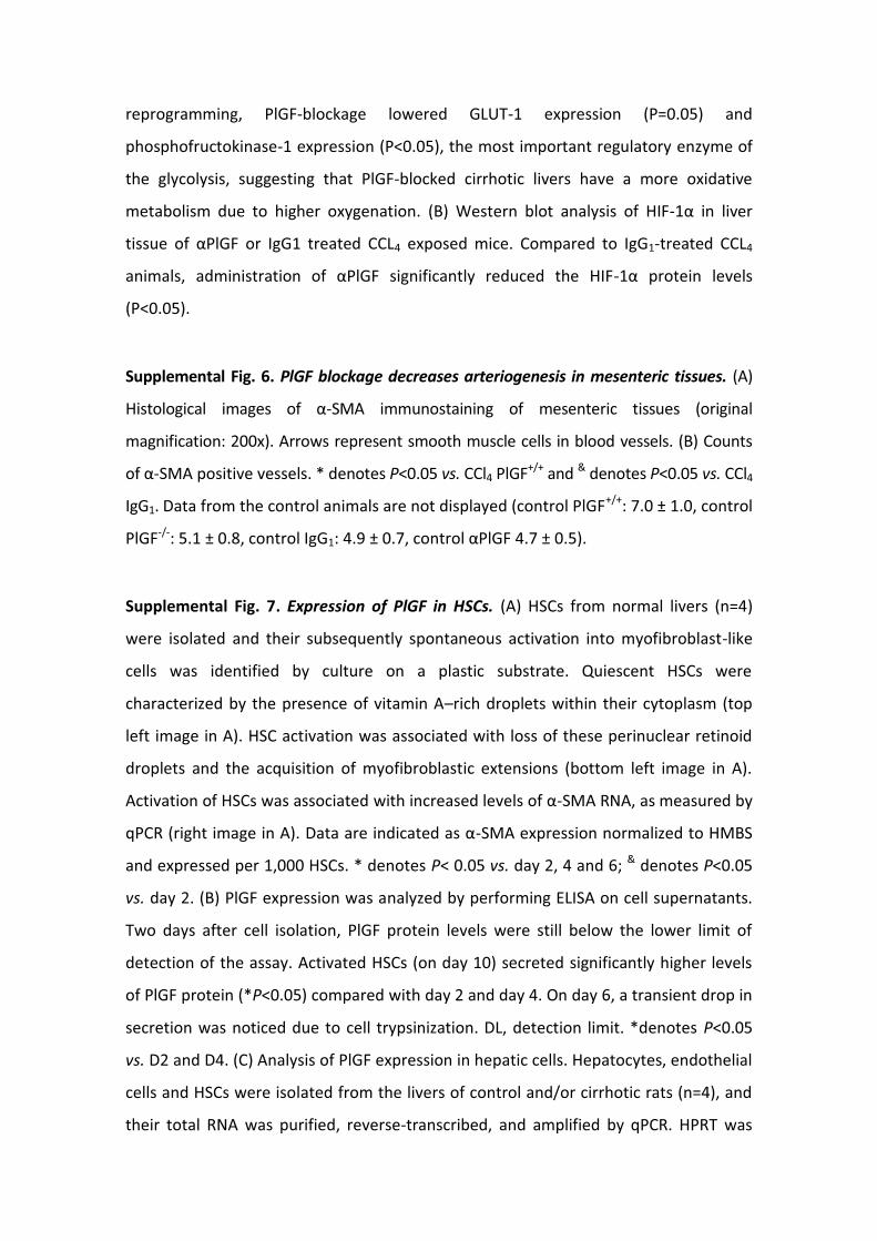
reprogramming, PlGF-blockage lowered GLUT-1 expression (P=0.05) and
phosphofructokinase-1 expression (P<0.05), the most important regulatory enzyme of
the glycolysis, suggesting that PlGF-blocked cirrhotic livers have a more oxidative
metabolism due to higher oxygenation. (B) Western blot analysis of HIF-1α in liver
tissue of αPlGF or IgG1 treated CCL4 exposed mice. Compared to IgG1-treated CCL4
animals, administration of αPlGF significantly reduced the HIF-1α protein levels
(P<0.05).
Supplemental Fig. 6. PlGF blockage decreases arteriogenesis in mesenteric tissues. (A)
Histological images of α-SMA immunostaining of mesenteric tissues (original
magnification: 200x). Arrows represent smooth muscle cells in blood vessels. (B) Counts
of α-SMA positive vessels. * denotes P<0.05 vs. CCl4 PlGF+/+ and & denotes P<0.05 vs. CCl4
IgG1. Data from the control animals are not displayed (control PlGF+/+: 7.0 ± 1.0, control
PlGF-/-: 5.1 ± 0.8, control IgG1: 4.9 ± 0.7, control αPlGF 4.7 ± 0.5).
Supplemental Fig. 7. Expression of PlGF in HSCs. (A) HSCs from normal livers (n=4)
were isolated and their subsequently spontaneous activation into myofibroblast-like
cells was identified by culture on a plastic substrate. Quiescent HSCs were
characterized by the presence of vitamin A–rich droplets within their cytoplasm (top
left image in A). HSC activation was associated with loss of these perinuclear retinoid
droplets and the acquisition of myofibroblastic extensions (bottom left image in A).
Activation of HSCs was associated with increased levels of α-SMA RNA, as measured by
qPCR (right image in A). Data are indicated as α-SMA expression normalized to HMBS
and expressed per 1,000 HSCs. * denotes P< 0.05 vs. day 2, 4 and 6; & denotes P<0.05
vs. day 2. (B) PlGF expression was analyzed by performing ELISA on cell supernatants.
Two days after cell isolation, PlGF protein levels were still below the lower limit of
detection of the assay. Activated HSCs (on day 10) secreted significantly higher levels
of PlGF protein (*P<0.05) compared with day 2 and day 4. On day 6, a transient drop in
secretion was noticed due to cell trypsinization. DL, detection limit. *denotes P<0.05
vs. D2 and D4. (C) Analysis of PlGF expression in hepatic cells. Hepatocytes, endothelial
cells and HSCs were isolated from the livers of control and/or cirrhotic rats (n=4), and
their total RNA was purified, reverse-transcribed, and amplified by qPCR. HPRT was
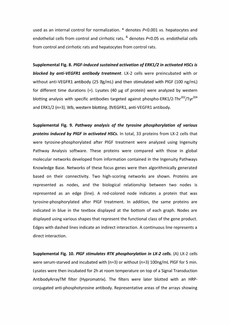
used as an internal control for normalization. * denotes P<0.001 vs. hepatocytes and
endothelial cells from control and cirrhotic rats. & denotes P<0.05 vs. endothelial cells
from control and cirrhotic rats and hepatocytes from control rats.
Supplemental Fig. 8. PlGF-induced sustained activation of ERK1/2 in activated HSCs is
blocked by anti-VEGFR1 antibody treatment. LX-2 cells were preincubated with or
without anti- PlGF (100 ng/mL)
for different time durations (+). Lysates (40 μg of protein) were analyzed by western
blotting analysis with specific antibodies targeted against phospho-ERK1/2-Thr202/Tyr204
and ERK1/2 (n=3). -VEGFR1 antibody.
Supplemental Fig. 9. Pathway analysis of the tyrosine phosphorylation of various
proteins induced by PlGF in activated HSCs. In total, 33 proteins from LX-2 cells that
were tyrosine-phosphorylated after PlGF treatment were analyzed using Ingenuity
Pathway Analysis software. These proteins were compared with those in global
molecular networks developed from information contained in the Ingenuity Pathways
Knowledge Base. Networks of these focus genes were then algorithmically generated
based on their connectivity. Two high-scoring networks are shown. Proteins are
represented as nodes, and the biological relationship between two nodes is
represented as an edge (line). A red-colored node indicates a protein that was
tyrosine-phosphorylated after PlGF treatment. In addition, the same proteins are
indicated in blue in the textbox displayed at the bottom of each graph. Nodes are
displayed using various shapes that represent the functional class of the gene product.
Edges with dashed lines indicate an indirect interaction. A continuous line represents a
direct interaction.
Supplemental Fig. 10. PlGF stimulates RTK phosphorylation in LX-2 cells. (A) LX-2 cells
were serum-starved and incubated with (n=3) or without (n=3) 100ng/mL PlGF for 5 min.
Lysates were then incubated for 2h at room temperature on top of a Signal Transduction
AntibodyArrayTM filter (Hypromatrix). The filters were later blotted with an HRP-
conjugated anti-phosphotyrosine antibody. Representative areas of the arrays showing
significantly increased intensity of the phosphotyrosine signals in the spots containing
antibodies against PDGFRA (these antibodies recognize both the unphosphorylated
and phosphorylated forms) and phospho-EGFR. The phosphotyrosine signal of cyclin D3
was used to normalize the results because PlGF does not affect its level of
phosphorylation. (B) In situ detection of VEGFR1/PDGFRA complexes by PLA (red spots) in
LX-2 cells before and after stimulation with PlGF for varying time periods. The cells were
counterstained with Alexa Fluor 488 conjugated to phalloidin (green) to visualize the cell
borders and with Hoechst 33258 (blue) to visualize the nuclei. Similar results were
obtained in 4 different experiments.
Supplemental Fig. 11. Deficiency of PlGF and αPlGF does not affect healthy blood
vessels in the thyroid gland. (A) Immunohistological images (CD31 staining) of mouse
thyroid glands (panel a and b). Vascular density quantification is shown in the
histogram. Deficiency of PlGF or treatment with αPlGF was not associated with vessel
regression when compared to vessel density in PlGF+/+ cirrhotic mice or CCl4 mice
treated with IgG1. (B) Levels of VEGF in the mesenteric tissues of αPlGF- or IgG1-treated
CCl4 mice, measured by ELISA
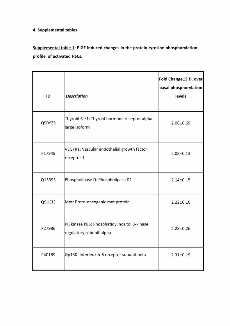
4. Supplemental tables
Supplemental table 1: PlGF-induced changes in the protein tyrosine phosphorylation
profile of activated HSCs.
ID Description
Fold Change��S.D. over
basal phosphorylation
levels
Q90Y25 Thyroid hormone receptor alpha
large isoform 2.06�0.69
P17948 VEGFR1: Vascular endothelial growth factor
receptor 1 2.08�0.13
Q13393 Phospholipase D: Phospholipase D1 2.14�0.15
Q9UEJ3 Met: Proto-oncogenic met protein 2.22�0.10
P27986 PI3kinase P85: Phosphatidylinositol 3-kinase
regulatory subunit alpha 2.28�0.26
P40189 Gp130: Interleukin-6 receptor subunit beta 2.31�0.19
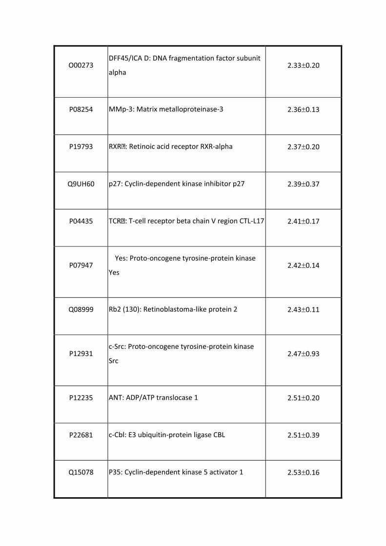
O00273 DFF45/ICA D: DNA fragmentation factor subunit
alpha 2.33�0.20
P08254 MMp-3: Matrix metalloproteinase-3 2.36�0.13
P19793 RXR : Retinoic acid receptor RXR-alpha 2.37�0.20
Q9UH60 p27: Cyclin-dependent kinase inhibitor p27 2.39�0.37
P04435 T-cell receptor beta chain V region CTL-L17 2.41�0.17
P07947 Yes: Proto-oncogene tyrosine-protein kinase
Yes 2.42�0.14
Q08999 Rb2 (130): Retinoblastoma-like protein 2 2.43�0.11
P12931 c-Src: Proto-oncogene tyrosine-protein kinase
Src 2.47�0.93
P12235 ANT: ADP/ATP translocase 1 2.51�0.20
P22681 c-Cbl: E3 ubiquitin-protein ligase CBL 2.51�0.39
Q15078 P35: Cyclin-dependent kinase 5 activator 1 2.53�0.16
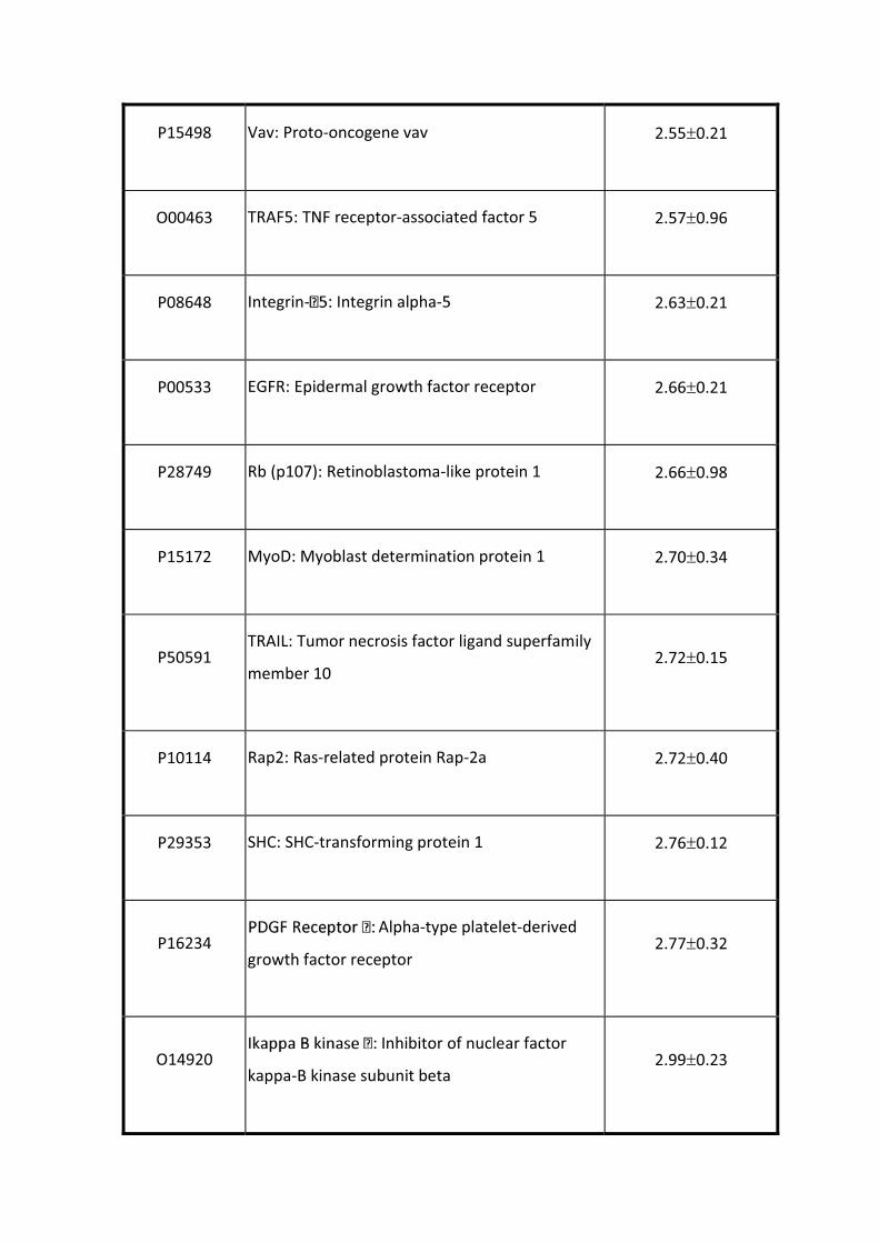
P15498 Vav: Proto-oncogene vav 2.55�0.21
O00463 TRAF5: TNF receptor-associated factor 5 2.57�0.96
P08648 Integrin- Integrin alpha-5 2.63�0.21
P00533 EGFR: Epidermal growth factor receptor 2.66�0.21
P28749 Rb (p107): Retinoblastoma-like protein 1 2.66�0.98
P15172 MyoD: Myoblast determination protein 1 2.70�0.34
P50591 TRAIL: Tumor necrosis factor ligand superfamily
member 10 2.72�0.15
P10114 Rap2: Ras-related protein Rap-2a 2.72�0.40
P29353 SHC: SHC-transforming protein 1 2.76�0.12
P16234 Alpha-type platelet-derived
growth factor receptor 2.77�0.32
O14920 Inhibitor of nuclear factor
kappa-B kinase subunit beta 2.99�0.23

O15524 SOCS-1: Suppressor of cytokine signaling 1 2.99�0.20
P10276 RARr: Retinoic acid receptor alpha 3.01�0.17
P07948 Lyn: Tyrosine-protein kinase Lyn 3.19�0.54
Q9Y4K3 TRAF6: TNF receptor-associated factor 6 3.19�0.12
P11473 VDR: Vitamin D3 receptor 3.50�0.36
Protein tyrosine phosphorylation profile of LX-2 cells treated with 100 ng/ml of Plgf for
five minutes. Proteins with a fold change greater than two are listed. For each of these
proteins, the Fold Change results are given as a mean�s.d. calculated from three
independent experiments.
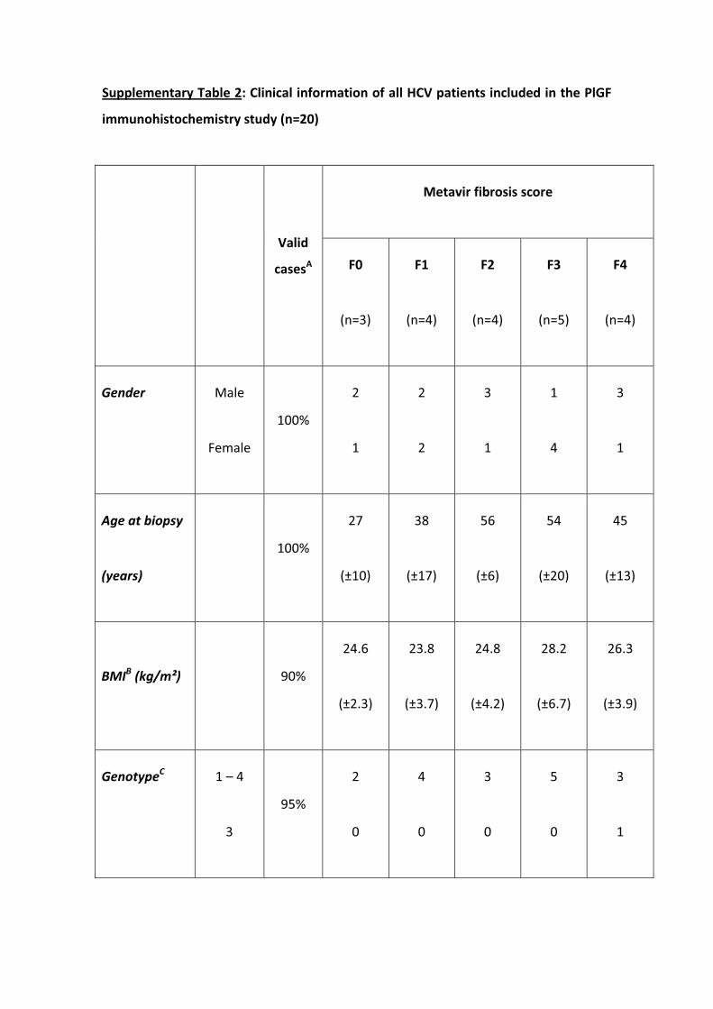
Supplementary Table 2: Clinical information of all HCV patients included in the PlGF
immunohistochemistry study (n=20)
Valid
casesA
Metavir fibrosis score
F0
(n=3)
F1
(n=4)
F2
(n=4)
F3
(n=5)
F4
(n=4)
Gender Male
Female
100%
2
1
2
2
3
1
1
4
3
1
Age at biopsy
(years)
100%
27
(±10)
38
(±17)
56
(±6)
54
(±20)
45
(±13)
BMIB (kg/m²) 90%
24.6
(±2.3)
23.8
(±3.7)
24.8
(±4.2)
28.2
(±6.7)
26.3
(±3.9)
GenotypeC 1 – 4
3
95%
2
0
4
0
3
0
5
0
3
1
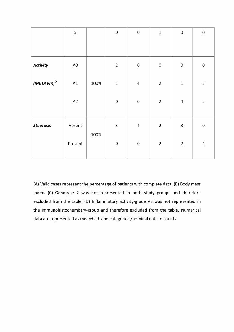
5 0 0 1 0 0
Activity
(METAVIR)D
A0
A1
A2
100%
2
1
0
0
4
0
0
2
2
0
1
4
0
2
2
Steatosis Absent
Present
100%
3
0
4
0
2
2
3
2
0
4
(A) Valid cases represent the percentage of patients with complete data. (B) Body mass
index. (C) Genotype 2 was not represented in both study groups and therefore
excluded from the table. (D) Inflammatory activity-grade A3 was not represented in
the immunohistochemistry-group and therefore excluded from the table. Numerical
data are represented as mean±s.d. and categorical/nominal data in counts.
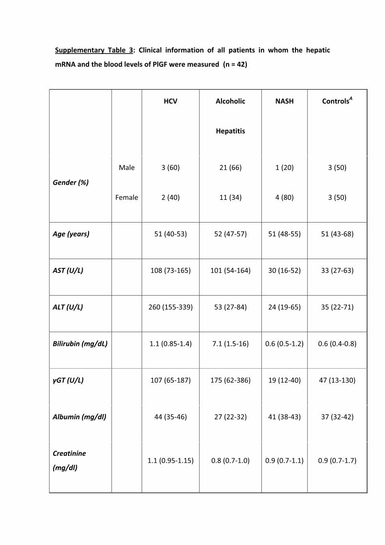
Supplementary Table 3: Clinical information of all patients in whom the hepatic
mRNA and the blood levels of PlGF were measured (n = 42)
HCV Alcoholic
Hepatitis
NASH ControlsA
Gender (%)
Male
Female
3 (60)
2 (40)
21 (66)
11 (34)
1 (20)
4 (80)
3 (50)
3 (50)
Age (years) 51 (40-53) 52 (47-57) 51 (48-55) 51 (43-68)
AST (U/L) 108 (73-165) 101 (54-164) 30 (16-52) 33 (27-63)
ALT (U/L) 260 (155-339) 53 (27-84) 24 (19-65) 35 (22-71)
Bilirubin (mg/dL) 1.1 (0.85-1.4) 7.1 (1.5-16) 0.6 (0.5-1.2) 0.6 (0.4-0.8)
γGT (U/L) 107 (65-187) 175 (62-386) 19 (12-40) 47 (13-130)
Albumin (mg/dl) 44 (35-46) 27 (22-32) 41 (38-43) 37 (32-42)
Creatinine
(mg/dl) 1.1 (0.95-1.15) 0.8 (0.7-1.0) 0.9 (0.7-1.1) 0.9 (0.7-1.7)
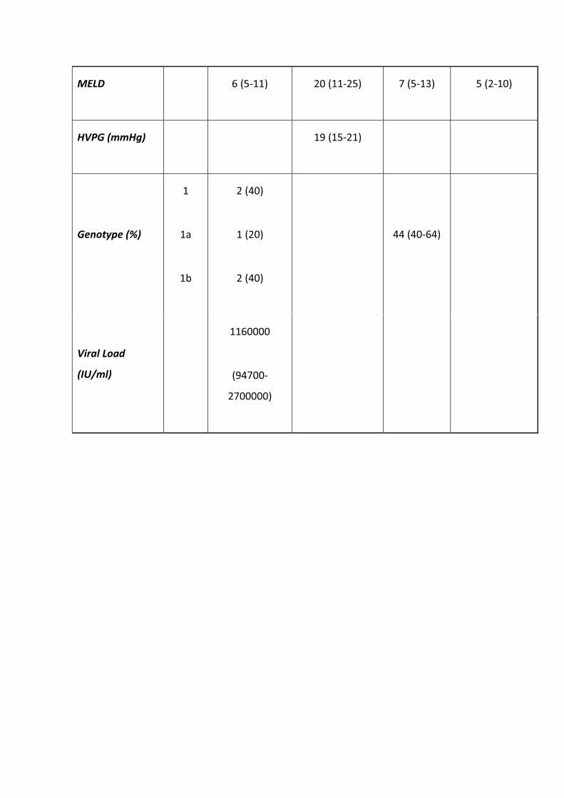
MELD 6 (5-11) 20 (11-25) 7 (5-13) 5 (2-10)
HVPG (mmHg) 19 (15-21)
Genotype (%)
1
1a
1b
2 (40)
1 (20)
2 (40)
44 (40-64)
Viral Load
(IU/ml)
1160000
(94700-
2700000)

Numerical data are represented as median values (interquartile range [IQR] 25-75) and
categorical / nominal data in counts. MELD: Model for End Stage Liver Disease; AST:
Aspartate- Aminotransferase; ALT: Alanin-Aminotransferase. ALiver samples of patients
with liver metastases from colorectal carcinoma except 1 who was affected by primary
hyperoxaluria.
Reference List
1. Geerts AM, Vanheule E, Praet M, Van Vlierberghe H., De Vos M., Colle I.
Comparison of three research models of portal hypertension in mice:
macroscopic, histological and portal pressure evaluation. Int J Exp Pathol 2008
Aug;89(4):251-263.
2. Tugues S, Fernandez-Varo G, Munoz-Luque J, Ros J, Arroyo V, Rodes J, et al.
Antiangiogenic treatment with sunitinib ameliorates inflammatory infiltrate,
fibrosis, and portal pressure in cirrhotic rats. Hepatology 2007 Dec;46(6):1919-
1926.
3. Colmenero J, Bataller R, Sancho-Bru P, Bellot P, Miquel R, Moreno M, et al.
Hepatic expression of candidate genes in patients with alcoholic hepatitis:
correlation with disease severity. Gastroenterology 2007 Feb;132(2):687-697.
4. Van Steenkiste C, Trachet B, Casteleyn D, van Loo L, Van Hoorebeke L, Segers P,
et al. Vascular corrosion casting: analyzing wall shear stress in the portal vein
and vascular abnormalities in portal hypertensive and cirrhotic rodents. Liver
Int. In press 2010.
5. Danese S, Sans M, Spencer DM, Beck I, Donate F, Plunkett ML, et al.
Angiogenesis blockade as a new therapeutic approach to experimental colitis.
Gut 2007 Jun;56(6):855-862.
6. Morales-Ruiz M, Cejudo-Martin P, Fernandez-Varo G, Tugues S, Ros J, Angeli P,
et al. Transduction of the liver with activated Akt normalizes portal pressure in
cirrhotic rats. Gastroenterology 2003 Aug;125(2):522-531.
7. Tugues S, Morales-Ruiz M, Fernandez-Varo G, Ros J, Arteta D, Munoz-Luque J,
et al. Microarray analysis of endothelial differentially expressed genes in liver of
cirrhotic rats. Gastroenterology 2005 Nov;129(5):1686-1695.
8. Xu L, Hui AY, Albanis E, Arthur MJ, O'Byrne SM, Blaner WS, et al. Human hepatic
stellate cell lines, LX-1 and LX-2: new tools for analysis of hepatic fibrosis. Gut
2005 Jan;54(1):142-151.
9. Van Steenkiste C., Geerts A, Vanheule E, Van Vlierberghe H., De Vos F., Olievier
K, et al. Role of placental growth factor in mesenteric neoangiogenesis in a
mouse model of portal hypertension. Gastroenterology 2009 Dec;137(6):2112-
2124.
10. Nakao S, Kuwano T, Ishibashi T, Kuwano M, Ono M. Synergistic effect of TNF-
alpha in soluble VCAM-1-induced angiogenesis through alpha 4 integrins. J
Immunol 2003 Jun 1;170(11):5704-5711.
11. Chen HX, Cleck JN. Adverse effects of anticancer agents that target the VEGF
pathway. Nat Rev Clin Oncol 2009 Aug;6(8):465-477.
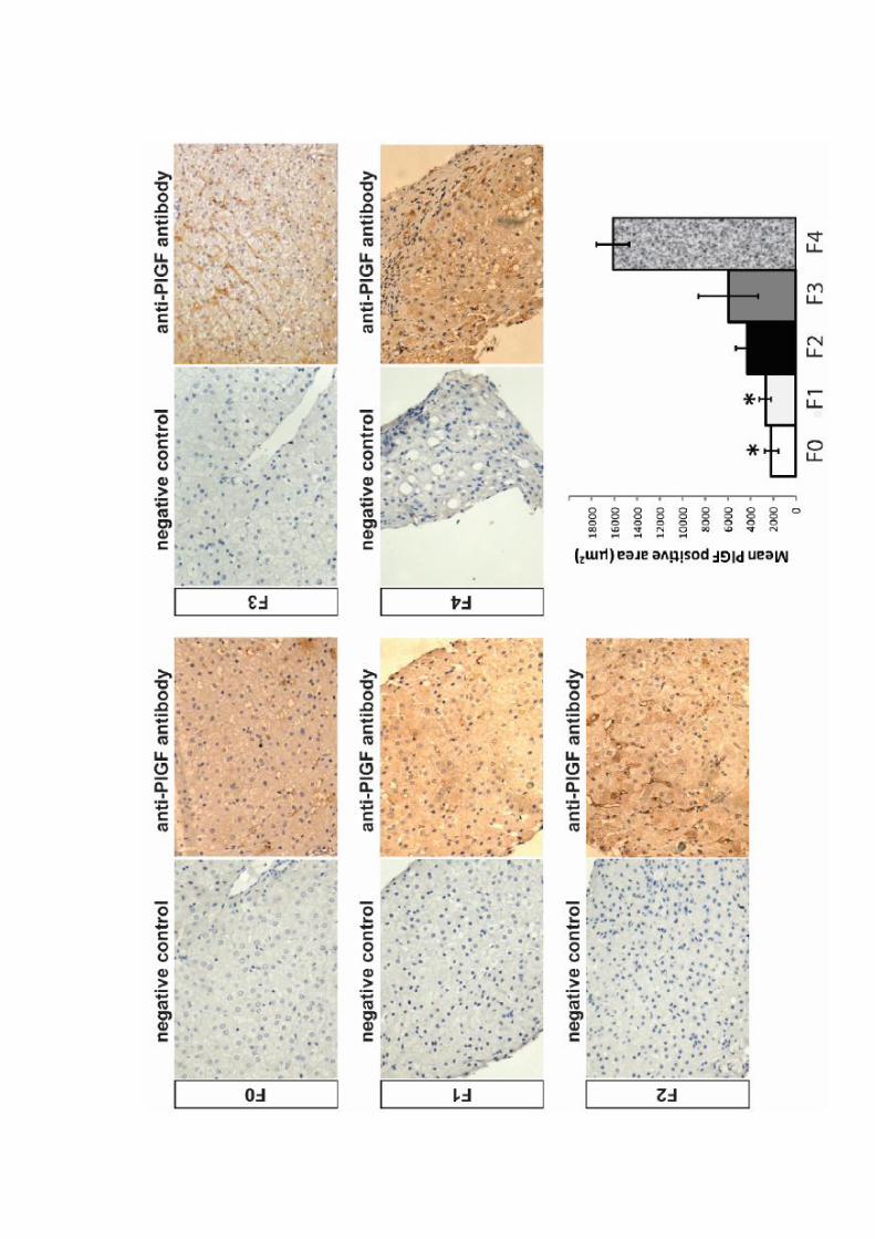
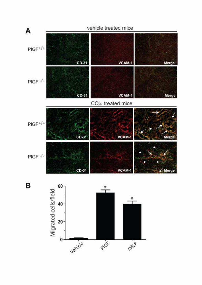
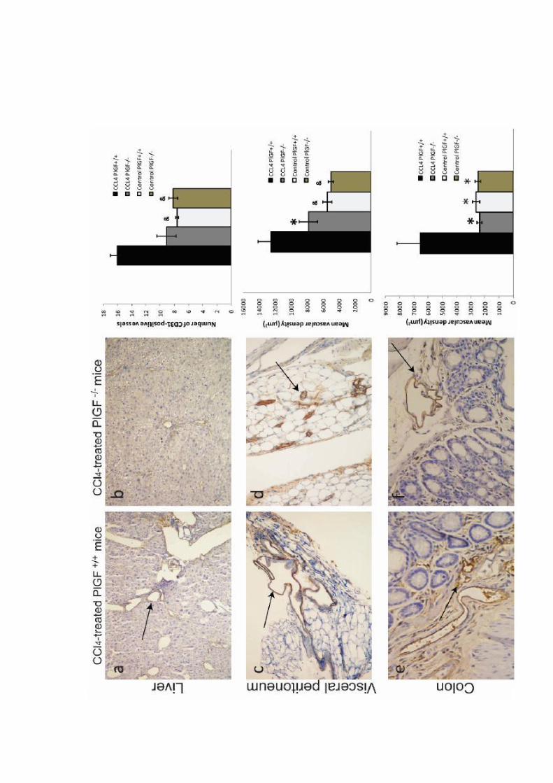
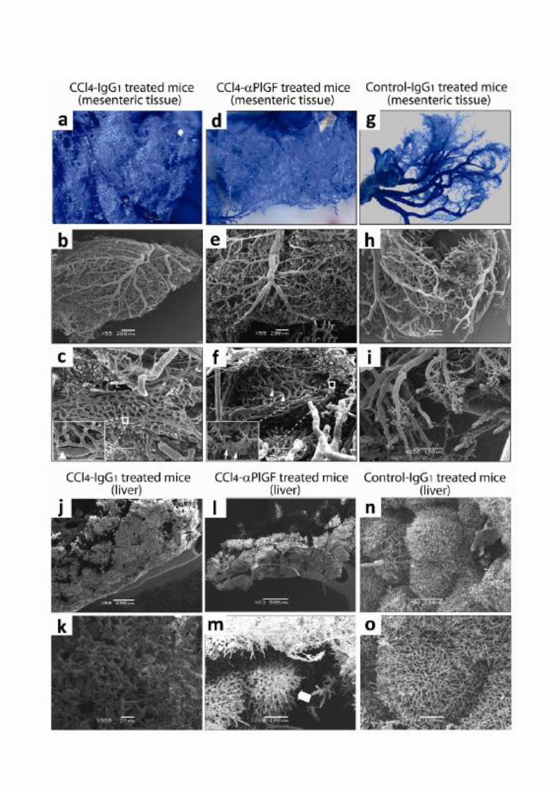
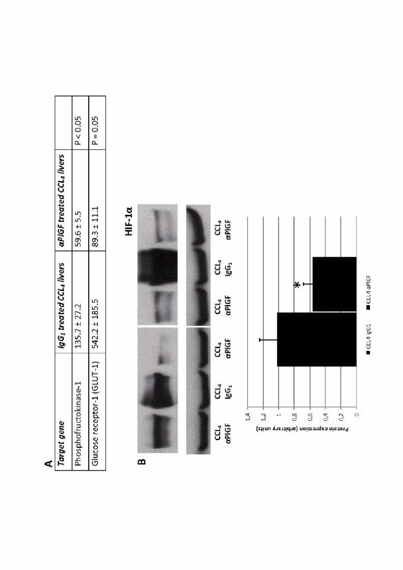
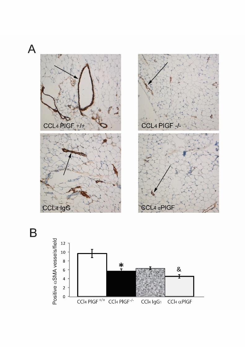
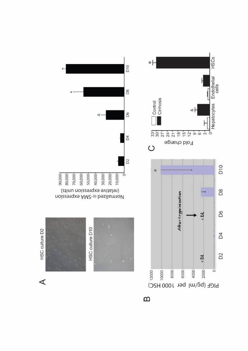
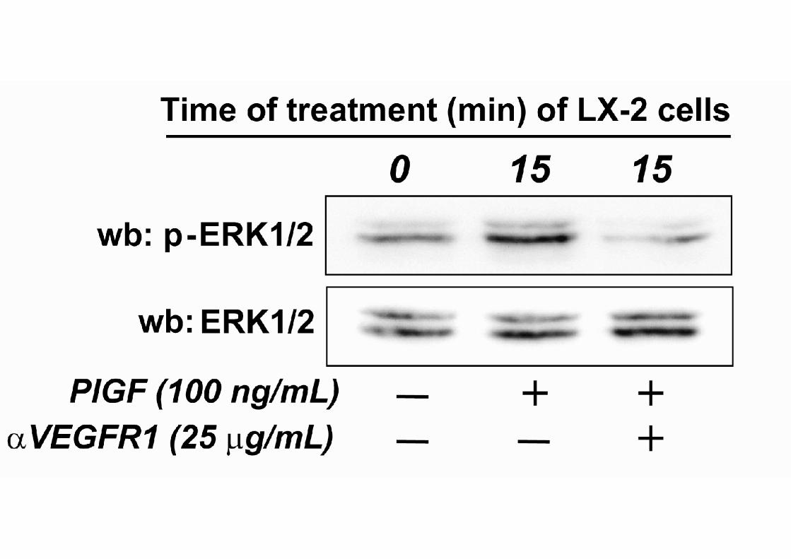
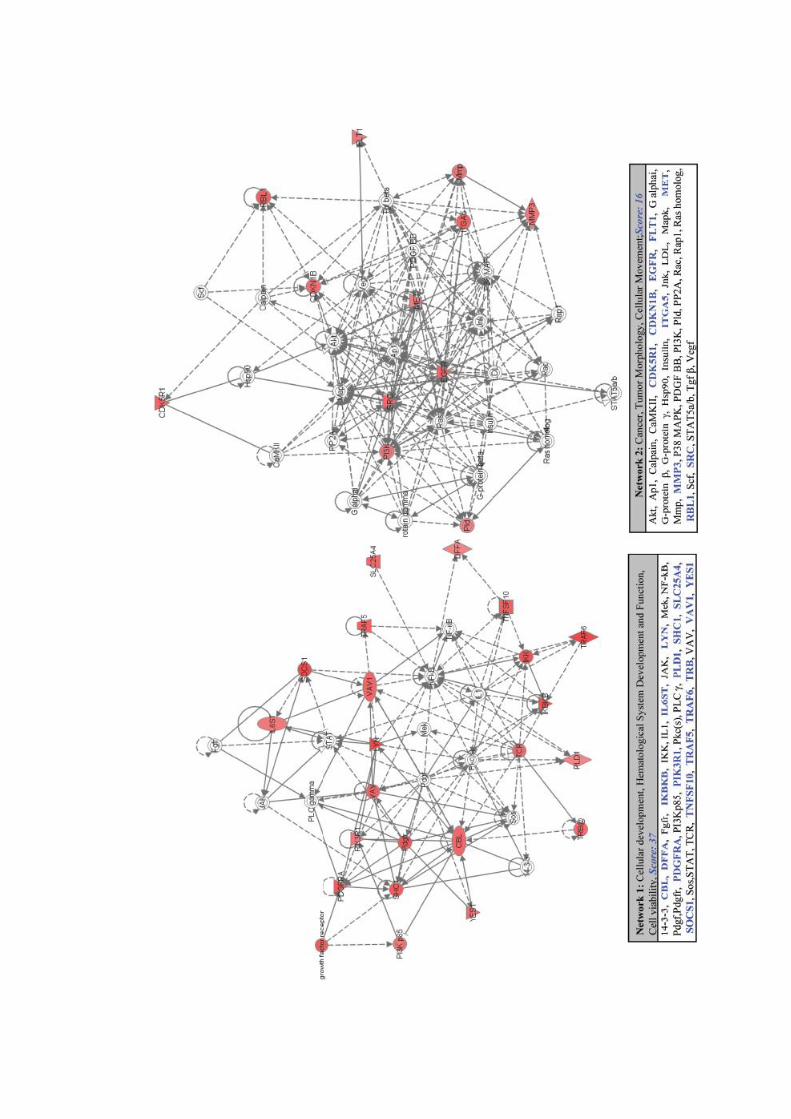
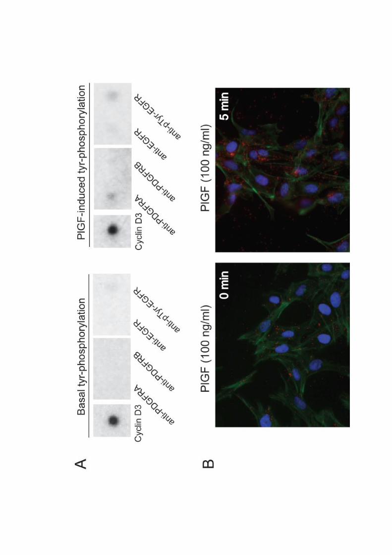
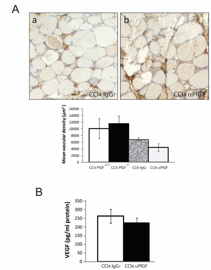


RESULTADOS _______________________________________________________________________
52
Artículo 2: La sobreproducción de óxido nítrico en células endoteliales linfáticas
provoca una disfunción en el drenaje linfático en ratas cirróticas.
Una de las complicaciones más severas durante la progresión de la cirrosis es la
formación de ascitis, que está asociada con un incremento de la morbilidad y la
mortalidad (Moore et al., 2003). Algunos de los mecanismos que contribuyen a la
aparición y al mantenimiento de la ascitis son: 1) el incremento de la presión
hidrostática intravascular, 2) el descenso de la presión oncótica en el plasma, 3) un
aumento de la angiogénesis en el territorio esplácnico y 4) la retención renal de sal y
agua (Gines et al., 2004; Van Steenkiste et al., 2009; Henriksen et al., 2005). Sin
embargo, la función del sistema linfático en la patogénesis de la ascitis no está bien
estudiada. Los primeros estudios en este campo demostraron que los pacientes
cirróticos presentaban una vasculatura linfática alterada y un incremento del flujo de
linfa en el conducto torácico (Dumont et al., 1960). A raíz de estos descubrimientos se
propuso la teoría del desequilibrio de linfa para explicar la formación de ascitis (Witte
et al., 1980). Según esta teoría, existiría un desequilibrio entre el fluido que se filtra a
las regiones intravasculares y el fluido que regresa a la circulación sanguínea, dando
lugar a una mala distribución de los fluidos corporales y como consecuencia la
formación de ascitis. En los pacientes cirróticos compensados, el sistema linfático
ayuda a prevenir la aparición de ascitis reabsorbiendo el exceso de fluido, tanto en el
hígado, como en las demás regiones de la zona esplácnica. Como consecuencia de esto
aumenta el flujo linfático (Witte et al., 1969), lo que estimula la aparición de
linfangiogénesis hepática (Tugues et al., 2005). A pesar de ello, este mecanismo
compensatorio no es capaz de evitar la formación de ascitis en pacientes con cirrosis
descompensada y esto hace hipotetizar que existe una disfunción del sistema linfático
en los estadíos más avanzados de la enfermedad. Por ello, en este estudio se investigó
si una incapacidad del sistema linfático para drenar el exudado tisular podría contribuir
a la formación de edema y ascitis características de la cirrosis. Además, y teniendo en
cuenta que ya se había demostrado que existía una sobreexpresión de la actividad de
eNOS en la vasculatura de ratas cirróticas (Ros et al., 1995; Fernández-Varo et al.,
2003), quisimos estudiar si una sobreproducción de NO en el endotelio linfático podría
RESULTADOS _______________________________________________________________________
53
ser responsable de esta disfunción linfática en la cirrosis. Los resultados más
importantes derivados de este estudio se enumeran a continuación:
1. Existe una disfunción del sistema linfático durante el progreso de la cirrosis.
En primer lugar, se analizó la funcionalidad del sistema linfático en cirrosis
utilizando la técnica de la linfangiografía con FITC-dextrano para las regiones
periféricas, o con la ingesta del lípido fluorescente BODIPY para la zona esplácnica. Las
ratas cirróticas presentaron una disfunción en el drenaje linfático comparadas con las
ratas control, tanto a nivel periférico como a nivel esplácnico. Para excluir una
disminución en el número total de vasos linfáticos en cirrosis como explicación a la
disfunción observada, comparamos los vasos teñidos en oreja entre ratas controles y
ratas cirróticas por la técnica de inmunofluorescencia whole-mount, usando la
podoplanina como marcador específico de vasos linfáticos. Esta tinción demostró que
la densidad de la red linfática estaba preservada en cirrosis, sin cambios significativos
en el número de vasos entre control y cirrosis (Figura 1).
2. La expresión proteica de eNOS y la producción de NO están incrementadas en LyECs
de ratas cirróticas.
Estudios previos ya habían demostrado que eNOS estaba sobreexpresada en la
vasculatura sanguínea de las ratas cirróticas (Ros et al., 1995; Fernández-Varo et al.,
2003). Con el objetivo de investigar si había un paralelismo en la vasculatura linfática
evaluamos por Western Blot los niveles de eNOS en LyECs aisladas del mesenterio, en
ratas controles y en ratas cirróticas. La expresión de eNOS estuvo aumentada del
orden del doble en LyECs de ratas cirróticas, además este aumento se correlacionó con
un incremento significativo en la acumulación de NO2- a 24 horas en el medio de
cultivo (Figura 2B y 2C).
RESULTADOS _______________________________________________________________________
54
3. La inhibición de eNOS mejora el drenaje linfático y reduce la acumulación de ascitis
en ratas cirróticas.
A continuación, evaluamos el efecto terapéutico que podría tener bloquear la
actividad de eNOS sobre la funcionalidad del sistema linfático con un tratamiento con
L-NMMA. Paralelamente, los animales también se trataron con Midodrina, una droga
hipertensiva que no afecta a la actividad de eNOS, para excluir el efecto hipertensivo
asociado al tratamiento con L-NMMA. Las mismas ratas cirróticas en las que se había
detectado un empeoramiento del drenaje linfático mostraron una mejora significativa
en la captación linfática después del tratamiento con L-NMMA, comparadas con el
grupo cirrótico tratado con Midodrina, tanto a nivel periférico como a nivel esplácnico.
El efecto beneficioso del tratamiento con L-NMMA no se debió a cambios en la presión
arterial media, ya que no se encontraron diferencias significativas al comparar los
valores de presión entre los tratamientos de L-NMMA y Midodrina (Figura 3).
Para estudiar si podía haber una relación entre la mejora en el drenaje linfático
y la reducción del volumen de ascitis, visualizamos el líquido acumulado en la cavidad
peritoneal de las ratas por resonancia magnética de imagen. Todas las ratas cirróticas
tratadas con L-NMMA mostraron una disminución significativa del volumen ascítico
después de 7 días de tratamiento. Por el contrario, el tratamiento con Midodrina no
afectó de una manera significativa al volumen total de ascitis. Esta reabsorción del
líquido ascítico se corroboró midiendo el volumen plasmático de ratas cirróticas con
ascitis, con o sin tratamiento de L-NMMA. En las ratas tratadas se observó que había
un incremento del volumen plasmático, comparadas con las ratas sin tratar (Figura 4).
4. La sobreexpresión de eNOS en LyECs de ratas cirróticas provoca un remodelado de
los vasos linfáticos caracterizado por la pérdida de recubrimiento de SMC.
Teniendo en cuenta que ya estaba demostrado que la expresión de eNOS está
relacionada con la permeabilidad vascular (Thibeault et al., 2010), quisimos investigar
si existía un incremento de la permeabilidad de los vasos linfáticos como consecuencia
de la sobreproducción de NO. Esto lo miramos usando la técnica de la linfangiografía
RESULTADOS _______________________________________________________________________
55
con un FITC-dextrano de 20 kDa. Este tamaño de partícula es lo suficientemente
grande como para no atravesar los vasos en condiciones normales, pero también es lo
suficientemente pequeño como para detectar si hay un aumento de permeabilidad
(Ono et al., 2005; Porter et al., 2001). No encontramos ningún tipo de escape en los
vasos linfáticos en ninguno de los grupos experimentales (Figura 5A).
Estudios previos de nuestro grupo ya habían demostrado que los cambios
crónicos en la producción vascular de NO están asociados a remodelado vascular
(Fernández-Varo et al., 2003). Por este motivo, y teniendo en cuenta la
sobreproducción de NO encontrada en el endotelio linfático en cirrosis, evaluamos si
un mecanismo similar podría ser responsable de la disfunción linfática observada en
las ratas cirróticas. El remodelado vascular linfático se evaluó en tejido mesentérico
por inmunohistoquímica usando la podoplanina como marcador específico. En el
mesenterio, los vasos teñidos con podoplanina tenían un mayor recubrimiento de SMC
en condiciones controles que en cirrosis. Este remodelado también se analizó en ratas
cirróticas tratadas con L-NMMA, donde encontramos que había un incremento del
porcentaje de vasos linfáticos recubiertos por SMC después del tratamiento,
comparado con el grupo cirrótico sin tratar (Figura 5B).
5. El NO inhibe la proliferación de las SMCs primarias.
Para profundizar más en el efecto que tiene el NO sobre las SMCs, estudiamos
el efecto que tenía un dador de NO (DETANONOate) en la proliferación de estas
células. Para ello, aislamos células musculares lisas de aorta de ratas controles e
hicimos un ensayo de proliferación con BrdU. Pudimos observar una disminución
significativa de la incorporación de BrdU cuando las células se incubaron con el dador
de NO, en comparación con la condición control, indicando que el NO tiene un efecto
antiproliferativo sobre estas células (Supplemental results Figura 2).
ORIGINAL ARTICLE
Increased nitric oxide production in lymphaticendothelial cells causes impairment of lymphaticdrainage in cirrhotic rats
Jordi Ribera,1 Montse Pauta,1 Pedro Melgar-Lesmes,1 Sonia Tugues,1,2
Guillermo Fernandez-Varo,1 Kara F Held,3 Guadalupe Soria,4 Raul Tudela,4,5
Anna M Planas,4,6 Carlos Fernandez-Hernando,7 Vicente Arroyo,8
Wladimiro Jimenez,1,9 Manuel Morales-Ruiz1
ABSTRACTBackground and aim The lymphatic network playsa major role in maintaining tissue fluid homoeostasis.Therefore several pathological conditions associated withoedema formation result in deficient lymphatic function.However, the role of the lymphatic system in thepathogenesis of ascites and oedema formation incirrhosis has not been fully clarified. The aim of this studywas to investigate whether the inability of the lymphaticsystem to drain tissue exudate contributes to theoedema observed in cirrhosis.Methods Cirrhosis was induced in rats by CCl4inhalation. Lymphatic drainage was evaluated usingfluorescent lymphangiography. Expression of endothelialnitric oxide synthase (eNOS) was measured in primarylymphatic endothelial cells (LyECs). Inhibition of eNOSactivity in cirrhotic rats with ascites (CH) was carried outby L-NG-methyl-L-arginine (L-NMMA) treatment(0.5 mg/kg/day).Results The (CH) rats had impaired lymphatic drainagein the splanchnic and peripheral regions compared withthe control (CT) rats. LyECs isolated from the CH ratsshowed a significant increase in eNOS and nitric oxide(NO) production. In addition, the lymphatic vessels of theCH rats showed a significant reduction in smooth musclecell (SMC) coverage compared with the CT rats. CH ratstreated with L-NMMA for 7 days showed a significantimprovement in lymphatic drainage and a significantreduction in ascites volume, which were associated withincreased plasma volume. This beneficial effect ofL-NMMA inhibition was also associated witha significant increase in lymphatic SMC coverage.Conclusions The upregulation of eNOS in the LyECs ofCH rats causes long-term lymphatic remodelling, whichis characterised by a loss of SMC lymphatic coverage.The amelioration of this lymphatic abnormality by chroniceNOS inhibition results in improved lymphatic drainageand reduced ascites.
The prognostic expectations for cirrhotic patientsare poor because of the severe complications thesepatients experience. One of the most commoncomplications is ascites, which is associated withincreased morbidity and mortality.1 Some of themechanisms that contribute to the pathogenesis ofascites include increased intravascular hydrostaticpressure, enlarged splanchnic arterial vasculature
and decreased plasma oncotic pressure.2e4 Thesechanges result in excessive fluid filtration, which isexacerbated by the concomitant transformation ofthe hepatic microvasculature into a capillarised anddefenestrated endothelium.5 6 In this scenario, theincrease in intrahepatic pressure is transmittedupstream by the blood to the splanchnic organs,resulting in further oedema.7 As the liver diseaseprogresses, the antidiuretic and antinatriureticproperties of certain neuroendocrine systems,which are activated secondary to the arterial vaso-dilation, worsen the oedematous condition and
< Additional materials arepublished online only. To viewthese files please visit thejournal online (http://gut.bmj.com/content/early/recent).
For numbered affiliations seeend of article.
Correspondence toDr Manuel Morales-Ruiz,Department of Biochemistry andMolecular Genetics, HospitalClinic Universitari, Villarroel 170,Barcelona 08036, Spain;[email protected]
Revised 3 December 2011Accepted 21 December 2011
Significance of this study
What is already known about this subject?< Under pathological conditions, four derange-
ments contribute to the formation of oedema:(1) an increase in intravascular hydrostaticpressure; (2) a decrease in plasma oncoticpressure; (3) renal retention of salt and water;and (4) impairment of lymphatic drainage.
< There is abundant literature establishing thepresence of most of these mechanisms incirrhosis and their role in the consequentoedema and ascites. However, few studieshave investigated whether there is impairedlymphatic functionality in cirrhosis.
< Lymphatic transport is promoted by extrinsicand intrinsic forces. The intrinsic forces arederived from the contractility of the smoothmuscle cells (SMCs) of the collecting lymphaticvessels. Regarding the molecular mechanismsgoverning the intrinsic contractility of thecollecting lymphatic vessels, there is evidencethat the lymphatic endothelial cells controllymph flow by regulating the contractility ofSMCs via the production of nitric oxide (NO).
< Increased endothelial nitric oxide synthase(eNOS) activity in the blood vessels of cirrhoticrats, along with its effect on vascular remodel-ling, has already been demonstrated. However,the changes in lymphatic NO production and itseffect on lymphatic remodelling have notpreviously been investigated in cirrhosis.
Ribera J, Pauta M, Melgar-Lesmes P, et al. Gut (2012). doi:10.1136/gutjnl-2011-300703 1 of 8
CirrhosisGut Online First, published on January 20, 2012 as 10.1136/gutjnl-2011-300703
Copyright Article author (or their employer) 2012. Produced by BMJ Publishing Group Ltd (& BSG) under licence.
group.bmj.com on February 1, 2012 - Published by gut.bmj.comDownloaded from
cause ascites.8 All of these mechanisms contribute to the onsetand maintenance of oedema and ascites. However, the role of thelymphatic system in the pathogenesis of ascites formation hasnot been fully clarified.
The lymphatic capillaries are formed by a thin layer of non-fenestrated lymphatic endothelial cells (LyECs) without base-ment membranes that form closed-ended structures. Perivascularsmooth muscle cells (SMCs) and pericytes are absent inlymphatic capillaries but are present in the collecting lymphaticvessels that conduct lymph within the lymphatic system.9 Inthe absence of a muscular pumping organ, such as the heart inthe cardiovascular system, lymphatic transport is promoted byextrinsic and intrinsic forces. The intrinsic forces are derivedfrom the contractility of the SMCs of the collecting lymphaticvessels, which has been proposed as one of the major drivingforces of lymph circulation.10 Regarding the molecular mecha-nisms governing the intrinsic contractility of the collectinglymphatic vessels, there is evidence that the LyECs controllymph flow by regulating the contractility of SMCs via theproduction of nitric oxide (NO).11
In cirrhotic patients, the lymphatic system helps to preventascites accumulation by reabsorbing excess fluid in the liver andsplanchnic regions. Lymph flow is increased as a consequence,12
which stimulates hepatic lymphangiogenesis.13 However, thiscompensatory mechanism is not sufficient to prevent ascitesformation in decompensated cirrhotic patients. This lymphaticdeficiency supports the hypothesis that advanced liver disease isassociated with impaired lymphatic function. Therefore ourstudy aimed to investigate whether the inability of the lymphaticsystem to drain tissue exudate contributes to the oedemaobserved in cirrhosis. In addition, increased eNOS activity in theblood vessels of cirrhotic rats, along with its effect on vascularremodelling, has already been demonstrated.14 15 Therefore we
also investigated whether NO overproduction by the LyECs maybe responsible for lymphatic impairment in cirrhosis.
MATERIALS AND METHODSThe experimental cirrhosis modelThe study was performed in cirrhotic (CH) and control (CT)male adult Wistar rats (CharleseRiver, Saint Aubin les Elseuf,France), following the guidelines of the investigation and ethicscommittees of the Hospital Clinic. Cirrhosis was induced byinhalation of CCl4, as described previously.13
Isolation of LyECsFreshly isolated primary LyECs were obtained from the mesen-tery of the CT and CH rats. Briefly, the atrium was cannulatedand the vascular system perfused with normal saline solution.Mesenteric lymphatic tissue mucosa was harvested, placed on35 mm plates containing ice-cold phosphate-buffered saline, andcut into small (1 mm) fragments. The fragments were incubatedin 0.25% collagenase A (Roche Diagnostics, Basel, Switzerland)at 378C. The suspension was passed through 100 mm nylon meshand centrifuged at 1800 rpm for 4 min at 48C. The cell pellet wasresuspended in Hank’s balanced salt solution. The LyECs wereisolated using rabbit antibody to rat podoplanin (Sigma Chem-ical, St Louis, Missouri, USA) in a 1:100 dilution as the primaryantibody and microbeads coupled with a secondary goat anti-rabbit antibody (MACS system, Miltenyi Biotec, Bergisch-Gladbach, Germany). The cells were grown in Dulbecco’smodified Eagle medium that was supplemented with 20% fetalcalf serum, 50 U/ml penicillin and 50 mg/ml streptomycin.
LymphangiographyTo evaluate lymphatic drainage in peripheral areas, 0.3 ml fluo-rescein isothiocyanate (FITC)-Dextran 2000 kDa or 20 kDa(Sigma Chemical) was subcutaneously injected into the ears,tails and footpads of the CTand CH rats. Immediately after theinjection, fluorescent images of the subcutaneous lymphaticdrainage were visualised using a fluorescence stereomicroscopesystem (Leica Microsystems, Heerbrugg, Switzerland). For thesplanchnic areas, a long-chain fatty acid (BODIPY FL C16;Invitrogen, San Diego, California, USA) was administeredintragastrically. After 2 h, fluorescent images of the mesentericlymphatic drainage were obtained.
L-NG-methyl-L-arginine (L-NMMA) and midodrine treatmentsThe CH rats were treated intragastrically with L-NMMA(Calbiochem, Gibbstown, New Jersey, USA) at a dose of 0.5 mg/kg/day for 1 week, or midodrine (Sigma Chemical) at a dose of5 mg/kg/day for 1 week. The untreated CH rats were given thedosing vehicle intragastrically as a treatment control.
Immunohistochemistry, immunocytofluorescence andwhole-mount immunofluorescence stainingFor immunohistochemistry, mesentery samples were fixed in10% buffered formaldehyde solution and embedded in paraffin.Immunolabelling was performed using a rabbit antibody to ratpodoplanin (Sigma Chemical). Immunolabelled cells weredetected with the Dako LSAB2 System, HRP (Dako, Glostrup,Denmark). Immunoreactivity was visualised using a lightmicroscope (Nikon Eclipse E600, Kawasaki, Kanagawa, Japan).For the immunocytofluorescence analyses, LyECs were fixed
with 4% paraformaldehyde. The cells were incubated with oneof the following primary antibodies for 1 h: rabbit anti-ratpodoplanin, mouse anti-rat CD31 (BD Pharmigen, FranklinLakes, New Jersey, USA) or goat anti-rat CD34 (LifeSpanBiosciences, Seattle, Washington, USA). The respective secondary
Significance of this study
What are the new findings?< Cirrhosis is accompanied by impairment of lymphatic drainage
of the splanchnic and peripheral regions.< The cirrhotic lymphatic endothelial cells overexpress eNOS
and overproduce NO.< NO overproduction causes lymphatic remodelling. The
remodelling process is characterised by a decrease incoverage of the lymphatic vessels by pericytes or SMCs.
< Long-term inhibition of eNOS activity with L-NMMA correctedlymphatic abnormalities, resulting in an increase in coverageof the lymphatic vessels by pericytes, an improvement inlymphatic drainage, and a significant decrease in ascitesvolume.
How might it impact on clinical practice in the foreseeablefuture?< Our observations have implications for the pathophysiology of
ascites and oedema formation in cirrhosis. We demonstratethat cirrhosis is accompanied by impairment of the lymphaticdrainage. In addition, we show that NO overproduction causeslymphatic remodelling. Both lymphatic abnormalities arereversed by L-NMMA treatment, resulting in improvedlymphatic drainage from the peritoneal cavity and reducedascites. Therefore eNOS inhibition is a new target forcorrecting lymphatic dysfunction in cirrhosis.
2 of 8 Ribera J, Pauta M, Melgar-Lesmes P, et al. Gut (2012). doi:10.1136/gutjnl-2011-300703
Cirrhosis
group.bmj.com on February 1, 2012 - Published by gut.bmj.comDownloaded from
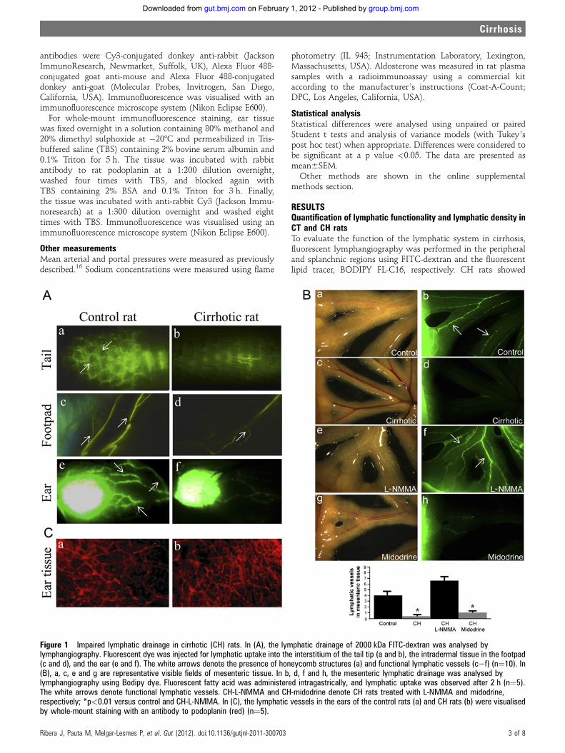
antibodies were Cy3-conjugated donkey anti-rabbit (JacksonImmunoResearch, Newmarket, Suffolk, UK), Alexa Fluor 488-conjugated goat anti-mouse and Alexa Fluor 488-conjugateddonkey anti-goat (Molecular Probes, Invitrogen, San Diego,California, USA). Immunofluorescence was visualised with animmunofluorescence microscope system (Nikon Eclipse E600).
For whole-mount immunofluorescence staining, ear tissuewas fixed overnight in a solution containing 80% methanol and20% dimethyl sulphoxide at �208C and permeabilized in Tris-buffered saline (TBS) containing 2% bovine serum albumin and0.1% Triton for 5 h. The tissue was incubated with rabbitantibody to rat podoplanin at a 1:200 dilution overnight,washed four times with TBS, and blocked again withTBS containing 2% BSA and 0.1% Triton for 3 h. Finally,the tissue was incubated with anti-rabbit Cy3 (Jackson Immu-noresearch) at a 1:300 dilution overnight and washed eighttimes with TBS. Immunofluorescence was visualised using animmunofluorescence microscope system (Nikon Eclipse E600).
Other measurementsMean arterial and portal pressures were measured as previouslydescribed.16 Sodium concentrations were measured using flame
photometry (IL 943; Instrumentation Laboratory, Lexington,Massachusetts, USA). Aldosterone was measured in rat plasmasamples with a radioimmunoassay using a commercial kitaccording to the manufacturer ’s instructions (Coat-A-Count;DPC, Los Angeles, California, USA).
Statistical analysisStatistical differences were analysed using unpaired or pairedStudent t tests and analysis of variance models (with Tukey’spost hoc test) when appropriate. Differences were considered tobe significant at a p value <0.05. The data are presented asmean6SEM.Other methods are shown in the online supplemental
methods section.
RESULTSQuantification of lymphatic functionality and lymphatic density inCT and CH ratsTo evaluate the function of the lymphatic system in cirrhosis,fluorescent lymphangiography was performed in the peripheraland splanchnic regions using FITC-dextran and the fluorescentlipid tracer, BODIPY FL-C16, respectively. CH rats showed
Figure 1 Impaired lymphatic drainage in cirrhotic (CH) rats. In (A), the lymphatic drainage of 2000 kDa FITC-dextran was analysed bylymphangiography. Fluorescent dye was injected for lymphatic uptake into the interstitium of the tail tip (a and b), the intradermal tissue in the footpad(c and d), and the ear (e and f). The white arrows denote the presence of honeycomb structures (a) and functional lymphatic vessels (cef) (n¼10). In(B), a, c, e and g are representative visible fields of mesenteric tissue. In b, d, f and h, the mesenteric lymphatic drainage was analysed bylymphangiography using Bodipy dye. Fluorescent fatty acid was administered intragastrically, and lymphatic uptake was observed after 2 h (n¼5).The white arrows denote functional lymphatic vessels. CH-L-NMMA and CH-midodrine denote CH rats treated with L-NMMA and midodrine,respectively; *p<0.01 versus control and CH-L-NMMA. In (C), the lymphatic vessels in the ears of the control rats (a) and CH rats (b) were visualisedby whole-mount staining with an antibody to podoplanin (red) (n¼5).
Ribera J, Pauta M, Melgar-Lesmes P, et al. Gut (2012). doi:10.1136/gutjnl-2011-300703 3 of 8
Cirrhosis
group.bmj.com on February 1, 2012 - Published by gut.bmj.comDownloaded from
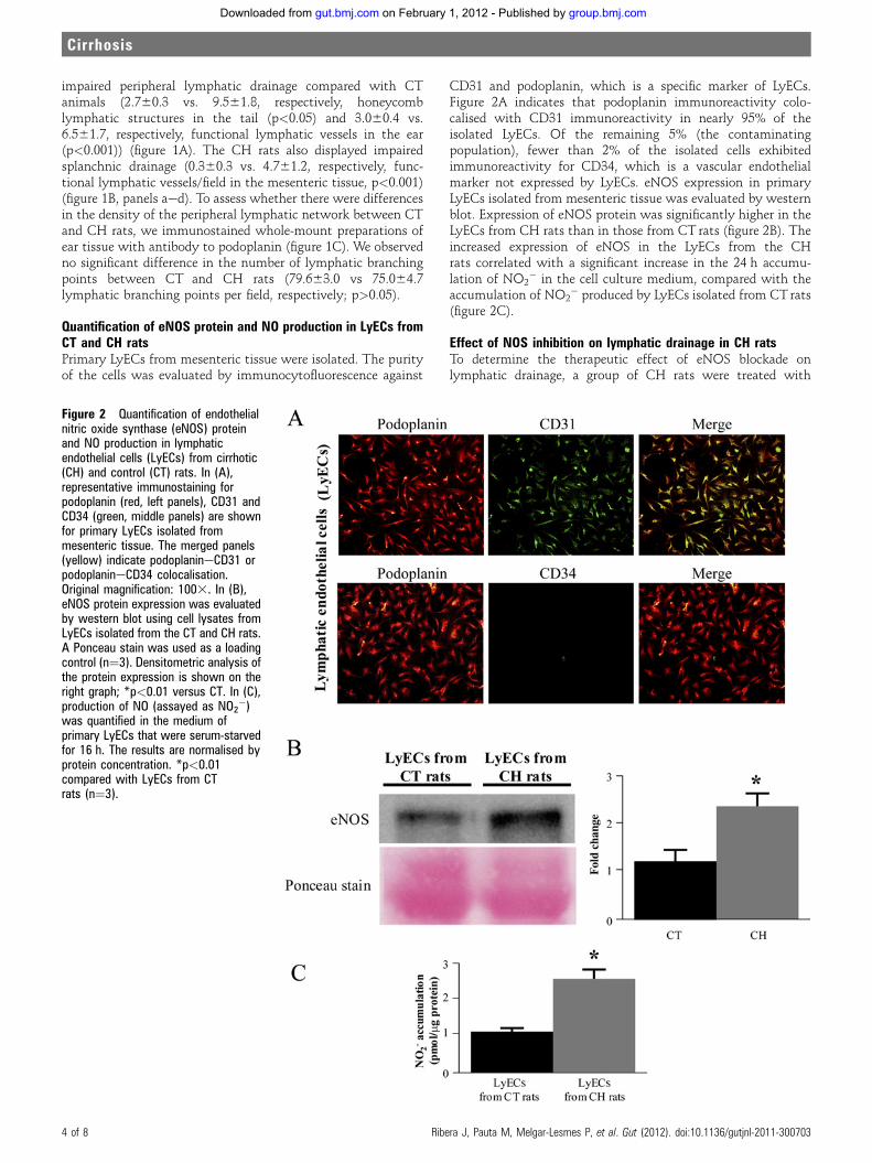
impaired peripheral lymphatic drainage compared with CTanimals (2.760.3 vs. 9.561.8, respectively, honeycomblymphatic structures in the tail (p<0.05) and 3.060.4 vs.6.561.7, respectively, functional lymphatic vessels in the ear(p<0.001)) (figure 1A). The CH rats also displayed impairedsplanchnic drainage (0.360.3 vs. 4.761.2, respectively, func-tional lymphatic vessels/field in the mesenteric tissue, p<0.001)(figure 1B, panels aed). To assess whether there were differencesin the density of the peripheral lymphatic network between CTand CH rats, we immunostained whole-mount preparations ofear tissue with antibody to podoplanin (figure 1C). We observedno significant difference in the number of lymphatic branchingpoints between CT and CH rats (79.663.0 vs 75.064.7lymphatic branching points per field, respectively; p>0.05).
Quantification of eNOS protein and NO production in LyECs fromCT and CH ratsPrimary LyECs from mesenteric tissue were isolated. The purityof the cells was evaluated by immunocytofluorescence against
CD31 and podoplanin, which is a specific marker of LyECs.Figure 2A indicates that podoplanin immunoreactivity colo-calised with CD31 immunoreactivity in nearly 95% of theisolated LyECs. Of the remaining 5% (the contaminatingpopulation), fewer than 2% of the isolated cells exhibitedimmunoreactivity for CD34, which is a vascular endothelialmarker not expressed by LyECs. eNOS expression in primaryLyECs isolated from mesenteric tissue was evaluated by westernblot. Expression of eNOS protein was significantly higher in theLyECs from CH rats than in those from CT rats (figure 2B). Theincreased expression of eNOS in the LyECs from the CHrats correlated with a significant increase in the 24 h accumu-lation of NO2
� in the cell culture medium, compared with theaccumulation of NO2
� produced by LyECs isolated from CTrats(figure 2C).
Effect of NOS inhibition on lymphatic drainage in CH ratsTo determine the therapeutic effect of eNOS blockade onlymphatic drainage, a group of CH rats were treated with
Figure 2 Quantification of endothelialnitric oxide synthase (eNOS) proteinand NO production in lymphaticendothelial cells (LyECs) from cirrhotic(CH) and control (CT) rats. In (A),representative immunostaining forpodoplanin (red, left panels), CD31 andCD34 (green, middle panels) are shownfor primary LyECs isolated frommesenteric tissue. The merged panels(yellow) indicate podoplanineCD31 orpodoplanineCD34 colocalisation.Original magnification: 1003. In (B),eNOS protein expression was evaluatedby western blot using cell lysates fromLyECs isolated from the CT and CH rats.A Ponceau stain was used as a loadingcontrol (n¼3). Densitometric analysis ofthe protein expression is shown on theright graph; *p<0.01 versus CT. In (C),production of NO (assayed as NO2
�)was quantified in the medium ofprimary LyECs that were serum-starvedfor 16 h. The results are normalised byprotein concentration. *p<0.01compared with LyECs from CTrats (n¼3).
4 of 8 Ribera J, Pauta M, Melgar-Lesmes P, et al. Gut (2012). doi:10.1136/gutjnl-2011-300703
Cirrhosis
group.bmj.com on February 1, 2012 - Published by gut.bmj.comDownloaded from
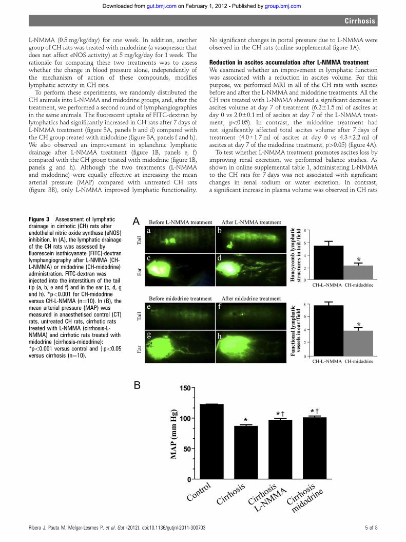
L-NMMA (0.5 mg/kg/day) for one week. In addition, anothergroup of CH rats was treated with midodrine (a vasopressor thatdoes not affect eNOS activity) at 5 mg/kg/day for 1 week. Therationale for comparing these two treatments was to assesswhether the change in blood pressure alone, independently ofthe mechanism of action of these compounds, modifieslymphatic activity in CH rats.
To perform these experiments, we randomly distributed theCH animals into L-NMMA and midodrine groups, and, after thetreatment, we performed a second round of lymphangiographiesin the same animals. The fluorescent uptake of FITC-dextran bylymphatics had significantly increased in CH rats after 7 days ofL-NMMA treatment (figure 3A, panels b and d) compared withthe CH group treated with midodrine (figure 3A, panels f and h).We also observed an improvement in splanchnic lymphaticdrainage after L-NMMA treatment (figure 1B, panels e, f)compared with the CH group treated with midodrine (figure 1B,panels g and h). Although the two treatments (L-NMMAand midodrine) were equally effective at increasing the meanarterial pressure (MAP) compared with untreated CH rats(figure 3B), only L-NMMA improved lymphatic functionality.
No significant changes in portal pressure due to L-NMMA wereobserved in the CH rats (online supplemental figure 1A).
Reduction in ascites accumulation after L-NMMA treatmentWe examined whether an improvement in lymphatic functionwas associated with a reduction in ascites volume. For thispurpose, we performed MRI in all of the CH rats with ascitesbefore and after the L-NMMA and midodrine treatments. All theCH rats treated with L-NMMA showed a significant decrease inascites volume at day 7 of treatment (6.261.5 ml of ascites atday 0 vs 2.060.1 ml of ascites at day 7 of the L-NMMA treat-ment, p<0.05). In contrast, the midodrine treatment hadnot significantly affected total ascites volume after 7 days oftreatment (4.061.7 ml of ascites at day 0 vs 4.362.2 ml ofascites at day 7 of the midodrine treatment, p>0.05) (figure 4A).To test whether L-NMMA treatment promotes ascites loss by
improving renal excretion, we performed balance studies. Asshown in online supplemental table 1, administering L-NMMAto the CH rats for 7 days was not associated with significantchanges in renal sodium or water excretion. In contrast,a significant increase in plasma volume was observed in CH rats
Figure 3 Assessment of lymphaticdrainage in cirrhotic (CH) rats afterendothelial nitric oxide synthase (eNOS)inhibition. In (A), the lymphatic drainageof the CH rats was assessed byfluorescein isothicyanate (FITC)-dextranlymphangiography after L-NMMA (CH-L-NMMA) or midodrine (CH-midodrine)administration. FITC-dextran wasinjected into the interstitium of the tailtip (a, b, e and f) and in the ear (c, d, gand h). *p<0.001 for CH-midodrineversus CH-L-NMMA (n¼10). In (B), themean arterial pressure (MAP) wasmeasured in anaesthetised control (CT)rats, untreated CH rats, cirrhotic ratstreated with L-NMMA (cirrhosis-L-NMMA) and cirrhotic rats treated withmidodrine (cirrhosis-midodrine):*p<0.001 versus control and yp<0.05versus cirrhosis (n¼10).
Ribera J, Pauta M, Melgar-Lesmes P, et al. Gut (2012). doi:10.1136/gutjnl-2011-300703 5 of 8
Cirrhosis
group.bmj.com on February 1, 2012 - Published by gut.bmj.comDownloaded from
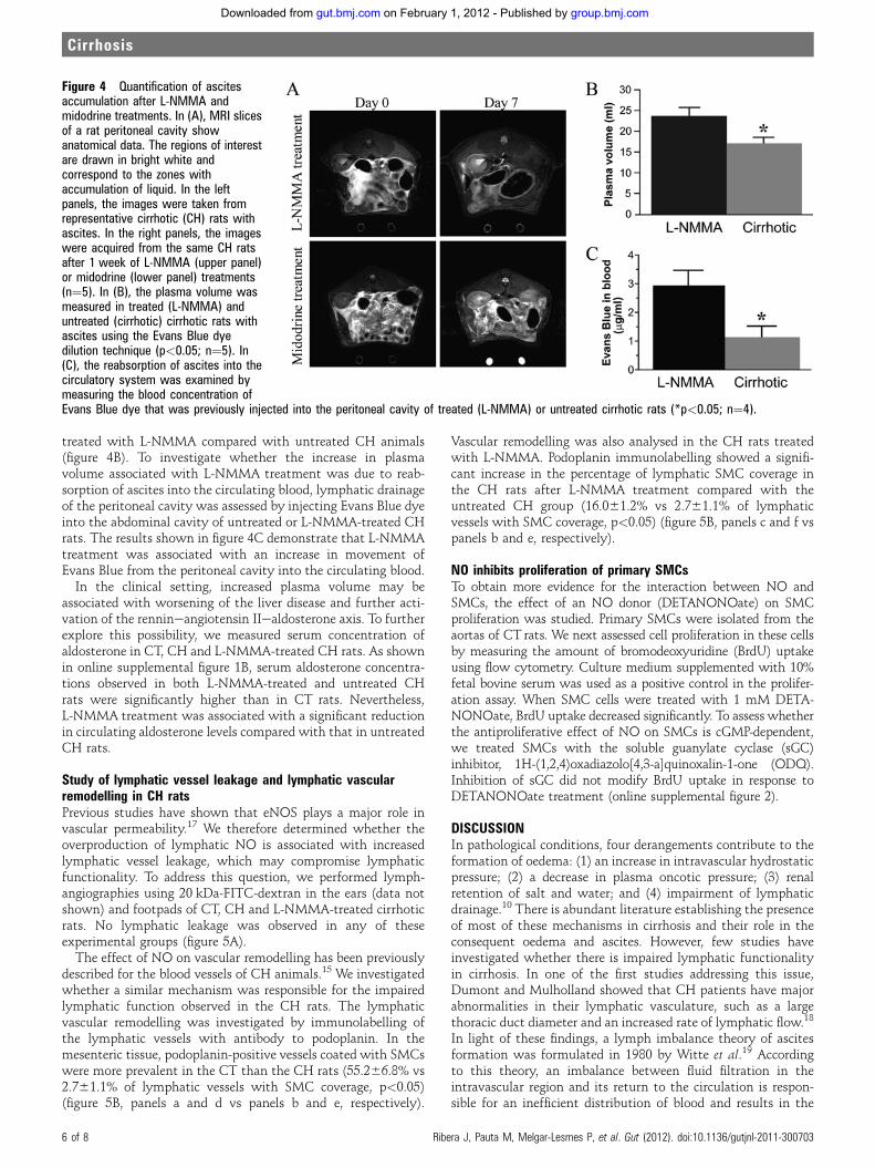
treated with L-NMMA compared with untreated CH animals(figure 4B). To investigate whether the increase in plasmavolume associated with L-NMMA treatment was due to reab-sorption of ascites into the circulating blood, lymphatic drainageof the peritoneal cavity was assessed by injecting Evans Blue dyeinto the abdominal cavity of untreated or L-NMMA-treated CHrats. The results shown in figure 4C demonstrate that L-NMMAtreatment was associated with an increase in movement ofEvans Blue from the peritoneal cavity into the circulating blood.
In the clinical setting, increased plasma volume may beassociated with worsening of the liver disease and further acti-vation of the rennineangiotensin IIealdosterone axis. To furtherexplore this possibility, we measured serum concentration ofaldosterone in CT, CH and L-NMMA-treated CH rats. As shownin online supplemental figure 1B, serum aldosterone concentra-tions observed in both L-NMMA-treated and untreated CHrats were significantly higher than in CT rats. Nevertheless,L-NMMA treatment was associated with a significant reductionin circulating aldosterone levels compared with that in untreatedCH rats.
Study of lymphatic vessel leakage and lymphatic vascularremodelling in CH ratsPrevious studies have shown that eNOS plays a major role invascular permeability.17 We therefore determined whether theoverproduction of lymphatic NO is associated with increasedlymphatic vessel leakage, which may compromise lymphaticfunctionality. To address this question, we performed lymph-angiographies using 20 kDa-FITC-dextran in the ears (data notshown) and footpads of CT, CH and L-NMMA-treated cirrhoticrats. No lymphatic leakage was observed in any of theseexperimental groups (figure 5A).
The effect of NO on vascular remodelling has been previouslydescribed for the blood vessels of CH animals.15 We investigatedwhether a similar mechanism was responsible for the impairedlymphatic function observed in the CH rats. The lymphaticvascular remodelling was investigated by immunolabelling ofthe lymphatic vessels with antibody to podoplanin. In themesenteric tissue, podoplanin-positive vessels coated with SMCswere more prevalent in the CT than the CH rats (55.266.8% vs2.761.1% of lymphatic vessels with SMC coverage, p<0.05)(figure 5B, panels a and d vs panels b and e, respectively).
Vascular remodelling was also analysed in the CH rats treatedwith L-NMMA. Podoplanin immunolabelling showed a signifi-cant increase in the percentage of lymphatic SMC coverage inthe CH rats after L-NMMA treatment compared with theuntreated CH group (16.061.2% vs 2.761.1% of lymphaticvessels with SMC coverage, p<0.05) (figure 5B, panels c and f vspanels b and e, respectively).
NO inhibits proliferation of primary SMCsTo obtain more evidence for the interaction between NO andSMCs, the effect of an NO donor (DETANONOate) on SMCproliferation was studied. Primary SMCs were isolated from theaortas of CT rats. We next assessed cell proliferation in these cellsby measuring the amount of bromodeoxyuridine (BrdU) uptakeusing flow cytometry. Culture medium supplemented with 10%fetal bovine serum was used as a positive control in the prolifer-ation assay. When SMC cells were treated with 1 mM DETA-NONOate, BrdU uptake decreased significantly. To assess whetherthe antiproliferative effect of NO on SMCs is cGMP-dependent,we treated SMCs with the soluble guanylate cyclase (sGC)inhibitor, 1H-(1,2,4)oxadiazolo[4,3-a]quinoxalin-1-one (ODQ).Inhibition of sGC did not modify BrdU uptake in response toDETANONOate treatment (online supplemental figure 2).
DISCUSSIONIn pathological conditions, four derangements contribute to theformation of oedema: (1) an increase in intravascular hydrostaticpressure; (2) a decrease in plasma oncotic pressure; (3) renalretention of salt and water; and (4) impairment of lymphaticdrainage.10 There is abundant literature establishing the presenceof most of these mechanisms in cirrhosis and their role in theconsequent oedema and ascites. However, few studies haveinvestigated whether there is impaired lymphatic functionalityin cirrhosis. In one of the first studies addressing this issue,Dumont and Mulholland showed that CH patients have majorabnormalities in their lymphatic vasculature, such as a largethoracic duct diameter and an increased rate of lymphatic flow.18
In light of these findings, a lymph imbalance theory of ascitesformation was formulated in 1980 by Witte et al.19 Accordingto this theory, an imbalance between fluid filtration in theintravascular region and its return to the circulation is respon-sible for an inefficient distribution of blood and results in the
Figure 4 Quantification of ascitesaccumulation after L-NMMA andmidodrine treatments. In (A), MRI slicesof a rat peritoneal cavity showanatomical data. The regions of interestare drawn in bright white andcorrespond to the zones withaccumulation of liquid. In the leftpanels, the images were taken fromrepresentative cirrhotic (CH) rats withascites. In the right panels, the imageswere acquired from the same CH ratsafter 1 week of L-NMMA (upper panel)or midodrine (lower panel) treatments(n¼5). In (B), the plasma volume wasmeasured in treated (L-NMMA) anduntreated (cirrhotic) cirrhotic rats withascites using the Evans Blue dyedilution technique (p<0.05; n¼5). In(C), the reabsorption of ascites into thecirculatory system was examined bymeasuring the blood concentration ofEvans Blue dye that was previously injected into the peritoneal cavity of treated (L-NMMA) or untreated cirrhotic rats (*p<0.05; n¼4).
6 of 8 Ribera J, Pauta M, Melgar-Lesmes P, et al. Gut (2012). doi:10.1136/gutjnl-2011-300703
Cirrhosis
group.bmj.com on February 1, 2012 - Published by gut.bmj.comDownloaded from
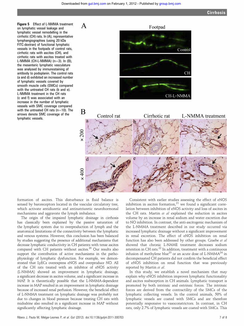
formation of ascites. This disturbance in fluid balance issensed by baroreceptors located in the vascular circulatory tree,which activate antidiuretic and antinatriuretic neurohormonalmechanisms and aggravate the lymph imbalance.
The origin of the impaired lymphatic drainage in cirrhosishas classically been explained by the passive saturation ofthe lymphatic system due to overproduction of lymph and theanatomical limitations of the connectivity between the lymphaticand venous systems. However, this conclusion has been balancedby studies suggesting the presence of additional mechanisms thatdecrease lymphatic conductivity in CH patients with tense ascitescompared with CH patients without ascites.20 Our results alsosupport the contribution of active mechanisms in the patho-physiology of lymphatic dysfunction. For example, we demon-strated that LyECs overexpress eNOS and overproduce NO. Allof the CH rats treated with an inhibitor of eNOS activity(L-NMMA) showed an improvement in lymphatic drainage,a significant decrease in ascites volume, and a significant increase inMAP. It is theoretically possible that the L-NMMA-dependentincrease in MAP resulted in an improvement in lymphatic drainagebecause of increased renal perfusion. However, the beneficial effectof L-NMMA treatment on lymphatic drainage was probably notdue to changes in blood pressure because treating CH rats withmidodrine also resulted in a significant increase in MAP withoutsignificantly affecting lymphatic drainage.
Consistent with earlier studies assessing the effect of eNOSinhibition in ascites formation,21 we found a significant corre-lation between inhibition of eNOS activity and loss of ascites inthe CH rats. Martin et al explained the reduction in ascitesvolume by an increase in renal sodium and water excretion dueto NO inhibition. In contrast, the anti-ascitogenic mechanism ofthe L-NMMA treatment described in our study occurred viaincreased lymphatic drainage without a significant improvementin renal excretion. The effect of eNOS inhibition on renalfunction has also been addressed by other groups. Graebe et alshowed that chronic L-NAME treatment decreases sodiumretention in CH rats.22 In addition, treatment with a continuousinfusion of methylene blue23 or an acute dose of L-NMMA24 indecompensated CH patients did not confirm the beneficial effectof eNOS inhibition on renal function that was previouslyreported by Martin et al.In this study, we establish a novel mechanism that may
explain why eNOS inhibition improves lymphatic functionalityand ascites reabsorption in CH animals. Lymphatic transport ispromoted by both intrinsic and extrinsic forces. The intrinsicforces are derived from the contractility of the SMCs of thelymphatic collecting vessels. In the control animals, 50% oflymphatic vessels are coated with SMCs and are thereforepotentially responsive to vasoconstrictors. In contrast, in CHrats, only 2.7% of lymphatic vessels are coated with SMCs. This
Figure 5 Effect of L-NMMA treatmenton lymphatic vessel leakage andlymphatic vessel remodelling in thecirrhotic (CH) rats. In (A), representativelymphangiographies (using 20 kDaFITC-dextran) of functional lymphaticvessels in the footpads of control rats,cirrhotic rats with ascites (CH), andcirrhotic rats with ascites treated withL-NMMA (CH-L-NMMA) (n¼3). In (B),the mesenteric lymphatic vasculaturewas analysed by immunostaining ofantibody to podoplanin. The control rats(a and d) exhibited an increased numberof lymphatic vessels covered bysmooth muscle cells (SMCs) comparedwith the untreated CH rats (b and e).L-NMMA treatment in the CH rats(c and f) was associated with anincrease in the number of lymphaticvessels with SMC coverage comparedwith the untreated CH rats (n¼10). Thearrows denote SMC coverage of thelymphatic vessels.
Ribera J, Pauta M, Melgar-Lesmes P, et al. Gut (2012). doi:10.1136/gutjnl-2011-300703 7 of 8
Cirrhosis
group.bmj.com on February 1, 2012 - Published by gut.bmj.comDownloaded from

percentage represents a significant loss of contractile capacity ofthe lymphatic system in cirrhosis. Regarding the pathologicalrole of lymphatic NO overproduction in lymphatic dysfunction,we demonstrated that NO inhibits SMC proliferation inprimary rat SMCs. In addition, the coating of lymphatic vesselsby SMCs was partially restored in CH rats by chronic admin-istration of L-NMMA, and this treatment was associated with(1) restoration of the lymphatic circulation in the mesentericregion (figure 1B) and (2) increased ascites drainage from theperitoneal cavity into the circulating blood (figure 4C). Impor-tantly, a significant increase in plasma volume with lower levelsof circulating aldosterone was noted in the L-NMMA-treatedanimals compared with untreated CH rats, supporting the ideathat the increase in plasma volume is not associated witha worsening of the cirrhosis but with the redistribution of bodyfluids into the circulation. eNOS inhibition in CH rats may alsorestore the contractility of the smooth muscle layer of thecollecting lymphatic vessels by suppressing lymphatic vasodila-tion via NO overproduction. This conclusion is supported bya previous study that found that eNOS inhibitors or the geneticdeletion of eNOS modify lymph flow in mice via NO-dependentregulation of the contractile driving force of collecting lymphaticvessels.11
Several studies have demonstrated increased NOS activity inthe blood vessels of systemic and splanchnic areas of bothcirrhotic patients and rats with experimental cirrhosis and/orportal hypertension. Our study leads us to further postulate thatthis overproduction is not limited to blood vessels but alsooccurs in lymphatic vessels. These previous publications suggestthat eNOS inhibitors may be a useful therapeutic strategy for thetreatment of the hyperdynamic abnormalities andoedema formation characteristic of cirrhosis. However, this situ-ation substantially differs from the defective eNOS activationdescribed in cirrhotic livers, where tissue-specific restoration ofintrahepatic eNOS activity is associated with reduced portalpressure in experimental models.25 26 Thus attempts to inhibitNOS activity must be undertaken carefully, and further researchshould be performed to improve both tissue-specific deliverystrategies for NO supplementation in cirrhotic livers and theinhibition of eNOS activity in extrahepatic blood and lymphaticvessels.
Author affiliations1Department of Biochemistry and Molecular Genetics, Hospital Clinic of Barcelona,IDIBAPS, CIBERehd, Barcelona, Spain2Department of Immunology, Genetics and Pathology, Rudbecklaboratoriet, Uppsala,Sweden3Department of Pharmacology, Vascular Biology and Therapeutics Program, YaleUniversity, New Haven, Connecticut, USA4Experimental 7T-MRI Unit, IDIBAPS, Barcelona, Spain5CIBER-BBN, Group of Biomedical Imaging of the University of Barcelona, Barcelona,Spain6Department of Brain Ischemia and Neurodegeneration, IIBB-CSIC, IDIBAPS,Barcelona, Spain7Departments of Medicine and Cell Biology, Leon H. Charney Division of Cardiology andthe Marc and Ruti Bell Vascular Biology and Disease Program, New York UniversitySchool of Medicine, New York, NY, USA8Liver Unit-Institut de Malalties Digestives, Hospital Clınic i Provincial de Barcelona,IDIBAPS, CIBERehd, University of Barcelona, Barcelona, Spain9Department of Physiological Sciences I, University of Barcelona, Barcelona, Spain10Department of Physiological Sciences I, University of Barcelona, Barcelona, Spain
Funding This work was supported by grants from the Ministerio de Ciencia eInnovacion-Plan Nacional de I+D+I (SAF2007-63069 and SAF2010-19025 to MM-Rand SAF2009-08039 to WJ) and AGAUR (2009 SGR 1496 to WJ). MP was supportedby MICINN (contract number BES-2007-16909). CIBERehd and CIBER-BBN arefinanced by the Instituto de Salud Carlos III.
Competing interests None.
Contributors JR, MP, PM-L, ST and GF-V contributed to the acquisition andinterpretation of data for cirrhosis induction, lymphatic drainage and vascular biology.VA and WJ helped draft the article and revised it critically for its scientific content. GS,RT and AMP contributed to the acquisition and interpretation of MRI data. MM-Rcontributed to the conception and design of the study as well as to the analysis andinterpretation of the data. KFH, CF-H, MP and JR were involved in the isolation ofsmooth muscle cells, the proliferation experiments, the aldosterone measurementsand the experiments on lymphatic drainage in the peritoneal cavity. GF-V carried outthe measurements of portal pressure.
Provenance and peer review Not commissioned; externally peer reviewed.
REFERENCES1. Moore KP,Wong F, Gines P, et al. The management of ascites in cirrhosis: report on
the consensus conference of the International Ascites Club. Hepatology2003;38:258e66.
2. Gines P, Cardenas A, Arroyo V, et al. Management of cirrhosis and ascites. N Engl JMed 2004;350:1646e54.
3. Van Steenkiste C, Geerts A, Vanheule E, et al. Role of placental growth factor inmesenteric neoangiogenesis in a mouse model of portal hypertension.Gastroenterology 2009;137:2112e24.
4. Henriksen JH, Moller S. Alterations of hepatic and splanchnic microvascularexchange in cirrhosis: local factors in the formation of ascites. In: Gines P, Arroyo V,Rodes J, et al, eds. Ascites and Renal Dysfunction in Liver Disease: Pathogenesis,Diagnosis and Treatment. Malden, MA: Blackwell Publishing, 2005:174e85.
5. Bhunchet E, Fujieda K. Capillarization and venularization of hepatic sinusoids inporcine serum-induced rat liver fibrosis: a mechanism to maintain liver blood flow.Hepatology 1993;18:1450e8.
6. Mori T, Okanoue T, Sawa Y, et al. Defenestration of the sinusoidal endothelial cell ina rat model of cirrhosis. Hepatology 1993;17:891e7.
7. Melgar-Lesmes P, Tugues S, Ros J, et al. Vascular endothelial growth factor andangiopoietin-2 play a major role in the pathogenesis of vascular leakage in cirrhoticrats. Gut 2009;58:285e92.
8. Cardenas A, Bataller R, Arroyo V. Mechanisms of ascites formation. Clin Liver Dis2000;4:447e65.
9. Schmid-Schonbein GW. Microlymphatics and lymph flow. Physiol Rev1990;70:987e1028.
10. Aukland K, Reed RK. Interstitial-lymphatic mechanisms in the control of extracellularfluid volume. Physiol Rev 1993;73:1e78.
11. Hagendoorn J, Padera TP, Kashiwagi S, et al. Endothelial nitric oxide synthaseregulates microlymphatic flow via collecting lymphatics. Circ Res 2004;95:204e9.
12. Witte MH, Dumont AE, Cole WR, et al. Lymph circulation in hepatic cirrhosis: effectof portacaval shunt. Ann Intern Med 1969;70:303e10.
13. Tugues S, Morales-Ruiz M, Fernandez-Varo G, et al. Microarray analysis ofendothelial differentially expressed genes in liver of cirrhotic rats. Gastroenterology2005;129:1686e95.
14. Ros J, Jimenez W, Lamas S, et al. Nitric oxide production in arterial vessels ofcirrhotic rats. Hepatology 1995;21:554e60.
15. Fernandez-Varo G, Ros J, Morales-Ruiz M, et al. Nitric oxide synthase 3-dependentvascular remodeling and circulatory dysfunction in cirrhosis. Am J Pathol2003;162:1985e93.
16. Morales-Ruiz M, Cejudo-Martin P, Fernandez-Varo G, et al. Transduction of the liverwith activated Akt normalizes portal pressure in cirrhotic rats. Gastroenterology2003;125:522e31.
17. Thibeault S, Rautureau Y, Oubaha M, et al. S-nitrosylation of beta-catenin byeNOS-derived NO promotes VEGF-induced endothelial cell permeability. Mol Cell2010;39:468e76.
18. Dumont AE, Mulholland JH. Flow rate and composition of thoracic-duct lymph inpatients with cirrhosis. N Engl J Med 1960;263:471e4.
19. Witte CL, Witte MH, Dumont AE. Lymph imbalance in the genesis and perpetuationof the ascites syndrome in hepatic cirrhosis. Gastroenterology 1980;78:1059e68.
20. Henriksen JH. Estimation of lymphatic conductance. A model based on protein-kinetic studies and haemodynamic measurements in patients with cirrhosis of theliver and in pigs. Scand J Clin Lab Invest 1985;45:123e30.
21. Martin PY, Ohara M, Gines P, et al. Nitric oxide synthase (NOS) inhibition for oneweek improves renal sodium and water excretion in cirrhotic rats with ascites. J ClinInvest 1998;101:235e42.
22. Graebe M, Brond L, Christensen S, et al. Chronic nitric oxide synthase inhibitionexacerbates renal dysfunction in cirrhotic rats. Am J Physiol Renal Physiol2004;286:288e97.
23. Cardenas A, Curry M, Afdhal NH. The not so good effects of nitric oxide inhibitionwith methylene blue in cirrhosis and ascites. Dig Dis Sci 2007;52:939e40.
24. Thiesson HC, Skott O, Jespersen B, et al. Nitric oxide synthase inhibition does notimprove renal function in cirrhotic patients with ascites. Am J Gastroenterol2003;98:180e6.
25. Morales-Ruiz M, Jimenez W. Pathophysiological role of nitric oxide in cirrhosis. In:Arroyo V, Gines P, Rodes J, et al, eds. Ascites and Renal Dysfunction in Liver Disease.Pathogenesis Diagnosis and Treatment. Malden, Massachussets: Blackwell ScienceInc, 2005:105e14.
26. Langer DA, Shah VH. Nitric oxide and portal hypertension: interface of vasoreactivityand angiogenesis. J Hepatol 2006;44:209e16.
PAGE fraction trail=88 of 8 Ribera J, Pauta M, Melgar-Lesmes P, et al. Gut (2012). doi:10.1136/gutjnl-2011-300703
Cirrhosis
group.bmj.com on February 1, 2012 - Published by gut.bmj.comDownloaded from
doi: 10.1136/gutjnl-2011-300703 published online January 20, 2012Gut
Jordi Ribera, Montse Pauta, Pedro Melgar-Lesmes, et al. cirrhotic ratsimpairment of lymphatic drainage inlymphatic endothelial cells causes Increased nitric oxide production in
http://gut.bmj.com/content/early/2012/01/20/gutjnl-2011-300703.full.htmlUpdated information and services can be found at:
These include:
Data Supplementhttp://gut.bmj.com/content/suppl/2012/01/20/gutjnl-2011-300703.DC1.html"Supplementary Data"
Referenceshttp://gut.bmj.com/content/early/2012/01/20/gutjnl-2011-300703.full.html#ref-list-1This article cites 24 articles, 5 of which can be accessed free at:
P<P Published online January 20, 2012 in advance of the print journal.
serviceEmail alerting
the box at the top right corner of the online article.Receive free email alerts when new articles cite this article. Sign up in
Notes
(DOIs) and date of initial publication. publication. Citations to Advance online articles must include the digital object identifier citable and establish publication priority; they are indexed by PubMed from initialtypeset, but have not not yet appeared in the paper journal. Advance online articles are Advance online articles have been peer reviewed, accepted for publication, edited and
http://group.bmj.com/group/rights-licensing/permissionsTo request permissions go to:
http://journals.bmj.com/cgi/reprintformTo order reprints go to:
http://group.bmj.com/subscribe/To subscribe to BMJ go to:
group.bmj.com on February 1, 2012 - Published by gut.bmj.comDownloaded from

1
SUPPLEMENTAL INFORMATION
1. Supplemental methods
2. References
3. Figure legends
4. Supplemental Table 1
2
1.- SUPPLEMENTAL METHODS
Western blot for endothelial nitric oxide (eNOS) detection and nitric oxide
(NO) quantification. The cell lysates were prepared in lysis buffer (Tris–HCl 20
mM pH 7.4 containing 1% Triton X-100, 0.1% SDS, 50 mM NaCl, 2.5 mM
EDTA, 1 mM Na4P2O7 10H2O, 20 mM NaF, 1 mM Na3VO4, 2 mM Pefabloc and
Complete® from Roche). The cell lysates were separated on a 7.5% SDS-
polyacrylamide gel (Mini Protean III, BioRad, Richmond, Ca) and transferred for
2 hours at 4ºC to nitrocellulose membranes (Transblot Transfer Medium,
BioRad, Richmond, CA) that were stained with Ponceau-S red as a control for
protein loading. The membranes were incubated at 4ºC with mouse monoclonal
anti-rat eNOS or anti-rat iNOS (BD Transduction Laboratories, Franklin Lakes,
NJ) overnight in a 1:1000 dilution. They were then incubated with goat anti-
mouse peroxidase-conjugated secondary antibody at a 1:2000 dilution (Cell
Signaling, Beverly, MA) for 1 hour at room temperature. The bands were
visualized by chemiluminescence (ECL western blotting analysis system;
Amersham Biosciences). To measure the NO concentration, the basal
accumulation of NO2- in the cell culture medium was quantified using a
chemiluminescence detector in a NO analyzer (Sievers, Buckinghamshire, UK).
Magnetic resonance imaging (MRI). The MRI experiments were conducted on
a 7.0 T BioSpec 70/30 horizontal animal scanner (Bruker BioSpin, Ettlingen,
Germany) that was equipped with a 12-cm inner diameter actively shielded
gradient system (400 mT/m). The receiver coil was a phased-array surface coil
for the rat brain. The animals were placed in a supine position in a Plexiglas
holder with a nose cone to administer anesthetic gases (isoflurane in a mixture
of 30% O2 and 70% CO2) and were held in position using a tooth bar, ear bars
3
and adhesive tape. Tripilot scans were used for accurate positioning of the
animal's head in the isocenter of the magnet. T2-weighted images were
acquired using a TurboRARE (rapid acquisition with rapid enhancement)
sequence using the following parameters: repetition time = 3236 ms, echo time
= 9 ms, RARE factor = 8, 4 averages, slice thickness = 1 mm with 1 mm gap
between slices, number of slices = 28, field of view = 60 x 60 mm3, matrix size =
256 x 256 pixels and a spatial resolution of 0.234 × 0.234 mm in a 1 mm slice
thickness. To acquire the total volume of the abdomen, the acquisition was
immediately repeated with the field of view shifted 1 mm in the z direction. The
total scan time was 30 minutes. Images were processed and analyzed using the
software Image J (National Institutes of Health, Bethesda, MD).
Plasma volume quantification. Plasma volume was measured using Evans
Blue dye, as previously described.[1] Briefly, 0.2 mL of Evans Blue (Sigma
Chemical, St. Louis, Missouri, USA) solution (3 mg) was injected through a
jugular vein catheter followed by physiological saline to clear the dye from the
catheter. Five minutes later, 1 mL of blood was withdrawn from a femoral artery
catheter. A 0.2-mL plasma aliquot was diluted in distilled water to 2 mL, and the
absorbance of the solution was read by a spectrophotometer at a 600-nm
wavelength. The plasma volume was calculated by the following formula:
plasma volume (mL) = Astandard/Asample x 10,
where the standard was 3 mg of Evans Blue dye in 10 mL of plasma diluted by
a factor of 10.
Evans Blue intraperitoneal drainage. The Evans Blue dye technique was
used to measure lymphatic drainage in the peritoneum. Thirty milligrams of
Evans Blue per kilogram (Sigma Chemical, St Louis, Missouri, USA) diluted in
4
physiological saline was injected in the peritoneal cavity. Thirty minutes later, an
aliquot of blood was extracted from heart and centrifuged 5 minutes at 3000
rpm. Next, 100 µl of serum was mixed with 900 µl of 15% trichloroacetic acid to
precipitate the dye. The mixture was then centrifuged, washed twice with 98%
ethanol, and resuspended in 1 mL of formamide. The dye was quantified by
spectrophotometry at 620 nm the results were calculated from a Evans Blue
standard curve (0.9–30 µg/mL) and expressed as µg of EB/mL of blood.
Isolation of smooth muscle cells (SMCs) from the thoracic aorta. Freshly
isolated primary SMCs were obtained from the thoracic aorta of CT rats. Briefly,
the atrium was cannulated, and the vascular system was perfused with normal
saline solution. Following removal of the adhering adventitia, thoracic aortas
were harvested and placed in a 1 mg/mL collagenase A (Roche Diagnostics,
Basel, Switzerland) solution at 37°C for 10 minutes. Next, the vessels were
rinsed with fresh HBSS and the remaining adventitia was removed under a
dissecting microscope. The vessels were cut into small (1 mm) fragments and
placed in a solution of 2 mg/mL collagenase A (Roche Diagnostics, Basel,
Switzerland) and 0.5 mg/mL elastase (Sigma Chemical, St Louis, Missouri,
USA) at 37ºC for 1 hour. Finally, the cell suspension was centrifuged at 1800
rpm for four minutes and resuspended in culture medium . The cells were grown
in Dulbecco’s Modified Eagle Medium (DMEM) that was supplemented with
10% fetal calf serum, 50 U/mL penicillin and 50 µg/mL streptomycin.
Balance studies of renal excretion. After developing ascites, rats were placed
in individual metabolic cages for one week before the balance study began.
Then, rats were treated with L-NMMA, Midodrine or vehicle for 7 days. Each rat
received the same diet and a sample of 24-hour urine was collected every day.
5
The diet consisted of 0.1% sodium and 0.6% potassium (Global Diet 2014,
Harlan, Indianapolis, Indiana, USA).
Cell proliferation assay. A BrdU cell proliferation assay kit (BrdU Flow kit; BD
Pharmigen, San Jose, CA, USA) was used to measure the incorporation of
BrdU during DNA synthesis following the manufacturer’s protocol. To assess
NO-dependent responses, the NO donor diethylenetriamine NONOate (DETA
NONOate) (Cayman Chemical Company, Ann Arbor, Michigan, USA) was used
at a final concentration of 1mM. In some experimental conditions, a dose of 50
µM 1H-[1,2,4]oxadiazolo-[4,3-a]quinoxalin-1-one (ODQ) (Sigma Chemical, St.
Louis, Missouri, USA) was used to inhibit soluble guanylate cyclase activity.
Briefly, primary rat SMCs isolated from thoracic aortas were cultivated in serum-
free medium for 24 hours. The cells were subsequently treated with 10% FBS,
10% FBS+1mM DETANONOate or 10% FBS+1mM DETANONOate+50µM
ODQ for an additional 24 hours. BrdU (10 µM) was added to the culture medium
for 5 h. The cells were fixed, and the DNA was denatured in a fixative solution
for 1 hour at 37°C. The cells were then incubated with a fluorescein
isothiocyanate (FITC)–conjugated anti-BrdU antibody for 30 minutes at room
temperature. BrdU uptake was quantified using flow cytometry (FACSCanto II,
BD Bioscience).
6
2.- REFERENCES
1. Li Y, Liu H, Gaskari SA, et al. Hepatic venous dysregulation contributes to
blood volume pooling in cirrhotic rats. Gut 2006;55:1030-1035.
7
3.- FIGURE LEGENDS
Supplemental_Figure_1: Quantification of portal pressure and aldosterone in
untreated and L-NMMA-treated cirrhotic ascitic rats. In (A), portal pressure was
measured in untreated and L-NMMA-treated CH rats as described in the
supplemental methods section. The data are presented as the mean±standard
error of the mean (n=5). In (B), the serum concentration of aldosterone was
measured in control, cirrhotic and L-NMMA-treated CH rats (L-NMMA) using a
radioimmune assay with a commercial kit as described in the Material and
Methods section. The data are presented as the mean±standard error of the
mean. *P<0.05 vs. control and L-NMMA and #P<0.05 vs. control (n=5)
Supplemental_Figure_2: Effect of an NO donors on SMC proliferation.
Representative figures are shown of a proliferation assay performed with
primary SMCs isolated from the aorta of CT rats treated with DETANONOate or
with DETANONOate+ODQ for 24 hours. The degree of BrdU incorporation was
quantified using flow cytometry. For each panel, the percentage of cells that
stained positively for BrdU is indicated. Similar results were obtained in five
independent experiments.
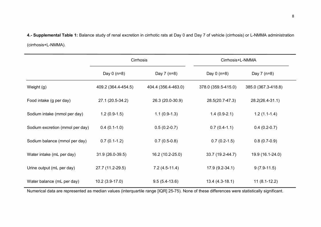
8
4.- Supplemental Table 1: Balance study of renal excretion in cirrhotic rats at Day 0 and Day 7 of vehicle (cirrhosis) or L-NMMA administration
(cirrhosis+L-NMMA).
Cirrhosis Cirrhosis+L-NMMA
Day 0 (n=8) Day 7 (n=8) Day 0 (n=8) Day 7 (n=8)
Weight (g) 409.2 (364.4-454.5) 404.4 (356.4-463.0) 378.0 (359.5-415.0) 385.0 (367.3-418.8)
Food intake (g per day) 27.1 (20.5-34.2) 26.3 (20.0-30.9) 28.5(20.7-47.3) 28.2(26.4-31.1)
Sodium intake (mmol per day) 1.2 (0.9-1.5) 1.1 (0.9-1.3) 1.4 (0.9-2.1) 1.2 (1.1-1.4)
Sodium excretion (mmol per day) 0.4 (0.1-1.0) 0.5 (0.2-0.7) 0.7 (0.4-1.1) 0.4 (0.2-0.7)
Sodium balance (mmol per day) 0.7 (0.1-1.2) 0.7 (0.5-0.8) 0.7 (0.2-1.5) 0.8 (0.7-0.9)
Water intake (mL per day) 31.9 (26.0-39.5) 16.2 (10.2-25.0) 33.7 (19.2-44.7) 19.9 (16.1-24.0)
Urine output (mL per day) 27.7 (11.2-29.5) 7.2 (4.5-11.4) 17.9 (9.2-34.1) 9 (7.9-11.5)
Water balance (mL per day) 10.2 (3.9-17.0) 9.5 (5.4-13.6) 13.4 (4.3-18.1) 11 (8.1-12.2)
Numerical data are represented as median values (interquartile range [IQR] 25-75). None of these differences were statistically significant.

9
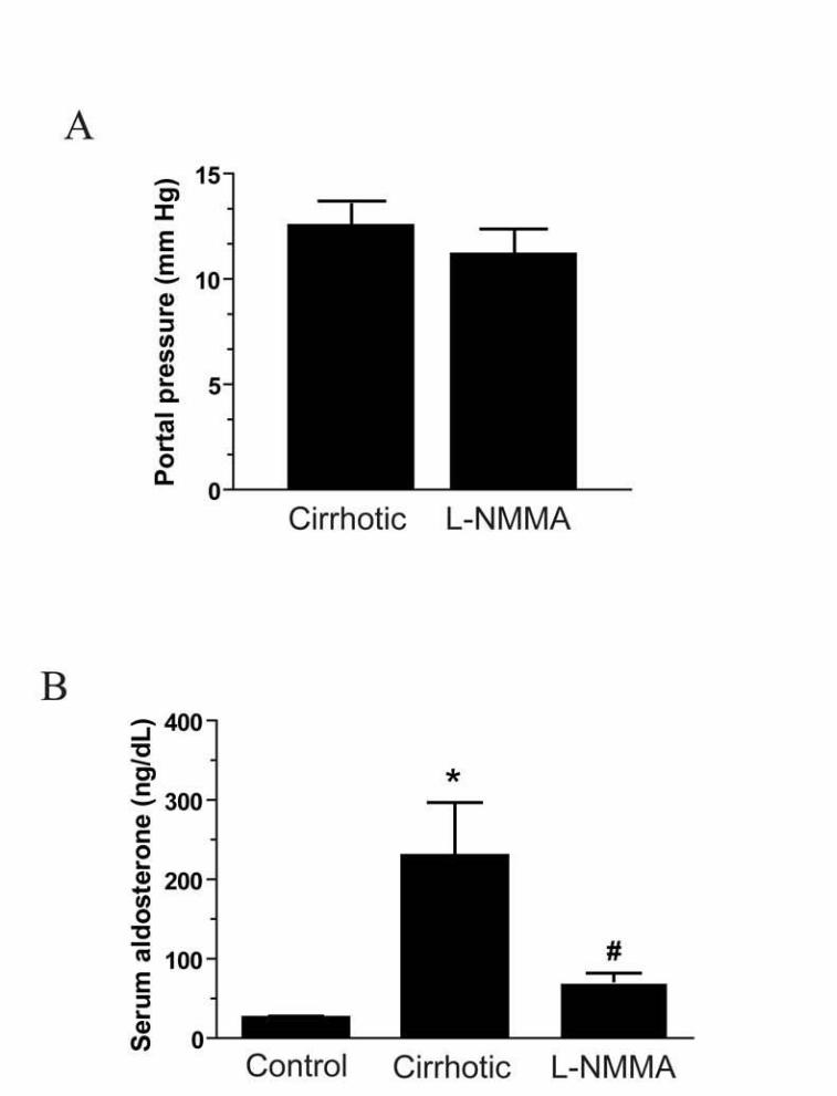
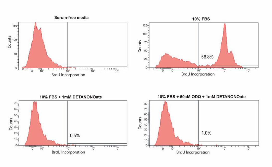

DISCUSIÓN

DISCUSIÓN _______________________________________________________________________
56
Las vasculaturas, tanto sanguínea como linfática, desempeñan funciones claves
para la viabilidad de los organismos complejos. La fisiología y la fisiopatología del
sistema vascular sanguíneo y la implicación de la angiogénesis (la formación de nuevos
vasos sanguíneos a partir de otros preexistentes) en múltiples enfermedades, ha sido
muy estudiada en las últimas décadas (Carmeliet, 2003). Hay muchas evidencias que
sugieren que el incremento incontrolado y desorganizado de la vasculatura sanguínea
podría ser la causa y el mantenimiento de algunas de las complicaciones asociadas a la
cirrosis hepática. En este contexto, las terapias antiangiogénicas clásicas dirigidas a
bloquear VEGF o las vías de los receptores tirosina quinasa, han demostrado ser
eficaces en disminuir el grado de angiogénesis, de fibrosis y de inflamación hepática,
además de disminuir también la presión portal (Mejias et al., 2009; Tugues et al.,
2007). Sin embargo, como ya se ha comentado anteriormente, este tipo de terapias se
han visto relacionadas con multitud de efectos adversos (Fischer et al., 2007; Wu et al.,
2008). En contraposición con los inhibidores clásicos de VEGF, los anticuerpos
monoclonales contra PlGF han demostrado que reducen la angiogénesis patológica en
varios tipos de cáncer sin afectar a la vasculatura sana y sin mayores efectos adversos
(Lassen et al., 2009; Riisbro et al., 2009; Van de Veire et al., 2010). Esto es así porque
PlGF no tiene ninguna función durante el desarrollo de angiogénesis fisiológica y no es
necesario para el mantenimiento de la quiescencia vascular en órganos sanos (Fischer
et al., 2007; Van de Veire et al., 2010). Sin embargo, estudios recientes en ratones
transgénicos demostraron que PlGF tiene actividad angiogénica sólo en condiciones
patológicas (Autiero et al., 2003; Van Steenkiste et al., 2009).
En el momento de empezar esta tesis doctoral se sabía que PlGF estimulaba el
crecimiento de las células endoteliales, su migración y supervivencia, así como la
angiogénesis patológica (Fischer et al., 2007; Van de Veire et al., 2010; Khurana et al.,
2005). Varias evidencias apuntaban hacia el posible uso terapéutico de la inhibición de
PlGF en la cirrosis: 1) las propiedades proangiogénicas y proinflamatorias de PlGF, y 2)
el efecto sinérgico que existe entre inflamación y angiogénesis, previamente
demostrado con el uso de otros inhibidores de RTKs en la cirrosis (Tugues et al., 2007;
Mejias et al., 2009). A pesar de esta información, en el periodo previo al diseño de
nuestro estudio existían pocas publicaciones científicas que avalasen esta hipótesis
DISCUSIÓN _______________________________________________________________________
57
(Van Steenkiste et al., 2009; Huang et al., 2007; Salcedo Mora et al., 2005). En estudios
previos se había demostrado que PlGF estaba sobreexpresado en la microvasculatura
esplácnica de ratones con hipertensión portal y que la deficiencia de PlGF en ratones
con ligadura parcial de la vena porta disminuía significativamente la angiogénesis
esplácnica, la formación de vasos porto-sistémicos, y el flujo arterial mesentérico (Van
Steenkiste et al., 2009). El primer estudio de esta tesis doctoral ha sido el primero en
describir la función de PlGF en el contexto de la cirrosis, caracterizando los efectos
fisiopatológicos y los mecanismos moleculares que tiene PlGF en hígados cirróticos y,
específicamente, en las HSCs. Tanto en un estudio de prevención como en un estudio
terapéutico, se ha demostrado que el bloqueo de la actividad de PlGF disminuye
significativamente la angiogénesis hepática y mesentérica, la arteriogénesis, la
inflamación hepática, el flujo arterial mesentérico, la fibrosis, y la hipertensión portal
en ratones cirróticos; todo ello en un perfil óptimo de bioseguridad. Seguidamente, se
ha corroborado la relevancia de estos resultados en humanos, demostrando que los
niveles de PlGF circulante en suero y su expresión proteica a nivel hepático están
incrementados en pacientes cirróticos y además su valor se correlaciona con el grado
de fibrosis. Finalmente, se han estudiado los efectos de PlGF en HSCs, que tienen una
importante función fisiopatológica en la fibrosis y la hipertensión portal.
Un hallazgo importante de este estudio ha sido la asociación que existe entre el
bloqueo de PlGF y la reducción significativa de la presión portal en ratones cirróticos.
También se ha encontrado una correlación positiva entre los niveles de PlGF circulante
en suero y el gradiente de presión venoso hepático en pacientes con cirrosis.
Basándonos en estas observaciones, podemos especular que el PlGF podría estar
involucrado también en la patogénesis de la presión portal en humanos. Hay bastantes
evidencias que sugieren que el incremento de presión portal no sólo se debe al
aumento del flujo sanguíneo y a la vasodilatación esplácnica, sino también a la
expansión de la red vascular esplácnica provocada por la angiogénesis (Van Steenkiste
et al., 2009). Teniendo esto en cuenta, la reducción de la angiogénesis y la
arteriogénesis en el área esplácnica debido al bloqueo de PlGF contribuiría a disminuir
el flujo venoso portal.
DISCUSIÓN _______________________________________________________________________
58
Otro punto importante del estudio ha sido la reducción de la fibrosis hepática al
bloquear PlGF. Esto está en concordancia con otros estudios que demuestran que la
inhibición de la angiogénesis frena la progresión de la fibrosis hepática (Taura et al.,
2008; Mejias et al., 2009; Tugues et al., 2007). Se ha demostrado que la
inmunoreactividad de PlGF en el hígado está muy acentuada en ratas y ratones
cirróticos. Además, las HCSs activadas son el tipo celular que más contribuye a la
producción de PlGF en roedores, y estas células expresan VEGFR1. Sin embargo, a
pesar de que el bloqueo de PlGF in vivo es antifibrogénico, no se han encontrado
cambios significativos en la expresión de genes profibrogénicos cuando las HCSs
humanas se han tratado con PlGF. Esta discrepancia se podría explicar considerando
que PlGF promueve un fenotipo angiogénico en HCSs caracterizado por una
fosforilación sostenida de ERK1/2 así como de un aumento en su migración y
proliferación. La adquisición de un fenotipo angiogénico por parte de las HSCs en
respuesta a PlGF ya había sido demostrado en otros estudios y conecta con el aumento
de recubrimiento de los sinusoides por HSCs característico de los hígados cirróticos
(Semela et al., 2008). Todos estos cambios llevan a una desorganización en la
vasculatura sanguínea hepática que compromete la regulación de la presión y la
perfusión intrahepáticas. Esta desorganización de la vasculatura hepática se analizó
obteniendo moldes vasculares utilizando la técnica de corrosión, y se vio que el
tratamiento con anticuerpos contra PlGF normaliza parcialmente la estructura
tridimensional de la red vascular hepática y reduce significativamente la presencia de
vasos proinflamatorios, caracterizados por la expresión de VCAM-1. Un mecanismo
similar de normalización de la vasculatura con el tratamiento contra PlGF se describió
recientemente en los nódulos del hepatocarcinoma (Semela et al., 2008). Es
interesante destacar que la reducción de la fibrosis sólo fue significativa cuando los
ratones se trataron con anticuerpos contra PlGF en fases tempranas de la enfermedad
(semanas de la 12 a la 20). En estadíos más avanzados (semanas de la 18 a la 25) no se
han encontrado efectos beneficiosos después de la terapia contra PlGF. Esto apoya la
idea de que en estadíos iniciales de la enfermedad, la adquisición de un fenotipo
angiogénico por parte de la HSCs en respuesta a PlGF, provoca un incremento en la
población de HSCs que se correlaciona con el grado de fibrosis. Sin embargo, cuando la
población de HSCs alcanza un punto crítico, la eficacia terapéutica del bloqueo de PlGF
DISCUSIÓN _______________________________________________________________________
59
queda limitada, ya que PlGF no tiene ningún efecto en la regulación de genes
profibrogénicos. Por lo tanto, esto limita la ventana de tiempo durante la que el
tratamiento contra PlGF es efectivo.
La activación sostenida de ERK en las HSCs en respuesta a PlGF nos llevó a
querer investigar los posibles mecanismos por los que esto podía estar pasando, ya
que VEGFR1 tiene una actividad tirosina quinasa relativamente débil. Fijándonos en el
perfil de proteínas fosforiladas en las HSCs estimuladas con PlGF, vimos que además de
VEGFR1 también se fosforilaban otros receptores tirosina quinasa, incluyendo PDGFRA
o EGFR. Esto podría sugerir que VEGFR1 amplía su señal a través de la asociación
molecular con otros receptores tirosina quinasa. En células endoteliales se describió un
mecanismo similar de asociación entre VEGFR1 y VEGFR2, a través del cual PlGF
aumentaba su potencial angiogénico (Autiero et al., 2003). VEGFR1 también puede
interaccionar con el receptor de las lipoproteínas de baja densidad y activarse
independientemente de su ligando por las LDL (Usui et al., 2007). Sin embargo, esta ha
sido la primera vez que se ha probado la existencia de una asociación molecular entre
VEGFR1 y otras familias de RTKs, que podría dar como resultado una ampliación de las
señales en respuesta a PlGF.
Aunque la terapia con drogas antiangiogénicas se ha usado clásicamente en
enfermedades con exceso de angiogénesis, su uso clínico no está del todo
implementado debido a que son terapias asociadas a efectos adversos, como
hipertensión, proteinuria, trombosis, o reducción de la vasculatura en órganos sanos.
Estudios en ratones transgénicos demostraron que la falta de PlGF no afectaba a su
desarrollo ni a su reproducción, pero sí que frenaba la angiogénesis patológica en
diferentes modelos de cáncer (Van de Veire et al., 2010). Además, la administración de
anticuerpos contra PlGF no se asoció con un deterioro vascular en órganos sanos
(Fischer et al., 2007) y es una terapia bien tolerada por pacientes humanos. En estos
momentos hay estudios en fase 2 con voluntarios sanos y pacientes con tumores
sólidos, y no se han encontrado mayores efectos secundarios (Lassen et al., 2009;
Riisbro et al., 2009). El presente estudio de esta tesis doctoral ha confirmado el perfil
de bioseguridad óptimo de la terapia contra PlGF y ha demostrado que no hay
DISCUSIÓN _______________________________________________________________________
60
resistencia al tratamiento por sobreexpresión de otros agentes angiogénicos como
VEGF.
El hecho de que el bloqueo de la actividad de PlGF consiga reducir la presión
portal es el dato más relevante del primer estudio, puesto que la hipertensión portal
es una de las manifestaciones clínicas más graves de la cirrosis, y lleva asociada un
incremento de la morbilidad y la mortalidad. El aumento patológico de la presión
hidrostática en el territorio venoso portal da lugar a la formación de una extensa red
de vasos colaterales que derivan parte del flujo sanguíneo portal hacia la circulación
sistémica y desvían su paso a través del hígado. Como consecuencia de este síndrome
pueden aparecer algunas de las complicaciones más importantes de la cirrosis, como
las varices gastroesofágicas o la formación de edema y ascitis (Abraldes and Bosch,
2002), que incrementan drásticamente la gravedad de la enfermedad. A pesar de ello,
actualmente no se conocen con precisión todos los mecanismos moleculares
implicados en la formación y mantenimiento de la ascitis en la cirrosis.
Los pacientes cirróticos desarrollan importantes alteraciones en la homeostasis
de los líquidos corporales, que dan lugar a la acumulación de ascitis y edema. Se sabe
que en condiciones patológicas existen cuatro desequilibrios que contribuyen a la
formación de edema: 1) un incremento de la presión hidrostática intravascular, 2) un
descenso de la presión oncótica del plasma, 3) una retención renal de sal y agua, y 4)
una disfunción en el drenaje linfático (Aukland et al., 1993). En el momento de
empezar a diseñar el segundo estudio la mayoría de artículos publicados abordaban la
implicación de los tres primeros mecanismos en la formación de edema y ascitis. Sin
embargo, pocos estudios estudiaban la existencia de una disfunción linfática en
cirrosis. En uno de ellos, Dumont y Mulholland demostraron que los pacientes CH
tenían una vasculatura linfática alterada, con el diámetro del conducto torácico
aumentado y un incremento del flujo linfático (Dumont et al., 1960). A raiz de estos
hallazgos, en el año 1980 se desarrolló una teoría que consideraba la existencia de un
desequilibrio linfático para explicar la formación de ascitis (Witte et al., 1980). Según
esta teoría, existiría un desequilibrio entre el fluido acumulado en las regiones
intravasculares y su retorno a la circulación sanguínea, lo que provocaría la formación
de ascitis.
DISCUSIÓN _______________________________________________________________________
61
Clásicamente, se había creído que la disfunción en el drenaje linfático durante
la cirrosis se daba por una saturación pasiva del sistema linfático. Esta hipótesis se
sustentaba en dos hechos: 1) la sobreproducción de linfa durante la evolución de la
enfermedad y 2) las limitaciones anatómicas de la conectividad entre los sistemas
linfático y venoso. Sin embargo, diversos estudios demostraron que existían otros
mecanismos que reducían la conductividad linfática en pacientes CH con ascitis en
comparación con pacientes CH sin ascitis (Henriksen et al., 1985). En el segundo
estudio de esta tesis doctoral demostramos que existen mecanismos activos que
contribuyen a la fisiopatología de la disfunción linfática. Por ejemplo, se ha
demostrado que las LyECs de ratas cirróticas sobreexpresan eNOS e incrementan la
producción de NO. Todas las ratas CH tratadas con un inhibidor de la actividad de
eNOS (L-NMMA) mejoraron su drenaje linfático y redujeron significativamente el
volumen de ascitis. Teóricamente, podría haber sido posible que el efecto beneficioso
de L-NMMA se debiera a un aumento en la MAP, ya que dicho incremento podría
mejorar el drenaje linfático mediante una mejora de la perfusión renal. Sin embargo,
los resultados de nuestro estudio excluyen esta posibilidad ya que ratas CH tratadas
con midodrina también aumentan significativamente su MAP sin tener ningún efecto
en el drenaje linfático.
De acuerdo con estudios recientes que habían evaluado el efecto de eNOS en la
formación de ascitis (Martin et al., 1998), se ha encontrado una correlación
significativa entre la inhibición de la actividad de eNOS y la pérdida de ascitis en ratas
CH. Martin et al explicaban la reducción de ascitis por un incremento de la excreción
renal de sodio y agua debido a la inhibición de NO. En el presente estudio,
demostramos que el mecanismo anti-ascitogénico desencadenado por el tratamiento
con L-NMMA se debió a una mejora del drenaje linfático en ausencia de cambios en la
excreción renal. Otros grupos habían descrito el efecto de la inhibición de eNOS en la
función renal. Graebe et al demostraron que el tratamiento crónico con L-NAME
reducía la retención de sodio en ratas CH (Graebe et al., 2004). Sin embargo, el
tratamiento con una infusión continua de azul de metileno (Cardenas et al., 2007) o
una dosis aguda de L-NMMA (Thiesson et al., 2003) en pacientes con CH
DISCUSIÓN _______________________________________________________________________
62
descompensada no confirmó los efectos beneficiosos de la inhibición de eNOS sobre la
función renal previamente descritos por Martin et al.
En este estudio, se ha establecido un nuevo mecanismo que podría explicar
porqué la inhibición de eNOS mejora la funcionalidad linfática y la reabsorción de
ascitis en animales CH. El transporte linfático está promovido por fuerzas extrínsecas e
intrínsecas. Las fuerzas intrínsecas vienen definidas por la capacidad contráctil de la
capa de SMCs que rodea los vasos linfáticos colectores. En los animales control, un
50% de los vasos linfáticos están recubiertos por SMC y, por lo tanto, son potenciales
respondedores a vasoconstrictores. Sin embargo, en las ratas CH sólo el 2.7% de los
vasos linfáticos están recubiertos por SMCs. Esto representa una pérdida significativa
de la capacidad contráctil del sistema linfático en cirrosis. Teniendo en cuenta la
función fisiopatológica de la sobreproducción linfática de NO en la disfunción linfática,
se ha demostrado que el NO inhibe la proliferación de las SMC primarias de rata. Es
más, la administración crónica de L-NMMA en ratas CH ha conseguido recuperar
parcialmente el recubrimiento de vasos linfáticos por SMC. Estos cambios se han
asociado a una restauración de la circulación linfática en la región mesentérica y a un
incremento del drenaje de ascitis desde la cavidad peritoneal hasta la circulación
sanguínea, observándose un incremento del volumen plasmático con niveles bajos de
aldosterona circulante. Esto indicaría que el aumento de volumen plasmático no se
asociaría con un empeoramiento de la condición de cirrosis sino con una redistribución
de los fluidos corporales. La inhibición de eNOS en ratas CH también restauraría la
capacidad contráctil de la capa de células musculares lisas de los vasos linfáticos
colectores, ya que se revertiría la vasodilatación producida por la sobreproducción de
NO. Esta conclusión se apoya en estudios previos, donde se demostró que los
inhibidores de eNOS modificaban el flujo linfático en ratones a través de la regulación
de las fuerzas contráctiles NO-dependientes de los vasos linfáticos colectores
(Hagendoorn et al., 2004).
Múltiples estudios habían demostrado que en cirrosis existía un incremento de
la actividad NOS en vasos sanguíneos, tanto sistémicos como esplácnicos. El presente
estudio ha demostrado que esta sobreproducción de NO no se limita sólo a los vasos
sanguíneos sino que afecta también a los vasos linfáticos. Sin embargo, esta situación

DISCUSIÓN _______________________________________________________________________
63
diferiría de la que ocurre en el hígado cirrótico, donde la actividad eNOS está muy
disminuida y su restauración en modelos experimentales se asocia con una reducción
de la presión portal (Morales-Ruiz et al., 2005; Langer et al., 2006). Por ello, la
inhibición de la actividad de NOS en la cirrosis es una terapia que requiere una
especificidad tisular. Por ejemplo, sería necesario desarrollar mecanismos que fueran
capaces de activar eNOS en el hígado cirrótico e inhibir su actividad en los vasos
sanguíneos y linfáticos extrahepáticos.

CONCLUSIONES

CONCLUSIONES _______________________________________________________________________
64
A partir de los resultados obtenidos en los dos estudios que componen esta
tesis doctoral, se puede concluir que:
1. Durante la progresión de la cirrosis se produce un aumento de la expresión de PlGF,
tanto a nivel génico como proteico. Además, esta sobreexpresión se correlaciona con
un aumento de la presión portal en pacientes con hepatitis alcohólica.
2. El bloqueo de la actividad de PlGF mejora la hemodinámica de los ratones cirróticos,
disminuyendo la presión portal alrededor de un 27%.
3. El tratamiento contra PlGF consigue disminuir la angiogénesis, el infiltrado
inflamatorio y el grado de fibrosis de los hígados cirróticos. Además, normaliza la
angioarquitectura tanto hepática como esplácnica.
4. En el hígado cirrótico la expresión de PlGF se localiza alrededor de los septos
fibrosos, siendo las HSC el tipo celular que más contribuye a su producción.
5. Las HCSs expresan el receptor específico de PlGF, VEGFR1, y PlGF tiene un efecto
autocrino sobre estas células activando diferentes vías de señalización relacionadas
con proliferación y migración celular, entre ellas la vía de ERK 1/2.
6. En las HSCs tratadas con PlGF se fosforilan en Tyr proteínas claves en señalización
celular, entre las que se encuentran otros receptores además de VEGFR1, incluyendo
PDGFRA o EGFR. Esto puede sugerir que VEGFR1 amplía su señal a través de la
asociación molecular con otros receptores tirosina quinasa.
CONCLUSIONES _______________________________________________________________________
65
7. A diferencia de las terapias antiangiogénicas clásicas, el bloqueo de PlGF no está
asociado a efectos secundarios adversos, ya que no afecta a la vasculatura de tejidos
sanos ni induce sobreexpresión de otros agentes proangiogénicos.
8. La cirrosis está asociada a una disfunción en el drenaje linfático tanto en las zonas
periféricas como en la zona esplácnica.
9. Las células endoteliales linfáticas presentan una sobreexpresión de eNOS y una
sobreproducción de NO.
10. La disfunción del sistema linfático no está provocada por un aumento de la
permeabilidad de los vasos, ya que la sobreproducción de NO en cirrosis no
incrementa la permeabilidad linfática.
11. La sobreproducción de NO en LyECs de ratas cirróticas provoca un remodelado
vascular a nivel linfático. Este remodelado se caracteriza por un descenso del
recubrimiento de los vasos linfáticos por pericitos o SMC.
12. La inhibición crónica de la actividad de eNOS con un tratamiento de L-NMMA
revierte las alteraciones funcionales y estructurales de la vasculatura linfática. Hay un
aumento del recubrimiento de los vasos por pericitos, mejorando el drenaje linfático y
disminuyendo el volumen de ascitis.
CONCLUSIONES _______________________________________________________________________
66
Globalmente, y según los resultados obtenidos en esta tesis doctoral, se puede
concluir que las vasculaturas, tanto la sanguínea como la linfática, contribuyen a la
fisiopatología de la cirrosis y sus complicaciones. Por un lado, PlGF contribuye de
manera significativa al intenso proceso de angiogénesis y fibrosis en la cirrosis
hepática, y por tanto debería tenerse en cuenta como diana terapéutica. El
tratamiento destinado a bloquear su actividad con anticuerpos anti-PlGF podría ser
una nueva estrategia para tratar en el futuro las enfermedades hepáticas crónicas. Por
otra parte, la cirrosis se asocia con una disfunción del sistema linfático provocada por
la sobreexpresión de eNOS en el endotelio linfático. Esta sobreproducción de NO
provoca una serie de alteraciones estructurales en la vasculatura linfática que
condicionan su funcionalidad. Estos datos apoyan la hipótesis de que modular la
funcionalidad del sistema linfático podría ser una nueva estrategia terapéutica para
evitar el acúmulo de líquido intersticial característico de las enfermedades hepáticas
crónicas. Futuros estudios permitirán valorar el impacto clínico de esta estrategia en el
tratamiento de la ascitis.


BIBLIOGRAFÍA

BIOBLIOGRAFÍA _______________________________________________________________________
67
Abraldes JG and Bosch J. Novel approaches to treat portal hypertension. J Gastroenterol Hepatol 2002;17:232-241.
Abtahian F, Guerriero A, Sebzda E, Lu MM, Zhou R, Mocsai A, Myers EE, Huang B, Jackson DG, Ferrari VA, Tybulewicz V, Lowell CA, Lepore JJ, Koretzky GA and Kahn ML. Regulation of blood and lymphatic vascular separation by signaling proteins SLP-76 and Syk. Science 2003;299:247-251.
Achen MG and Stacker SA. Molecular control of lymphatic metastasis. Ann N Y Acad Sci 2008;1131:225-234.
Ahmad A and Alvarez F. Role of NK and NKT cells in the immunopathogenesis of HCV-induced hepatitis. J Leukoc Biol 2004;76:743-759.
Alberts B, Johnson A, Lewis J, Raff M, Roberts K, and Walter P. Molecular Biology of the Cell. Garland Science Editorial 2004. Alcolado R, Arthur MJ, and Iredale JP. Pathogenesis of liver fibrosis. Clin Sci 1997;92:103-112. Alderton WK, Cooper CE, and Knowles RG. Nitric oxide synthases: structure, function and inhibition. Biochem J 2001;357:593-615. Aleffi S, Petrai I, Bertolani C, Parola M, Colombatto S, Novo E, Vizzutti F, Anania FA, Milani S, Rombouts K et al. Upregulation of proinflammatory and proangiogenic cytokines by leptin in human hepatic stellate cells. Hepatology 2005;42:1339-1348. Alitalo K, Tammela T, Petrova TV. Lymphangiogenesis in development and human disease. Nature 2005;438:946-953.
Alitalo K. The lymphatic vasculature in disease. Nat Med 2011;17:1371-1380.
Almakdisi T, Massoud S, and Makdisi G. Lymphomas and chylous ascites: review of the literature. Oncologist 2005;10:632-635. Anderson RN and Smith BL. Deaths: leading causes for 2001. Natl Vital Stat Rep 2003;52:1-85. Anthony PP, Ishak KG, Nayak NC, Poulsen HE, Scheuer PJ, and Sobin LH. The morphology of cirrhosis. Recommendations on definition, nomenclature, and classification by a working group sponsored by the World Health Organization J Clin Pathol 1978;31:395-414. Arroyo V and Jimenez W. Complications of cirrhosis II. Renal and circulatory dysfunction. Lights and shadows in an important clinical problem. J Hepatol 2000;32:157-170.
BIBLIOGRAFÍA _______________________________________________________________________
68
Aukland K and Reed RK. Interstitial-lymphatic mechanisms in the control of extracellular fluid volume. Physiol Rev 1993;73:1-78.
Aurrand-Lions M, Duncan L, Ballestrem C, and Imhof BA. JAM-2, a novel immunoglobulin superfamily molecule, expressed by endothelial and lymphatic cells. J Biol Chem 2001;276:2733-2741.
Autiero M, Luttun A, Tjwa M, and Carmeliet P. Placental growth factor and its receptor, vascular endothelial growth factor receptor-1: novel targets for stimulation of ischemic tissue revascularization and inhibition of angiogenic and inflammatory disorders. J. Thromb. Haemost 2003;1:1356-1370. Autiero M, Waltenberger J, Communi D, KRanz A, Moons L, Lambrechts D et al. Role of PlGF in the intra- and intermolecular cross talk between the VEGF receptors Flt1 and Flk1. Nat Med 2003;9:936-943. Backhed F, Crawford PA, O'Donnell D and Gordon JI. Postnatal lymphatic partitioning from the blood vasculature in the small intestine requires fasting-induced adipose factor. Proc Natl Acad Sci USA 2007;104:606-611. Baluk P, Tammela T, Ator E, Lyubynska N, Achen MG, Hicklin DJ, Jeltsch M, Petrova TV, Pytowski B, Stacker SA, Yla-Herttuala S, Jackson DG, Alitalo K, and McDonald DM. Pathogenesis of persistent lymphatic vessel hyperplasia in chronic airway inflammation. J Clin Invest 2005;115:247–257. Banerji S, Ni J, Wang SX, Clasper S, Su J, Tammi R, Jones M and Jackson DG. LYVE-1, a new homologue of the CD44 glycoprotein, is a lymph-specific receptor for hyaluronan. J Cell Biol 1999;144:789-801. Bataller R and Brenner DA. Liver fibrosis. J Clin Invest 2005;115:209-218.
Bellik L, Vinci MC, Filippi S, Ledda F, and Parenti A. Intracellular pathways triggered by the selective FLT-1-agonist placental growth factor in vascular smooth muscle cells exposed to hypoxia. Br J Pharmacol 2005;146:568-575.
Benyon RC and Arthur MJ. Extracellular matrix degradation and the role of hepatic stellate cells. Semin Liver Dis 2001;21:373-384. Bhunchet E and Fujieda K. Capillarization and venularization of hepatic sinusoids in porcine serum-induced rat liver fibrosis: a mechanism to maintain liver blood flow. Hepatology 1993;18:1450-1458. Bischoff J. Cell adhesion and angiogensis. J Clin Invest 1997;196:713-718. Bissell DM. Hepatic fibrosis as wound repair: a progress report. J Gastroenterol 1998;33:295-302.
BIOBLIOGRAFÍA _______________________________________________________________________
69
Bouwens L, De Bleser P, Vanderkerken K, Geerts B, and Wisse E. Liver cell heterogeneity: functions of non-parenchymal cells. Enzyme 1992;46:155-168. Brice G, Child AH, Evans A, Bell R, Mansour S, Burnand K, Sarfarazi M, Jeffery S, and Mortimer P.Milroy disease and the VEGFR-3 mutation phenotype. J Med Genet 2005;42:98-102. Bromley SK, Thomas SY, Luster AD. Chemokine receptor CCR7 guides T cell exit from peripheral tissues and entry into afferent lymphatics. Nat Immunol 2005;6:895-901. Bruyere F and Noel A. Lymphangiogenesis: in vitro and in vivo models. FASEB J 2010;24:8-21. Busconi L and Michel T. Endothelial nitric oxide synthase. N-terminal myristoylation determines subcellular localization. J Biol Chem 1993;268:8410-8413. Butler MG, Dagenais SL, Rockson SG, and Glover TW. A novel VEGFR3 mutation causes Milroy disease. Am J Med Genet A 2007;143:1212-1217. Callewaert BL, Loeys BL, Casteleyn C, Willaert A, Dewint P, De BJ, Sedlmeier R, Simoens P, De Paepe AM, and Coucke PJ. Absence of arterial phenotype in mice with homozygous slc2A10 missense substitutions. Genesis 2008;46:385-389. Cao S, Yaqoob U, Das A, Shergill U, Jagavelu K, Huebert RC, Routray C, Abdelmoneim S, Vasdev M, Leof E, Charlton M, Watts RJ, Mukhopadhyay D, and Shah VH. Neuropilin-1 promotes cirrhosis of the rodent and human liver by enhancing PDGF/TGF-β signaling in hepatic stellate cells. J Clin Invest 2010;120:2379–2394.
Cardenas A, Bataller R, Arroyo V. Mechanisms of ascites formation. Clin Liver Dis 2000;4:447-465. Cardenas A, Curry M, Afdhal NH. The not so good effects of nitric oxide inhibition with methylene blue in cirrhosis and ascites. Dig Dis Sci 2007;52:939-940. Carmeliet P, Ferreira V, Breier G, Pollefety S, Kieckens L, Gertsenstein M, Fahrig M, Vandenhoeck A, Harpal K et al. Abnormal blood vessel development and lethality in embryos lacking a single VEGF allele. Nature 1996;380:435-439. Carmeliet P, Lampugnani MG, Moons L, Breviario F, Compernolle V, Bono F, Balconi G, Spagnuolo R, Oosthuyse B, Dewerchin M, Zanetti A, Angellilo A, Mattot V, Nuyens D, Lutgens E, Clotman F, de Ruiter MC, Gittenberger-de Groot A, Poelmann R, Lupu F, Herbert JM, Collen D, and Dejana E. Targeted deficiency or cytosolic truncation of the VE-cadherin gene in mice impairs VEGF-mediated endothelial survival and angiogenesis. Cell 1999;98:147-157.
BIBLIOGRAFÍA _______________________________________________________________________
70
Carmeliet P, Moons L, Luttun A, Vincenti V, Compernolle V, De MM, Wu Y, Bono F, Devy L, Beck H et al. Synergism between vascular endothelial growth factor and placental growth factor contributes to angiogenesis and plasma extravasation in pathological conditions. Nat Med 2001;7:575-583. Carmeliet P. Angiogenesis in life, disease and medicine. Nature 2005;438:932-936. Cassella M and Skobe M. Lymphatic vessel activation in cancer. Ann N Y Acad Sci 2002;979:120-130. Chen HX and Cleck JN. Adverse effects of anticancer agents that target the VEGF pathway. Nat Rev Clin Oncol 2009;6:465-477. Cheng WS, Gough IR, Ward M et al. Chylous ascites in cirrhosis: A case report and review of the literature. J Gastroenterol Hepatol 1989;4:95–99. Cheng SL,Wang HC, Yu CJ, Tsao PN, Carmeliet P, Cheng SJ, and Yang PC. Prevention of elastase-induced emphysema in placenta growth factor knock-out mice. Respir Res 2009;10:115–125. Christou CM, Pearce AC, Watson AA, Mistry AR, Pollitt AY, Fenton-May AE, Johnson LA, Jackson DG, Watson SP and O'Callaghan CA. Renal cells activate the platelet receptor CLEC-2 through podoplanin. Biochem J 2008;411:133-140. Claria J, Jimenez W, Ros J, Asbert M, Castro A, Arroyo V, Rivera F, and Rodes J. Pathogenesis of arterial hypotension in cirrhotic rats with ascites: role of endogenous nitric oxide. Hepatology 1992;15:343-349. Claria J and Titos E. Kupffer cell. Gastroenterol Hepatol 2004;27:264-273. Clark B, Sitzia J, and Harlow W. Incidence and risk of arm oedema following treatment for breast cancer: a three-year follow-up study. QJM 2005;98:343-348. Colmenero J, Bataller R, Sancho-Bru P, Bellot P, Miquel R, Moreno M, Jares P, Bosch J, Arroyo V, Caballeria J et al. Hepatic expression of candidate genes in patients with alcoholic hepatitis: correlation with disease severity. Gastroenterology 2007;132:687-697. Costa C, Inicio J, and Soares R. Angiogenesis and chronic inflammation: cause or consequence? Angiogenesis 2007;10:149-166. Conn HO and Atterbury CE. Cirrhosis. In: Schiff L and Schiff ER, eds. Diseases of the liver. JB Lippincott, Philadelphia, 725-864.
Cueni LN and Detmar M. New insights into the molecular control of the lymphatic vascular system and its role in disease. J Invest Dermatol 2006;126:2167-2177.
BIOBLIOGRAFÍA _______________________________________________________________________
71
Danese S, Sans M, Spencer DM, Beck I, Donate F, Plunkett ML, De la MC, Redline R, Shaw DE, Levine AD et al. Angiogenesis blockade as a new therapeutic approach to experimental colitis. Gut 2007;56:855-862. Dasarathy S. Inflammation and liver. JPEN J Parenter Enteral Nutr 2008;32:660-666. De FS, Gigante B, and Persico MG. Structure and function of placental growth factor. Trends Cardiovasc Med 2002;12:241-246.
Debes GF, Arnold CN, Young AJ, Krautwald S, Lipp M, Hay JB and Butcher EC. Chemokine receptor CCR7 required for T lymphocyte exit from peripheral tissues. Nat Immunol 2005;6:889-894.
Detmar M and Hirakawa S. The formation of lymphatic vessels and its importance in the setting of malignancy. J Exp Med 2002;196:713-8. Dimmeler S, Fleming I, Fisslthaler B, Hermann C, Busse R and Zeiher AM. Activation of nitric oxide synthase in endothelial cells by Akt-dependent phosphorylation. Nature 1999;399:601-605.
DiPalma T, Tucci M, Russo G, Maglione D, Lago CT, Romano A, Saccone S, Della Valle G, De Gregorio L, Dragani TA, Viglietto G, and Persico MG. The placenta growth factor gene of the mouse. Mamm Genome 1996;7:6-12.
Dumont AE, Mulholland JH. Flow rate and composition of thoracic-duct lymph in patients with cirrhosis. N Engl J Med 1960;263:471-474. Eliceiri BP, Klemke R, Stromblad S, and Cheresh DA. Integrin alphavbeta3 requirement for sustained mitogen-activated protein kinase activity during angiogenesis. J Cell Biol 1998;140:1255-1263. Erber R, Thurnher A, Katsen AD, Groth G, Kerger H, Hammes HP, Menger MD, Ullrich A, and Vajkoczy P. Combined inhibition of VEGF and PDGF signaling enforces tumor vessel regression by interfering with pericytes-mediated endothelial cell survival mechanisms. FASEB J 2004;18:338-340. Erlinger S and Benhamou JP. Cirrhosis clinical aspects. In: Bircher J, Benhamou JP, McIntyre N, Rizzetto M, and Rodes J, eds. Oxford textbook of clinical hepatology. Oxford: Oxford University Press, chapter 6.2, 629-641 (1999). Ezaki T, Baluk P, Thurston G, La Barbara A, Woo C, and McDonald DM. Time course of endothelial cell proliferation and microvascular remodeling in chronic inflammation. Am J Pathol 2001;158:2043-2055.
BIBLIOGRAFÍA _______________________________________________________________________
72
Fang J, Dagenais SL, Erickson RP, Arlt MF, Glynn MW, Gorski JL, Seaver LH, and Glover TW. Mutations in FOXC2 (MFH-1), a forkhead family transcription factor, are responsible for the hereditary lymphedema-distichiasis syndrome. Am J Hum Genet 2000;67:1382-1388.
Fattovich G, Giustina G, Degos F, Tremolada F, Diodati G, Almasio P, Nevens F, Solinas A, Mura D, Brouwer JT, Thomas H, Njapoum C, Casarin C, Bonetti P, Fuschi P, Basho J, Tocco A, Bhalla A, Galassini R, Noventa F, Schalm SW, and Realdi G. Morbidity and mortality in compensated cirrhosis type C: a retrospective follow-up study of 384 patients. Gastroenterology 1997;112:463-472.
Fernandez M, Semela D, Bruix J, Colle I, Pinzani M, and Bosch J. Angiogenesis in liver disease. J Hepatol 2009;50:604-620. Fernandez-Varo G, Ros J, Morales-Ruiz M, Cejudo-Martin P, Arroyo V, Sole M, et al. Nitric oxide synthase 3-dependent vascular remodeling and circulatory dysfunction in cirrhosis. Am J Pathol 2003;162:1985-1993. Ferrara N, Carver-Moore K, Chen H, Dowd M, Lu L, O'Shea KS, Powell-Braxton L, Hillan KJ, Moore MW. Heterozygous embryonic lethality induced by targeted inactivation of the VEGF gene. Nature 1996;380:439-442.
Ferrara N and Alitalo K. Clinical applications of angiogenic growth factors and their inhibitors. Nat Med 1999;5:1359-1364. Ferrara, N, Gerber, HP, and LeCouter, J. The biology of VEGF and its receptors. Nat Med 2003; 9:669-676. Fischer C, Jonckx B, Mazzone M, Zacchigna S, Loges S, Pattarini L, Chorianopoulos E, Liesenborghs L, Koch M, De Mol M et al. Anti-PlGF inhibits growth of VEGF(R)-inhibitor-resistant tumors without affecting healthy vessels. Cell 2007;131:463-475. Flisiak R. Role of Ito cells in the liver function. Pol J Pathol 1997;48:139-45. Friedman SL and Arthur MJ. Activation of cultured rat hepatic lipocytes by Kupffer cell conditioned medium. Direct enhancement of matrix synthesis and stimulation of cell proliferation via induction of PDGF receptors. J Clin Invest 1989;84:1780-1767. Friedman SL. Molecular regulation of hepatic fibrosis, an integrated cellular response to tissue injury. J Biol Chem 2000;275:2247-2250. Friedman SL. Liver fibrosis – from bench to bedside. J Hepatol 2003;38:S38-S53. Friedman SL. Mechanisms of hepatic fibrosis and therapeutic implications. Nat Clin Pract Gastroenterol Hepatol 2004;1:98-105.
BIOBLIOGRAFÍA _______________________________________________________________________
73
Fritz-Six KL, Dunworth WP, Li M and Caron KM. Adrenomedullin signaling is necessary for murine lymphatic vascular development. J Clin Invest 2008;118:40-50. Fu YX and Chaplin DD. Development and maturation of secondary lymphoid tissues. Annu Rev Immunol 1999;17:399.
Fulton D, Gratton JP, McCabe TJ, Fontana J, Fujio Y, Walsh K, Franke TF, Papapetropoulos A and Sessa WC. Regulation of endothelium-derived nitric oxide production by the protein kinase Akt. Nature 1999;399:597-601.
Ghalamkarpour A, Morlot S, Raas-Rothschild A, Utkus A, Mulliken JB, Boon LM, and Vikkula M. Hereditary lymphedema type I associated with VEGFR3 mutation: the first de novo case and atypical presentations. Clin Genet 2006;70:330-335.
Gashev AA and Zawieja DC. Physiology of human lymphatic contractility: a historical perspective. Lymphology 2001;34:124-134.
Geerts AM, Vanheule E, Praet M, Van VH, De VM, and Colle I. Comparison of three research models of portal hypertension in mice: macroscopic, histological and portal pressure evaluation. Int J Exp Pathol 2008;89:251-263.
Gerli R, Solito R, Weber E, Aglianó M. Specific adhesion molecules bind anchoring filaments and endothelial cells in human skin initial lymphatics. Lymphology 2000;33:148-157.
Gines P, Cardenas A, Arroyo V, Rodes J. Management of cirrhosis and ascites. N Engl J Med 2004;350:1646-1654. González D, Herrera B, Beltrán A, Otero K, Quintero G, and Rojas A. Nitric oxide disrupts VE-cadherin complex in murine microvascular endothelial cells. Biochem Biophys Res Commun 2003;304:113-118. Graebe M, Brond L, Christensen S, Nielsen S, Olsen NV, Jonassen TE. Chronic nitric oxide synthase inhibition exacerbates renal dysfunction in cirrhotic rats. Am J Physiol Renal Physiol 2004;286:288-297. Gupta TK, Toruner M, Chung MK, and Groszmann RJ. Endothelial dysfunction and decreased production of nitric oxide in the intrahepática microcirculation of cirrhotic rats. Hepatology 1998;28:926-931. Gutierrez-Ruiz MC and Gomez-Quiroz LE. Liver fibrosis: searching for cell model answers. Liver Int 2007;27:434-439. Gunn MD, Tangemann K, Tam C, Cyster JG, Rosen SD and Williams LT. A chemokine expressed in lymphoid high endothelial venules promotes the adhesion and chemotaxis of naive T lymphocytes. Proc Natl Acad Sci USA 1998;95:258–263.
BIBLIOGRAFÍA _______________________________________________________________________
74
Hagendoorn J, Padera TP, Kashiwagi S, Isaka N, Noda F, Lin MI, et al. Endothelial nitric oxide synthase regulates microlymphatic flow via collecting lymphatics. Circ Res 2004;95:204-209. Halin C and Detmar M. An unexpected connection: lymph node lymphangiogenesis and dendritic cell migration. Immunity 2006;24:129-131. Halin C, Tobler NE, Vigl B, Brown LF, and Detmar M. VEGF-A produced by chronically inflamed tissue induces lymphangiogenesis in draining lymph nodes. Blood 2007;110:3158–3167. Hartmann JT, Haap M, Kopp HG, and Lipp HP. Tyrosine Kinase Inhibitors - A Review on Pharmacology, Metabolism and Side Effects. Curr Drug Metab 2009. Heidaran MA, Pierce JH, Yu JC, Lombardi D, Artrip JE, Fleming TP, Thomason A, and Aaronson SA. Role of alpha beta receptor heterodimer formation in beta platelet-derived growth factor (PDGF) receptor activation by PDGF-AB. J Biol Chem 1991;266:20232-20237. Hendrickson H, Chatterjee S, Caos S, Morales-Ruiz M, Sessa WC, and Shah V. Influence of caveolin on constitutively activated recombinant eNOS: insights into eNOS dysfunction in BDL rat liver. Am J Physiol Gastrointest Liver Physiol 2003;285:652-660. Henriksen JH. Estimation of lymphatic conductance. A model based on protein-kinetic studies and haemodynamic measurements in patients with cirrhosis of the liver and in pigs. Scand J Clin Lab Invest 1985;45:123-130. Henriksen JH, Moller S. Alterations of Hepatic and Splanchnic microvascular exchange in cirrhosis: Local factors in the formation of ascites. In: Gines P, Arroyo V, Rodes J, Schrier RW, eds. Ascites and renal dysfunction in liver disease: Pathogenesis, diagnosis and treatment. Malden, MA: Blackwell Publishing 2005:174-185. Hoeben A, Landuyt B, Highley MS, Wildiers H, Van Oosterom AT, and De Bruijn EA. Vascular endothelial growth factor and angiogenesis. Pharmacol Rev 2004;56:549-580. Holmes K, Roberts OL, Thomas AM, Cross MJ. Vascular endothelial growth factor receptor-2: structure, function, intracellular signalling and therapeutic inhibition. Cell Signal 2007;19:2003-12. Holopainen T, Huang H, Chen C, Kim KE, Zhang L, Zhou F, Han W, Li C, Yu J, Wu J, Koh GY, Alitalo K, and He Y. Angiopoietin-1 overexpression modulates vascular endothelium to facilitate tumor cell dissemination and metastasis establishment. Cancer Res 2009;69:4656-64.
BIOBLIOGRAFÍA _______________________________________________________________________
75
Huang XX, McCaughan GW, Shackel NA, and Gorrell MD. Upregulation of proproliferative genes and the ligand/receptor pair placental growth factor and vascular endothelial growth factor receptor 1 in hepatitis C cirrhosis. Liver Int 2007;27:960-968. Ikomi F and Schmid-Schönbein GW. Lymph pump mechanics in the rabbit hind leg. Am J Physiol 1996;271:173-183. Iredale, JP. Cirrhosis: new research provides a basis for rational and targeted treatments. BMJ 2003;327:143-147. Iredale, JP. Models of liver fibrosis: exploring the dynamic nature of inflammation and repair in a solid organ. J Clin Invest 2007;117:539-548.
Irjala H, Johansson EL, Grenman R, Alanen K, Salmi M, and Jalkanen S. Mannose receptor is a novel ligand for L-selectin and mediates lymphocyte binding to lymphatic endothelium. J Exp Med 2001;194:1033-1042.
Irjala H, Alanen K, Grénman R, Heikkilä P, Joensuu H, and Jalkanen S. Mannose receptor (MR) and common lymphatic endothelial and vascular endothelial receptor (CLEVER)-1 direct the binding of cancer cells to the lymph vessel endothelium. Cancer Res 2003;63:4671-4676. Irrthum A, Devriendt K, Chitayat D, Matthijs G, Glade C, Steijlen PM, Fryns JP, Van Steensel MA, and Vikkula M. Mutations in the transcription factor gene SOX18 underlie recessive and dominant forms of hypotrichosis-lymphedema-telangiectasia. Am J Hum Genet 2003;72:1470-1478. Jackson DG. The lymphatics revisited: new perspectives from the hyaluronan receptor LYVE-1. Trends Cardiovasc Med 2003;13:1-7.
Jackson JR, Bolognese B, Kircher CH, Marshall LA, and Winkler JD. Modulation of angiogenesis in a model of chronic inflammation. Inflamm Res 1997;46:129-130.
Johnson LA, Clasper S, Holt AP, Lalor PF, Baban D and Jackson DG. An inflammation-induced mechanism for leukocyte transmigration across lymphatic vessel endothelium. J Exp Med 2006;203:2763–2777.
Johnson-Leger CA, Aurrand-Lions M, Beltraminelli N, Fasel N, and Imhof BA. Junctional adhesion molecule-2 (JAM-2) promotes lymphocyte transendothelial migration. Blood 2002;100:2479-2486.
Julenius K, Molgaard A, Gupta R, and Brunak S. Prediction, conservation analysis, and structural characterization of mammalian mucin-type O-glucosylation sites. Glycobiology 2005;15:153-164.
Jurisic G and Detmar M. Lymphatic endothelium in health and disease. Cell Tissue Res 2009;335:97-108.
BIBLIOGRAFÍA _______________________________________________________________________
76
Kaipainen A, Korhonen J, Mustonen T, van Hinsbergh VW, Fang GH, Dumont D, Breitman M and Alitalo K. Expression of the fms-like tyrosine kinase 4 gene becomes restricted to lymphatic endothelium during development. Proc Natl Acad Sci USA 1995;92:3566-3570. Kajiya K, Hirakawa S, and Detmar M. Vascular endothelial growth factor-A mediates ultraviolet B-induced impairment of lymphatic vessel function. Am J Pathol 2006;169:1496–1503. Karkkainen MJ, Ferrell RE, Lawrence EC, Kimak MA, Levinson KL, McTigue MA, Alitalo K, and Finegold DN. Missense mutations interfere with VEGFR-3 signalling in primary lymphoedema. Nat Genet 2000;25:153-159. Karpanen T, Wirzenius M, Mäkinen T, Veikkola T, Haisma HJ, Achen MG, Stacker SA, Pytowski B, Ylä-Herttuala S and Alitalo K. Lymphangiogenic growth factor responsiveness is modulated by postnatal lymphatic vessel maturation. Am J Pathol 2006;169:708-718. Karpanen T and Alitalo K. Molecular biology and pathology of lymphangiogenesis. Annu Rev Pathol 2008;3:367-397. Kerjaschki D, Regele HM, Moosberger I, Nagy-Bojarski K, Watschinger B, Soleiman A, Birner P, Krieger S, Hovorka A, Silberhumer G, Laakkonen P, Petrova T, Langer B, and Raab I. Lymphatic neoangiogenesis in human kidney transplants is associated with immunologically active lymphocytic infiltrates. J Am Soc Nephrol 2004;15:603–612. Kershenobich SD and Bonder WA. Liver fibrosis and inflammation. A review. Ann Hepatol 2003;2:159-163. Khurana R, Moons L, Shafi S, Luttun A, Collen D, Martin JF, Carmeliet P, and Zachary IC. Placental growth factor promotes atherosclerotic intimal thickening and macrophage accumulation. Circulation 2005;111:2828-2836. Klabunde RE. Cardiovascular Physiology Concepts. Lippincott Williams & Wilkins Publisher 2008. Kmiec Z. Cooperation of liver cells in heatlh and disease. Adv Anat Embryol Cell Biol 2001;161:1-151. Knowles RG and Moncada S. Nitric oxide synthase in mammals. Biochem J 1994;298:249-258. Lamas S, Marsden PA, Li GK, Tempst P, and Michel T. Endothelial nitric oxide synthase: molecular cloning and characterization of a distinct constitutive enzyme isoforma. Proc Natl Acad Sci USA 1992;89:6348-6352.
BIOBLIOGRAFÍA _______________________________________________________________________
77
Langer DA and Shah VH. Nitric oxide and portal hypertension: interface of vasoreactivity and angiogenesis. J Hepatol 2006; 44: 209-16. Lassen U, Nielsen D, Sorensen M, Ronnengart E, Eldrup K, Bentzon K et al. A phase I, dose escalation study of TB-403, a monoclonal antibody directed against PlGF, in patients with solid tumors. Mol Cancer Ther 2009;8:111. Lindahl P, Johansson BR, Leveen P, and Betsholtz C. Pericyte loss and microaneurysm formation in PDGF-B-deficient mice. Science 1997;277:242-245. Liu H, Honmou O, Harada K, Nakamura K, Houkin K, Hamada H, and Kocsis JD. Neuroprotection by PlGF gene-modified human mesenchymal stem cells after cerebral ischaemia. Brain 2006;129:2734–2745. Luckey SW and Petersen DR. Activation of Kuppfer cells during the course of carbon tetrachloride-induced liver injury and fibrosis in rats. Exp Mol Pathol 2001;71:226-240.
Luttun A, Tjwa M, Moons L, Wu Y, Angelillo-Scherrer A, Liao F, Nagy JA, Hooper A, Priller J, De Klerck B, Compernolle V, Daci E, Bohlen P, Dewerchin M, Herbert JM, Fava R, Matthys P, Carmeliet G, Collen D, Dvorak HF, Hicklin DJ, and Carmeliet P. Revascularization of ischemic tissues by PlGF treatment, and inhibition of tumor angiogenesis, arthritis and atherosclerosis by anti-Flt1. Nat Med 2002;8:831-840.
Maby-El Hajjami H, and Petrova TV. Developmental and pathological lymphangiogenesis: from models to human disease. Histochem Cell Biol 2008;130:1063-1078.
Maglione D, Guerriero V, Viglietto G, Li-Bovi P, and Persico MG. Isolation of a human placenta cDNA coding for a protein related to the vascular permeability factor. Proc Natl Acad Sci USA 1991;88:9267-9271. Maher JJ. Interactions between hepatic stellate cells and the immune system. Semin Liver Dis 2001;21:417-426. Mangion J, Rahman N, Mansour S, Brice G, Rosbotham J, Child AH, Murday VA, Mortimer PS, Barfoot R, Sigurdsson A, Edkins S, Sarfarazi M, Burnand K, Evans AL, Nunan TO, Stratton MR, and Jeffery S. A gene for lymphedema-distichiasis maps to 16q24.3. Am J Hum Genet 1999;65:427-432. Martin PY, Ohara M, Gines P, Xu DL, St John J, Niederberger M, et al. Nitric oxide synthase (NOS) inhibition for one week improves renal sodium and water excretion in cirrhotic rats with ascites. J Clin Invest 1998;101:235-242. Matsumoto T and Claesson-Welsh L. VEGF receptor signal transduction. Sci STKE 2001;112:re21.
BIBLIOGRAFÍA _______________________________________________________________________
78
Mejias M, Garcia-Pras E, Tiani C, Miquel R, Bosch J, and Fernandez M. Beneficial effects of sorafenib on splanchnic, intrahepatic, and portocolateral circulations in portal hypertensive and cirrhotic rats. Hepatology 2009;49:1245-1256. Melgar-Lesmes P, Tugues S, Ros J, Fernandez-Varo G, Morales-Ruiz M, Rodes J, et al. Vascular endothelial growth factor and angiopoietin-2 play a major role in the pathogenesis of vascular leakage in cirrhotic rats. Gut 2009;58:285-292. Melgert BN, Olinga P, Van Der Laan JM, Weert B, Cho J, Schuppan D et al. Targeting dexamethasone to Kuppfer cells: effects on liver inflammation and fibrosis in rats. Hepatology 2001;34:719-728.
Melrose WD. Lymphatic filariasis: new insights into an old disease. Int J Parasitol 2002;32:947-960.
Moncada S, Palmer RM, and Higgs EA. Nitric oxide: physiology, pathophysiology, and pharmacology. Pharmacol Rev 1991;43:109-142.
Monshouwer M and Hoebe KH. Hepatic dysfunction during inflammation. Toxicol In Vitro 2003;17:681-686.
Mor F, Quintana FJ, and Cohen IR. Angiogenesis-inflammation cross-talk: vascular endothelial growth factor is secreted by activated T cells and induces Th1 polarization. J Immunol 2004;172:4618-4623. Morales-Ruiz M, Cejudo-Martin P, Fernandez-Varo G, Tugues S, Ros J, Angeli P, Rivera F, Arroyo V, Rodes J, Sessa WC, and Jimenez W. Transduction of the liver with activated Akt normalizes portal pressure in cirrhotic rats. Gastroenterology 2003;125:522-531. Morales-Ruiz M and Jimenez W. Neovascularization, angiogenesis, and vascular remodeling in portal hypertension. In Portal Hypertension: Pathobiology, evaluation, and treatment. Sanyal AJ, and Shah VH, eds. Humana Press. Totowa, New Jersey. 2005:99-112. Morales-Ruiz M and Jimenez W. Pathophysiological role of Nitric Oxide in cirrhosis. In: Arroyo V, Ginés P, Rodés J, Schrier RW, eds. Ascites and renal dysfunction in liver disease. Pathogenesis Diagnosis and Treatment. Malden, Massachussets: Blackwell Science Inc 2005:105-114. Morales-Ruiz M, Tugues S, Cejudo-Martin P, Ros J, Melgar-Lesmes P, Fernandez-Llama P, Arroyo V, Rodes J, and Jimenez W. Ascites from cirrhotic patients induces angiogenesis through the phosphoinositide 3-kinase/Akt signaling pathway. J Hepatol 2005;43:85-91.
BIOBLIOGRAFÍA _______________________________________________________________________
79
Mori T, Okanoue T, Sawa Y, Hori N, Ohta M, and Kagawa K. Defenestration of the sinusoidal endothelial cell in a rat model of cirrhosis. Hepatology 1993;17:891-897. Moore KP, Wong F, Gines P, Bernardi M, Ochs A, Salerno F, et al. The management of ascites in cirrhosis: report on the consensus conference of the International Ascites Club. Hepatology 2003;38:258-266.
Müller A, Homey B, Soto H, Ge N, Catron D, Buchanan ME, McClanahan T, Murphy E, Yuan W, Wagner SN, Barrera JL, Mohar A, Verástegui E, and Zlotnik A. Involvement of chemokine receptors in breast cancer metastasis. Nature 2001;410:50-56.
Mumprecht V and Detmar M. Lymphangiogenesis and cancer metastasis. J Cell Mol Med 2009;13:1405-16.
Murad F. Shattuck Lecture. Nitric oxide and cyclic GMP in cell signaling and drug development. N Engl J Med 2006;355:2003-2011.
Olszewski WL. The innate reaction of the human skin lymphatic system to foreign and self-antigens. Lymphat Res Biol 2005;3:50-57.
Ono N, Mizuno R, and Ohhashi T. Effective permeability of hydrophilic substances through walls of lymph vessels: roles of endothelial barrier. Am J Physiol Heart Circ Physiol 2005;289:1676-1682.
Patsenker E, Popov Y, Stickel F, Schneider V, Ledermann M, Sagesser H et al. Pharmacological inhibition of integrin alphavbeta3 aggravates experimental liver fibrosis and suppresses hepatic angiogenesis. Hepatology 2009;50:1501-1511.
Pepper MS and Skobe M. Lymphatic endothelium: morphological, molecular and functional properties. J Cell Biol 2003;163:209-213.
Petrova TV, Mäkinen T, Mäkelä TP, Saarela J, Virtanen I, Ferrell RE, Finegold DN, Kerjaschki D, Ylä-Herttuala S, and Alitalo K. Lymphatic endothelial reprogramming of vascular endothelial cells by the Prox-1 homeobox transcription factor. EMBO J 2002;21:4593-4599.
Pinzani M, Rombouts K, and Colagrande S. Fibrosis in chronic liver diseases: diagnosis and management. J Hepatol 2005;42:22-36.
Podgrabinska S, Braun P, Velasco P, Kloos B, Pepper MS, Skobe M. Molecular characterization of lymphatic endothelial cells. Proc Natl Acad Sci USA 2002;99:16069-16074.
Porter CJ, Edwards GA, and Charman SA. Lymphatic transport of proteins after s.c. injection: implications of animal model selection. Adv Drug Deliv Rev 2001;50:157-171.

BIBLIOGRAFÍA _______________________________________________________________________
80
Prewett M, Huber J, Li Y, Santiago A, O'Connor W, King K, Overholser J, Hooper A, Pytowski B, Witte L, Bohlen P, and Hicklin DJ. Antivascular endothelial growth factor receptor (fetal liver kinase 1) monoclonal antibody inhibits tumor angiogenesis. Cancer Res. 1999;59:5209–5218.
Radhakrishnan K and Rockson SG. Gorham's disease: an osseous disease of lymphangiogenesis? Ann N Y Acad Sci 2008;1131:203-205.
Ramadori G, Moriconi F, Malik I, and Dudas J. Physiology and pathophysiology of liver inflammation, damage and repair. J Physiol Pharmacol 2008;59:S107-S117.
Randolph GJ, Angeli V and Swartz MA. Dendritic-cell trafficking to lymph nodes through lymphatic vessels. Nat Rev Immunol 2005;5:617-628.
Rees DD, Palmer RM, Schulz R, Hodson HF, and Moncada S. Characterization of three inhibitors of endothelial nitric oxide synthase in vitro and in vivo. Br J Pharmacol 1990;101:746-752. Rector WG. Spontaneous chylous ascites of cirrhosis. J Clin Gastroenterol 1984;6:369-372.
Riisbro R, Larsson L, Winsted L, Niskanen T, Pakola S, Stassen JM et al. A first-in-man phase I dose escalation study of TB403, a monoclonal antibody directed against PlGF in healthy male subjects. Mol Cancer Ther 2009;8:3. Ristimaki A, Narko K, Enholm B, Joukov V, and Alitalo K. Proinflammatory cytokines regulate expression of the lymphatic endothelial mitogen vascular endothelial growth factor-C. J Biol Chem 1998;273:8413–8418. Rockey DC and Chung JJ. Reduced nitric oxide production by endothelial cells in cirrhotic rat liver: endothelial dysfunction in portal hypertension. Gastroenterology 1998;114:344-351. Rockey DC. Vascular mediators in the injured liver. Hepatology 2003;37:4-12.
Rockson SG. Therapeutics for lymphatic disease: the role of the pharmaceutical and biotechnology sector. Lymphat Res Biol 2005;3:103-104.
Rolny C, Mazzone M, Tugues S, Laoui D, Johansson I, Coulon C, Squadrito ML, Segura I, Li X, Knevels E, et al. HRG inhibits tumor growth and metastasis by inducing macrophage polarization and vessel normalization through downregulation of PlGF. Cancer Cell 2011;19:31–44.
Roncal C, Buysschaert I, Chorianopoulos E, Georgiadou M, Meilhac O, Demol M, Michel JB, Vinckier S, Moons L, and Carmeliet P. Beneficial effects of prolonged systemic administration of PlGF on late outcome of post-ischaemic myocardial performance. J Pathol 2008;216:236-244.
BIOBLIOGRAFÍA _______________________________________________________________________
81
Roovers K, Davey G, Zhu X, Bottazzi ME, and Assoian RK. Alpha5beta1 integrin controls cyclin D1 expression by sustaining mitogen-activated protein kinase activity in growth factor-treated cells. Mol Biol Cell 1999;10:3197-3204. Ros J, Jimenez W, Lamas S, Claria J, Arroyo V, Rivera F, et al. Nitric oxide production in arterial vessels of cirrhotic rats. Hepatology 1995;21:554-560. Sabin FR. On the origin of the lymphatic system from the veins and the development of the lymph hearts and thoracic duct in the pig. Am J Anat 1902;1:367-389.
Saito N, Hamada J, Furukawa H, Tsutsumida A, Oyama A, Funayama E, Saito A, Tsuji T, Tada M, Moriuchi T, Yamamoto Y. Laminin-421 produced by lymphatic endothelial cells induces chemotaxis for human melanoma cells. Pigment Cell Melanoma Res 2009;22:601-610.
Salcedo MX, Sanz-Cameno P, Medina J, Martin-Vilchez S, Garcia-Buey L, Borque MJ, and Moreno-Otero R. Association between angiogenesis soluble factors and disease progression markers in chronic hepatitis C patients. Rev Esp Enferm Dig 2005;97:699-706. Sarem M, Znaidak R, Macias M, and Rey M. Hepatic stellate cells: it’s role in normal and pathological conditions. Gastroenterol Hepatol 2006;29:93-101.
Schacht V, Ramirez MI, Hong YK, Hirakawa S, Feng D, Harvey N, Williams M, Dvorak AM, Dvorak HF, Oliver G and Detmar M. T1alpha/podoplanin deficiency disrupts normal lymphatic vasculature formation and causes lymphedema. EMBO J 2003;22:3546-3556.
Schmid-Schonbein GW. Microlymphatics and lymph flow. Physiol Rev 1990;70:987-1028.
Schrier RW, Arroyo V, Bernardi M, Epstein M, Henriksen JH, Rodes J. Peripheral arterial vasodilation hypothesis: a proposal for the initiation of renal sodium and water retention in cirrhosis. Hepatology 1988;8:1151-1157.
Schuppan D. Structure of the extracellular matrix in normal and fibrotic liver: collagens and glycoproteins. Semin Liver Dis 1990;10:1-10.
Sebzda E, Hibbard C, Sweeney S, Abtahian F, Bezman N, Clemens G, Maltzman JS, Cheng L, Liu F, Turner M, Tybulewicz V, Koretzky GA and Kahn ML. Syk and Slp-76 mutant mice reveal a cell-autonomous hematopoietic cell contribution to vascular development. Dev Cell 2006;11:349-361.
Selvaraj SK, Giri RK, Perelman N, Johnson C, Malik P, and Kalra VK. Mechanism of monocyte activation and expression of proinflammatory cytochemokines by placenta growth factor. Blood 2003;102:1515-1524.
BIBLIOGRAFÍA _______________________________________________________________________
82
Semela D, Das A, Langer D, Kang N, Leof E, and Shah V. Plateletderived growth factor signaling through ephrin-b2 regulates hepatic vascular structure and function. Gastroenterology 2008;135:671-679. Shah V, Toruner M, Haddad F, Cadelina G, Papapetropoulos A, Choo K, Sessa WC, and Groszmann RJ. Impaired endothelial nitric oxide synthase activity associated with enhanced caveolin binding in experimental cirrhosis in the rat. Grastroenterology 1999;117:1222-1228. Shin WS and Rockson SG. Animal models for the molecular and mechanistic study of lymphatic biology and disease. Ann N Y Acad Sci 2008;1131:50-74. Sieber CC, Lopez-Talavera JC, and Groszmann RJ. Role of nitric oxide in the in vitro splanchnic vascular hyporeactivity in ascitic cirrhotic rats. Gastroenterology 1993;104:1750-1754.
Sleeman JP, Krishnan J, Kirkin V, and Baumann P. Markers for the lymphatic endothelium: in search of the holy grail? Microsc Res Tech 2001;55:61-69.
Sleeman JP and Thiele W. Tumor metastasis and the lymphatic vasculature. Int J Cancer 2009;125:2747-2756.
Song E, Ouyang N, Horbelt M, Antus B, Wang M, and Exton MS. Influence of alternatively and classically activated macrophages on fibrogenic activities of human fibroblasts. Cell Immunol 2000;5:512-518.
Stacker SA, Achen MG, Jussila L, Baldwin ME, and Alitalo K. Lymphangiogenesis and cancer metastasis. Nat Rev Cancer 2002;2:573-583.
Suzuki-Inoue K, Kato Y, Inoue O, Kaneko MK, Mishima K, Yatomi Y, Yamazaki Y, Narimatsu H and Ozaki Y. Involvement of the snake toxin receptor CLEC-2, in podoplanin-mediated platelet activation, by cancer cells. J Biol Chem 2007;282:25993-256001.
Szuba A and Rockson SG. Lymphedema: anatomy, physiology and pathogenesis. Vasc Med 1997;2:321-326.
Szuba A, Skobe M, Karkkainen MJ, Shin WS, Beynet DP, Rockson NB, Dakhil N, Spilman S, Goris ML, Strauss HW, Quertermous T, Alitalo K, and Rockson SG. Therapeutic lymphangiogenesis with human recombinant VEGF-C. FASEB J 2002;16:1985-1987.
Tammela T, Petrova TV and Alitalo K. Molecular lymphangiogenesis: new players. Trends Cell Biol 2005;15:434-441.
Tammela T, Saaristo A, Holopainen T, Lyytikkä J, Kotronen A, Pitkonen M, Abo-Ramadan U, Ylä-Herttuala S, Petrova TV, and Alitalo K. Therapeutic differentiation and maturation of lymphatic vessels after lymph node dissection and transplantation. Nat Med 2007;13:1458-1466.
BIOBLIOGRAFÍA _______________________________________________________________________
83
Taura K, De Minicis S, Seki E, Hatano E, Iwaisako K, Osterreicher CH, Kodama Y, Miura K, Ikai I, Uemoto S et al. Hepatic stellate cells secrete angiopoietin 1 that induces angiogenesis in liver fibrosis. Gastroenterology 2008;135:1729-1738. Thabut D and Shah V. Intrahepatic angiogenesis and sinusoidal remodeling in chronic liver disease: new targets for the treatment of portal hypertension? J Hepatol 2010;53:976-980. Thibeault S, Rautureau Y, Oubaha M, Faubert D, Wilkes BC, Delisle C, et al. S-nitrosylation of beta-catenin by eNOS-derived NO promotes VEGF-induced endothelial cell permeability. Mol Cell 2010;39:468-476. Thiesson HC, Skott O, Jespersen B, Schaffalitzky de Muckadell OB. Nitric oxide synthase inhibition does not improve renal function in cirrhotic patients with ascites. Am J Gastroenterol 2003;98:180-186. Titos E, Claria J, Planaguma A, Lopez-Parra M, Villamor N, Parrizas M et al. Inhibition of 5-lipoxygenase induces cell growth arrest and apoptosis in rat Kupffer cells: implications for liver fibrosis. FASEB J 2003;17:1745-1747. Tugues S, Morales-Ruiz M, Fernandez-Varo G, Ros J, Arteta D, Munoz-Luque J, et al. Microarray analysis of endothelial differentially expressed genes in liver of cirrhotic rats. Gastroenterology 2005;129:1686-1695. Tugues S, Fernandez-Varo G, Munoz-Luque J, Ros J, Arroyo V, Rodes J, Friedman SL, Carmeliet P, Jimenez W, and Morales-Ruiz M. Antiangiogenic treatment with sunitinib ameliorates inflammatory infiltrate, fibrosis, and portal pressure in cirrhotic rats. Hepatology 2007;46:1919-1926. Usui R, Shibuya M, Ishibashi S, and Maru Y. Ligand-independent activation of vascular endothelial growth factor receptor 1 by low-density lipoprotein. EMBO Rep 2007;8:1155-1161. Van de Veire S, Stalmans I, Heindryckx F, Oura H, Tijeras-Raballand A, Schmidt T, Loges S, Albrecht I, Jonckx B, Vinckier S et al. Further Pharmacological and Genetic Evidence for the Efficacy of PlGF Inhibition in Cancer and Eye Disease. Cell 2010;141:178-190. Van Steenkiste C, Geerts A, Vanheule E, Van Vlierberghe H, De Vos F, Olievier K, et al. Role of placental growth factor in mesenteric neoangiogenesis in a mouse model of portal hypertension. Gastroenterology 2009;137:2112-2124.
Van Steenkiste C, Ribera J, Geerts A, Pauta M, Tugues S, Casteleyn C, Libbrecht L, Olievier K, Schroyen B, Reynaert H, van Grunsven LA, Blomme B, Coulon S, Heindryckx F, De Vos M, Stassen JM, Vinckier S, Altamirano J, Bataller R, Carmeliet P, Van Vlierberghe H, Colle I, and Morales-Ruiz M. Inhibition of placental growth factor activity reduces the severity of fibrosis, inflammation, and portal hypertension in cirrhotic mice. Hepatology 2011;53:1629-1640.
BIBLIOGRAFÍA _______________________________________________________________________
84
Vinay K, Ramzi SC, and Stanley LR. Robbins Patología Humana. 2005. Elsevier. Wang YQ, Ikeda K, Ikebe T, Hirakawa K, Sowa M, Nakatani K, Kawada N, and Kaneda K. Inhibition of hepatic stellate cell proliferation and activation by the semisynthetic analogue of fumagillin TNP-470 in rats. Hepatology 2000;32:980-989. Wei W, Chen ZW, Yang Q, Jin H, Furnary A, Yao XQ, Yim AP, and He GW. Vasorelaxation induced by vascular endothelial growth factor in the human internal mammary artery and radial artery. Vascul Pharmacol 2007;46:253-259.
Wigle JT and Oliver G. Prox1 function is required for the development of the murine lymphatic system. Cell 1999;98:769-78.
Wisse E, Braet F, Luo D, De Zanger R, Jans D, Crabbé E, and Vermoesen A. Structure and function of sinusoidal lining cells in the liver. Toxicol Pathol 1996;24:100-111.
Witte CL, Witte MH, Dumont AE. Lymph imbalance in the genesis and perpetuation of the ascites syndrome in hepatic cirrhosis. Gastroenterology 1980;78:1059-1068. Witte MH, Dumont AE, Cole WR, Witte CL, Kintner K. Lymph circulation in hepatic cirrhosis: effect of portacaval shunt. Ann Intern Med 1969;70:303-310. Wong L, Yamasaki G, Johnson RJ, and Friedman SL. Induction of PDGF receptor in hepatic lipocytes during cellular activation in vivo and in culture. J Clin Invest 1994;94:1563-1569. Wu HC, Huang CT, and Chang DK. Anti-angiogenic therapeutic drugs for treatment of human cancer. J Cancer Mol 2008;4:37-45. Yla-Herttuala S, Rissanen TT, Vajanto I, and Hartikainen J. Vascular endothelial growth factors: biology and current status of clinical applications in cardiovascular medicine. J Am Coll Cardiol 2007;49:1015-1026. Yonekura H, Sakurai S, Liu X, Migita H, Wang H, Yamagishi S, Nomura M, Abedin MJ, Unoki H, Yamamoto Y, and Yamamoto H. Placenta growth factor and vascular endothelial growth factor B and C expression in microvascular endothelial cells and pericytes. Implication in autocrine and paracrine regulation of angiogenesis. J Biol Chem 1999;274:35172-35178. Yoshiji H, Kuriyama S, Yoshii J, Ikenaka Y, Noguchi R, Hicklin DJ, Wu Y, Yanase K, Namisaki T, Yamazaki M et al. Vascular endothelial growth factor and receptor interaction is a prerequisite for murine hepatic fibrogenesis. Gut 2003;52:1347-1354. Yuan L, Moyon D, Pardanaud L, Breant C, Karkkainen MJ, Alitalo K, and Eichmann A. Abnormal lymphatic vessel development in neuropilin 2 mutant mice. Development 2002;129:4797–4806.

BIOBLIOGRAFÍA _______________________________________________________________________
85
Ziche M, Maglione D, Ribatti D, Morbidelli L, Lago CT, Battisti M, Paoletti I, Barra A, Tucci M, Parise G, Vincenti V, Granger HJ, Viglietto G, and Persico MG. Placenta growth factor-1 is chemotactic, mitogenic, and angiogenic. Lab Invest 1997;76:517-31.


AGRADECIMIENTOS

(Manuel, Sonia, Jordi i Montse)
(Laura i Jordi)
(Jordi i Montse)
2007
2008
2009
(Montse, Jordi i Cristina)
(Montse, Jordi, Bibi i Ana)
(Jordi, Vedrana, Cristina, Santi i Eva)
2010
2011
2012
AGRADECIMIENTOS _______________________________________________________________________
86
AGRAÏMENTS
Tot i que en el seu moment vaig decidir escriure la tesi en castellà, aquest últim
apartat no em sortiria igual si no l’escrigués en la meva llengua, el català. La veritat és
que aquests 5 anys de tesi m’han passat volant, malgrat que alguns dies i setmanes es
fessin eternes!!! Els que heu estat a prop meu durant tot aquests temps sabíeu les
ganes que tenia de tancar aquest capítol, i ara que per fi arriba el dia m’agradaria
donar-vos les gràcies d’una manera no gaire extensa (sempre vaig dir que intentaria fer
uns agraïments curts!!!) i lo més sincera possible (així que disculpeu-me si amb alguns
em poso una mica sentimental o dramàtic!!! :-D). Intentaré en recordar-me de tothom,
però si no fos així perdoneu-me!!!
En primer lloc voldria donar-li les gràcies al meu director de tesi, el Dr. Manuel
Morales Ruiz, el Manuel, especialista del Servei de Bioquímica i Genètica Molecular,
per haver-me donat aquesta oportunitat i per haver confiat sempre en mi. Aún me
acuerdo de la entrevista que me hiciste en Junio de 2007 y la alegría que sentí cuando
me llamaste para decirme que contabas conmigo, gracias por tu apoyo y ayuda
durante todos estos años. Aquest agraïment el voldria fer extensible al Dr. Wladimiro
Jiménez, cap del Servei de Bioquímica i Genètica Molecular, pels seus consells i la seva
aportació a la meva feina durant tota la tesi.
El grup dels “Manuels”, del que he format part, és un grup molt petit i potser
per això m’hi he sentit tan bé. Als meus companys de grup són als qui vull agrair primer
que res la companyia dia a dia durant tot aquest temps: per sobre de tot a la Montse,
recentment al Jon (el nostre post-doc!!!) i també al Pedro durant el temps que vam
compartir. Pedrín, creo que tenías la capacidad tanto de divertirme mucho como de
sacarme de mis casillas, pero tú sabes que lo pasamos muy bien, gracias por toda tu
ayuda, consejos y todas las veces que me hiciste reír, algunas contigo, otras de ti �.
Espero que tengas mucha suerte en la vida.
Tot va començar al Juliol de 2007 amb la Sònia, la Dra. Sònia Tugues!!! A qui
voldria agrair el haver sigut la meva mestra durant els primers mesos de tesi. Gràcies
per haver-me ensenyat gran part del que sé avui, per haver-me guiat durant els primers
temps, la teva paciència front les meves desgràcies, però sobretot per haver-me fet
AGRADECIMIENTOS _______________________________________________________________________
87
sentir entre amics i com a casa des de el primer dia, i per tots aquells riures i bons
moments que vam passar junts. Amb la Sònia em vaig incorporar al mític laboratori
502 d’IDIBAPS. En aquest laboratori és on vaig passar més de la meitat d’aquest temps
de tesi, els 3 primers anys, i en guardo molt bons records. El “lab patera”, compartit
amb el grup dels Clària, un grup amb el que sempre dic que estic agermanat i que ha
continuat sent així malgrat la nostra separació al traslladar-nos al CEK: des de les
últimes que van arribar com la Vero, la Bibi o la Cris (Cris no oblidaré mai un juliol a
punt d’agafar vacances cantant i ballant per sobre les taules, vaig riure moltíssim!!!
Espero que tot et vagi molt bé!) a la gent més veterana com l’Eva, l’Ana o l’Esther
(Esther ets una gran persona, sempre m’has escoltat i aconsellat quan m’has vist
preocupat, i m’has transmès molta tranquil·litat, gràcies per tot!!!) , o els que ja van
marxar com el Marcos i la Rachel.
Voldria en recordar-me també d’altres grups i gent que he tingut molt a la vora,
sobretot el grup dels “Wladis”, amb el que també estem agermanats: el Guillermo, el
Santi, la Denise (me ha encantado conocerte en esta última etapa, espero que la tesis
te vaya genial Denissssssss), la Vedrana i les noies de pràctiques, que només van estar
una temporada però també els hi agafes afecte, com la Silvia o la Teresa. El grup dels
Bataller, sobretot amb els que he tingut més contacte, com la Silvia, l’Uri, la Cristina o
el Jose. El Javi (Javier), que sempre sembla que va per lliure però que ha estat present
des del primer dia (siempre poniendo la oreja xD), o la Pepa, sempre disposada a donar
un cop de mà, tot i algunes petites enganxades � (a les 09:00 en punt baixa la
guillotina!!!).
I abans d’acabar amb la gent del clínic / CEK m’agradaria escriure unes paraules
especials a les meves noies!!!
_ A l’Eva (Evis), recordo perfectament el dia que vas entrar, bàsicament perquè
jo feia res que havia començat. Recordo estar sol al lab 502 esperant la Sònia i va
entrar la Marta per presentar-me l’última incorporació Clària, i eres tu!!! Sempre t’he
vist com un torbellino, d’aquí cap allà, desbordant energia. Per mi ha sigut un plaer
compartir gran part dels meus dies aquí amb tu, sempre seràs la meva companya als
genials concerts d’Amaral!!! Ànims!!!
AGRADECIMIENTOS _______________________________________________________________________
88
_ A la Vedrana (Vedri), gràcies per tot, pels bons moments, els riures, les
converses i l’ajuda, m’ha agradat molt poder passar més temps amb tu des de que vam
venir al CEK i haver pogut compartir l’últim congrés espanyol, per mi van ser els millors
dies a Madrid de tots els cops que he anat, vas ser una excel·lent companya
d’habitació xD!!!
_ A la Bibi, tot i haver-nos conegut en la meva última etapa (bueno, els últims
dos anys i mig, que tampoc son quatre dies...) vam connectar molt ràpid. Què t’he de
dir? Ja saps que ets una de les persones que més m’ha alegrat els dies durant aquests
últims anys, no oblidaré mai les nostres converses impossibles sobre compensacions i
les nostres ganes infinites de retrobar-nos amb la nostra “amiga” dels caps de
setmana... Tot i ser molt diferents en alguns aspectes crec que som molt iguals en
d’altres, sobretot en la nostra petita ment perversa, els nostres pensaments totalment
censurables, mira que n’hem arribat a dir de grosses!!! Però apart d’això, lo més
important és que sé que ets dels “meus”... Gràcies per haver-me fet riure tantes i
tantes vegades, i sobretot per haver estat sempre amb mi.
_ A la Montse, tu has sigut gairebé la meva única companya en tot aquest camí,
ens n’han passat de tots colors... Com l’hem liat!!! Si em poso a pensar en totes les
“chapuces” que hem fet no acabaria mai, la PCR que va córrer al revés, el mític celo
porós, el Western que va perdre una banda, transferir proteïnes al paper enlloc de la
membrana (d’això no fa gaire xD)... Però sobretot ens hem fet costat en els bons i els
mals moments, i crec que malgrat tot ens ho hem passat molt bé. Part d’aquesta
aventura que hem viscut junts ha quedat gravada en les nostres grans frases cèlebres i
filosòfiques, i m’agradaria que sabessis que més que un company de grup t’endús un
amic. Gràcies per haver-me creat records que em duraran per sempre, i passi el que
passi en el futur espero que no oblidis mai aquests anys i que tinguis clar que si algun
dia em necessites sempre podràs comptar amb mi.
AGRADECIMIENTOS _______________________________________________________________________
89
_ A l’Ana (Anitín). T’he deixat pel final per què no tinc paraules, ets la persona a
la que tindria més coses que dir-li i no sé ni com començar. Recordo un dels meus
primers dies aquí, era l’hora de dinar i la Sònia em va dir: nosaltres dinarem a les
14:00, que esperarem a l’Anitita. Jo en aquell moment no sabia qui eres i em va fer
molt de pal haver-me d’esperar per dinar amb algú que no coneixia. Ja havia conegut
prou gent en pocs temps, no en volia conèixer més (mai m’ha entusiasmat tenir que
conèixer gent nova!!!). Qui m’havia de dir aleshores que acabaries sent lo més
important que m’emporto de tots aquests anys. Un dia em vas dir que tot passa per
alguna cosa i que si l’univers ens havia fet viure aquest camí potser havia sigut perquè
ens trobéssim. Si ho penso en aquests termes llavors no tinc cap dubte de que tot això
ha valgut la pena. Tu saps perfectament tot el que hem compartit i tot el que ens ha
unit, com ens sentim, com pensem, la manera de veure la vida i de sentir-la. Les
nostres converses infinites sobre el sentit de tot, sense les quals molts dies haguessin
sigut una mica més foscos... M’has fet volar molt lluny d’aquí quan més ho he
necessitat i en aquests moments he sigut molt feliç. A vegades, l’únic que un necessita
per sentir-se una mica menys perdut només és algú que sigui capaç d’entendre’l, i tu
m’has entès tant... Veure’t sempre era com una sensació de refugi, de “tornar a casa”,
potser per això ets l’única persona que he sentit realment part del meus aquí.
M’agradaria dir-te de tot cor que sense tu res d’això hagués estat igual, i vull que
sàpigues que durant aquests anys, sense cap mena de dubte, has format part de la
meva “manada”, i això si que no ho oblidaré mai. Gràcies per tot.
Per acabar, voldria fer una petita menció al meu món real, al que he tingut i tinc
fora de les parets de la ciència. Sempre he criticat que als agraïments de les tesis es
comenci agraint als companys de grup i la cosa vagi marxant fins acabar nombrant a
persones que res tenen a veure amb la teva feina aquí. Però si que és cert que la gent
que no té res a veure amb aquest món a vegades t’ajuden molt, i arribats a aquest
punt no m’agradaria acabar sense fer una petita menció a la meva gent, de la que tant
heu sentit a parlar alguns. Com no als meus pares, amb els que em retrobo cada
divendres amb un bon sopar, i al Teo, el meu estimadíssim gos!!! I ara sí, a la meva
petita manada (tant els que hi van ser com els que encara hi són): Edu, Gonzalo, Isaac,

AGRADECIMIENTOS _______________________________________________________________________
90
Jordi (ya sabes que somos muy iguales! Gracias por las confidencias, por la compañía y
por tenderme siempre una mano, en mis momentos altos, pero sobre todo en mis
momentos más bajos. Y por haberme escuchado y aconsejado tantas veces, tanto en
temas laborales como en temas sentimentales y de vida, aunque luego ni me acuerde
de lo que te he contado xD. Espero que siga así!!!) y sobre todo Álvaro (porque como
dice una “amiga”: al principio, en medio o al final... pero siempre nos acabamos
encontrando. Sobran las palabras, tú eres el que más presente está en mi vida real,
aunque no somos nada iguales, al menos en teoría! Aun así sabes la gran conexión que
tenemos, contigo más que con nadie he vivido mis mejores días, y los que quedan!!!).
Entre todos me habéis ayudado mucho sin saberlo, con vosotros me he sentido
realmente feliz, y saber que pase lo que pase siempre se tiene un sitio al que poder
volver, porque encajas, no tiene precio...
I finalment, vull acabar aquests agraïments donant-me les gràcies a MI per
haver-ho fet xD!!!
Quan recordi tots aquests anys de tesi sempre aniran lligats als vostres noms,
així que a tots/es els que heu estat amb mi durant aquests 5 anys,
moltíssimes gràcieeeesssss!!!