Embed Size (px)
Citation preview
Case-Control Study:
ABO-Incompatible Plasma Causing
Hepatic Veno-Occlusive Disease
in HSCT
Erin Meyer, DO, MPH
Assistant Medical Director of Blood, Tissue, and Apheresis Services
Children’s Healthcare of Atlanta
Assistant Professor of Pathology and Lab Medicine
Emory University School of Medicine
3/7/13
Outline
• Background
▫ VOD
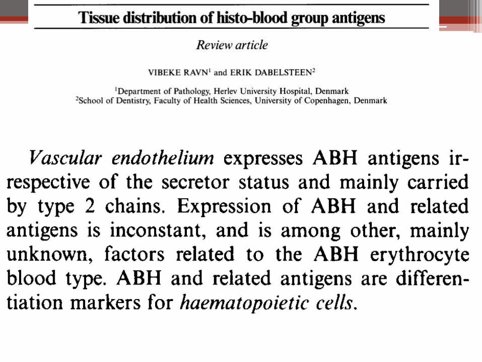
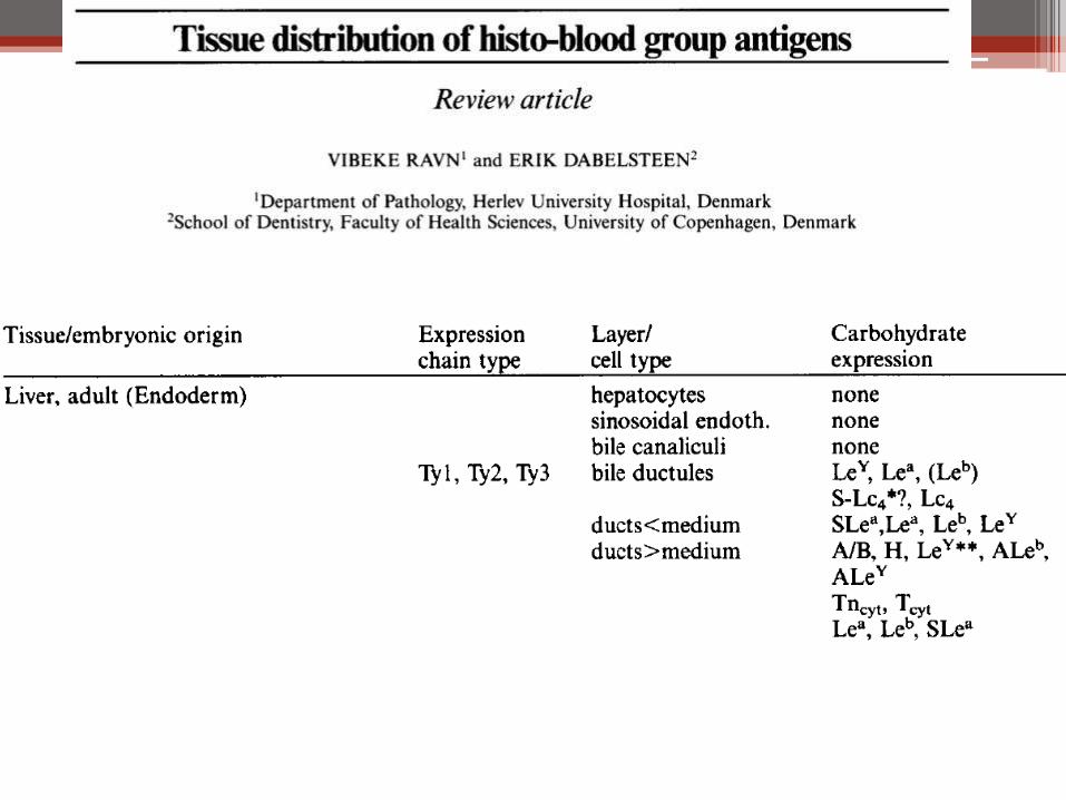
▫ ABO expression
▫ VOD and Platelet transfusion
• Study
▫ Methods
▫ Patient Population
• Conclusions
• Future Directions
Hematopoietic Stem Cell Transplant
• Reconstitute bone marrow after high-dose chemotherapy
▫ Allogeneic or autologous
▫ Sources: cord, bone marrow, peripheral
▫ Diseases: leukemia, lymphoma, solid organ tumors
• Pediatrics: Improved prognosis in solid organ tumors refractory to conventional treatment
▫ High-dose chemo with stem cell rescue
Complications of High-Dose Chemo and
HSCT • Graft versus host disease (GVHD)
• Graft rejection
• Disease relapse
• Anemia
• Neutropenia and infection
• Hepatic veno-occlusive disease
• Thrombocytopenia
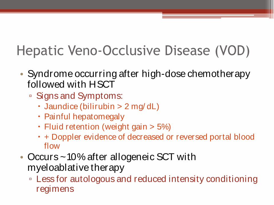
Hepatic Veno-Occlusive Disease (VOD)
• Syndrome occurring after high-dose chemotherapy followed with HSCT ▫ Signs and Symptoms: Jaundice (bilirubin > 2 mg/dL)
Painful hepatomegaly
Fluid retention (weight gain > 5%)
+ Doppler evidence of decreased or reversed portal blood flow
• Occurs ~10% after allogeneic SCT with myeloablative therapy ▫ Less for autologous and reduced intensity conditioning
regimens
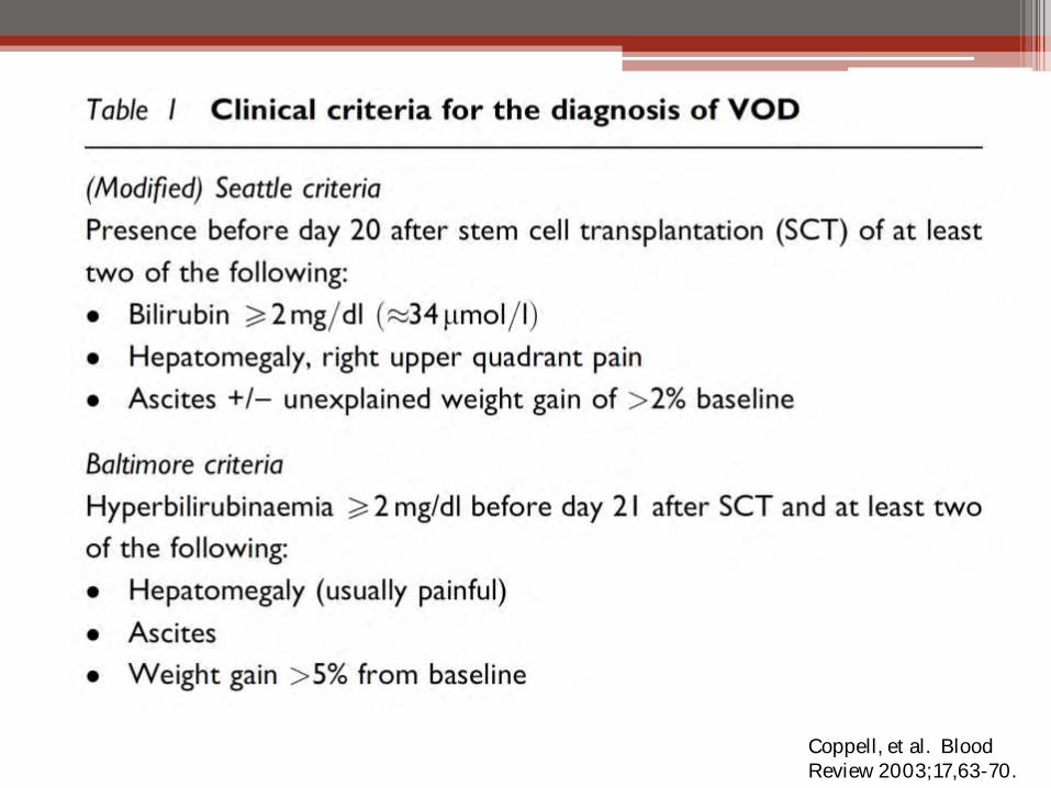
Coppell, et al. Blood Review 2003;17,63-70.
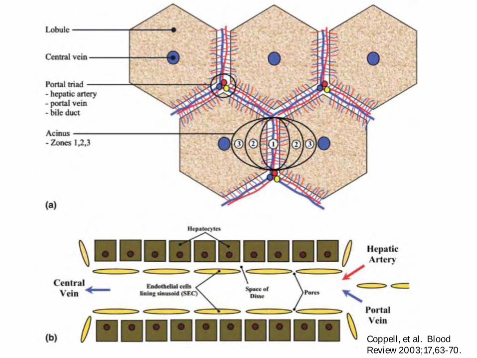
Coppell, et al. Blood Review 2003;17,63-70.
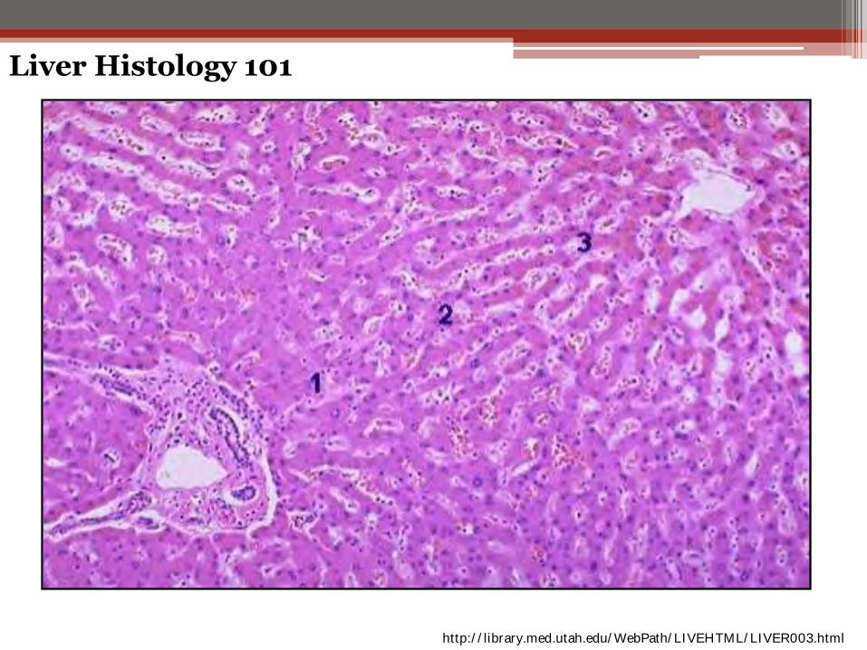
Liver Histology 101
http://library.med.utah.edu/WebPath/LIVEHTML/LIVER003.html
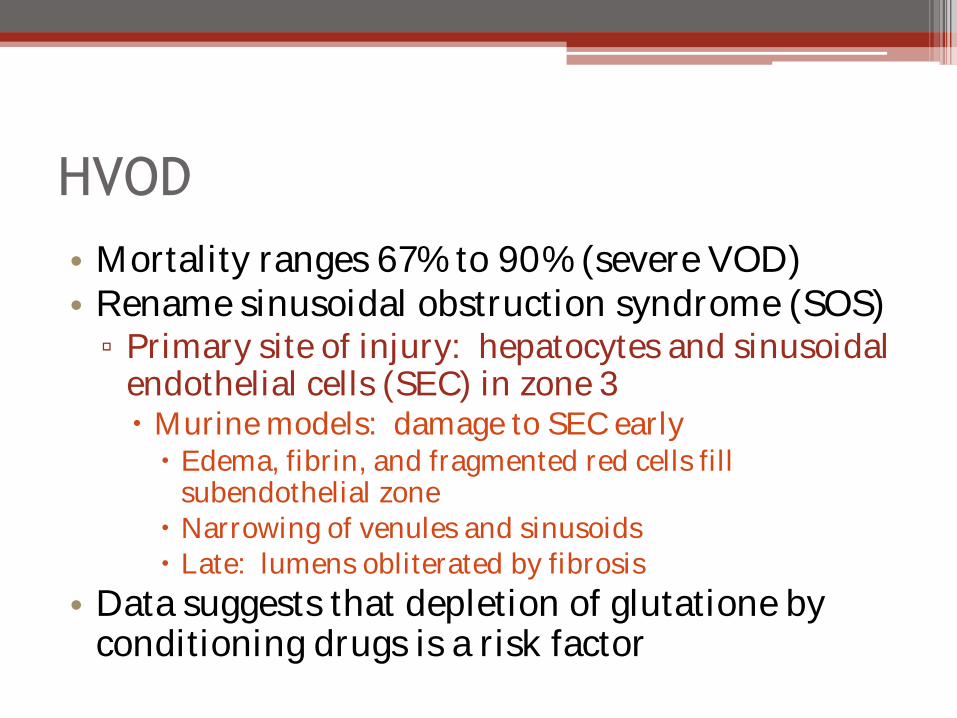
HVOD
• Mortality ranges 67% to 90% (severe VOD) • Rename sinusoidal obstruction syndrome (SOS)
▫ Primary site of injury: hepatocytes and sinusoidal endothelial cells (SEC) in zone 3 Murine models: damage to SEC early Edema, fibrin, and fragmented red cells fill
subendothelial zone
Narrowing of venules and sinusoids
Late: lumens obliterated by fibrosis
• Data suggests that depletion of glutatione by conditioning drugs is a risk factor
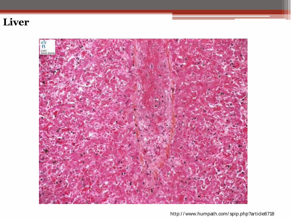
http://www.humpath.com/spip.php?article8718
Liver
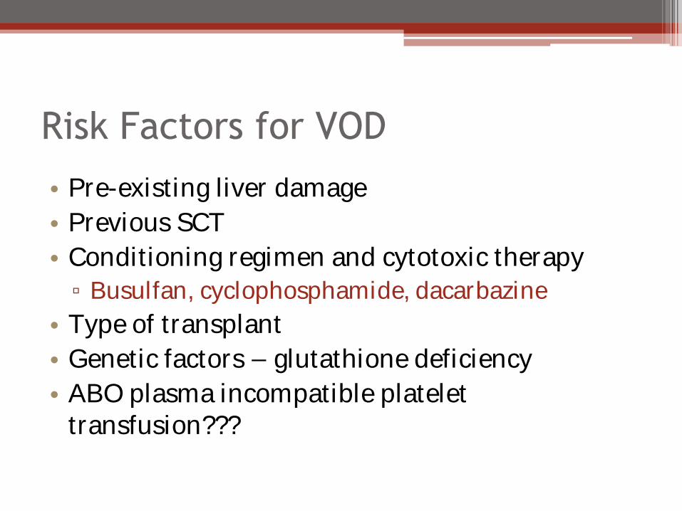
Risk Factors for VOD
• Pre-existing liver damage
• Previous SCT
• Conditioning regimen and cytotoxic therapy
▫ Busulfan, cyclophosphamide, dacarbazine
• Type of transplant
• Genetic factors – glutathione deficiency
• ABO plasma incompatible platelet transfusion???
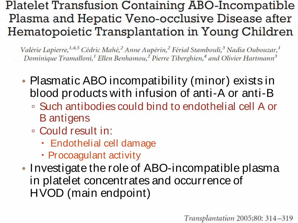
• Plasmatic ABO incompatibility (minor) exists in blood products with infusion of anti-A or anti-B ▫ Such antibodies could bind to endothelial cell A or
B antigens ▫ Could result in: Endothelial cell damage Procoagulant activity
• Investigate the role of ABO-incompatible plasma in platelet concentrates and occurrence of HVOD (main endpoint)
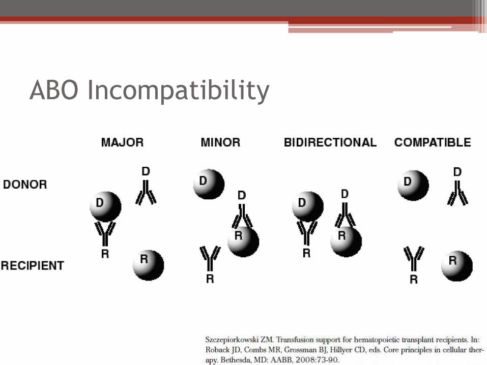
ABO Incompatibility
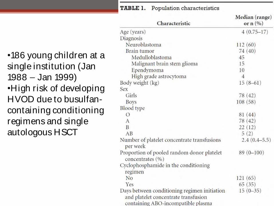
•186 young children at a single institution (Jan 1988 – Jan 1999) •High risk of developing HVOD due to busulfan-containing conditioning regimens and single autologous HSCT
Results
• 73 of 186 kids developed HVOD at median time of 27 days (r 14-52) after initiation of conditioning regimen
• ABO plasma (minor) incompatible PC were transfused in 47% (87/186) of all kids
• HVOD occurred in 42 of 87 who received at least 1 ABO minor PC versus 31 of 99 (31%) who always got ABO compatible PC (P=0.02)
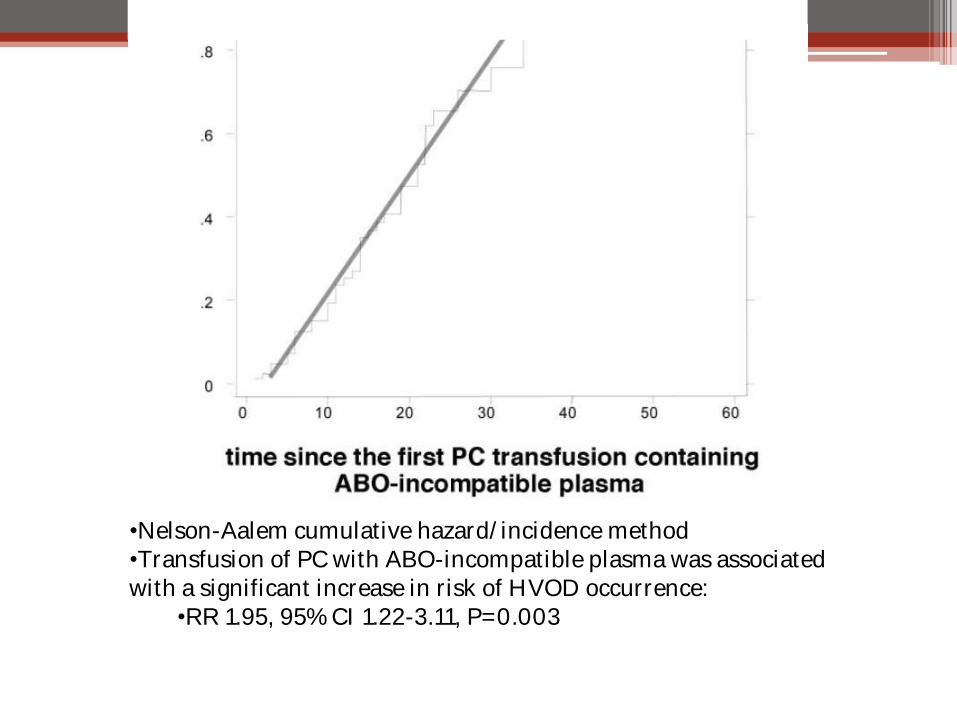
•Nelson-Aalem cumulative hazard/incidence method •Transfusion of PC with ABO-incompatible plasma was associated with a significant increase in risk of HVOD occurrence:
•RR 1.95, 95% CI 1.22-3.11, P=0.003

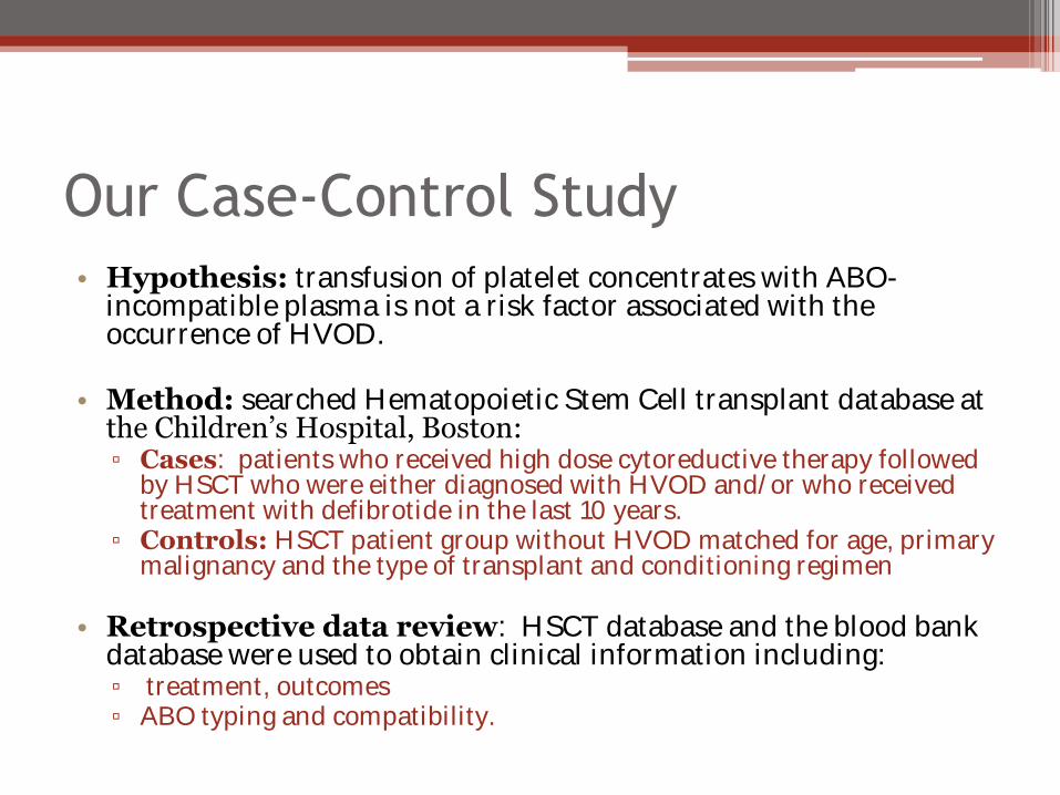
Our Case-Control Study
• Hypothesis: transfusion of platelet concentrates with ABO-incompatible plasma is not a risk factor associated with the occurrence of HVOD.
• Method: searched Hematopoietic Stem Cell transplant database at
the Children’s Hospital, Boston: ▫ Cases: patients who received high dose cytoreductive therapy followed
by HSCT who were either diagnosed with HVOD and/or who received treatment with defibrotide in the last 10 years.
▫ Controls: HSCT patient group without HVOD matched for age, primary malignancy and the type of transplant and conditioning regimen
• Retrospective data review: HSCT database and the blood bank
database were used to obtain clinical information including: ▫ treatment, outcomes ▫ ABO typing and compatibility.
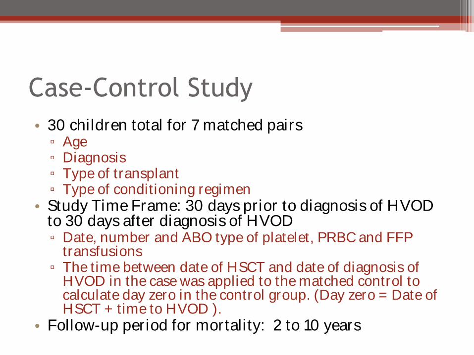
Case-Control Study
• 30 children total for 7 matched pairs ▫ Age ▫ Diagnosis ▫ Type of transplant ▫ Type of conditioning regimen
• Study Time Frame: 30 days prior to diagnosis of HVOD to 30 days after diagnosis of HVOD ▫ Date, number and ABO type of platelet, PRBC and FFP
transfusions ▫ The time between date of HSCT and date of diagnosis of
HVOD in the case was applied to the matched control to calculate day zero in the control group. (Day zero = Date of HSCT + time to HVOD ).
• Follow-up period for mortality: 2 to 10 years
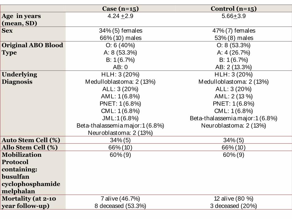
Case (n=15) Control (n=15)
Age in years (mean, SD)
4.24 +2.9 5.66+3.9
Sex 34% (5) females 66% (10) males
47% (7) females 53% (8) males
Original ABO Blood Type
O: 6 (40%) A: 8 (53.3%) B: 1 (6.7%)
AB: 0
O: 8 (53.3%) A: 4 (26.7%) B: 1 (6.7%)
AB: 2 (13.3%)
Underlying Diagnosis
HLH: 3 (20%) Medulloblastoma: 2 (13%)
ALL: 3 (20%) AML: 1 (6.8%) PNET: 1 (6.8%) CML: 1 (6.8%) JML:1 (6.8%)
Beta-thalassemia major:1 (6.8%) Neuroblastoma: 2 (13%)
HLH: 3 (20%) Medulloblastoma: 2 (13%)
ALL: 3 (20%) AML: 2 (13 %)
PNET: 1 (6.8%) CML: 1 (6.8%)
Beta-thalassemia major:1 (6.8%) Neuroblastoma: 2 (13%)
Auto Stem Cell (%) 34% (5) 34% (5)
Allo Stem Cell (%) 66% (10) 66% (10)
Mobilization Protocol containing: busulfan cyclophosphamide melphalan
60% (9) 60% (9)
Mortality (at 2-10 year follow-up)
7 alive (46.7%) 8 deceased (53.3%)
12 alive (80 %) 3 deceased (20%)
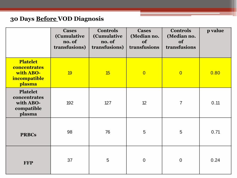
Cases (Cumulative
no. of transfusions)
Controls (Cumulative
no. of transfusions)
Cases (Median no.
of transfusions
Controls (Median no.
of transfusions
p value
Platelet concentrates
with ABO-incompatible
plasma
19 15 0 0 0.80
Platelet concentrates
with ABO- compatible
plasma
192 127 12 7 0.11
PRBCs
98 76 5 5 0.71
FFP
37 5 0 0 0.24
30 Days Before VOD Diagnosis

Cases (Cumulative
no. of transfusions)
Controls (Cumulative
no. of transfusions)
Cases (Median no.
of transfusions
Controls (Median no.
of transfusions
p value
Platelet concentrates with
ABO-incompatible
plasma
77 21 3 0 0.008
Platelet concentrates with ABO- compatible
plasma
520 208 19 1 0.003
PRBCs
221 114 8 2 0.003
FFP
270 23 13 0 0.003
30 Days After VOD Diagnosis
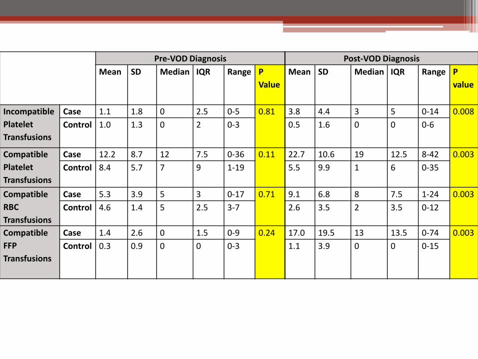
Pre-VOD Diagnosis Post-VOD Diagnosis
Mean SD Median IQR Range P
Value
Mean SD Median IQR Range P
value
Incompatible
Platelet
Transfusions
Case 1.1 1.8 0 2.5 0-5 0.81 3.8 4.4 3 5 0-14 0.008
Control 1.0 1.3 0 2 0-3 0.5 1.6 0 0 0-6
Compatible
Platelet
Transfusions
Case 12.2 8.7 12 7.5 0-36 0.11 22.7 10.6 19 12.5 8-42 0.003
Control 8.4 5.7 7 9 1-19 5.5 9.9 1 6 0-35
Compatible
RBC
Transfusions
Case 5.3 3.9 5 3 0-17 0.71 9.1 6.8 8 7.5 1-24 0.003
Control 4.6 1.4 5 2.5 3-7 2.6 3.5 2 3.5 0-12
Compatible
FFP
Transfusions
Case 1.4 2.6 0 1.5 0-9 0.24 17.0 19.5 13 13.5 0-74 0.003
Control 0.3 0.9 0 0 0-3 1.1 3.9 0 0 0-15
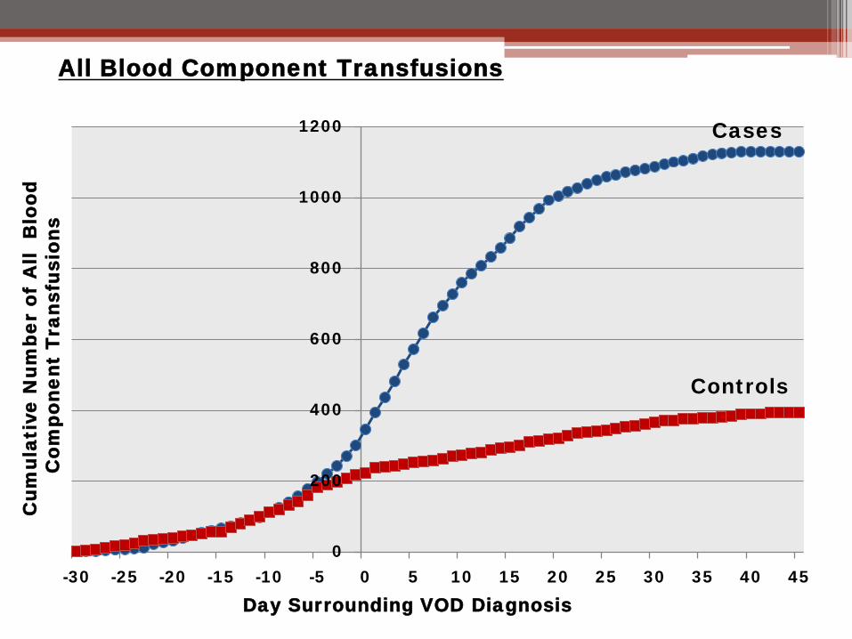
All Blood Component Transfusions
Day Surrounding VOD Diagnosis
0
200
400
600
800
1000
1200
-30 -25 -20 -15 -10 -5 0 5 10 15 20 25 30 35 40 45
Cu
mu
lative
N
um
be
r o
f A
ll B
lo
od
C
om
po
ne
nt T
ra
nsfu
sio
ns
Cases
Controls
0
10
20
30
40
50
60
70
80
-3
0
-2
5
-2
0
-1
5
-1
0
-5
0
5
10
15
20
25
30
35
40
45
Day Surrounding VOD Diagnosis
Cu
mu
lative
N
um
be
r o
f A
BO
p
la
sm
a
In
co
mp
atib
le
P
late
le
t
T
ra
nsfu
sio
ns
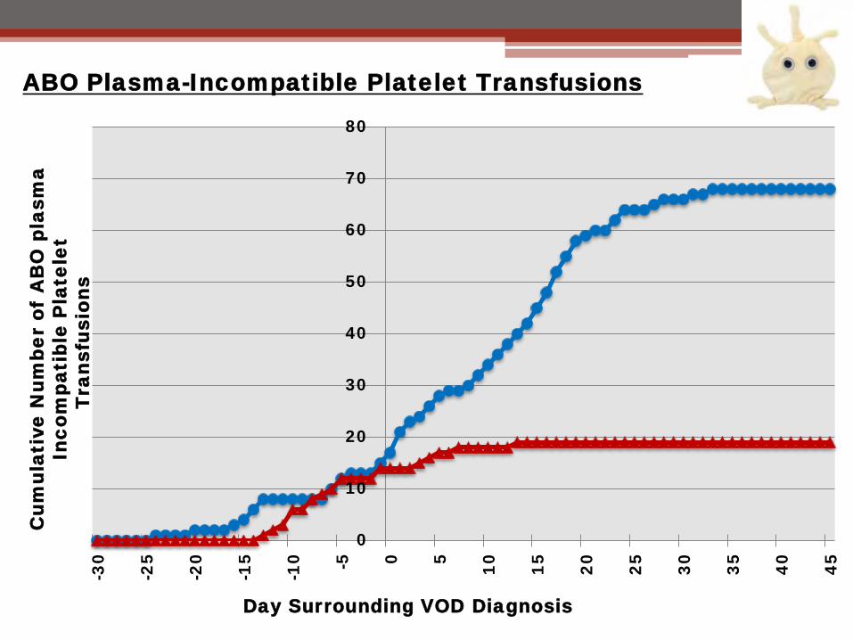
ABO Plasma-Incompatible Platelet Transfusions
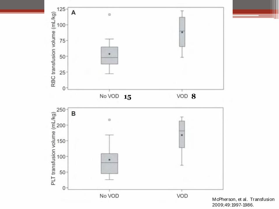
Results Summary
• There was no statistically significant difference between the cases and controls in terms of the number of transfusions of the blood components before the development of HVOD ▫ All blood components
• The number of transfusions increased significantly in the HVOD group (cases) after the diagnosis of HVOD as compared to the control group. ▫ For all blood components!!
8 15
McPherson, et al. Transfusion 2009;49:1997-1986.
Conclusion
• In our case-control study, ABO incompatible PC transfusions were not associated with an increased incidence of VOD
▫ BUT…..
Small N
Consistent diagnosis of VOD – criteria for discerning
Blood type?

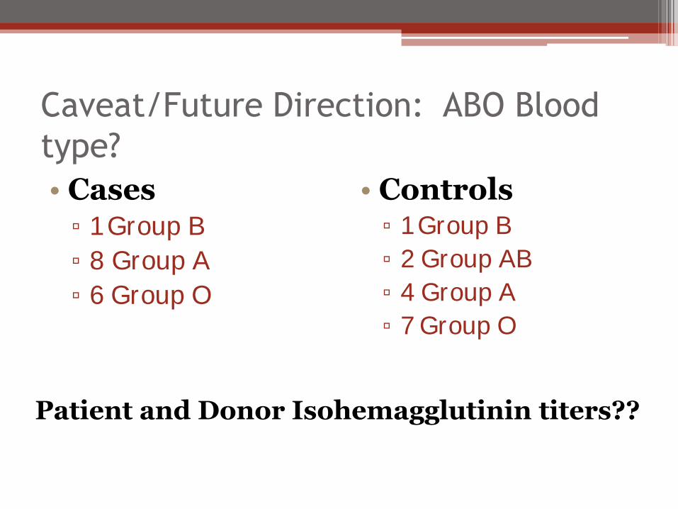
Caveat/Future Direction: ABO Blood
type?
• Cases ▫ 1 Group B
▫ 8 Group A
▫ 6 Group O
• Controls ▫ 1 Group B
▫ 2 Group AB
▫ 4 Group A
▫ 7 Group O
Patient and Donor Isohemagglutinin titers??
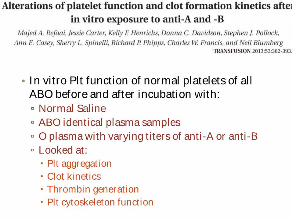
• In vitro Plt function of normal platelets of all ABO before and after incubation with:
▫ Normal Saline
▫ ABO identical plasma samples
▫ O plasma with varying titers of anti-A or anti-B
▫ Looked at:
Plt aggregation
Clot kinetics
Thrombin generation
Plt cytoskeleton function
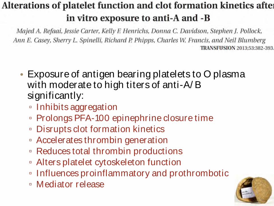
• Exposure of antigen bearing platelets to O plasma with moderate to high titers of anti-A/B significantly: ▫ Inhibits aggregation ▫ Prolongs PFA-100 epinephrine closure time ▫ Disrupts clot formation kinetics ▫ Accelerates thrombin generation ▫ Reduces total thrombin productions ▫ Alters platelet cytoskeleton function ▫ Influences proinflammatory and prothrombotic ▫ Mediator release
Questions Please!