Embed Size (px)
Citation preview
668
Correspondence to:Zlatan ELEKDžona Kenedija 19/1538220 Kosovska [email protected]
Received • Примљено: February 14, 2018
Revised • Ревизија: June 25, 2018
Accepted • Прихваћено: June 27, 2018
Online first: June 29, 2018
SUMMARYIntroduction Bilateral abdominoscrotal hydrocele is a rare entity in childhood. The etiology of abdomino-scrotal hydrocele has not been fully clarified. The diagnosis is based on clinical examination, ultrasound and magnetic resonance imaging. The treatment is surgery.Case outline This paper presents an eight-month-old boy who was admitted at the department of pediatric surgery due to bilateral swellings in the scrotum area. The changes were first noticed when he was three months old and the diagnosis of bilateral abdominoscrotal hydrocele was confirmed after the scrotum ultrasound examination. When the patient was six and eight months old, the symptoms have significantly increased, the magnetic resonance of the abdomen and the lesser pelvis was performed, and the bilateral abdominoscrotal hydrocele was successfully treated with inguinal surgery. The opera-tive and postoperative course was uneventful.Conclusion In this paper, we presented a rare form of hydrocele in children, as well as diagnostic evalu-ation that involved ultrasound and magnetic resonance examination. Surgical treatment by inguinal approach is also presented.Keywords: bilateral; abdominoscrotal hydrocele; magnetic resonance
DOI: https://doi.org/10.2298/SARH180214045E
UDC: 617.55-007.43-053.2-089
CASE REPORT / ПРИКАЗ БОЛЕСНИКА
Bilateral abdominoscrotal hydrocele in childhood Zlatan Elek1, Boban Mitrović1, Saša Dimić1, Aleksandar Božović1, Jovan Mladenović2, Zoran Marjanović3
1Kosovska Mitrovica Clinical Hospital Centre, Department of Pediatric Surgery, Kosovska Mitrovica, Serbia;2University of Priština, Faculty of Medicine, Kosovska Mitrovica, Serbia;3Clinical Centre Niš, Clinic for Children's Surgery and Orthopedics, Niš, Serbia
INTRODUCTION
Abdominoscrotal hydrocele (ASH) is a rare form of hydrocele in children. Dupuytren was the first to describe it in 1834 [1]. Bickel intro-duced the term ASH in 1919 [1]. The reported incidence of ASH is 0.17–3.1% of hydroceles.
ASH has the shape of an hourglass and is made up of two parts: inguinoscrotal part and abdominal part which intercommunicate through the deep (internal) inguinal ring. The etiology and pathogenesis of ASH have not been clarified yet. The diagnosis is based on clinical appearance, ultrasound, and magnetic resonance imaging [2].
The treatment of ASH is surgery. Generally, as in all hydroceles in children, the inguinal ap-proach is considered standard, although there are cases in which the operation was done by the scrotal approach. The paper presents a case
of bilateral ASH that was surgically treated by the inguinal approach [3, 4, 5].
CASE REPORT
We present an eight-month-old boy who was admitted to the department of pediatric surgery for an intervention.
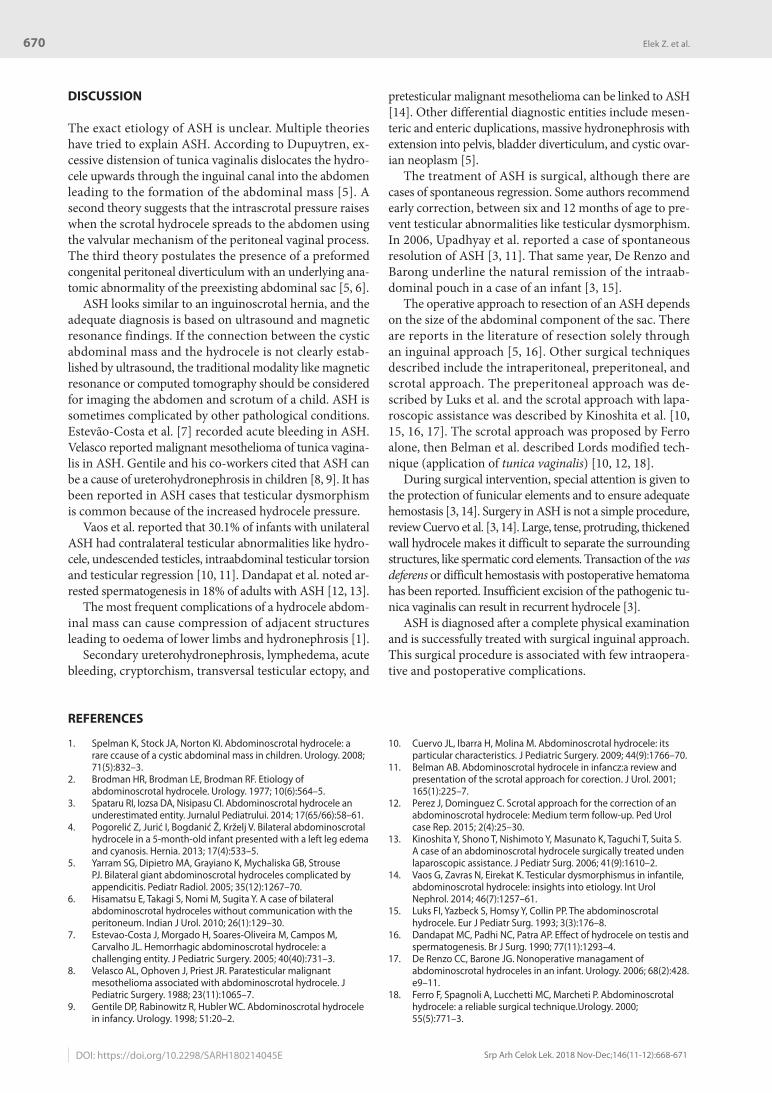
The first time the patient was admitted for examination at the age of three months because of a bilateral scrotum swelling. Clinical exami-nation showed both testicles in the scrotum and bilateral hydrocele (Figure 1). An ultrasound exam of the scrotum, pelvis, and urinary tract was performed. The diagnosis of ASH on the left side, and hydrocele on the right side was established. Urinary tract was normal. Control exam was scheduled in three months. An ul-trasound check-up exam of the scrotum and
Figure 1. Bilateral hydrocele was diagnosed in the third month of age
Figure 2. Bilateral hydrocele in the eighth month of age

669
Srp Arh Celok Lek. 2018 Nov-Dec;146(11-12):668-671 www.srpskiarhiv.rs
pelvis were done when the patient was six months old. A 36-milimeter wide bilateral ASH was diagnosed, extending upwards through the inguinal canal into the abdominal cav-ity, with the diameter of 56 mm on the left side and 38 mm on the right side. The proximal part of hydrocele covered and compressed the urinary bladder. Since the hydroceles have grown from the previous examination, MRI of the abdomen and pelvis were performed. The MRI findings showed liquid deposits in the scrotum with a maximum diameter of 58 mm on the left side and 40 mm on the right
side. The described changes extended through the inguinal canal, compressing within the abdominal cavity the urinary bladder and reaching the common iliac artery branches, corresponding to ASH (Figures 2 and 3).
The child was admitted to the department and prepared for surgical intervention. The surgical intervention was done under the usual conditions of endotracheal anesthesia for the inguinal approach (Figures 4 and 5).
Operative and postoperative courses were uneventful. Wounds healed per primam intentionem. Stiches were re-moved on the seventh postoperative day, when the child was discharged. Regular checkups on the seventh, 14th, and 30th day after the operation showed regular clinical findings (Figure 6).
Figure 3. Magnetic resonance imaging findings of bilateral abdom-inoscrotal hydrocele
Figure 4. Operative finding after resection of right-sided abdomino-scrotal hydrocele
Figure 5. Operative finding after resection of left-sided abdomino-scrotal hydrocele – abdominal component
Figure 6. Postoperative finding six months after surgery
Bilateral abdominoscrotal hydrocele in childhood
670
Srp Arh Celok Lek. 2018 Nov-Dec;146(11-12):668-671
DOI: https://doi.org/10.2298/SARH180214045E
DISCUSSION
The exact etiology of ASH is unclear. Multiple theories have tried to explain ASH. According to Dupuytren, ex-cessive distension of tunica vaginalis dislocates the hydro-cele upwards through the inguinal canal into the abdomen leading to the formation of the abdominal mass [5]. A second theory suggests that the intrascrotal pressure raises when the scrotal hydrocele spreads to the abdomen using the valvular mechanism of the peritoneal vaginal process. The third theory postulates the presence of a preformed congenital peritoneal diverticulum with an underlying ana-tomic abnormality of the preexisting abdominal sac [5, 6].
ASH looks similar to an inguinoscrotal hernia, and the adequate diagnosis is based on ultrasound and magnetic resonance findings. If the connection between the cystic abdominal mass and the hydrocele is not clearly estab-lished by ultrasound, the traditional modality like magnetic resonance or computed tomography should be considered for imaging the abdomen and scrotum of a child. ASH is sometimes complicated by other pathological conditions. Estevão-Costa et al. [7] recorded acute bleeding in ASH. Velasco reported malignant mesothelioma of tunica vagina-lis in ASH. Gentile and his co-workers cited that ASH can be a cause of ureterohydronephrosis in children [8, 9]. It has been reported in ASH cases that testicular dysmorphism is common because of the increased hydrocele pressure.
Vaos et al. reported that 30.1% of infants with unilateral ASH had contralateral testicular abnormalities like hydro-cele, undescended testicles, intraabdominal testicular torsion and testicular regression [10, 11]. Dandapat et al. noted ar-rested spermatogenesis in 18% of adults with ASH [12, 13].
The most frequent complications of a hydrocele abdom-inal mass can cause compression of adjacent structures leading to oedema of lower limbs and hydronephrosis [1].
Secondary ureterohydronephrosis, lymphedema, acute bleeding, cryptorchism, transversal testicular ectopy, and
pretesticular malignant mesothelioma can be linked to ASH [14]. Other differential diagnostic entities include mesen-teric and enteric duplications, massive hydronephrosis with extension into pelvis, bladder diverticulum, and cystic ovar-ian neoplasm [5].
The treatment of ASH is surgical, although there are cases of spontaneous regression. Some authors recommend early correction, between six and 12 months of age to pre-vent testicular abnormalities like testicular dysmorphism. In 2006, Upadhyay et al. reported a case of spontaneous resolution of ASH [3, 11]. That same year, De Renzo and Barong underline the natural remission of the intraab-dominal pouch in a case of an infant [3, 15].
The operative approach to resection of an ASH depends on the size of the abdominal component of the sac. There are reports in the literature of resection solely through an inguinal approach [5, 16]. Other surgical techniques described include the intraperitoneal, preperitoneal, and scrotal approach. The preperitoneal approach was de-scribed by Luks et al. and the scrotal approach with lapa-roscopic assistance was described by Kinoshita et al. [10, 15, 16, 17]. The scrotal approach was proposed by Ferro alone, then Belman et al. described Lords modified tech-nique (application of tunica vaginalis) [10, 12, 18].
During surgical intervention, special attention is given to the protection of funicular elements and to ensure adequate hemostasis [3, 14]. Surgery in ASH is not a simple procedure, review Cuervo et al. [3, 14]. Large, tense, protruding, thickened wall hydrocele makes it difficult to separate the surrounding structures, like spermatic cord elements. Transaction of the vas deferens or difficult hemostasis with postoperative hematoma has been reported. Insufficient excision of the pathogenic tu-nica vaginalis can result in recurrent hydrocele [3].
ASH is diagnosed after a complete physical examination and is successfully treated with surgical inguinal approach. This surgical procedure is associated with few intraopera-tive and postoperative complications.
REFERENCES
1. Spelman K, Stock JA, Norton KI. Abdominoscrotal hydrocele: a rare ccause of a cystic abdominal mass in children. Urology. 2008; 71(5):832–3.
2. Brodman HR, Brodman LE, Brodman RF. Etiology of abdominoscrotal hydrocele. Urology. 1977; 10(6):564–5.
3. Spataru RI, Iozsa DA, Nisipasu CI. Abdominoscrotal hydrocele an underestimated entity. Jurnalul Pediatrului. 2014; 17(65/66):58–61.
4. Pogorelić Z, Jurić I, Bogdanić Ž, Krželj V. Bilateral abdominoscrotal hydrocele in a 5-month-old infant presented with a left leg edema and cyanosis. Hernia. 2013; 17(4):533–5.
5. Yarram SG, Dipietro MA, Grayiano K, Mychaliska GB, Strouse PJ. Bilateral giant abdominoscrotal hydroceles complicated by appendicitis. Pediatr Radiol. 2005; 35(12):1267–70.
6. Hisamatsu E, Takagi S, Nomi M, Sugita Y. A case of bilateral abdominoscrotal hydroceles without communication with the peritoneum. Indian J Urol. 2010; 26(1):129–30.
7. Estevao-Costa J, Morgado H, Soares-Oliveira M, Campos M, Carvalho JL. Hemorrhagic abdominoscrotal hydrocele: a challenging entity. J Pediatric Surgery. 2005; 40(40):731–3.
8. Velasco AL, Ophoven J, Priest JR. Paratesticular malignant mesothelioma associated with abdominoscrotal hydrocele. J Pediatric Surgery. 1988; 23(11):1065–7.
9. Gentile DP, Rabinowitz R, Hubler WC. Abdominoscrotal hydrocele in infancy. Urology. 1998; 51:20–2.
10. Cuervo JL, Ibarra H, Molina M. Abdominoscrotal hydrocele: its particular characteristics. J Pediatric Surgery. 2009; 44(9):1766–70.
11. Belman AB. Abdominoscrotal hydrocele in infancz:a review and presentation of the scrotal approach for corection. J Urol. 2001; 165(1):225–7.
12. Perez J, Dominguez C. Scrotal approach for the correction of an abdominoscrotal hydrocele: Medium term follow-up. Ped Urol case Rep. 2015; 2(4):25–30.
13. Kinoshita Y, Shono T, Nishimoto Y, Masunato K, Taguchi T, Suita S. A case of an abdominoscrotal hydrocele surgically treated unden laparoscopic assistance. J Pediatr Surg. 2006; 41(9):1610–2.
14. Vaos G, Zavras N, Eirekat K. Testicular dysmorphismus in infantile, abdominoscrotal hydrocele: insights into etiology. Int Urol Nephrol. 2014; 46(7):1257–61.
15. Luks FI, Yazbeck S, Homsy Y, Collin PP. The abdominoscrotal hydrocele. Eur J Pediatr Surg. 1993; 3(3):176–8.
16. Dandapat MC, Padhi NC, Patra AP. Effect of hydrocele on testis and spermatogenesis. Br J Surg. 1990; 77(11):1293–4.
17. De Renzo CC, Barone JG. Nonoperative managament of abdominoscrotal hydroceles in an infant. Urology. 2006; 68(2):428.e9–11.
18. Ferro F, Spagnoli A, Lucchetti MC, Marcheti P. Abdominoscrotal hydrocele: a reliable surgical technique.Urology. 2000; 55(5):771–3.
Elek Z. et al.
671
Srp Arh Celok Lek. 2018 Nov-Dec;146(11-12):668-671 www.srpskiarhiv.rs
САЖЕТАКУвод Билатерална абдоминоскротална хидроцела је редак ентитет у дечјем узрасту. Етиологија абдоминоскроталне хидроцеле није у потпуности разјашњена. Дијагноза се по-ставља на основу клиничке слике, ултразвучног прегледа и магнетне резонанце. Лечење је хируршко.Приказ болесника У раду је приказан дечак стар осам месеци који је примљен на одељење дечје хирургије због oбостраног отока у пределу скротума. Први пут су промене уочене у трећем месецу старости, а дијагноза је потврђена на основу ултразвучног прегледа скротума и мале карлице. На контролним прегледима у шестом и осмом месецу ста-
рости промене су се значајно увећале, урађена је магнетна резонанца абдомена и мале карлице и предузето је опера-тивно лечење. Оперативни и постоперативни ток протекли су уредно.Закључак У раду смо приказали један од ретких случајева хидроцеле у дечјем узрасту, као и дијагностичку евалуацију која је обухватала ултразвучну дијагностику и магнетну ре-зонанцу. Такође је приказано хируршко лечење ингвинал-ним приступом.
Kључне речи: билатерална; абдоминоскротална хидроцела; магнетна резонанца
Билатерална абдоминоскротална хидроцела у дечјем узрастуЗлатан Елек1, Бобан Митровић1, Саша Димић1, Александар Божовић1, Јован Младеновић2, Зоран Марјановић3
1Kлиничко-болнички центар Косовска Митровица, Oдељење дечје хирургије, Косовска Митровица, Србија;2Универзитет у Приштини, Mедицински факултет, Косовска Митровица, Србија;3Kлинички центар Ниш, Клиника за дечју хирургију и ортопедију, Ниш, Србија
Bilateral abdominoscrotal hydrocele in childhood