Embed Size (px)
Citation preview
Case ReportHarmful Effects of Synthetic Surface-ActiveDetergents against Atopic Dermatitis
Hajime Deguchi,1,2 Riho Aoyama,1,2 Hideaki Takahashi,1,2
Yoshinari Isobe,3 and Yutaka Tsutsumi2
1Fujita Health University School of Medicine, Toyoake, Aichi 470-1192, Japan2Department of Pathology, Fujita Health University School of Medicine, Toyoake, Aichi 470-1192, Japan3Isobe Clinic, Anjo, Aichi 446-0026, Japan
Correspondence should be addressed to Yutaka Tsutsumi; [email protected]
Received 31 October 2014; Accepted 30 December 2014
Academic Editor: Jeung-Hoon Lee
Copyright © 2015 Hajime Deguchi et al.This is an open access article distributed under theCreativeCommonsAttribution License,which permits unrestricted use, distribution, and reproduction in any medium, provided the original work is properly cited.
We report herein two cases of intractable atopic dermatitis successfully treated by simply avoiding the contact with surface-active detergents in the daily life and living. The detergents were closely related to the exacerbation and remission of the disease.Steroid ointment was no longer used. We discuss that the removal of horny layer lipids by surface-active detergents accelerates thetransepidermal water loss and disturbs the barrier function of the epidermis and thus is intimately involved in the pathogenesis ofatopic dermatitis.
1. Introduction
Atopic dermatitis is etiologically related to abnormalitiesin physiologic functions of the skin, resulting in chronicpersistent and irritating inflammation of type I and/or typeIV allergic reactions: atopic dermatitis is a disease of alteredepidermal barrier [1–4]. Allergens are usually not specified.Infants aged below 2 years show the lowest epidermalbarrier function and are susceptible to atopic dermatitis [1–3]. The main victims are thus infants and young children,but the long-lasting disease is also seen in the adulthood.The treatment strategy against atopic dermatitis includes theexternal use of steroids or tacrolimus ointment, in additionto moisturizing and protective skin cares [5]. Internal use ofantihistamines and antiallergic drugs and the elimination ofexacerbating factors are also employed.
Dry skin is one of the major symptoms in atopic dermati-tis. The abnormality of the epidermis, especially the hornylayer (stratum corneum), is closely linked to loss of the barrierfunction.The transepidermal water loss is caused by reducedlipids in the horny layer. The lipid bilayers intermit betweenthe horny keratinocytes (corneocytes).When the corneocytesare thought of as bricks, the lipids filling the spaces between
the cells are the mortar or cement (brick and mortar model)[2, 4]. The lipid bilayers consist of ceramides, cholesteroland long-chained fatty acids, and impede penetration oflipophilic as well as hydrophilic substances [6–9]. Soap anddetergents acting as surfactants may provoke skin damagesuch as scaling, dryness, tightness, roughness, erythema,and swelling. An itch-scratch cycle accelerates damaging theepidermal barrier [1–4].
We report herein two representative adult patients whoshowed exacerbation of atopic dermatitis after the contactwith surface-active detergents and the disuse led to theremission. We propose that the removal of horny layer lipidsby surface-active detergents is intimately involved in thepathogenesis of atopic dermatitis, as one of the authorshave published Japanese-written books for promoting thegeneral public and dermatitis patients to avoid using soap anddetergents [10, 11].
2. Case Presentation
Case 1. Case 1 is a 50-year-old male, an office worker in a gasstation. After a 10-month history of chronic prurigo treatedwith steroid ointment, he visited Isobe Clinic in Anjo, Aichi,
Hindawi Publishing CorporationCase Reports in Dermatological MedicineVolume 2015, Article ID 898262, 5 pageshttp://dx.doi.org/10.1155/2015/898262
2 Case Reports in Dermatological Medicine
(a) (b) (c)
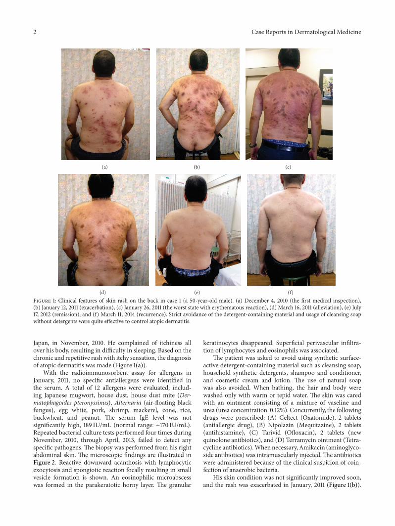
(d) (e) (f)Figure 1: Clinical features of skin rash on the back in case 1 (a 50-year-old male). (a) December 4, 2010 (the first medical inspection),(b) January 12, 2011 (exacerbation), (c) January 26, 2011 (the worst state with erythematous reaction), (d) March 16, 2011 (alleviation), (e) July17, 2012 (remission), and (f) March 11, 2014 (recurrence). Strict avoidance of the detergent-containing material and usage of cleansing soapwithout detergents were quite effective to control atopic dermatitis.
Japan, in November, 2010. He complained of itchiness allover his body, resulting in difficulty in sleeping. Based on thechronic and repetitive rashwith itchy sensation, the diagnosisof atopic dermatitis was made (Figure 1(a)).
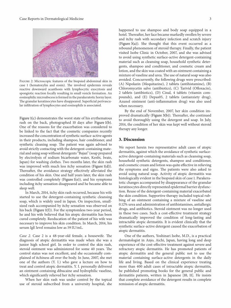
With the radioimmunosorbent assay for allergens inJanuary, 2011, no specific antiallergens were identified inthe serum. A total of 12 allergens were evaluated, includ-ing Japanese mugwort, house dust, house dust mite (Der-matophagoides pteronyssinus), Alternaria (air-floating blackfungus), egg white, pork, shrimp, mackerel, cone, rice,buckwheat, and peanut. The serum IgE level was notsignificantly high, 189 IU/mL (normal range: ∼170 IU/mL).Repeated bacterial culture tests performed four times duringNovember, 2010, through April, 2013, failed to detect anyspecific pathogens. The biopsy was performed from his rightabdominal skin. The microscopic findings are illustrated inFigure 2. Reactive downward acanthosis with lymphocyticexocytosis and spongiotic reaction focally resulting in smallvesicle formation is shown. An eosinophilic microabscesswas formed in the parakeratotic horny layer. The granular
keratinocytes disappeared. Superficial perivascular infiltra-tion of lymphocytes and eosinophils was associated.
The patient was asked to avoid using synthetic surface-active detergent-containing material such as cleansing soap,household synthetic detergents, shampoo and conditioner,and cosmetic cream and lotion. The use of natural soapwas also avoided. When bathing, the hair and body werewashed only with warm or tepid water. The skin was caredwith an ointment consisting of a mixture of vaseline andurea (urea concentration: 0.12%). Concurrently, the followingdrugs were prescribed: (A) Celtect (Oxatomide), 2 tablets(antiallergic drug), (B) Nipolazin (Mequitazine), 2 tablets(antihistamine), (C) Tarivid (Ofloxacin), 2 tablets (newquinolone antibiotics), and (D) Terramycin ointment (Tetra-cycline antibiotics).Whennecessary, Amikacin (aminoglyco-side antibiotics) was intramuscularly injected.The antibioticswere administered because of the clinical suspicion of coin-fection of anaerobic bacteria.
His skin condition was not significantly improved soon,and the rash was exacerbated in January, 2011 (Figure 1(b)).
Case Reports in Dermatological Medicine 3
100𝜇m
Figure 2: Microscopic features of the biopsied abdominal skin incase 1 (hematoxylin and eosin). The involved epidermis revealsreactive downward acanthosis with lymphocytic exocytosis andspongiotic reaction focally resulting in small vesicle formation. Aneosinophilicmicroabscess is formed in the parakeratotic horny layer.The granular keratinocytes have disappeared. Superficial perivascu-lar infiltration of lymphocytes and eosinophils is associated.
Figure 1(c) demonstrates the worst state of his erythematousrash on the back, photographed 10 days after Figure 1(b).One of the reasons for the exacerbation was considered tobe linked to the fact that the cosmetic companies recentlyincreased the concentration of synthetic surface-active agentsin their products, including shampoo, hair conditioner, andsynthetic cleaning soap. The patient was again advised toavoid strictly contacting with the detergent-containing mate-rial and using soapwithout detergents “Bajan” (soap preparedby electrolysis of sodium bicarbonate water, Kenbi, Iwate,Japan) for washing clothes. Two months later, the skin rashwas improved with much less itchy sensation (Figure 1(d)).Thereafter, the avoidance strategy effectively alleviated thecondition of his skin. One and half years later, the skin rashwas controlled completely (Figure 1(e)), and his symptomsincluding itchy sensation disappeared and he became able tosleep well.
In March, 2014, itchy skin rash recurred, because his wifestarted to use the detergent-containing synthetic cleansingsoap, which is widely used in Japan. On inspection, small-sized rash accompanied by itchy sensation was observed onhis back (Figure 1(f)). For the symptomless two-year period,he and his wife believed that his atopic dermatitis has beencured completely. Reeducation of the patient of his wife wasnecessary to improve his skin condition. In March, 2014, hisserum IgE level remains low as 59 IU/mL.
Case 2. Case 2 is a 48-year-old female, a housewife. Thediagnosis of atopic dermatitis was made when she was ajunior high school girl. In order to control the skin rash,steroid ointment was administered for some 40 years. Thecontrol status was not excellent, and she occasionally com-plained of itchiness all over the body. In June, 2007, she metone of the authors (Y. I.) who gave a lecture on how totreat and control atopic dermatitis. Y. I. personally deliveredan ointment containing dibucaine and hydrophilic vaseline,which significantly relieved her itchy sensation.
When her skin rash was under control by the topicaluse of steroid subscribed from a university hospital, she
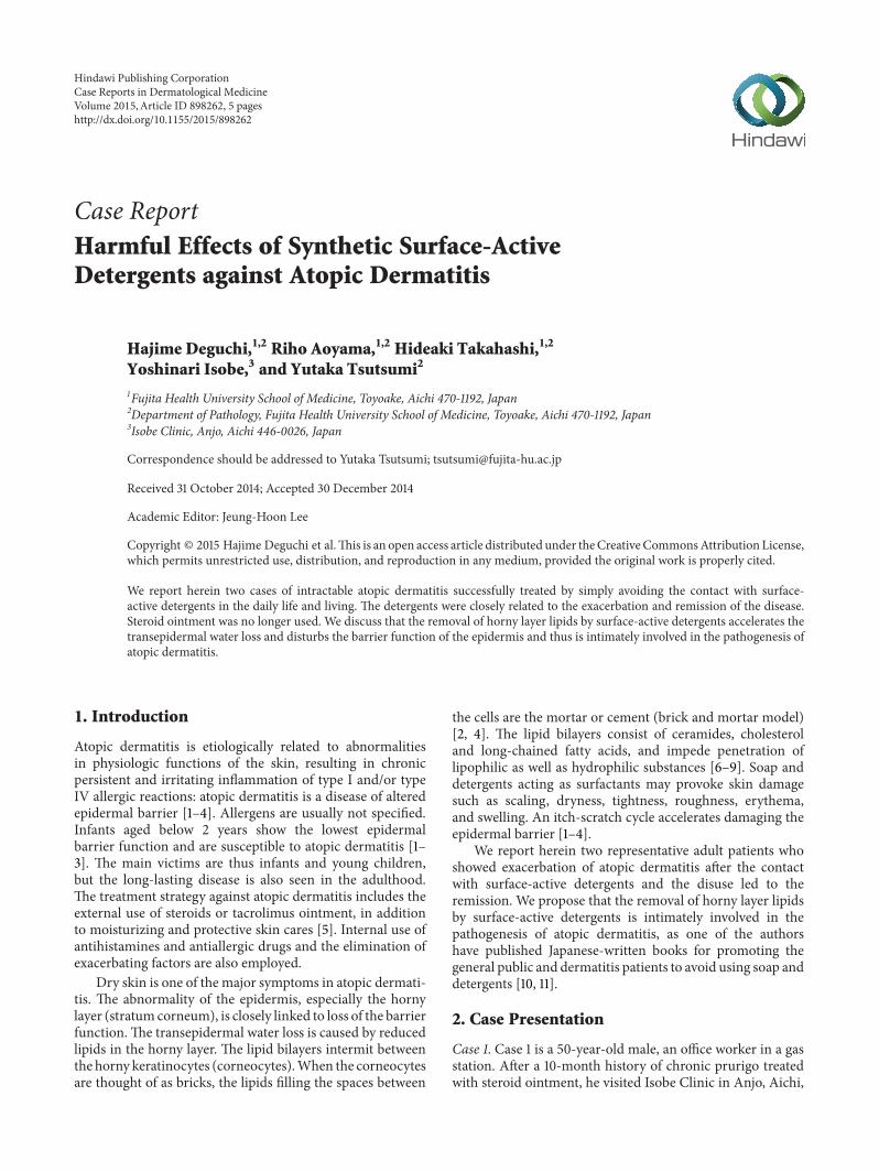
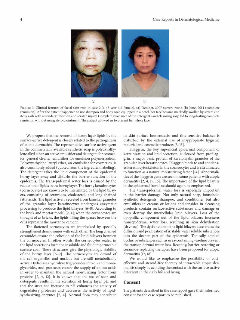
happened to use shampoo and body soap equipped in ahotel.Thereafter, her face becamemarkedly swollen by severeand itchy rash with secondary infection and scratch injury(Figure 3(a)). She thought that this event occurred as arebound phenomenon of steroid therapy. Finally, the patientvisited Isobe Clinic in October, 2007, and she was advisedto avoid using synthetic surface-active detergent-containingmaterial such as cleansing soap, household synthetic deter-gents, shampoo and conditioner, and cosmetic cream andlotion, and the skin was coated with an ointment containing amixture of vaseline and urea.The use of natural soap was alsoavoided. Concurrently, the following drugs were prescribed:(A) Nipolazin (Mequitazine), 2 tablets (antihistamine), (B)Chloromycetin salve (antibiotics), (C) Tarivid (Ofloxacin),2 tablets (antibiotics), (D) Cinal, 4 tablets (vitamin com-pounds), and (E) Depas05, 2 tablets (antianxiety drug).Azunol ointment (anti-inflammation drug) was also usedwhen necessary.
By the end of November, 2007, her skin condition im-proved dramatically (Figure 3(b)). Thereafter, she continuedto avoid thoroughly using the detergent and soap. In July,2014, the condition of her skin was kept well without steroidtherapy any longer.
3. Discussion
We report herein two representative adult cases of atopicdermatitis, against which the avoidance of synthetic surface-active detergent-containing materials such as cleansing soap,household synthetic detergents, shampoo and conditioner,and cosmetic cream and lotion was quite effective in relievingthe symptoms and signs. The patients were also asked toavoid using natural soap. Activity of atopic dermatitis washistologically evident in the biopsied skin of case 1. Parakera-totic changes accompanied by disappearance of the granularkeratinocytes directly represented epidermal barrier dysfunc-tion. Reuse of the detergent-containing material exacerbatedthe skin condition. Supportive therapy included topical rub-bing of an ointment containing a mixture of vaseline and0.12% urea and administration of antihistamines, antiallergicdrugs, and antibiotics. Steroid ointment was no longer usedin these two cases. Such a cost-effective treatment strategydramatically improved the condition of long-lasting andintractable atopic dermatitis. It is evident clinically that thesynthetic surface-active detergent caused the exacerbation ofatopic dermatitis.
One of the authors, Yoshinari Isobe, M.D., is a practicaldermatologist in Anjo, Aichi, Japan, having long and deepexperience of the cost-effective treatment against severe andrefractory atopic dermatitis. He has promoted patients ofatopic dermatitis and the general public not to use thematerial containing surface-active detergents in the dailylife and living. Based on the clinical experience treatingmore than 400 adult cases of intractable atopic dermatitis,he published promoting books for the general public anddermatitis patients, written in Japanese [10, 11]. He insiststhat complete avoidance of the detergent results in completeremission of atopic dermatitis.
4 Case Reports in Dermatological Medicine
(a) (b)
Figure 3: Clinical features of facial skin rash in case 2 (a 48-year-old female). (a) October, 2007 (severe rash), (b) June, 2014 (completeremission). After the patient happened to use shampoo and body soap equipped in a hotel, her face became markedly swollen by severe anditchy rash with secondary infection and scratch injury. Complete avoidance of the detergents and cleansing soap led to long-lasting completeremission without using steroid ointment. The patient allowed us to present her whole face.
We propose that the removal of horny layer lipids by thesurface-active detergent is closely related to the pathogenesisof atopic dermatitis. The representative surface-active agentin the commercially available synthetic soap is polyoxyalky-lene alkyl ether, an active emulsifier and detergent for cosmet-ics, general cleaner, emulsifier for emulsion polymerization.Polyoxyethylene lauryl ether, an emulsifier for cosmetics, isalso commonly added (quoted from the ingredient labeling).The detergent takes the lipid component of the epidermalhorny layer away and disturbs the barrier function of theepidermis. The transepidermal water loss is caused by thereduction of lipids in the horny layer.Thehorny keratinocytes(corneocytes) are known to be intermitted by the lipid bilay-ers, consisting of ceramides, cholesterol, and long-chainedfatty acids. The lipid actively secreted from lamellar granulesof the granular layer keratinocytes undergoes enzymaticprocessing to produce the lipid bilayers [6–8]. According tothe brick and mortar model [2, 4], when the corneocytes arethought of as bricks, the lipids filling the spaces between thecells represent the mortar or cement.
The flattened corneocytes are interlocked by speciallystrengthened desmosomes with each other.The long chainedceramides ensure the cohesion of the lipid bilayers betweenthe corneocytes. In other words, the corneocytes sealed inthe lipid secretions form the insoluble and fluid impermeablesurface coat. These structures give the physiologic stabilityof the horny layer [6–9]. The corneocytes are devoid ofthe cell organelles and nucleus but are still metabolicallyactive. Hydrolases hydrolyze triglycerides into di- andmono-glycerides, and proteases ensure the supply of amino acidsin order to maintain the natural moisturizing factor fromproteins [2, 4, 12]. It is known that the use of soap anddetergents results in the elevation of horny layer pH andthat the sustained increase in pH enhances the activity ofdegradatory proteases and decreases the activity of lipid-synthesizing enzymes [2, 4]. Normal flora may contribute
to skin surface homeostasis, and this sensitive balance isdisturbed by the external use of inappropriate hygienicmaterial and cosmetic products [3, 13].
Filaggrin, the key superficial epidermal component ofkeratinization and lipid secretion, is cleaved from profilag-grin, a major basic protein of keratohyalin granules of thegranular layer keratinocytes. Filaggrin binds to and condens-es keratin cytoskeleton in the corneocytes and is citrullinatedto function as a natural moisturizing factor [14]. Abnormali-ties of the filaggrin gene are seen in some patients with atopicdermatitis [2, 4, 15, 16]. The importance of the lipid bilayersin the epidermal frontline should again be emphasized.
The transepidermal water loss is especially importantin the barrier damage. Not only natural soap, householdsynthetic detergents, shampoo, and conditioner but alsoemulsifiers in creams or lotions and tensides in cleansingproducts contain surface-active substances and damage oreven destroy the intercellular lipid bilayers. Loss of thelipophilic component out of the lipid bilayers increasestransepidermal water loss, resulting in skin dehydration(dryness).The dysfunction of the lipid bilayers accelerates thediffusion and permeation of irritablewater soluble substancesinto the deeper part of the epidermis. Topically appliedocclusive substances such as urea-containing vaseline preventthe transepidermal water loss. Recently, barrier-restoring orceramide-replacing therapies have been proposed for atopicdermatitis [17, 18].
We would like to emphasize the possibility of cost-effective and steroid-free therapy of intractable atopic der-matitis simply by avoiding the contact with the surface-activedetergent in the daily life and living.
Consent
The patients described in the case report gave their informedconsent for the case report to be published.
Case Reports in Dermatological Medicine 5
Conflict of Interests
The authors declare that there is no conflict of interestsregarding the publication of this paper.
References
[1] T. Bieber, “Atopic dermatitis,”The New England Journal of Med-icine, vol. 358, no. 14, pp. 1483–1494, 2008.
[2] M. J. Cork, S. G. Danby, Y. Vasilopoulos et al., “Epidermalbarrier dysfunction in atopic dermatitis,” Journal of InvestigativeDermatology, vol. 129, no. 8, pp. 1892–1908, 2009.
[3] M. Boguniewicz and D. Y. M. Leung, “Atopic dermatitis: adisease of altered skin barrier and immune dysregulation,”Immunological Reviews, vol. 242, no. 1, pp. 233–246, 2011.
[4] F. Thawer-Esmail, “Skin barrier function and atopic eczema,”Current Allergy and Clinical Immunology, vol. 24, no. 4, pp. 193–198, 2011.
[5] H. Saeki, M. Furue, F. Furukawa et al., “Guidelines for manage-ment of atopic dermatitis,”The Journal of Dermatology, vol. 36,no. 10, pp. 563–577, 2009.
[6] T. Doering, W. M. Holleran, A. Potratzt et al., “Sphingolipidactivator proteins are required for epidermal permeabilitybarrier formation,”The Journal of Biological Chemistry, vol. 274,no. 16, pp. 11038–11045, 1999.
[7] P. W. Wertz, “Lipids and barrier function of the skin,” ActaDermato-Venereologica, vol. 208, pp. 7–11, 2000.
[8] D. Tsuruta, K. J. Green, S. Getsios, and J. C. R. Jones, “Thebarrier function of skin: how to keep a tight lid on water loss,”Trends in Cell Biology, vol. 12, no. 8, pp. 355–357, 2002.
[9] J. Segre, “Complex redundancy to build a simple epidermalpermeability barrier,” Current Opinion in Cell Biology, vol. 15,no. 6, pp. 776–782, 2003.
[10] Y. Isobe, Atopic Dermatitis. You Can Cure without SteroidTherapy, Waseda Publishing, Tokyo, Japan, 2001, (Japanese).
[11] Y. Isobe, No Washing and No Atopic Dermatitis, KodanshaPublishing, Tokyo, Japan, 2011, (Japanese).
[12] H. Lautenschlager, “Nitrosamine in Kosmetika-Haut inGefahr?” Kosmetische Praxis, vol. 6, pp. 14–15, 2006.
[13] M. Boguniewicz and D. Y. M. Leung, “Recent insights intoatopic dermatitis and implications for management of infec-tious complications,” The Journal of Allergy and ClinicalImmunology, vol. 125, no. 1–3, pp. 4–13, 2010.
[14] A. Sandilands, C. Sutherland, A. D. Irvine, andW.H. I.McLean,“Filaggrin in the frontline: role in skin barrier function anddisease,” Journal of Cell Science, vol. 122, no. 9, pp. 1285–1294,2009.
[15] I. Nemoto-Hasebe, M. Akiyama, T. Nomura, A. Sandilands, W.H. I. McLean, and H. Shimizu, “FLG mutation p.Lys4021X inthe C-terminal imperfect filaggrin repeat in Japanese patientswith atopic eczema,” British Journal of Dermatology, vol. 161, no.6, pp. 1387–1390, 2009.
[16] M. Akiyama, “FLG mutations in ichthyosis vulgaris and atopiceczema: spectrum of mutations and population genetics,”British Journal of Dermatology, vol. 162, no. 3, pp. 472–477, 2010.
[17] D. Sajic, R. Asiniwasis, and S. Skotnicki-Grant, “A look at epi-dermal barrier function in atopic dermatitis: physiologic lipidreplacement and the role of ceramides,” SkinTherapy Letter, vol.17, no. 7, pp. 6–9, 2012.
[18] Y. Valdman-Grinshpoun, D. Ben-Amitai, and A. Zvulunov,“Barrier-restoring therapies in atopic dermatitis: current ap-proaches and future perspectives,” Dermatology Research andPractice, vol. 2012, Article ID 923134, 6 pages, 2012.

Submit your manuscripts athttp://www.hindawi.com
Stem CellsInternational
Hindawi Publishing Corporationhttp://www.hindawi.com Volume 2014
Hindawi Publishing Corporationhttp://www.hindawi.com Volume 2014
MEDIATORSINFLAMMATION
of
Hindawi Publishing Corporationhttp://www.hindawi.com Volume 2014
Behavioural Neurology
EndocrinologyInternational Journal of
Hindawi Publishing Corporationhttp://www.hindawi.com Volume 2014
Hindawi Publishing Corporationhttp://www.hindawi.com Volume 2014
Disease Markers
Hindawi Publishing Corporationhttp://www.hindawi.com Volume 2014
BioMed Research International
OncologyJournal of
Hindawi Publishing Corporationhttp://www.hindawi.com Volume 2014
Hindawi Publishing Corporationhttp://www.hindawi.com Volume 2014
Oxidative Medicine and Cellular Longevity
Hindawi Publishing Corporationhttp://www.hindawi.com Volume 2014
PPAR Research
The Scientific World JournalHindawi Publishing Corporation http://www.hindawi.com Volume 2014
Immunology ResearchHindawi Publishing Corporationhttp://www.hindawi.com Volume 2014
Journal of
ObesityJournal of
Hindawi Publishing Corporationhttp://www.hindawi.com Volume 2014
Hindawi Publishing Corporationhttp://www.hindawi.com Volume 2014
Computational and Mathematical Methods in Medicine
OphthalmologyJournal of
Hindawi Publishing Corporationhttp://www.hindawi.com Volume 2014
Diabetes ResearchJournal of
Hindawi Publishing Corporationhttp://www.hindawi.com Volume 2014
Hindawi Publishing Corporationhttp://www.hindawi.com Volume 2014
Research and TreatmentAIDS
Hindawi Publishing Corporationhttp://www.hindawi.com Volume 2014
Gastroenterology Research and Practice
Hindawi Publishing Corporationhttp://www.hindawi.com Volume 2014
Parkinson’s Disease
Evidence-Based Complementary and Alternative Medicine
Volume 2014Hindawi Publishing Corporationhttp://www.hindawi.com


![îàñî ¯l Ó¶¤9^tNÔ ä~ {'$ ]]lK j - KAKEN€¦ · conversely, growth without microbiota has harmful effects on CKD progression. We examined the effect of canagliflozin, a SGLT](https://img.pdfslide.tips/doc/110x75/60971cb1d0d01c1761033561/-l-9tn-lk-j-kaken-conversely-growth-without-microbiota.jpg)