Embed Size (px)
Citation preview
Canine Demodicosis caused by Demodex canis and
short opisthosomal Demodex cornei in Shi Tzu dog
from Bangkok Metropolitan Thailand
Rangsan Sakulploy1 and Arkom Sangvaranond2
∫∑§—¥¬àÕ
ÿπ—¢æ—π∏ÿå Shi Tzu ‡æ»ºŸâ Õ“¬ÿ 4 ªï∂Ÿ°π”¡“∑’˧≈‘π‘° —µ«·æ∑¬å·ÀàßÀπ÷Ëß´÷Ëßµ—ÈßÕ¬Ÿà„π‡¢µ¿“…’‡®√‘≠
°√ÿ߇∑æ¡À“π§√ ‚¥¬¡’ªí≠À“‡°’ˬ«°—∫‚√§º‘«Àπ—ß ÷́Ë߉¡àµÕ∫ πÕßµàÕ°“√√—°…“¡“‡ªìπ‡«≈“π“πª√–¡“≥
2 ªï ÿπ—¢¡’ generalised skin erythema ·≈– greasy skin ‚¥¬‡©æ“–Õ¬à“߬‘Ëß∫π‡∑â“∑—Èß 4 ¢â“ß ∫√‘‡«≥Àπâ“
·≈–„∫ÀŸ¥â“π„π∑—Èß 2 ¢â“ß ∫πæ◊Èπº‘«¥â“π∫π¢Õß≈”µ—« ÿπ—¢®–æ∫ follicular papules ·≈– furuncles ®”π«π¡“°
´÷Ëß·ºàª°§≈ÿ¡æ◊Èπ∑’Ë∑—ÈßÀ¡¥ ÿπ—¢¡’Õ“°“√§—πÕ¬à“ß√ÿπ·√ß∑’Ë„∫ÀŸ∑—Èß 2 ¢â“ß ·≈–æ∫ hot spots ∫π·°â¡∑—Èß 2
¢â“ß´÷Ëß¡’‡ âπºà“»Ÿπ¬å°≈“ߪ√–¡“≥ 2 ́ ¡. º≈°“√∑” cytology ¢Õß™àÕßÀŸ∑—Èß 2 ¢â“ߢÕß ÿπ—¢æ∫ cocci ®”π«π
¡“° ·≈–æ∫ yeasts „π®”π«ππâÕ¬ intracellular cocci ∂Ÿ°µ√«®æ∫®“°°“√∑” cytology ¢Õߺ‘«Àπ—ß ÿπ—¢
·≈–¬—ßæ∫°“√µ‘¥‰√¢’ȇ√◊ÈÕπ 2 ™π‘¥„π ÿπ—¢¥—ß°≈à“«‚¥¬‰√∑’Ëæ∫ à«π„À≠à‰¥â·°à Demodex cornei ·≈– à«π
πâÕ¬‰¥â·°à D. canis ‚¥¬°“√„™â«‘∏’°“√µ√«®·∫∫ hair-plucking ®– “¡“√∂µ√«®æ∫ D. canis ·≈–‚¥¬«‘∏’
°“√µ√«®·∫∫ tape preparation technique ®“° „∫ÀŸ¥â“π„π∑—Èß 2 ¢â“ߢÕß ÿπ—¢ “¡“√∂µ√«®æ∫√–¬–µ—«
‡µÁ¡«—¬¢Õ߉√ short-tail Demodex ®”π«π¡“° ‰√ short-tail Demodex ‰¥â∂Ÿ°·¬°™π‘¥‚¥¬°“√§“¥°“√≥å«à“
‡ªìπ Demodex cornei „π°“√»÷°…“§√—Èßπ’ȉ¥â√“¬ß“π°“√«—¥¢π“¥§«“¡¬“«¢Õß gnathosoma podosoma
opisthosoma total body length ·≈– body width ¢Õß Demodex ∑—Èß 2 ™π‘¥ º≈°“√«—¥¢π“¥®“°µ—«‡µÁ¡«—¬
®”π«π 30 µ—«¢Õß D. cornei ¡’¥—ßµàÕ‰ªπ’È opisthosoma length ‡∑à“°—∫ 62.50 › 102.50 ‰¡§√Õπ (59.25 ±
9.68 ‰¡§√Õπ) («—¥®“° 30 µ—«Õ¬à“ß) body width ‡∑à“°—∫ 32.50 › 42.50 ‰¡§√Õπ (39.06 ± 2.31 ‰¡§√Õπ)
(«—¥®“° 24 µ—«Õ¬à“ß ) ·≈– total body length ‡∑à“°—∫ 132.50 › 187.50 ‰¡§√Õπ (156.92 ± 11.12 ‰¡§√Õπ)
(«—¥®“° 30 µ—«Õ¬à“ß) à«πº≈°“√«—¥¢π“¥µ—«‡µÁ¡«—¬®”π«π 16 µ—«¢Õß D. canis æ∫«à“ total body length
‡∑à“°—∫ 175.00 › 262.50 ‰¡§√Õπ (217.83 ± 30.06 ‰¡§√Õπ) («—¥®“° 15 µ—«Õ¬à“ß) body width ‡∑à“°—∫
Kasetsart Veterinarians vol. 20 No. 1. 2010 «“√ “√ —µ«·æ∑¬å ªï∑’Ë Ú ©∫—∫∑’Ë Ò ÚııÛ
1 Baan Rak Sat Veterinary Clinic, Bhasricharoen, Bangkok 10160, Thailand2 Department of Parasitology, Faculty of Veterinary Medicine, Kasetsart University, Bangkhen Campus, Bangkok 10900,
Thailand.

«“√ “√ —µ«·æ∑¬å ªï∑’Ë Ú ©∫—∫∑’Ë Ò ÚııÛ28
37.50 › 47.50 ‰¡§√Õπ (42.50 ± 2.89 ‰¡§√Õπ) («—¥®“° 13 µ—«Õ¬à“ß) ·≈– opisthosoma length ‡∑à“°—∫
120.00 › 175.00 ‰¡§√Õπ (147.50 ± 19.56 ‰¡§√Õπ) («—¥®“° 11 µ—«Õ¬à“ß) Õ—µ√“ à«π¢Õß§à“‡©≈’ˬ¢Õß
total body length ·≈– opisthosoma length √–À«à“ß D. canis ·≈– D. cornei ‡∑à“°—∫ 1.39 ·≈– 2.49 µ“¡
≈”¥—∫ ÿπ—¢∑’ˇªìπ¢’ȇ√◊ÈÕπ demodectic ¥—ß°≈à“«‰¥â√—∫°“√√—°…“Õ¬à“ߪ√– ∫§«“¡ ”‡√Á®¥â«¬ 3.3% lime
sulphur ·≈– daily oral ivermectin ‚¥¬„™â¢π“¥¬“ 600 ‰¡‚§√°√—¡/°°. √à«¡°—∫°“√√—°…“‡ √‘¡¥â«¬¬“ªÆ‘
™’«π– ·≈–°“√„™â antimicrobial shampoo
§” ”§—≠: °“√ª√“°Ø Demodex cornei Demodex canis ÿπ—¢æ—π∏ÿå Shi Tzu °√ÿ߇∑æ¡À“π§√
ª√–‡∑»‰∑¬
ABSTRACT
A 4 year old male Shi Tzu was brought to a
veterinary clinic in Bhasricharoen, Bangkok with a
problem of skin disease which was unresponsive
to any treatments for about 2 years. The dog had
generalised skin erythema and greasy skin
especially on all feet, face and both inner pinnae.
On dorsum of body of the dog, there were
numerous follicular papules and furuncles which
spreaded to all areas. The dog had severe pruritus
on both ear pinnae and hot spots with diameter
about 2 cm on both cheeks were found. Cytology
of both ear canals revealed a large number of cocci
and small number of yeasts. Intracellular cocci were
found from cytology of skin. Mixed infestations of
Demodex cornei (majority) and D. canis (minority)
were detected in the same dog. By using hair-
plucking examination, D. canis were detected and
by tape preparation technique from both inner
pinnae, a large number of adult short-tail Demodex
mites were found. Short-tail Demodex mites were
identified tentatively as Demodex cornei.
Measurements of lengths of gnathosoma,
podosoma, opisthosoma, total body length and body
width of both Demodex spp. were done. Thirty
adult D. cornei were measured, opisthosoma length
was 62.50 › 102.50 microns (59.25 ± 9.68 microns)
(n = 30), body width was 32.50 › 42.50 microns
(39.06 ± 2.31 microns) (n = 24) and total body
length was 132.50 › 187.50 microns (156.92 ±
11.12 microns) (n = 30). Total body length of sixteen
adult D. canis was 175.00 › 262.50 microns (217.83
± 30.06 microns) (n = 15), body width was 37.50
› 47.50 microns (42.50 ± 2.89 microns) (n = 13)
and opisthosoma length was 120.00 › 175.00
microns (147.50 ± 19.56 microns) (n = 11). The
ratio of mean of total body length and opisthosoma
length between D. canis and D. cornei were 1.39
and 2.49 respectively. The demodectic mangy dog
was treated successfully with 3.3% lime sulphur,
daily oral ivermectin with the dosage of 600 µg/
kg, and supportive treatments with antibiotic and
topical antimicrobial shampoo.
Key words: prevalence, Demodex cornei,
Demodex canis, Shi Tzu dog, Bangkok, Thailand

«“√ “√ —µ«·æ∑¬å ªï∑’Ë Ú ©∫—∫∑’Ë Ò ÚııÛ 29
INTRODUCTION
Canine demodicosis is one of well known
skin diseases encountered in veterinary practice
(Scott et al., 2001). Recently, Demodex canis was
found that can cause folliculitis and furunculosis in
dogs; however, during last 2 decades, reports about
immerging of new species of Demodex mites were
published in many countries of the world (Scott et
al., 2001; Chen, 1995; Mason, 1993; Mueller and
Bettenay, 1999). Currently, there are 3 species of
Demodex mites that are able to cause demodicosis
in dogs. D. canis causes demodectic folliculitis and/
or furunculosis in dogs, D. injai induces oily skin
and hair coat on trunk of dogs; however, D. cornei
can cause a pruritic canine skin disease.
The morphology of adult D. canis, which is
considered as a slender and elongate mite, the
opisthosoma length is 91-115 microns, body width
is 40-45 microns and total body length is 167-244
microns (Robson et al, 2003) compared with the
short-tail Demodex mite (D. cornei), which is shorter
especially of its opisthosoma. The opisthosoma
length was about 56.40 microns, and the total body
length was 139.00 microns (Tamura,1999);
therefore, body length of adult D. cornei was about
50% shorter than those of D. canis. Moreover,
terminal end of opisthosoma of adult D. cornei
was blunted whereas those of adult D. canis were
more tapered. In addition, morphology of D. cornei
looks similar with D. criceti of hamsters and D.
gatoi of cats (Mason, 1993).
The diagnostic approaches included
superficial and deep skin scrapings to detect mites,
and cytology to reveal concurrent infections, while
treatment of canine demodicosis comprised of (1)
application of topical miticides (e.g. amitraz and/or
lime sulphur) and/or systemic treatment using
macrocyclic lactones (e.g. ivermectin, doramectin,
milbemycin oxime and moxidectin), (2) control of
secondary bacterial infections and (3) managing
predisposing factors or underlying causes. (Scott
et al., 2001; Chen, 1995; Foster and Foil, 2003;
Mueller, 2004; Gortel, 2006; Tater and Patterson,
2008; Mecklenburg et al, 2009)
This paper was aim to report the occurrence
of mixed Demodex infestations between D. cornei
and D. canis in a Shi Tzu dog in Bangkok
metropolitan, Thailand, especially of the short form
Demodex which was firstly detected in one
domesticated dog in Thailand.
MATERIALS AND METHODS
Case history
A 4 years old male Shi Tzu dog was brought
to a veterinary clinic located in Bangkok metropolitan
area with a problem of its skin disease that was
unresponsive to any treatments. According to the
past history, this dog had suffered from
demodicosis during its puppy life, and was received
some treatments until clinically resolved. Since its
second year of life, the dog has suffered from
pruritic skin diseases associated with folliculitis and
furunculosis and the skin condition got worse during
the rainy season. The dog received several
«“√ “√ —µ«·æ∑¬å ªï∑’Ë Ú ©∫—∫∑’Ë Ò ÚııÛ30
treatments including various flea and tick controls,
daily oral ivermectin 400 µg/kg and various types
of antibiotics and also a herbal spray.
Cytology and skin examination technique
for demonstrating mites
Cytology of both ear canals and skin cytology
of the mangy dog were performed. Dermatological
examination techniques for demonstrating mites
in this study consisted of hair-plucking examination
technique and tape preparation technique. The
acetate tape was used to investigate short bodied
Demodex mites. The sticky surface of the tape
was pressed on the suspected lesions for several
times in order to collect the superficial short bodied
Demodex mites. The tape was then mounted
directly on a glass slide without any mounting
medium. The glass slides were brought to examine
for the causative demodectic mites under
compound microscopes with 400 and 1000 times
of magnification.
Mite Identification
Species identification of Demodex mites in
this study was based on data concerning habitats
of the mites, clinical signs and confirmed by
examination of sizes and morphology of the mites
which was ever reported formerly by Patterson,
2008; Chesney, 1999; Tamura et al., 2001 and
Mason, 1993.
RESULTS
On physical examination, this dog had
generalised erythema and greasy skin especially
on all feet, face and both inner pinnae. The lesions
were severe on both inner pinnae of the dog, and
skin colour change to purple. On dorsum of body
of the dog, there were numerous follicular papules
and furuncles which spread over whole areas of
the dorsal surface (generalised folliculitis and
furunculosis). The mangy dog had severe pruritus
on both ears (scratches his ear 20 times in 2 hours)
and hot spots with diameter of 2 cm. were found
on both cheeks.
Both ears cytology revealed a large number
of cocci and a minor number of yeasts in both ear
canals of the dog. Skin cytology demonstrated also
intracellular cocci. The dog was infested by both
D. canis and D. cornei. By using hair-plucking
examination technique, a large number of D. canis
were found. The tape preparation technique of both
inner pinnae of the dog revealed a numerous
number of short-tail Demodex mites (D. cornei)
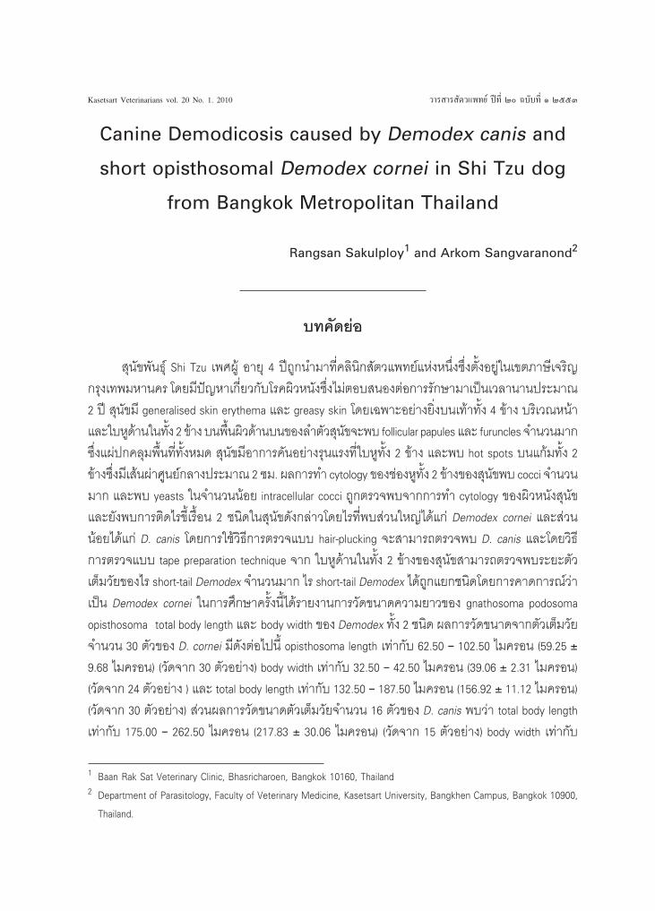
and also less number of D. canis with many spindle
shape eggs of Demodex spp.
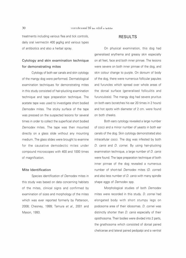
Morphological studies of both Demodex
mites were recorded in this study. D. cornei had
elongated body with short stumpy legs on
podosoma area of their idiosomas. D. cornei was
distinctly shorter than D. canis especially of their
opisthosoma. Their bodies were divided into 2 parts,
the gnathosoma which consisted of dorsal paired
chelicerae and lateral paired pedipalpi and a ventral
«“√ “√ —µ«·æ∑¬å ªï∑’Ë Ú ©∫—∫∑’Ë Ò ÚııÛ 31
single median hypostome. The short opisthosoma
of D. cornei had blunted posterior end and distinct
transverse striations. The size of opisthosoma which
measured from one specimen was 62.50 × 40
microns. The fourth coxisternal plate of D. cornei
was rectangular in shape
Measurements of both D. canis and D.
cornei were done in this study. The measured mite
specimens were mounted under Scotch-tapes. The
measurement data consisted of the followings:
gnathosoma length, podosoma length, opisthosoma
length, body width and total body length. The adult
Figure 1 Ventral view of two adult Demodex
cornei from the infested Shi Tzu dog
from Bangkok metropolitan area
Figure 2 Ventral view of two adult Demodex
cornei and mite egg from the infested
Shi Tzu dog from Bangkok metropolitan
area
Figure 3 Ventral view of adult Demodex cornei
(right) and Demodex canis (left) from
the infested Shi Tzu dog from Bangkok
metropolitan area
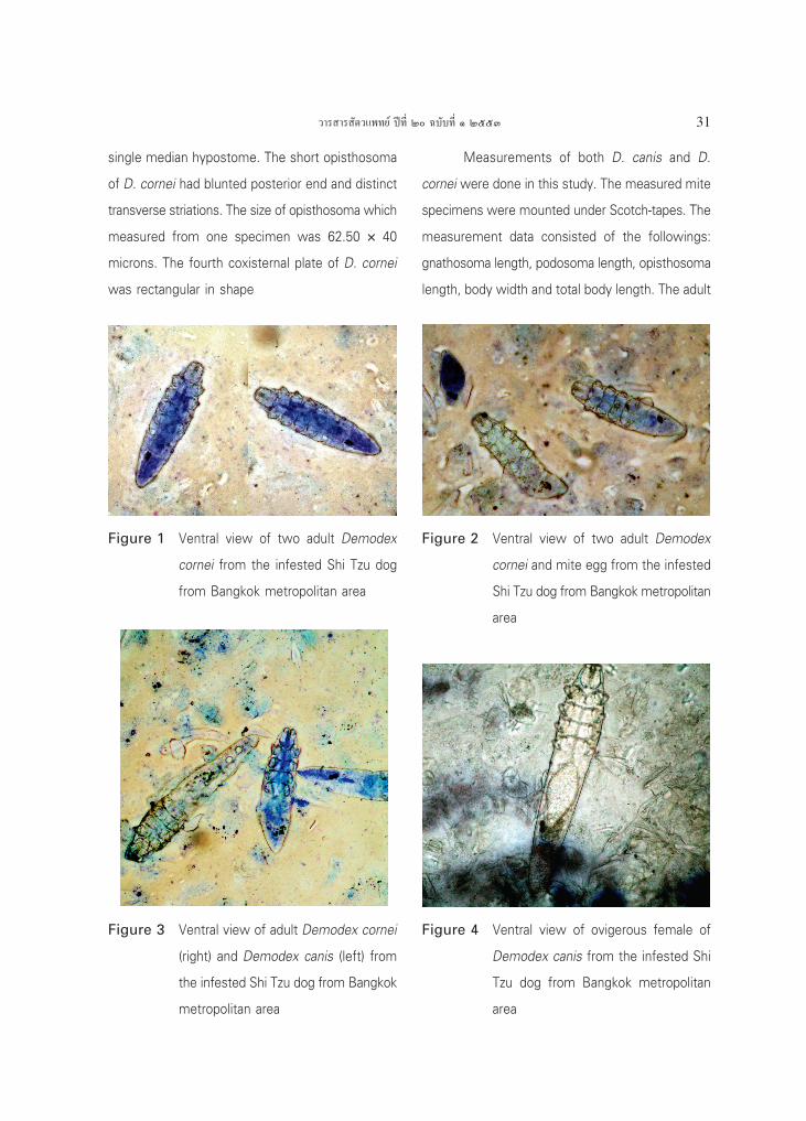
Figure 4 Ventral view of ovigerous female of
Demodex canis from the infested Shi
Tzu dog from Bangkok metropolitan
area
«“√ “√ —µ«·æ∑¬å ªï∑’Ë Ú ©∫—∫∑’Ë Ò ÚııÛ32
mites were measured in microns by using ocular
micrometers and under compound microscopes.
Measurement data of thirty adult (males
and females) D. cornei from this study were
reported. Gnathosoma length was 17.50 › 25.00
microns (23.50 ± 1.93 microns) (n = 30) (mean ±
standard deviation), podosoma length was 55.00 ›
62.50 microns (60.00 ± 2.50 microns) (n = 29),
opisthosoma length was 62.50 › 102.50 microns
(59.25 ± 9.68 microns) (n = 30), body width was
32.50 › 42.50 microns (39.06 ± 2.31 microns) (n =
24), total body length was 132.50 › 187.50 microns
(156.92 ± 11.12 microns) (n = 30).
Sixteen mounted adults of D. canis were
measured under the microscopes and the following
measurement data were also recorded: gnathosoma
length was 22.50 › 27.50 microns (24.84 ± 1.70
microns) (n = 16), podosoma length was 57.50 ›
65.00 microns ( 60.89 ± 2.10 microns) (n = 14),
opisthosoma length was 120.00 › 175.00 microns
(147.50 ± 19.56 microns) (n = 11), body width
was 37.50 › 47.50 microns (42.50 ± 2.89 microns)
(n = 13), total body length was 175.00 › 262.50
microns (217.83 ± 30.06 microns) ( n = 15). The
ratio of mean of total body length and opisthosoma
length between D. canis and D. cornei were 1.39
and 2.49 respectively.
The diagnosis for this Shi Tzu dogûs skin
problems was a combination of generalised surface
and superficial demodicosis due to D. cornei,
generalised follicular demodicosis due to D. canis
and infections of skin and ears concurrent with
allergic skin diseases.
For treatment of the case, 3.3 % lime-
sulphur was used in the form of topical application
on mangy areas of the dog with weekly interval
for eliminating surface and superficial D. cornei.
Ivermectin was given orally with dosage of 600
µg/kg/day to cure mite infestations cause by D.
canis and D. cornei. Adjunctive therapy was done
together with the treatment of miticides. Cephalexin
was used to control secondary bacterial infection
by using dosage of 30 mg/kg twice a day. The
combination formula of gentamicin, clotrimazole
and betamethasone was applied as ear-ointment
by interval of twice a day for controlling otitis
externa.
One week after application of 3.3 % lime-
sulphur, pruritus on both pinnae of the dog was
decreased and general skin condition was obviously
improved. On the second week after treatment,
the numbers of surface Demodex mites which were
detected by the tape preparation technique were
gradually decreased and the general skin condition
was improved. Hot spots on both cheeks
disappeared and pruritic degree was reduced
significantly. On one month after treatment, the
general skin condition was obviously improved
when comparing with those of the second week,
the pruritic degree was reduced, and numbers of
surface Demodex mites were also decreased
gradually.
DISCUSSION
According to the observation in this study,
«“√ “√ —µ«·æ∑¬å ªï∑’Ë Ú ©∫—∫∑’Ë Ò ÚııÛ 33
the clinical presentation caused by D. cornei
infestation often present in the form of a scaly and
pruritic skin diseases that is relevant to the previous
report and literature (Mason,1993; Tater and
Patterson, 2008). Moreover, it was observed by
authors that degree of pruritus due to D. cornei
infestations depended on the number of infested
mites. In comparison, the clinical presentation of
canine demodicosis which caused by D. canis
infestation was often present in the form of alopecic
skin diseases rather than pruritic skin diseases
except in the case associated with secondary
bacterial infection in which the degree of pruritus
will depend on degree of the secondary infection.
In Taiwan, Chen (1995) reported that the
demodectic dog showed major clinical sign as mild
pruritus. In addition, other clinical signs were
alopecia and dry scaling on ventral surface of
abdomen and all four limbs, ventral aspect of neck
and around eyes of the affected dog. While the
study from the United Kingdom (Chesney, 1999)
indicated that the early skin sign had first been
observed at a mean age of about 7 months and
the major clinical sign were alopecia and scaling.
D. cornei infestation is considered by authors as a
pruritic skin disease and differential diagnostic list
may include the following skin conditions: scabies,
allergic skin diseases, bacterial infection, and
malassezia dermatitis.
The diagnostic approach for D. cornei
infestation is superficial skin scraping or using tape
preparation techniques (Mason,1993; Tater and
Patterson, 2008; Guaguere et al., 2008); however,
the authors prefer to use the tape preparation
techniques because of less invasive, decreasing
chance to damage examined mites and also
available for cytology in the same examined slide.
According to the mite identification and reports
(Chen, 1995; Mason, 1993; Tater and Patterson,
2008; Chesney, 1999; Tamura et al., 2001;
Patterson, 2008; Guaguere et al., 2008) the simple
ways of differentiation between D. cornei and D.
canis were reported. From mite sizes, D. cornei is
seemed obviously shorter than D. canis. The total
body length of D. cornei is about 50 › 70 % of
those of D. canis. The mite collection technique
can also give a useful diagnostic data, because D.
cornei inhabits in stratum corneum of epidermis,
the suitable collection technique for D. cornei is
superficial skin scraping or using tape preparation
techniques (Tater and Patterson, 2008) while the
habitat of D. canis is hair follicles and sebaceous
glands which deeper into layer of dermis, so it
may concluded that the suitable collection
techniques for D. canis are deep skin scraping or
hair-plucking examination (Tater and Patterson,
2008).
Currently, the information about D. cornei
infestation in dogs was quite limit including
information about the treatment, although the first
report was published since 1998 (Scott et al, 2001).
The treatment protocol for D. cornei infestation
could modify from the protocol for feline
demodicosis and D. canis infestation (Scott et al,
2001; Chen, 1995; Foster and Foil, 2003; Mueller,
2004; Gortel, 2006; Tater and Patterson, 2008;
«“√ “√ —µ«·æ∑¬å ªï∑’Ë Ú ©∫—∫∑’Ë Ò ÚııÛ34
Mecklenburg et al, 2009). For D. canis infestation,
there is good evidence for recommending systemic
daily ivermectin, or moxidectin or milbemycin oxime
at the dosages of 300-600 µg/kg, 400 µg/kg and 2
mg/kg respectively for treatment of generalised
canine demodicosis, and using topical application
of amitraz rinse at the dosage of 0.025-0.06%
weekly interval or fortnightly can also be
recommended for generalised canine demodicosis
as supported by good evidences (Mueller, 2004).
In authorsû opinion, these protocols may be also
benefit in cases of D. cornei infestation.
In addition, in cat, there was good evidence
to recommend lime sulphur dips at 2% every week
for therapy of feline demodicosis (Mueller, 2004).
Moreover, there was fair evidence for the use of
higher concentration of amitraz rinse at higher
frequency, using weekly doramectin (400 µg/kg
subcutaneously) for treatment of generalised canine
demodicosis caused by D. canis infestation, and
using amitraz rinse at 0.0125-0.025% weekly and
weekly doramectin 600 µg/kg subcutaneously can
be benefit for treatment of feline demodicosis
(Mueller, 2004). These protocols may be useful in
cases of D. cornei infestation too.
It may be concluded that the short-bodied
Demodex mites from this study were D. cornei.
Because of their characters of short and wide body
when compared with D. canis and from Tater and
Patterson (2008), the short tailed Demodex mites
was tentatively identified as D. cornei. The other
diagnostic data of the short bodied mites was the
location on body of the mangy dog which the mites
inhabit. The mean total body length of short form
of Demodex spp. was 156.92 microns while the
mean total body length of D. canis was 217.83
microns, so the short form adult Demodex mites
from this study was about 72 % of the mean total
body length of adult D. canis. However, the total
body length was measured from both adult male
and female mites in this study, and the male mites
were shorter than the female mites. From result
of this study, it was revealed that mean
opisthosoma length (tail) of adult D. cornei was
about 40.17% of the mean opisthosoma length of
adult D. canis and this result was important
evidence of the diagnostic data.
ACKNOWLEDGMENTS
The authors wish to express appreciation
to Mr. Jaray Tongtoyai, Mrs. Natthaga Sakulploy
and Mrs. Sirilak Wiriyaukaradecha for their
photographing of the mites. Moreover, the authors
would like to give a special thank to Dr. Sonya
Bettenay, Prof. Ralf Mueller and Dr. Linda Vogelnest
who always give a lot of valuable knowledge,
comments and supporting.
REFERENCES
Chen, C. 1995. A short-tailed demodectic mite and
Demodex canis infestation in a Chihuahua dog.
Veterinary Dermatology. 6(4) : 227-9
Chesney, C.J. 1999. Short form of Demodex
species mite in the dog: occurrence and

«“√ “√ —µ«·æ∑¬å ªï∑’Ë Ú ©∫—∫∑’Ë Ò ÚııÛ 35
measurements. J. Small. Anim. Pract. 40(2) :
58-61.
Craig, M. 2003. Demodicosis. p. 153-158. In BSAVA
Manual of Small Animal Dermatology (edited
by Foster, A.P. and C.S. Foil) 2nd ed. British
Small Animal Veterinary Association, Waterwell,
UK.
Gortel, K. 2006. Update on Canine Demodicosis.
Veterinary Clinic of North America (Small Animal
Practice). 36(1) : 229-241
Guaguere, E. and Beugnet, F. 2008. Parasitic skin
conditions. p. 194-203 In Merialûs A Practical
Guide to Canine Dermatology, (edited by
Guaguere, E., Prelaud, P. and Craig, M.)
Kalianxis, Italy.
Hillier, A. and C.E. Desch. 2002. Large-Body
Demodex mite infestation in 4 dogs. JAVMA.
220(5) : 623-627
Mason, K.V. 1993. A new species of Demodex
mite with D. canis causing canine demodicosis:
a case report. Veterinary Dermatology. 4 : 37
Mecklenburg, L., Linek, M. and D.J. Tobin. 2009.
Hair Loss Disorders in Domestic Animals.
Wiley-Blackwell, Iowa, USA. 276 pp.
Mueller, R.S. 2004. Treatment protocols for
demodicosis: an evidence-based Review.
Veterinary Dermatology. 15 : 75-89
Mueller, R.S. and S.V. Bettenay.1999. An unusual
presentation of canine demodicosis caused by
a long-bodied Demodex mite in a Lakeland
Terrier. Aust. Vet. Pract . 29(3) : 128-131
Patterson, S. 2008. Manual of Skin Diseases of
Dog and Cat 2nd ed. Blackwell Publishing,
Oxford, UK. 355 pp.
Robson, D.C,. Burton, G.G., Basset, R., Shipstone,
M. and R. Mueller. 2003. Eight case of
Demodecosis caused by a long-bodied
Demodex species (1997-2002). Aust .Vet. Pract.
33(2) : 64-72
Scott, D.W., Miller, W.H. and C.E. Griffin. 2001.
Small Animal Dermatology, 6th.ed. W.B.
Saunders, Philadelphia. 1528 pp.
Tamura, Y., Kawamura, Y., Inoue, I, and S. Ishino.
2001. Scanning electron Microscopy description
of a new species of Demodex canis spp.
Veterinary Dermatology. 12 : 275-278
Tater, K.C. and A.P. Patterson. 2008. Canine and
Feline Demodicosis. Veterinary Medicine.
103(8) : 444-461