Embed Size (px)
Citation preview
causes of dizziness and vestibular dysfunction
Herman Kingma, department of ORL, Maastricht University Medical Centre
Maastricht Research Institute Mental Health and Neuroscience
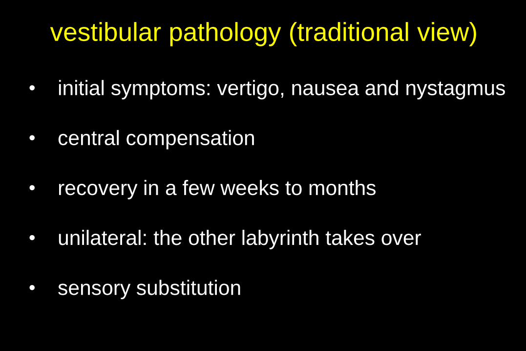
vestibular pathology (traditional view)
• initial symptoms: vertigo, nausea and nystagmus
• central compensation
• recovery in a few weeks to months
• unilateral: the other labyrinth takes over
• sensory substitution
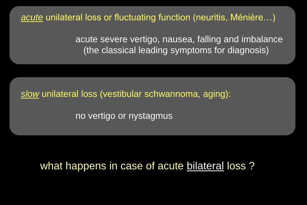
acute unilateral loss or fluctuating function (neuritis, Ménière…)
acute severe vertigo, nausea, falling and imbalance
(the classical leading symptoms for diagnosis)
slow unilateral loss (vestibular schwannoma, aging):
no vertigo or nystagmus
what happens in case of acute bilateral loss ?
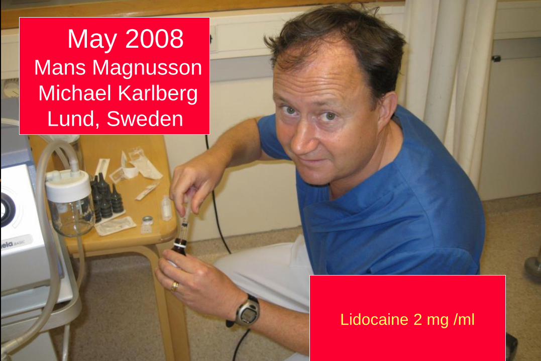
Lidocaine 2 mg /ml
May 2008Mans Magnusson
Michael Karlberg
Lund, Sweden
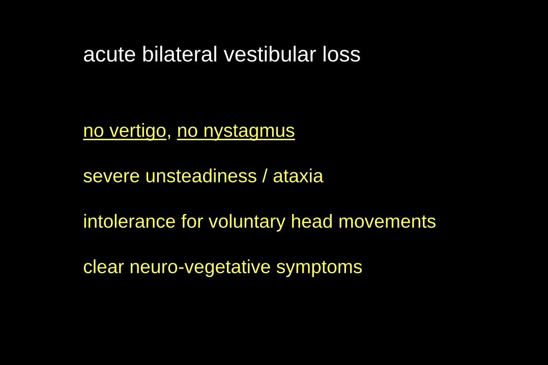
acute bilateral vestibular loss
no vertigo, no nystagmus
severe unsteadiness / ataxia
intolerance for voluntary head movements
clear neuro-vegetative symptoms
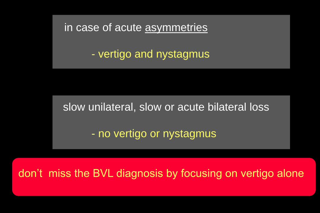
in case of acute asymmetries
- vertigo and nystagmus
slow unilateral, slow or acute bilateral loss
- no vertigo or nystagmus
don’t miss the BVL diagnosis by focusing on vertigo alone

take home message:
vertigo is only 1 aspect of vestibular loss
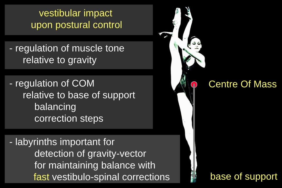
base of support
Centre Of Mass
vestibular impact
upon postural control
- regulation of muscle tone
relative to gravity
- regulation of COM
relative to base of support
balancing
correction steps
- labyrinths important for
detection of gravity-vector
for maintaining balance with
fast vestibulo-spinal corrections
for maintaining balance
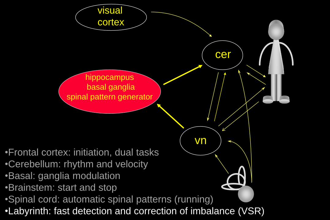
vn
cer
hippocampus
basal ganglia
spinal pattern generator
visual
cortex
•Frontal cortex: initiation, dual tasks
•Cerebellum: rhythm and velocity
•Basal: ganglia modulation
•Brainstem: start and stop
•Spinal cord: automatic spinal patterns (running)
•Labyrinth: fast detection and correction of imbalance (VSR)
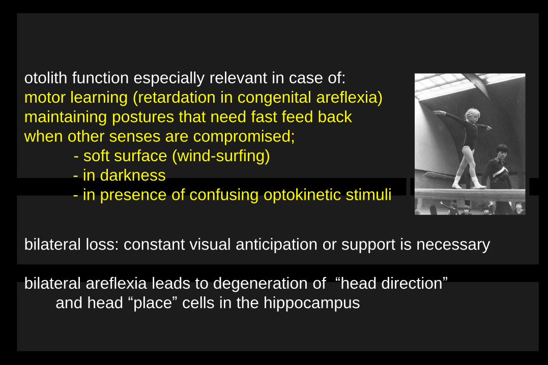
otolith function especially relevant in case of:
motor learning (retardation in congenital areflexia)
maintaining postures that need fast feed back
when other senses are compromised;
- soft surface (wind-surfing)
- in darkness
- in presence of confusing optokinetic stimuli
bilateral loss: constant visual anticipation or support is necessary
bilateral areflexia leads to degeneration of “head direction”
and head “place” cells in the hippocampus
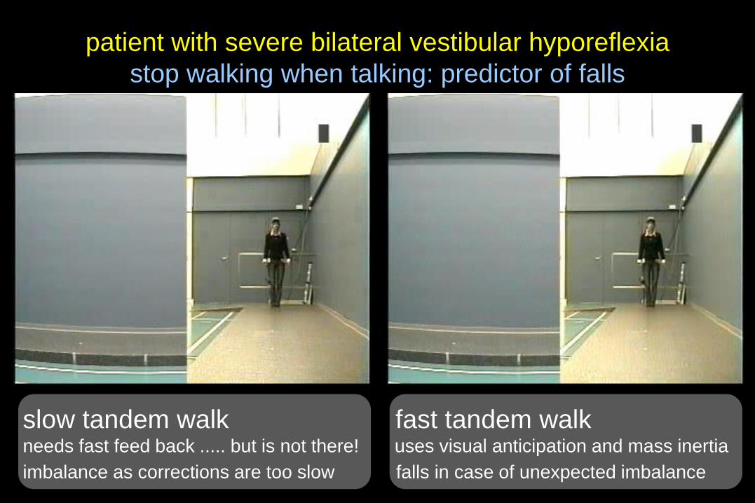
patient with severe bilateral vestibular hyporeflexia
stop walking when talking: predictor of falls
slow tandem walk fast tandem walkneeds fast feed back ..... but is not there! uses visual anticipation and mass inertia
imbalance as corrections are too slow falls in case of unexpected imbalance
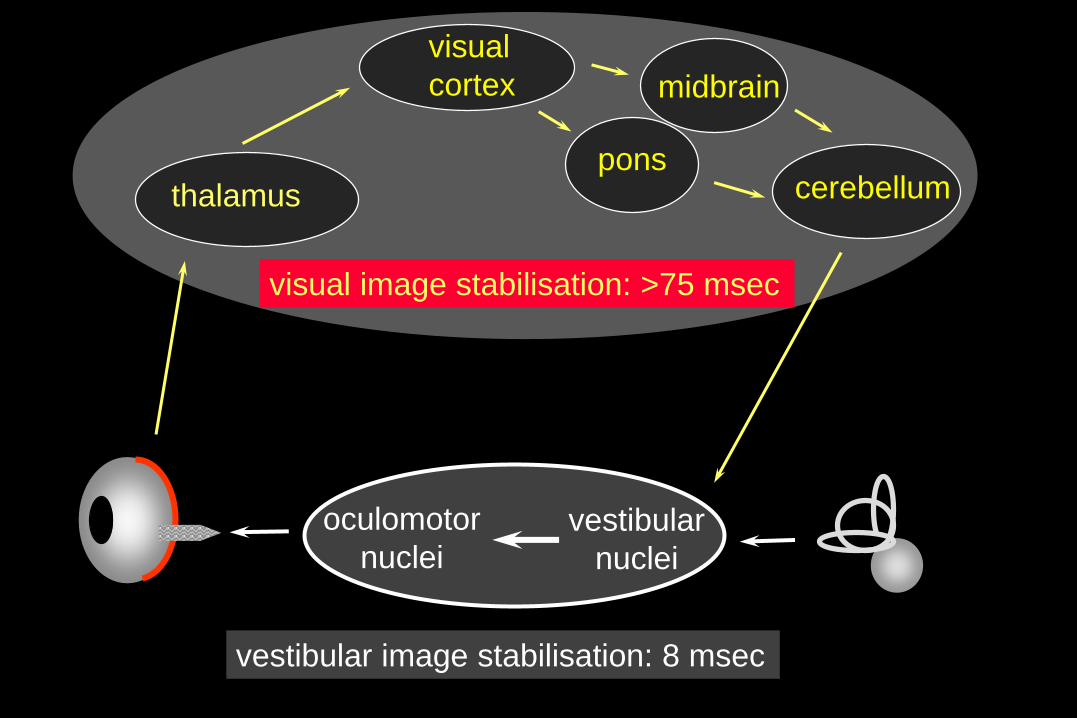
thalamus
visual
cortex
ponscerebellum
oculomotor
nuclei
vestibular image stabilisation: 8 msec
midbrain
vestibular
nuclei
visual image stabilisation: >75 msec
head impulse test in unilateral loss
standard video (50 Hz)
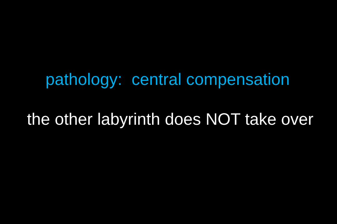
pathology: central compensation
the other labyrinth does NOT take over
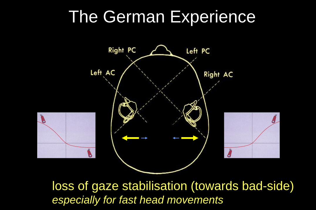
loss of gaze stabilisation (towards bad-side)especially for fast head movements
The German Experience
head impulse test in bilateral loss
CASIO Exilem: high speed recording (300 Hz)
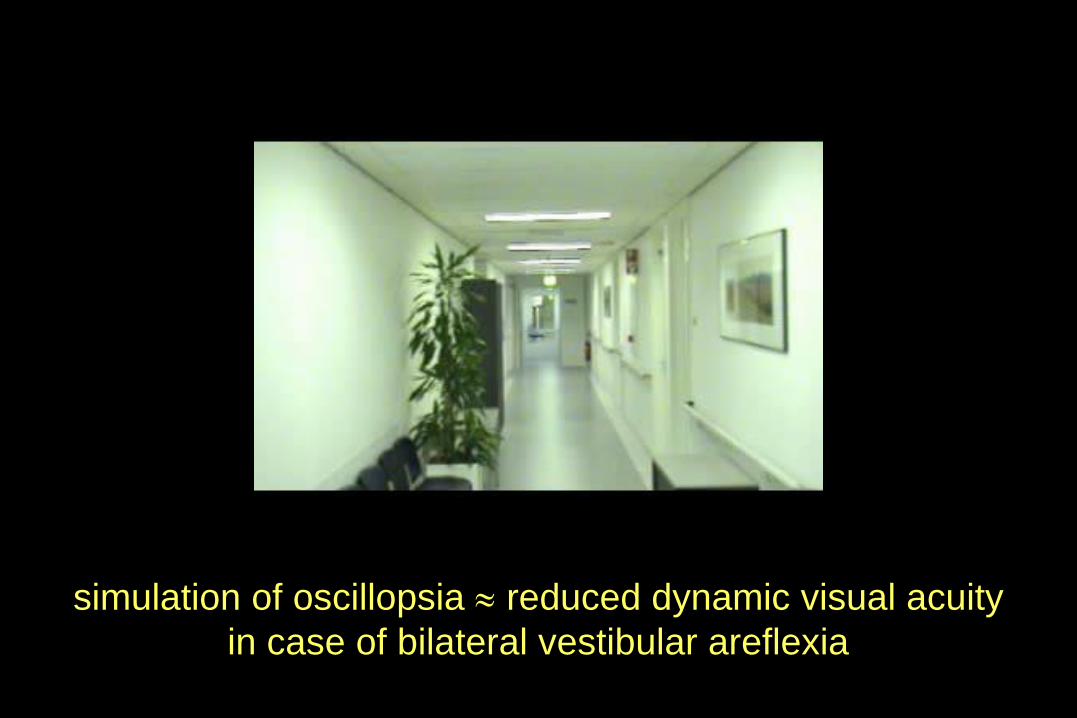
simulation of oscillopsia reduced dynamic visual acuity
in case of bilateral vestibular areflexia
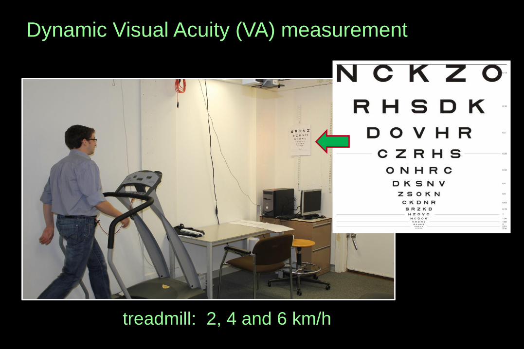
Dynamic Visual Acuity (VA) measurement
treadmill: 2, 4 and 6 km/h
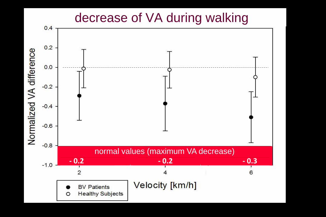
decrease of VA during walking
- 0.2 - 0.2 - 0.3normal values (maximum VA decrease)
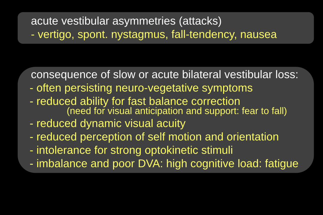
acute vestibular asymmetries (attacks)
- vertigo, spont. nystagmus, fall-tendency, nausea
consequence of slow or acute bilateral vestibular loss:
- often persisting neuro-vegetative symptoms
- reduced ability for fast balance correction(need for visual anticipation and support: fear to fall)
- reduced dynamic visual acuity
- reduced perception of self motion and orientation
- intolerance for strong optokinetic stimuli
- imbalance and poor DVA: high cognitive load: fatigue
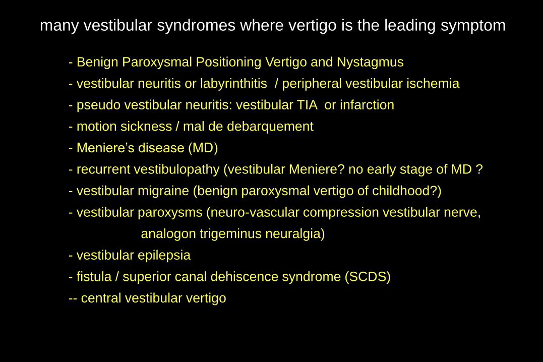
many vestibular syndromes where vertigo is the leading symptom
- Benign Paroxysmal Positioning Vertigo and Nystagmus
- vestibular neuritis or labyrinthitis / peripheral vestibular ischemia
- pseudo vestibular neuritis: vestibular TIA or infarction
- motion sickness / mal de debarquement
- Meniere’s disease (MD)
- recurrent vestibulopathy (vestibular Meniere? no early stage of MD ?
- vestibular migraine (benign paroxysmal vertigo of childhood?)
- vestibular paroxysms (neuro-vascular compression vestibular nerve,
analogon trigeminus neuralgia)
- vestibular epilepsia
- fistula / superior canal dehiscence syndrome (SCDS)
-- central vestibular vertigo
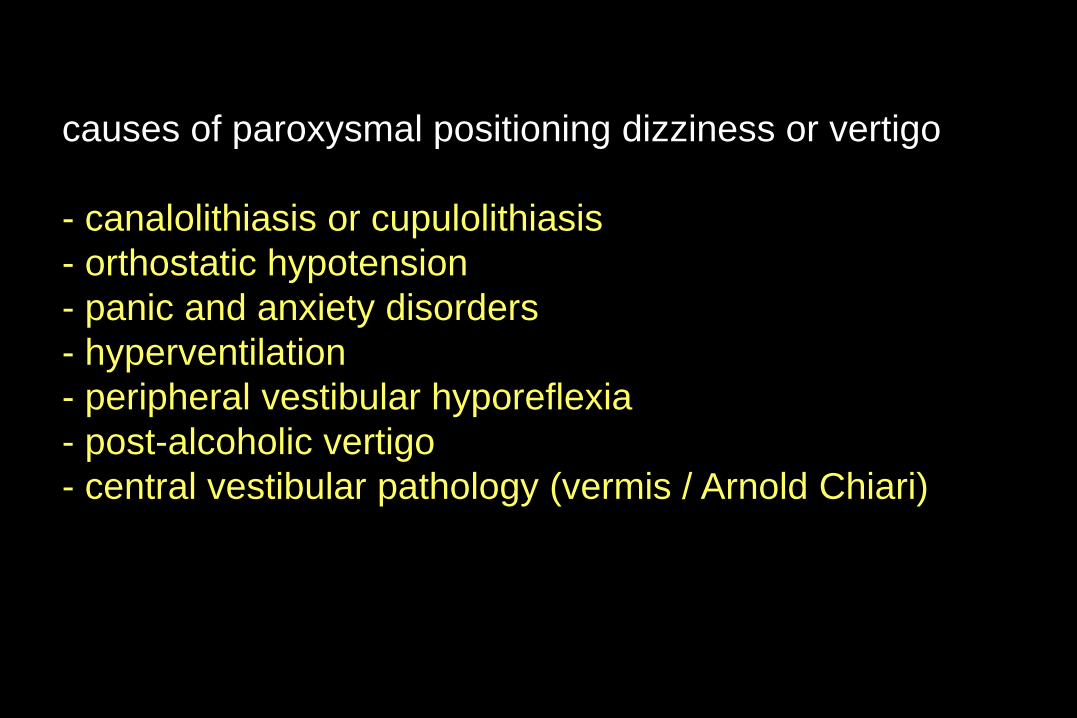
causes of paroxysmal positioning dizziness or vertigo
- canalolithiasis or cupulolithiasis
- orthostatic hypotension
- panic and anxiety disorders
- hyperventilation
- peripheral vestibular hyporeflexia
- post-alcoholic vertigo
- central vestibular pathology (vermis / Arnold Chiari)
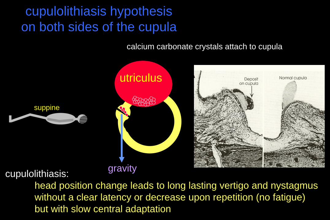
utriculus
suppine
gravitycupulolithiasis:
head position change leads to long lasting vertigo and nystagmus
without a clear latency or decrease upon repetition (no fatigue)
but with slow central adaptation
calcium carbonate crystals attach to cupula
cupulolithiasis hypothesis
on both sides of the cupula
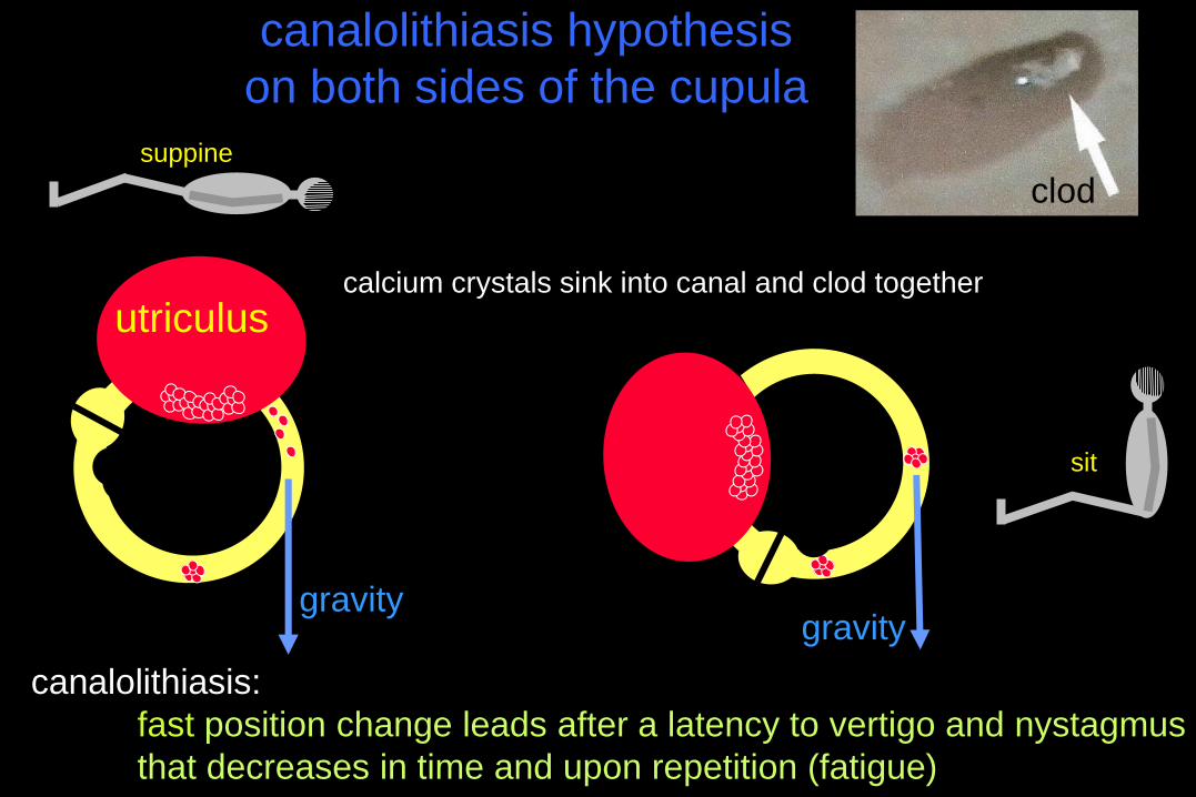
calcium crystals sink into canal and clod together
canalolithiasis hypothesis
on both sides of the cupula
canalolithiasis:
fast position change leads after a latency to vertigo and nystagmus
that decreases in time and upon repetition (fatigue)
suppine
utriculus
sit
gravitygravity
clod
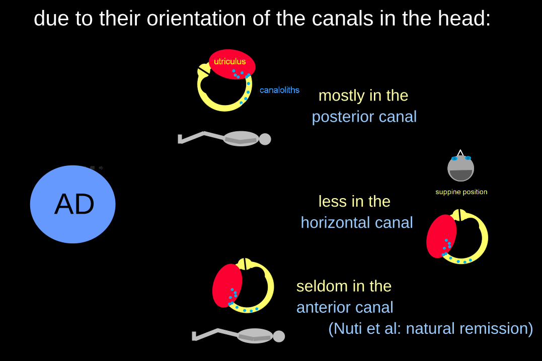
due to their orientation of the canals in the head:
mostly in the
posterior canal
less in the
horizontal canal
seldom in the
anterior canal
(Nuti et al: natural remission)
AD
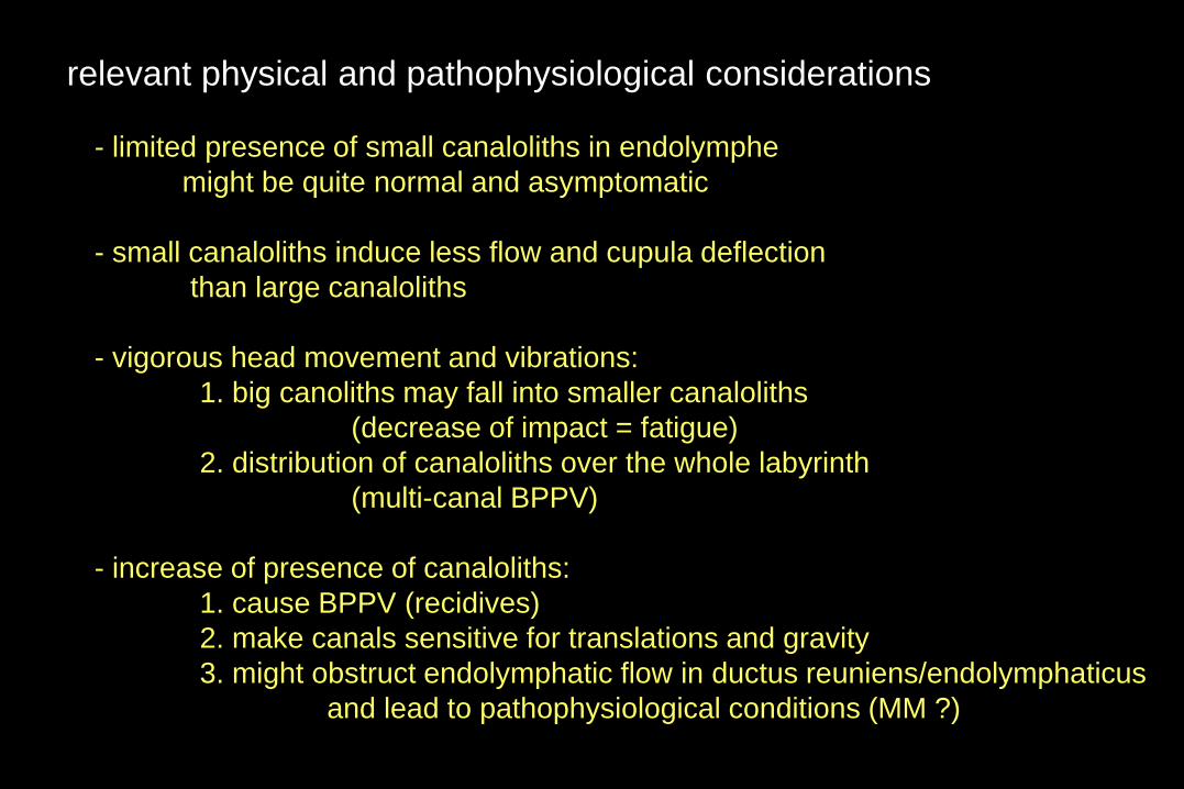
relevant physical and pathophysiological considerations
- limited presence of small canaloliths in endolymphe
might be quite normal and asymptomatic
- small canaloliths induce less flow and cupula deflection
than large canaloliths
- vigorous head movement and vibrations:
1. big canoliths may fall into smaller canaloliths
(decrease of impact = fatigue)
2. distribution of canaloliths over the whole labyrinth
(multi-canal BPPV)
- increase of presence of canaloliths:
1. cause BPPV (recidives)
2. make canals sensitive for translations and gravity
3. might obstruct endolymphatic flow in ductus reuniens/endolymphaticus
and lead to pathophysiological conditions (MM ?)
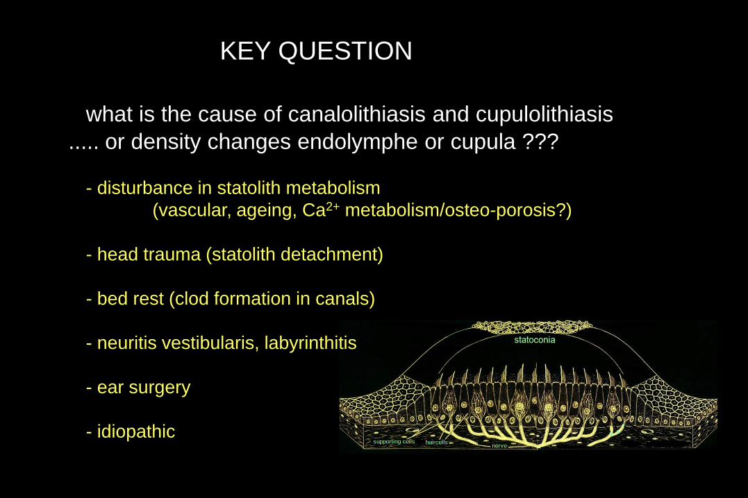
KEY QUESTION
what is the cause of canalolithiasis and cupulolithiasis
..... or density changes endolymphe or cupula ???
- disturbance in statolith metabolism
(vascular, ageing, Ca2+ metabolism/osteo-porosis?)
- head trauma (statolith detachment)
- bed rest (clod formation in canals)
- neuritis vestibularis, labyrinthitis
- ear surgery
- idiopathic

AVOR: free APP for Iphone/Ipad
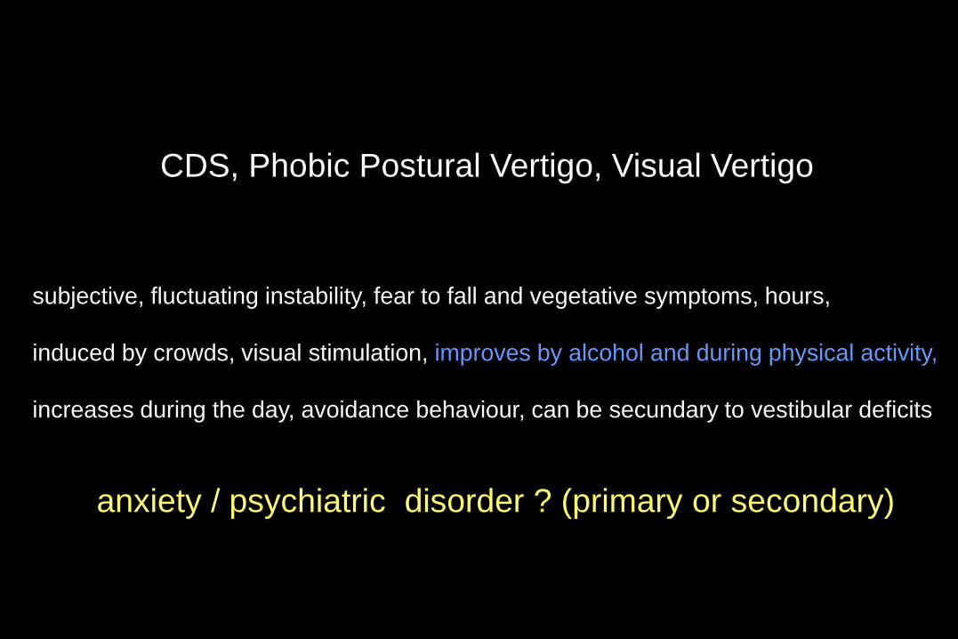
CDS, Phobic Postural Vertigo, Visual Vertigo
subjective, fluctuating instability, fear to fall and vegetative symptoms, hours,
induced by crowds, visual stimulation, improves by alcohol and during physical activity,
increases during the day, avoidance behaviour, can be secundary to vestibular deficits
anxiety / psychiatric disorder ? (primary or secondary)
motion sickness
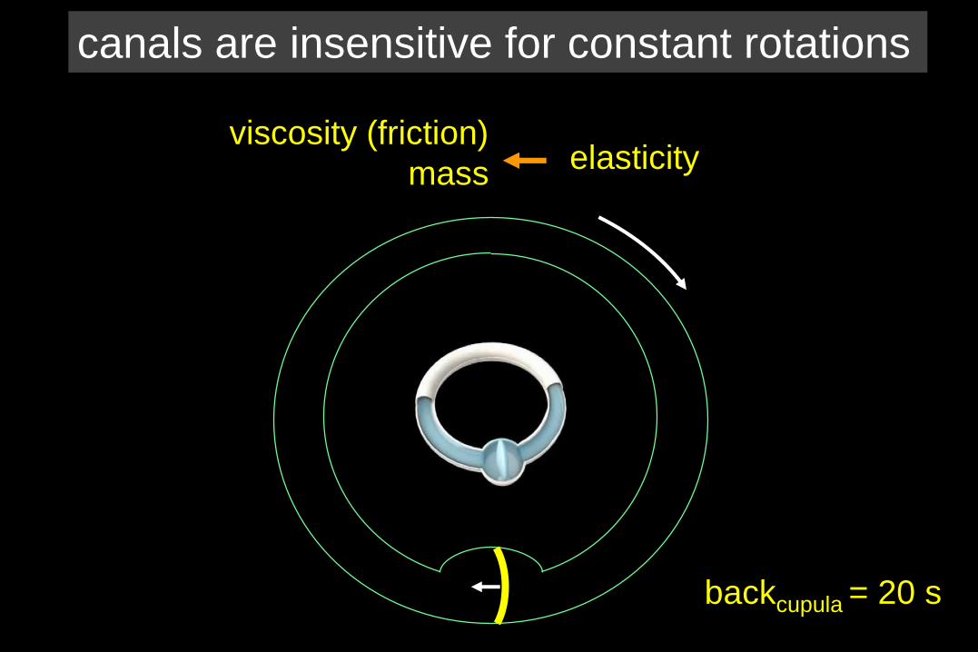
elasticityviscosity (friction)
mass
backcupula = 20 s
canals are insensitive for constant rotations
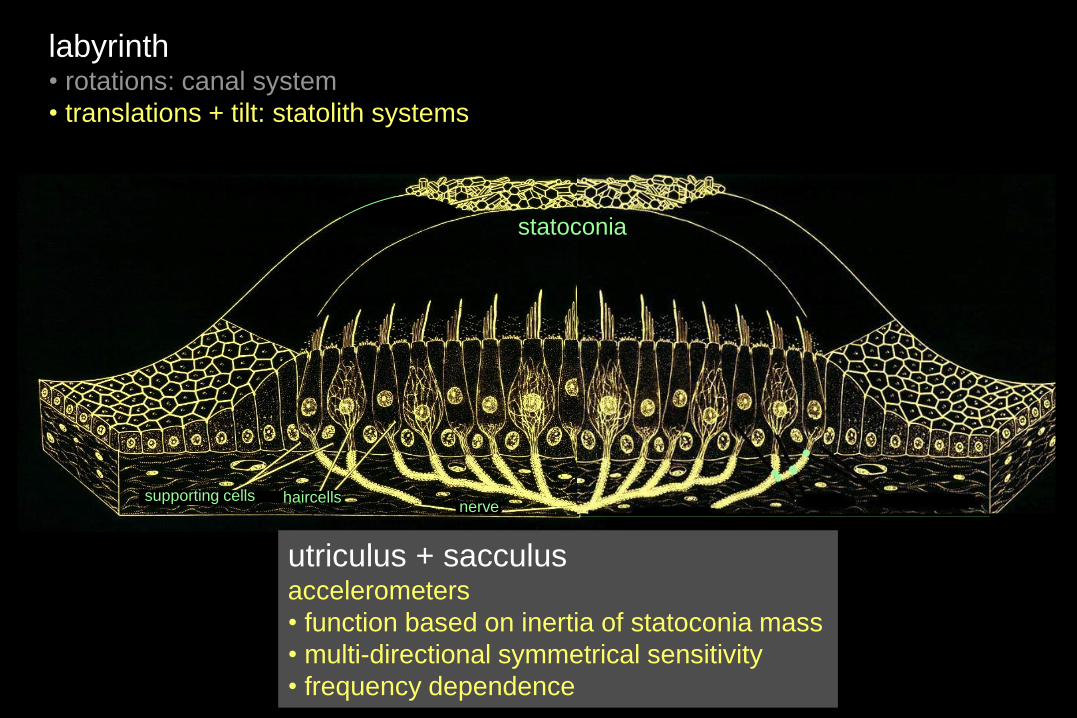
labyrinth• rotations: canal system
• translations + tilt: statolith systems
statolithsstatoconia
supporting cells haircellsnerve
utriculus + sacculusaccelerometers
• function based on inertia of statoconia mass
• multi-directional symmetrical sensitivity
• frequency dependence
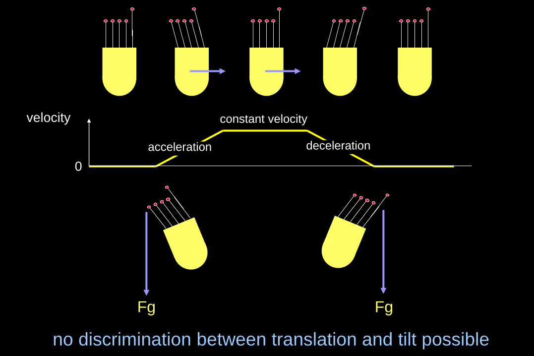
Fg
0
velocity
Fg
no discrimination between translation and tilt possible
constant velocity
acceleration deceleration
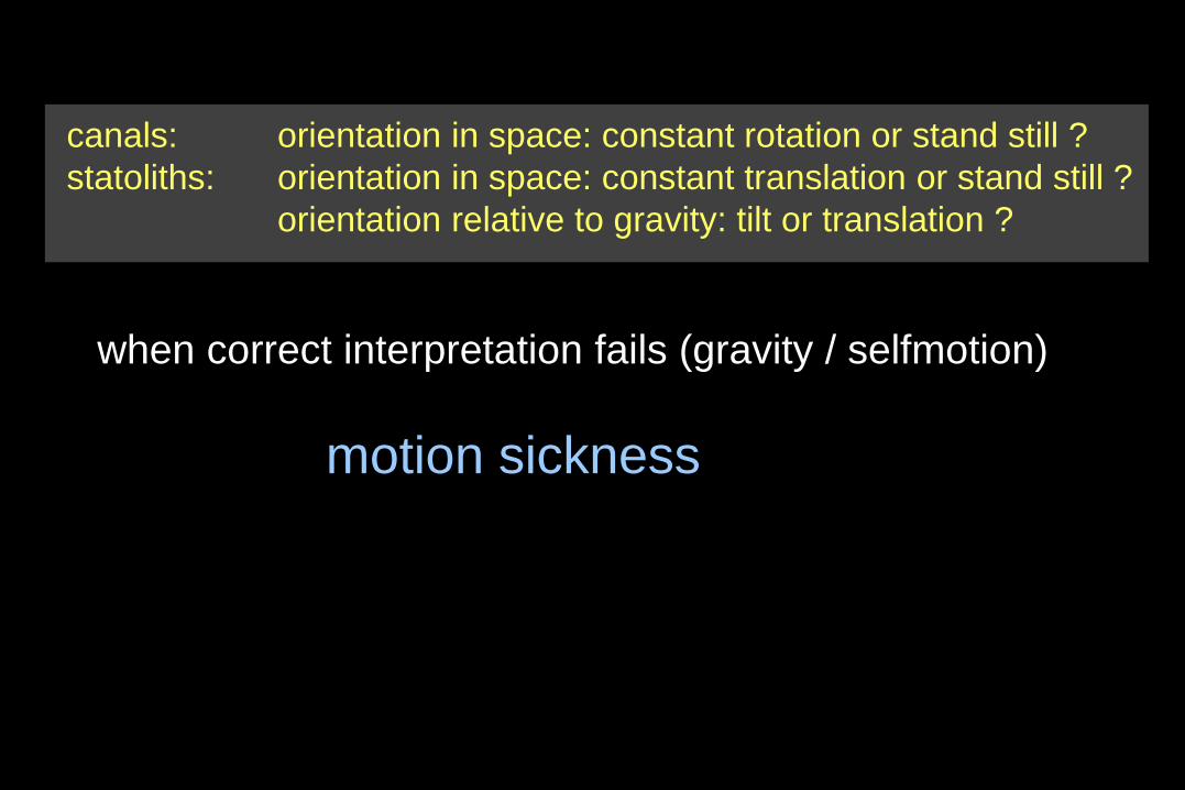
canals: orientation in space: constant rotation or stand still ?
statoliths: orientation in space: constant translation or stand still ?
orientation relative to gravity: tilt or translation ?
when correct interpretation fails (gravity / selfmotion)
motion sickness
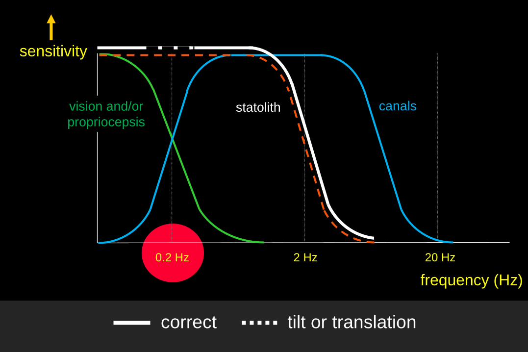
0.2 Hz 2 Hz 20 Hz
sensitivity
frequency (Hz)
canalsstatolithvision and/or
propriocepsis
correct tilt or translation
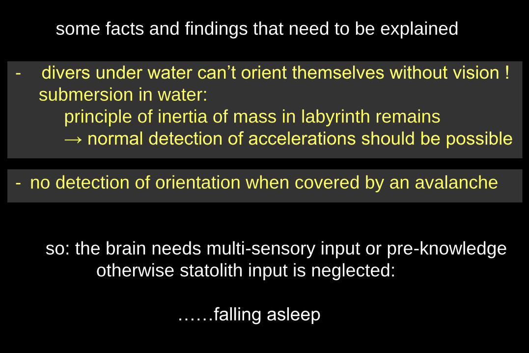
some facts and findings that need to be explained
- divers under water can’t orient themselves without vision !
submersion in water:
principle of inertia of mass in labyrinth remains
→ normal detection of accelerations should be possible
- no detection of orientation when covered by an avalanche
so: the brain needs multi-sensory input or pre-knowledge
otherwise statolith input is neglected:
……falling asleep
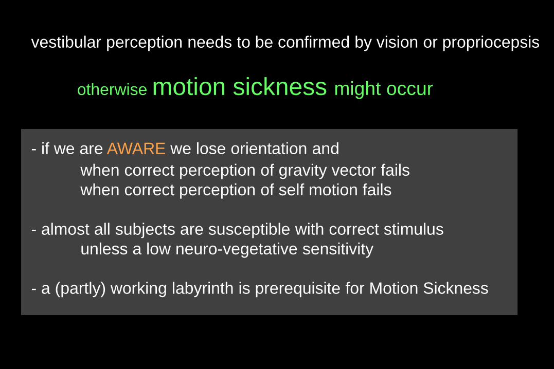
vestibular perception needs to be confirmed by vision or propriocepsis
otherwise motion sickness might occur
- if we are AWARE we lose orientation and
when correct perception of gravity vector fails
when correct perception of self motion fails
- almost all subjects are susceptible with correct stimulus
unless a low neuro-vegetative sensitivity
- a (partly) working labyrinth is prerequisite for Motion Sickness
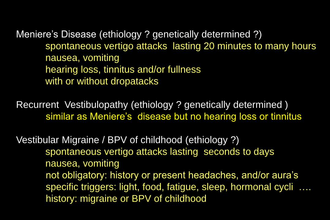
Meniere’s Disease (ethiology ? genetically determined ?)
spontaneous vertigo attacks lasting 20 minutes to many hours
nausea, vomiting
hearing loss, tinnitus and/or fullness
with or without dropatacks
Recurrent Vestibulopathy (ethiology ? genetically determined )
similar as Meniere’s disease but no hearing loss or tinnitus
Vestibular Migraine / BPV of childhood (ethiology ?)
spontaneous vertigo attacks lasting seconds to days
nausea, vomiting
not obligatory: history or present headaches, and/or aura’s
specific triggers: light, food, fatigue, sleep, hormonal cycli ….
history: migraine or BPV of childhood
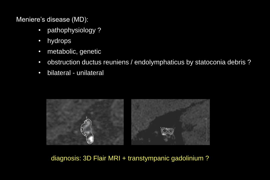
Meniere’s disease (MD):
• pathophysiology ?
• hydrops
• metabolic, genetic
• obstruction ductus reuniens / endolymphaticus by statoconia debris ?
• bilateral - unilateral
diagnosis: 3D Flair MRI + transtympanic gadolinium ?
fistula / superior canal dehiscence syndrome (SCDS)
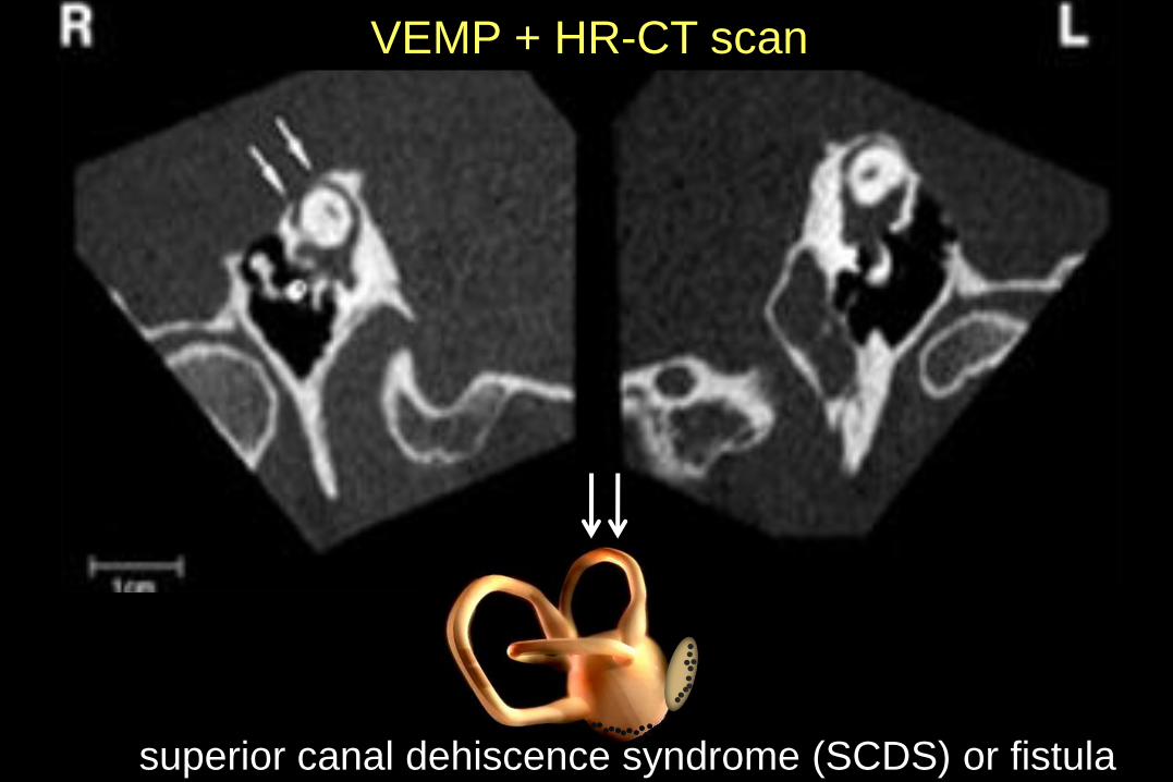
VEMP + HR-CT scan
superior canal dehiscence syndrome (SCDS) or fistula
WHY
fistula / superior canal dehiscence syndrome (SCDS)
study of skulls: SCDS common feature, often asymptomatic
I hope this was
useful to you and help you
with the management of your patients
thank you
all slides: www.hermankingma.com
contact: [email protected]