Embed Size (px)
Citation preview
Journal of Clinical and Analytical Medicine | 1
Edit
ör’e Mektup
Letters to Editor
Servikal Ektopik Timus Dokusu İçindeki Paratiroid Dokusu / Ectopic Thymic Tissue with Parathyroid Elements
Esra Karakuş1, Müjdem Nur Azılı2, Atilla Şenaylı21Ankara Çocuk Sağlığı ve Hastalıkları, 2Çocuk Cerrahisi,
Hematoloji Onkoloji Eğitim ve Araştırma Hastanesi, Ankara, Türkiye
Cervical Ectopic Thymic Tissue with Parathyroid Elements Mimicking Papillary Carcinoma Metastasis
Papiller Karsinom Metastazını Taklit Eden Servikal Ektopik Timus Dokusu İçindeki Paratiroid Dokusu
DOI: 10.4328/JCAM.3327 Received: 18.02.2015 Accepted: 07.04.2015 Publihed Online: 08.04.2015Corresponding Author: Esra Karakuş, Ankara Çocuk Sağlığı ve Hastalıkları, Hematoloji Onkoloji Eğitim ve Araştırma Hastanesi, Dışkapı, Ankara, Türkiye.T.: +90 3125969746 GSM: +905071636748 F.: +90 3123472330 E-Mail: [email protected]
To the editor:
A 16 year-old female patient was presented with a cervical mass and pain. An ultrasound imaging detected hypoechoic nodules
and calcifications at the right and left thyroid lobe. Preoperative blood examples were within the normal limits, except for
autoantibodies (TgAb, TPOAb and TrAb). The nodule at the lower pole of the middle part of the right thyroid lobe was aspirated
with a single pass using a 22-gauge needle. Fine needle aspiration biopsy performed on the left lesion revealed cellular, three-
dimensional and papillary groups. Some of these groups showed follicular organization. The cells had voluminous, pleomorphic
nuclei with rough chromatin. Pseudoinclusions were extensive and some nuclei had prominent nucleoli. Foamy macrophages and
mature lymphocytes were sparsely seen in the background. The patient underwent bilateral total thyroidectomy. Thyroidectomy
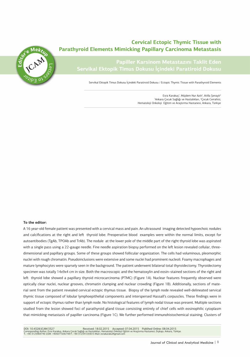
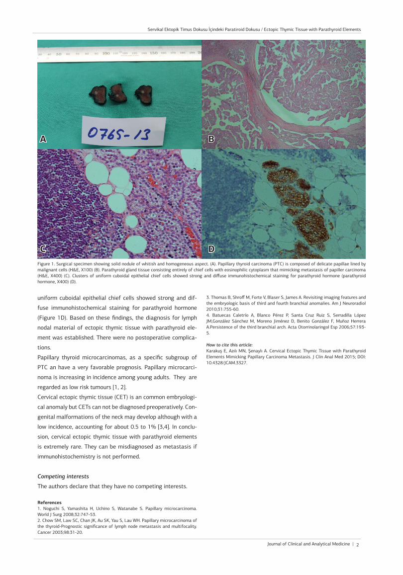
specimen was totally 14x9x4 cm in size. Both the macroscopic and the hematoxylin and eosin–stained sections of the right and
left thyroid lobe showed a papillary thyroid microcarcinoma (PTMC) (Figure 1A). Nuclear features frequently observed were
optically clear nuclei, nuclear grooves, chromatin clumping and nuclear crowding (Figure 1B). Additionally, sections of mate-
rial sent from the patient revealed cervical ectopic thymus tissue. Biopsy of the lymph node revealed well-delineated servical
thymic tissue composed of lobular lymphoepithelial components and interspersed Hassall’s corpuscles. These findings were in
support of ectopic thymus rather than lymph node. No histological features of lymph nodal tissue was present. Multiple sections
studied from the lesion showed foci of parathyroid gland tissue consisting entirely of chief cells with eosinophilic cytoplasm
that mimicking metastasis of papiller carcinoma (Figure 1C). We further performed immunohistochemical staining. Clusters of
Journal of Clinical and Analytical Medicine |
Servikal Ektopik Timus Dokusu İçindeki Paratiroid Dokusu / Ectopic Thymic Tissue with Parathyroid Elements
2
uniform cuboidal epithelial chief cells showed strong and dif-
fuse immunohistochemical staining for parathyroid hormone
(Figure 1D). Based on these findings, the diagnosis for lymph
nodal material of ectopic thymic tissue with parathyroid ele-
ment was established. There were no postoperative complica-
tions.
Papillary thyroid microcarcinomas, as a specific subgroup of
PTC an have a very favorable prognosis. Papillary microcarci-
noma is increasing in incidence among young adults. They are
regarded as low risk tumours [1, 2].
Cervical ectopic thymic tissue (CET) is an common embryologi-
cal anomaly but CETs can not be diagnosed preoperatively. Con-
genital malformations of the neck may develop although with a
low incidence, accounting for about 0.5 to 1% [3,4]. In conclu-
sion, cervical ectopic thymic tissue with parathyroid elements
is extremely rare. They can be misdiagnosed as metastasis if
immunohistochemistry is not performed.
Competing interests
The authors declare that they have no competing interests.
References1. Noguchi S, Yamashita H, Uchino S, Watanabe S. Papillary microcarcinoma. World J Surg 2008;32:747-53.2. Chow SM, Law SC, Chan JK, Au SK, Yau S, Lau WH. Papillary microcarcinoma of the thyroid-Prognostic significance of lymph node metastasis and multifocality. Cancer 2003;98:31-20.
3. Thomas B, Shroff M, Forte V, Blaser S, James A. Revisiting imaging features and the embryologic basis of third and fourth branchial anomalies. Am J Neuroradiol 2010;31:755-60.4. Batuecas Caletrío A, Blanco Pérez P, Santa Cruz Ruiz S, Serradilla López JM,González Sánchez M, Moreno Jiménez D, Benito González F, Muñoz Herrera A.Persistence of the third branchial arch. Acta Otorrinolaringol Esp 2006;57:193-5.
How to cite this article:Karakuş E, Azılı MN, Şenaylı A. Cervical Ectopic Thymic Tissue with Parathyroid Elements Mimicking Papillary Carcinoma Metastasis. J Clin Anal Med 2015; DOI: 10.4328/JCAM.3327.
Figure 1. Surgical specimen showing solid nodule of whitish and homogeneous aspect. (A). Papillary thyroid carcinoma (PTC) is composed of delicate papillae lined by malignant cells (H&E, X100) (B). Parathyroid gland tissue consisting entirely of chief cells with eosinophilic cytoplasm that mimicking metastasis of papiller carcinoma (H&E, X400) (C). Clusters of uniform cuboidal epithelial chief cells showed strong and diffuse immunohistochemical staining for parathyroid hormone (parathyroid hormone, X400) (D).
A B
C D