Embed Size (px)
Citation preview
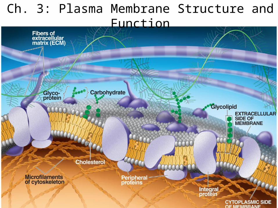
Ch. 3: Plasma Membrane Structure and Function
δ +
δ +
δ -
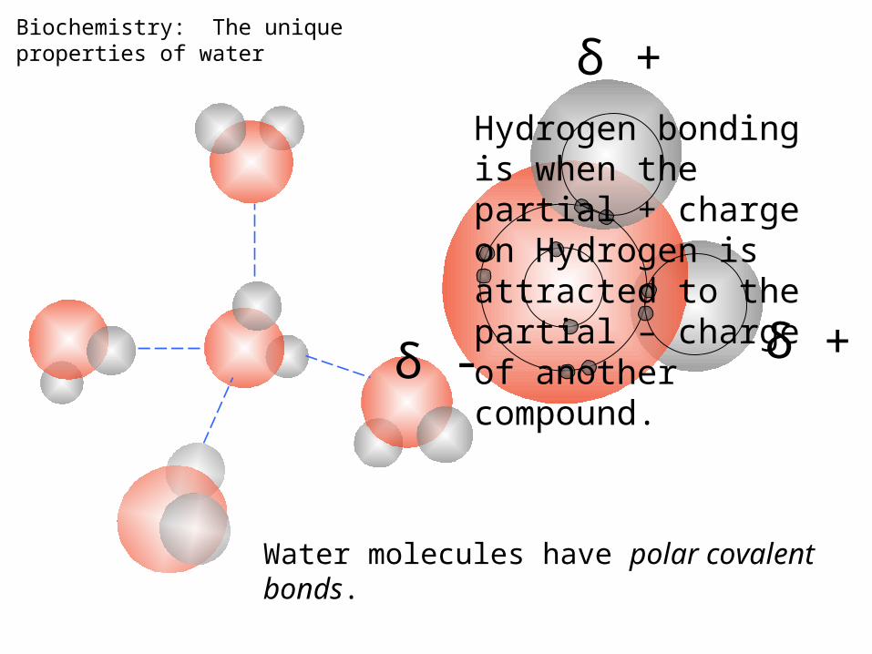
Water molecules have polar covalent bonds.
Biochemistry: The unique properties of water
Hydrogen bonding is when the partial + charge on Hydrogen is attracted to the partial – charge of another compound.
Properties of PhospholipidsPhospholipids
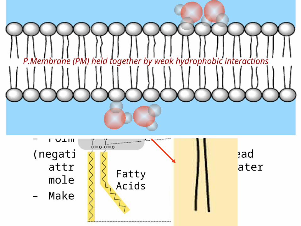
– Glycerol with Phosphate Head + 2 Fatty Acid Chains
– Amphiphilic (“Both” “lover”)• Hydrophilic head• Hydrophobic tail
– Forms 2 layers in water
(negatively charged phosphate head attracted to + end of polar water molecules)
– Makes up cell membranes
Phosphate
Glycerol
Fatty Acids
P.Membrane (PM) held together by weak hydrophobic interactions
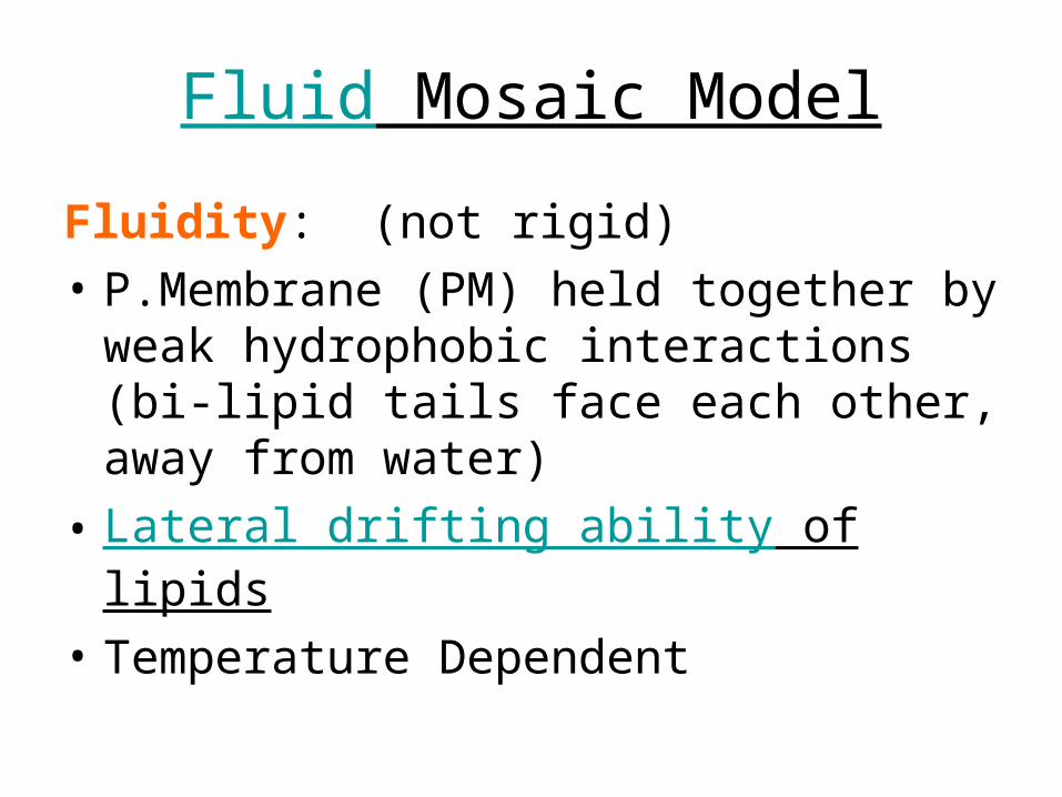
Fluid Mosaic Model
Fluidity: (not rigid)
• P.Membrane (PM) held together by weak hydrophobic interactions (bi-lipid tails face each other, away from water)
• Lateral drifting ability of lipids
• Temperature Dependent
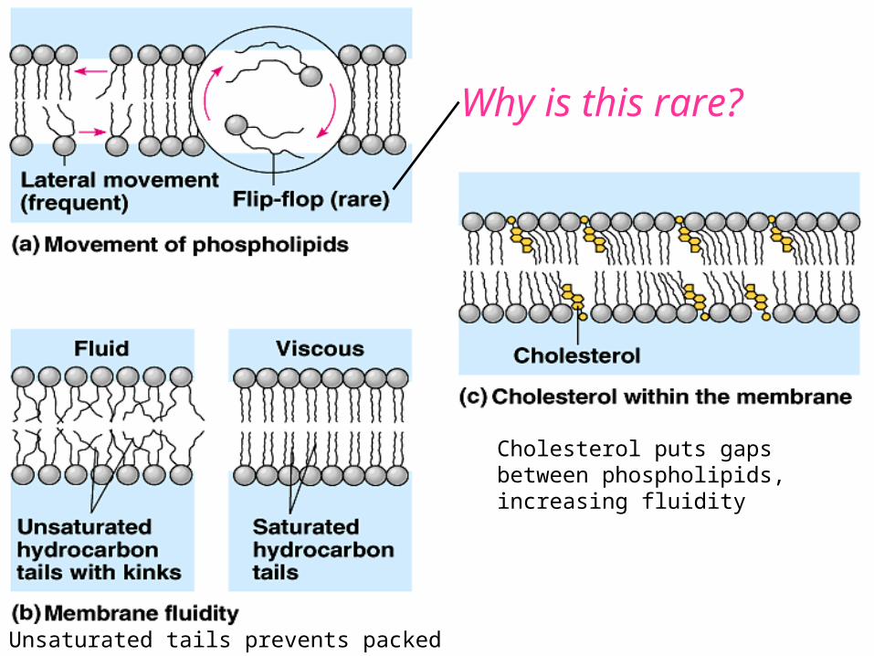
Cholesterol puts gaps between phospholipids, increasing fluidity
Unsaturated tails prevents packed phospho-lipid rafts
Why is this rare?
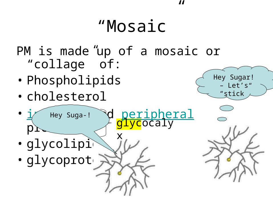
“Mosaic”PM is made up of a mosaic or “collage” of:• Phospholipids• cholesterol• integral and peripheral proteins• glycolipids• glycoproteins
glycocalyx
Hey Sugar! – Let’s “stick”
Hey Suga-!
Integral or Transmembrane Proteins
• Penetrate hydrophobic core of membrane
Surface or Peripheral Proteins
• Loosely bound to surface
• Some attaches to cyto-skeleton or ECM (Extracellular matrix)

Review: What organelles are responsible for creating membrane proteins?
Membrane Transport
• Cells NEED to be able to:– remove waste– take in necessary nutrients from interstitial
fluids– Send out signals to other cells– Receive signals from other cells
• Transport Classified as:– Passive– Active
Selective Permeability of Plasma Membrane
General rule: like dissolves like
• Non-polar, hydrophobic solutes dissolve in lipid
• Ions, hydrophillic, or polar solutes dissolve in water
Selective Permeability of Plasma Membrane
Selective Permeability: some substances can pass through lipid core or membrane more easily than others
1. CO2, O2, non-polar molecules, and other lipids, are hydrophobic and can pass hydrophobic lipid membrane core easily
2. Water, sugars, charged ions, or polar molecules cannot pass lipid core easily so must use hydrophillic transport proteins to pass (ex. Aquaporins)
3. Small molecules are more permeable than larger ones

Passive Transport
• Molecules move down [gradient] (from high to low
concentration) until equilibrium is reached• Spontaneous process• No ATP needed; uses Kinetic energy (KE)
or Hydrostatic Pressure as E source• Types of Passive Transport:
– Simple Diffusion– Facilitated Diffusion– Osmosis– Filtration
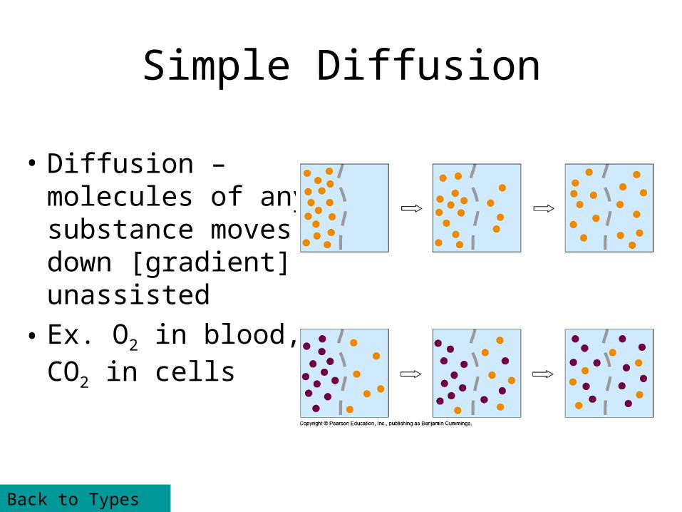
Simple Diffusion
• Diffusion – molecules of any substance moves down [gradient], unassisted
• Ex. O2 in blood, CO2 in cells
Back to Types of PT
Facilitated Diffusion
• Assisted diffusion of molecules with help from channels or carriers
Channels specific for particular molecule, like sugars, amino acids
Carriers move substances like ions, water. Selective by size and charge
Back to Types of PT
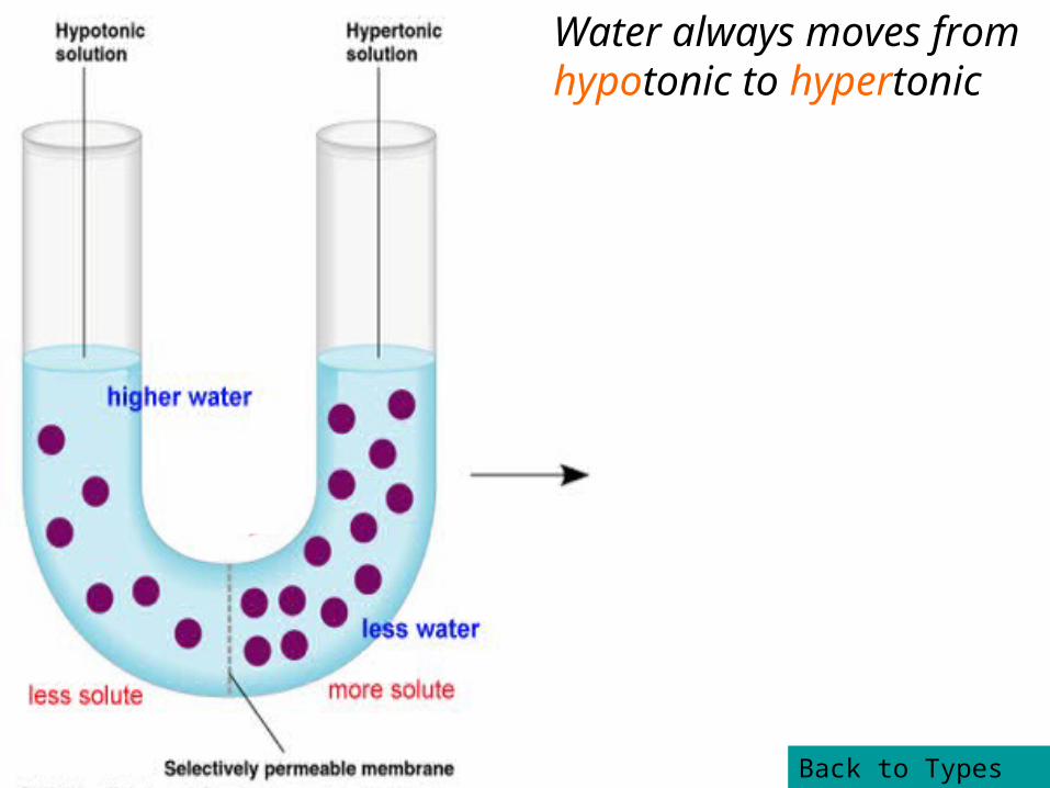
Osmosis
• Diffusion of WATER across the membrane
• Tonicity dependent– Isotonic solution: solution in equilibrium to another
solution across the membrane– Hypotonic: solution with less dissolved [solute], higher
[water] compared to another solution– Hypertonic: solution with more dissolved [solute],
lower [water] compared to another solution
Water always moves from hypotonic to hypertonic
Back to Types of PT
Filtration
• “Forcing” of water and solutes through membrane by hydrostatic pressure
• Selective only by SIZE
• Ex. only blood cells/proteins too large to pass are held back
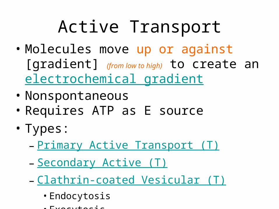
Active Transport• Molecules move up or against [gradient] (from
low to high) to create an electrochemical gradient
• Nonspontaneous• Requires ATP as E source
• Types:– Primary Active Transport (T)– Secondary Active (T)– Clathrin-coated Vesicular (T)
• Endocytosis• Exocytosis
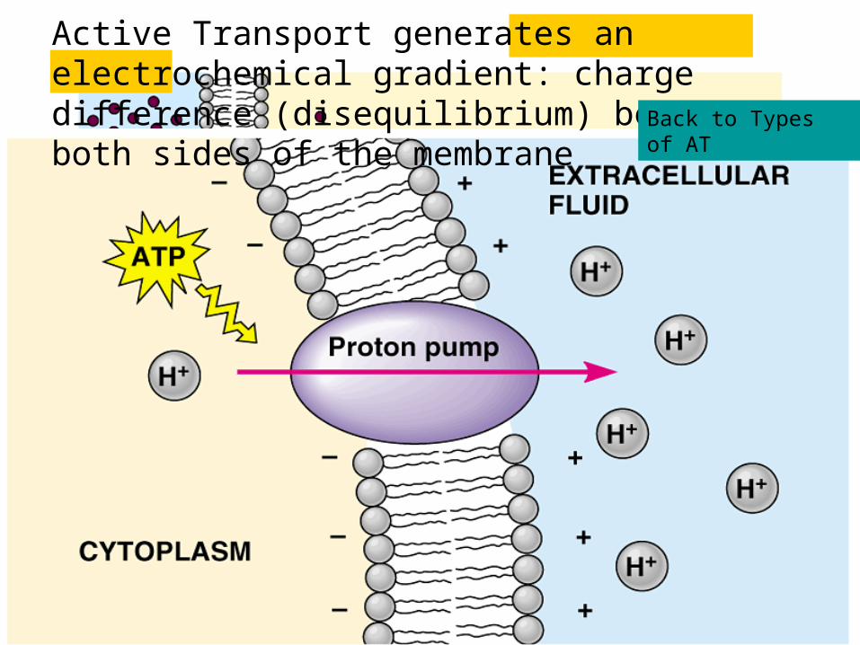
Active Transport generates an electrochemical gradient: charge difference (disequilibrium) between both sides of the membrane Back to Types of AT
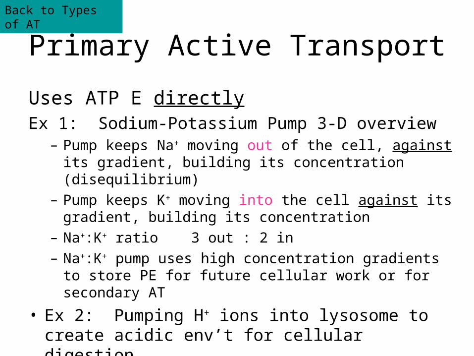
Primary Active Transport
Uses ATP E directlyEx 1: Sodium-Potassium Pump 3-D overview
– Pump keeps Na+ moving out of the cell, against its gradient, building its concentration (disequilibrium)
– Pump keeps K+ moving into the cell against its gradient, building its concentration
– Na+:K+ ratio 3 out : 2 in– Na+:K+ pump uses high concentration gradients to store
PE for future cellular work or for secondary AT
• Ex 2: Pumping H+ ions into lysosome to create acidic env’t for cellular digestion
Back to Types of AT
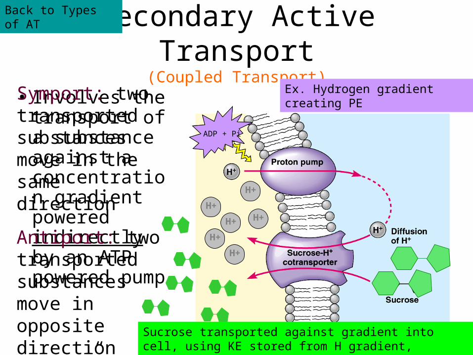
Secondary Active Transport(Coupled Transport)
• Involves the transport of a substance against a concentration gradient powered indirectly by an ATP powered pump
H+
ATP
H+
H+
H+
H+
H+
ADP + Pi
Ex. Hydrogen gradient creating PE
Sucrose transported against gradient into cell, using KE stored from H gradient, falling back down gradient
Symport: two transported substances move in the same direction
Antiport: two transported substances move in opposite direction (“wave” to each other)
Back to Types of AT
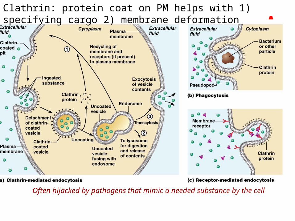
Endocytosis
• The engulfing of substances by pseudopods extensions of the plasma membrane
• Three types:– Phagocytosis (cell eating – lg. particles
engulfed)– Pinocytosis (cell drinking – sm. ions and
liquids engulfed)– Receptor Mediated Endocytosis (use of
surface proteins to engulf a specific substrate)
Clathrin: protein coat on PM helps with 1) specifying cargo 2) membrane deformation
Often hijacked by pathogens that mimic a needed substance by the cell
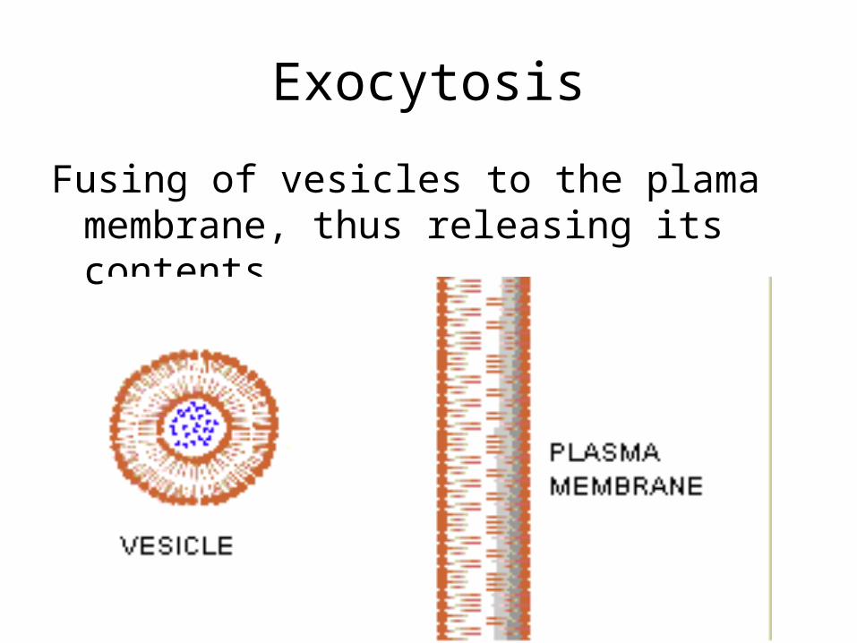
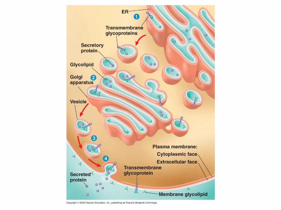
Exocytosis
Fusing of vesicles to the plama membrane, thus releasing its contents
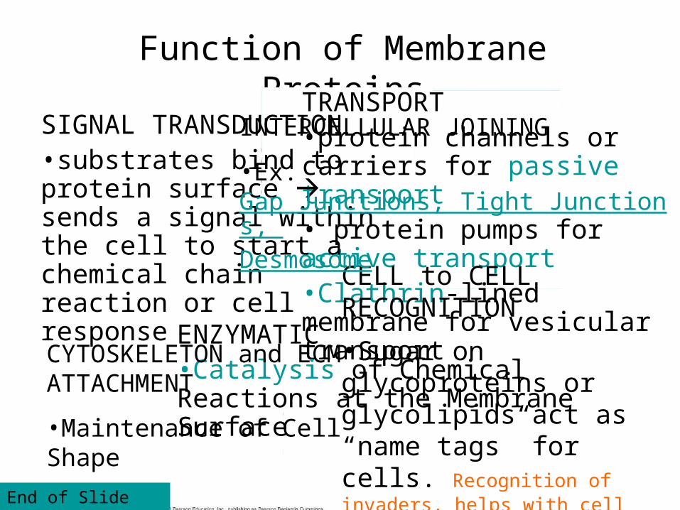
Function of Membrane ProteinsTRANSPORT
•protein channels or carriers for passive transport• protein pumps for active transport•Clathrin-lined membrane for vesicular transport
ENZYMATIC •Catalysis of Chemical Reactions at the Membrane Surface
SIGNAL TRANSDUCTION•substrates bind to protein surface sends a signal within the cell to start a chemical chain reaction or cell response CELL to CELL
RECOGNITION
•Sugar on glycoproteins or glycolipids act as “name tags” for cells. Recognition of invaders, helps with cell communication and coordination
INTERCELLULAR JOINING
•Ex. Gap Junctions, Tight Junctions, Desmosome
CYTOSKELETON and ECM ATTACHMENT
•Maintenance of Cell Shape
End of Slide Show
Signal Transduction
3 Stages of Signal Transduction
1) Reception: A ligand or substrate binds to receptor protein. Receptor proteins can be on the cell
surface, but not always. Receptor protein changes shape
2) Transduction: Amplifies and sends the signal through chemical relay
3) Cell Response: Specific response is triggered
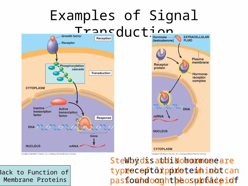
Examples of Signal Transduction
Why is this hormone-receptor protein not found on the surface of the plasma membrane?
Steroids and Hormones are types of lipids, which can pass through phospholipid membranes easily.
Back to Function of Membrane Proteins
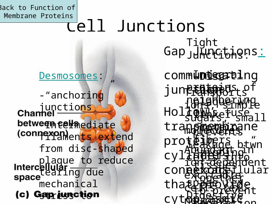
Cell JunctionsTight Junctions:
-Integral proteins of neighboring cells fuse
-prevents leakage btwn cells into extracellular space (ex. Digestive tract)
Back to Function of Membrane Proteins
-Disc-shaped plaque w/ linker protein fibers “zipping” tissues together to prevent separation
Gap Junctions:
communicating junction
Hollow, transmembrane protein cylinders, connexons, that provide cytoplasmic channels btwn cells
Transports ions, simple sugars, small molecules
Abundant in ion-dependent excitable cells (ex. neurons)
Desmosomes:
-“anchoring” junctions
-Intermediate filaments extend from disc-shaped plaque to reduce tearing due mechanical stress to prevent separation