Embed Size (px)
Citation preview
⏐13
Chapter 1 General introduction

General introduction⏐15
Introduction Interstitial lung diseases Interstitial lung diseases (ILD) are a rapidly growing and increasingly complex component of clinical practice. They represent a group of heterogeneous disorders that diffusely involve the lung parenchyma. The term ’interstitial‘ was originally applied to these disorders because they are associated with thickening of the alveolar septum. The ’interstitium‘ is that part of the alveolar structures bounded by the alveolar epithelial and endothelial basement membranes. The normal alveolar interstitium is composed of connective tissue components (e.g. collagen, elastic fibers, mesenchymal cells), and inflammatory and immune effector cells (monocytes/macrophages and lymphocytes). Generally, ILD involve alveolar epithelial and endothelial cells as well. In addition, although these diseases primarily attack the alveolar structures (inflammation and fibrosis), many also involve airways, arteries and veins.1,2 ILD can lead to diffuse remodelling and architectural damage to normal lung tissue and progressive loss of lung function. Although idiopathic pulmonary fibrosis (IPF) and sarcoidosis are the two most common forms of ILD encountered in clinical practice, well over 100 different types of ILD have been identified on the basis of clinical presentation, radiographic findings, and histopathologic examination.3-5 The patient’s age, cigarette-smoking status, and gender will provide useful clues. IPF for example is almost always an adult disorder, typically occurring in patients over 60 years of age. Patients with idiopathic non-specific interstitial pneumonia (NSIP) of the fibrotic variety are usually younger than 60 years of age. Although pulmonary sarcoidosis can manifest in the elderly patient, it is more common in young adults and middle-aged people. Respiratory bronchiolitis associated with ILD (RB-ILD) is seen almost exclusively in cigarette smokers, but it can occur in both men and women of all ages. In contrast, the very rare disorder Lymphangioleiomyomatosis (LAM) occurs exclusively in women of childbearing age. A detailed occupational history and changes in domestic environment are also essential, as it may lead to identification of a specific inhalation cause or trigger for ILD. At-risk occupations for ILD include miners (pneumoconiosis); sandblasters and granite workers (silicosis); dental workers (dental workers’ pneumoconiosis); welders, shipyard workers, pipe fitters, electricians, and mechanics (asbestosis); farm workers, poultry workers, bird fanciers, and bird breeders (hypersensitivity pneumonitis); and workers in aerospace, nuclear, computer, and electronics industries (berylliosis). History of existing, persistent and/or altered environmental ‘fibrogenic’ factors at home, the workplace, in automobiles or frequently visited facilities/homes or hobbies such as exposure
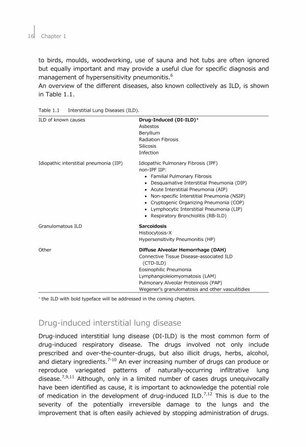
16⏐Chapter 1
to birds, moulds, woodworking, use of sauna and hot tubs are often ignored but equally important and may provide a useful clue for specific diagnosis and management of hypersensitivity pneumonitis.6 An overview of the different diseases, also known collectively as ILD, is shown in Table 1.1. Table 1.1 Interstitial Lung Diseases (ILD). ILD of known causes Drug-Induced (DI-ILD)* Asbestos Beryllium Radiation Fibrosis Silicosis Infection
Idiopathic interstitial pneumonia (IIP) Idiopathic Pulmonary Fibrosis (IPF) non-IPF IIP: • Familial Pulmonary Fibrosis • Desquamative Interstitial Pneumonia (DIP) • Acute Interstitial Pneumonia (AIP) • Non-specific Interstitial Pneumonia (NSIP) • Cryptogenic Organizing Pneumonia (COP) • Lymphocytic Interstitial Pneumonia (LIP) • Respiratory Bronchiolitis (RB-ILD)
Granulomatous ILD Sarcoidosis Histiocytosis-X Hypersensitivity Pneumonitis (HP)
Other Diffuse Alveolar Hemorrhage (DAH) Connective Tissue Disease-associated ILD
(CTD-ILD) Eosinophilic Pneumonia Lymphangioleiomyomatosis (LAM) Pulmonary Alveolar Proteinosis (PAP) Wegener’s granulomatosis and other vasculitidies * the ILD with bold typeface will be addressed in the coming chapters.
Drug-induced interstitial lung disease Drug-induced interstitial lung disease (DI-ILD) is the most common form of drug-induced respiratory disease. The drugs involved not only include prescribed and over-the-counter-drugs, but also illicit drugs, herbs, alcohol, and dietary ingredients.7-10 An ever increasing number of drugs can produce or reproduce variegated patterns of naturally-occurring infiltrative lung disease.7,9,11 Although, only in a limited number of cases drugs unequivocally have been identified as cause, it is important to acknowledge the potential role of medication in the development of drug-induced ILD.7,12 This is due to the severity of the potentially irreversible damage to the lungs and the improvement that is often easily achieved by stopping administration of drugs.
General introduction⏐17
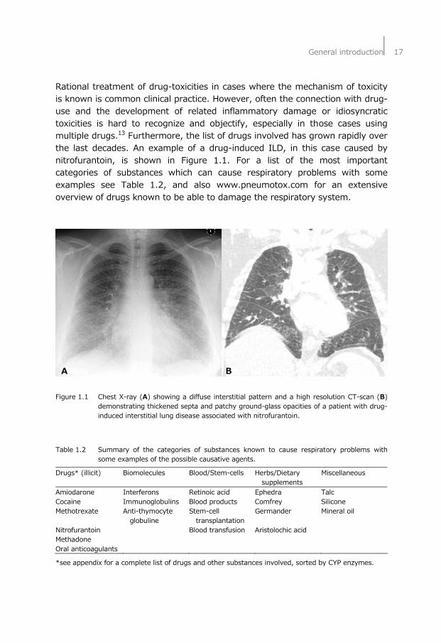
Rational treatment of drug-toxicities in cases where the mechanism of toxicity is known is common clinical practice. However, often the connection with drug-use and the development of related inflammatory damage or idiosyncratic toxicities is hard to recognize and objectify, especially in those cases using multiple drugs.13 Furthermore, the list of drugs involved has grown rapidly over the last decades. An example of a drug-induced ILD, in this case caused by nitrofurantoin, is shown in Figure 1.1. For a list of the most important categories of substances which can cause respiratory problems with some examples see Table 1.2, and also www.pneumotox.com for an extensive overview of drugs known to be able to damage the respiratory system. A B Figure 1.1 Chest X-ray (A) showing a diffuse interstitial pattern and a high resolution CT-scan (B)
demonstrating thickened septa and patchy ground-glass opacities of a patient with drug-induced interstitial lung disease associated with nitrofurantoin.
Table 1.2 Summary of the categories of substances known to cause respiratory problems with
some examples of the possible causative agents. Drugs* (illicit) Biomolecules Blood/Stem-cells Herbs/Dietary
supplements Miscellaneous
Amiodarone Interferons Retinoic acid Ephedra Talc Cocaine Immunoglobulins Blood products Comfrey Silicone Methotrexate Anti-thymocyte
globuline Stem-cell transplantation
Germander Mineral oil
Nitrofurantoin Blood transfusion Aristolochic acid Methadone Oral anticoagulants *see appendix for a complete list of drugs and other substances involved, sorted by CYP enzymes.
18⏐Chapter 1
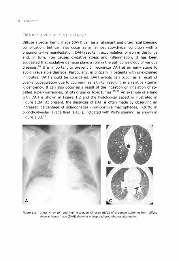
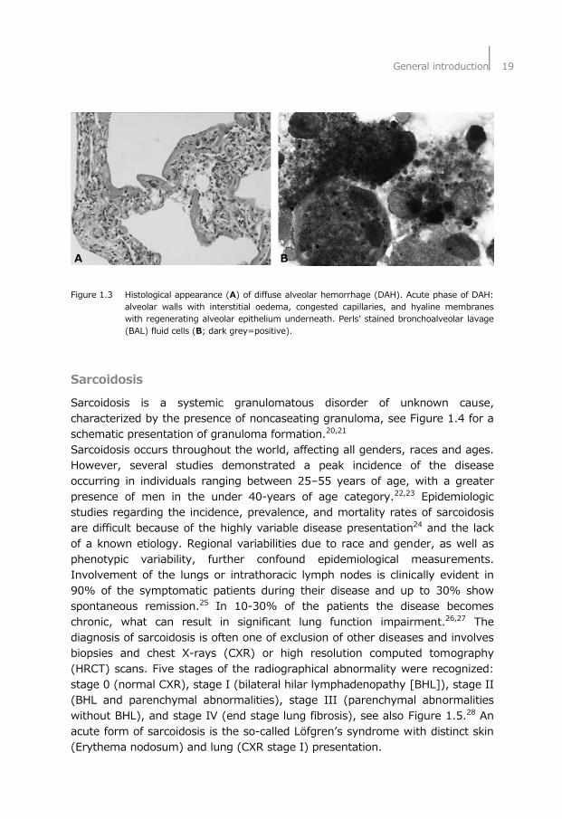
Diffuse alveolar hemorrhage Diffuse alveolar hemorrhage (DAH) can be a fulminant and often fatal bleeding complication, but can also occur as an almost sub-clinical condition with a pneumonia-like manifestation. DAH results in accumulation of iron in the lungs and, in turn, iron causes oxidative stress and inflammation. It has been suggested that oxidative damage plays a role in the pathophysiology of various diseases.14 It is important to prevent or recognize DAH at an early stage to avoid irreversible damage. Particularly, in critically ill patients with unexplained infiltrates, DAH should be considered. DAH events can occur as a result of over-anticoagulation due to coumarin sensitivity, resulting in a relative vitamin K deficiency. It can also occur as a result of the ingestion or inhalation of so-called super-warfarines, (illicit) drugs or toxic fumes.15-18 An example of a lung with DAH is shown in Figure 1.2 and the histological aspect is illustrated in Figure 1.3A. At present, the diagnosis of DAH is often made by observing an increased percentage of siderophages (iron-positive macrophages, >20%) in bronchoalveolar lavage fluid (BALF), indicated with Perl’s staining, as shown in Figure 1.3B.19
B A C Figure 1.2 Chest X-ray (A) and high resolution CT-scan (B/C) of a patient suffering from diffuse
alveolar hemorrhage (DAH) showing widespread ground-glass attenuation.
General introduction⏐19
A B Figure 1.3 Histological appearance (A) of diffuse alveolar hemorrhage (DAH). Acute phase of DAH:
alveolar walls with interstitial oedema, congested capillaries, and hyaline membranes with regenerating alveolar epithelium underneath. Perls’ stained bronchoalveolar lavage (BAL) fluid cells (B; dark grey=positive).
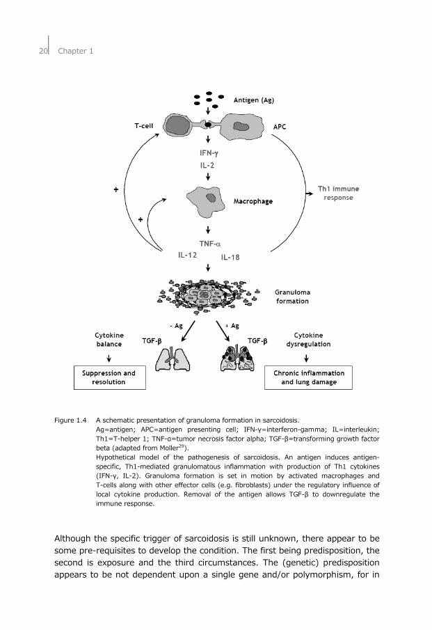
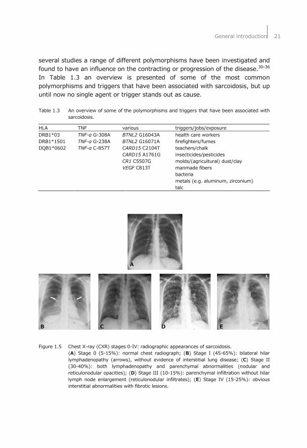
Sarcoidosis Sarcoidosis is a systemic granulomatous disorder of unknown cause, characterized by the presence of noncaseating granuloma, see Figure 1.4 for a schematic presentation of granuloma formation.20,21 Sarcoidosis occurs throughout the world, affecting all genders, races and ages. However, several studies demonstrated a peak incidence of the disease occurring in individuals ranging between 25–55 years of age, with a greater presence of men in the under 40-years of age category.22,23 Epidemiologic studies regarding the incidence, prevalence, and mortality rates of sarcoidosis are difficult because of the highly variable disease presentation24 and the lack of a known etiology. Regional variabilities due to race and gender, as well as phenotypic variability, further confound epidemiological measurements. Involvement of the lungs or intrathoracic lymph nodes is clinically evident in 90% of the symptomatic patients during their disease and up to 30% show spontaneous remission.25 In 10-30% of the patients the disease becomes chronic, what can result in significant lung function impairment.26,27 The diagnosis of sarcoidosis is often one of exclusion of other diseases and involves biopsies and chest X-rays (CXR) or high resolution computed tomography (HRCT) scans. Five stages of the radiographical abnormality were recognized: stage 0 (normal CXR), stage I (bilateral hilar lymphadenopathy [BHL]), stage II (BHL and parenchymal abnormalities), stage III (parenchymal abnormalities without BHL), and stage IV (end stage lung fibrosis), see also Figure 1.5.28 An acute form of sarcoidosis is the so-called Löfgren’s syndrome with distinct skin (Erythema nodosum) and lung (CXR stage I) presentation.
20⏐Chapter 1
Figure 1.4 A schematic presentation of granuloma formation in sarcoidosis. Ag=antigen; APC=antigen presenting cell; IFN-γ=interferon-gamma; IL=interleukin;
Th1=T-helper 1; TNF-α=tumor necrosis factor alpha; TGF-β=transforming growth factor beta (adapted from Moller29).
Hypothetical model of the pathogenesis of sarcoidosis. An antigen induces antigen-specific, Th1-mediated granulomatous inflammation with production of Th1 cytokines (IFN-γ, IL-2). Granuloma formation is set in motion by activated macrophages and T-cells along with other effector cells (e.g. fibroblasts) under the regulatory influence of local cytokine production. Removal of the antigen allows TGF-β to downregulate the immune response.
Although the specific trigger of sarcoidosis is still unknown, there appear to be some pre-requisites to develop the condition. The first being predisposition, the second is exposure and the third circumstances. The (genetic) predisposition appears to be not dependent upon a single gene and/or polymorphism, for in
TNF-α
General introduction⏐21
several studies a range of different polymorphisms have been investigated and found to have an influence on the contracting or progression of the disease.30-36 In Table 1.3 an overview is presented of some of the most common polymorphisms and triggers that have been associated with sarcoidosis, but up until now no single agent or trigger stands out as cause. Table 1.3 An overview of some of the polymorphisms and triggers that have been associated with
sarcoidosis.
HLA TNF various triggers/jobs/exposure DRB1*03 TNF-α G-308A BTNL2 G16043A health care workers DRB1*1501 TNF-α G-238A BTNL2 G16071A firefighters/fumes DQB1*0602 TNF-α C-857T CARD15 C2104T teachers/chalk CARD15 A1761G insecticides/pesticides CR1 C5507G molds/(agricultural) dust/clay VEGF C813T manmade fibers bacteria metals (e.g. aluminum, zirconium) talc A B C D E Figure 1.5 Chest X-ray (CXR) stages 0-IV: radiographic appearances of sarcoidosis. (A) Stage 0 (5-15%): normal chest radiograph; (B) Stage I (45-65%): bilateral hilar
lymphadenopathy (arrows), without evidence of interstitial lung disease; (C) Stage II (30-40%): both lymphadenopathy and parenchymal abnormalities (nodular and reticulonodular opacities); (D) Stage III (10-15%): parenchymal infiltration without hilar lymph node enlargement (reticulonodular infiltrates); (E) Stage IV (15-25%): obvious interstitial abnormalities with fibrotic lesions.
22⏐Chapter 1
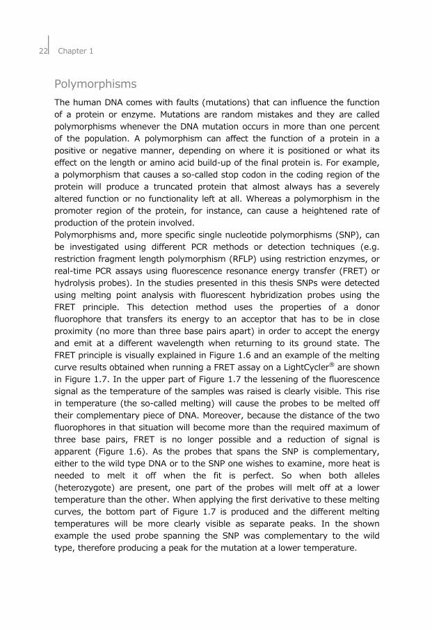
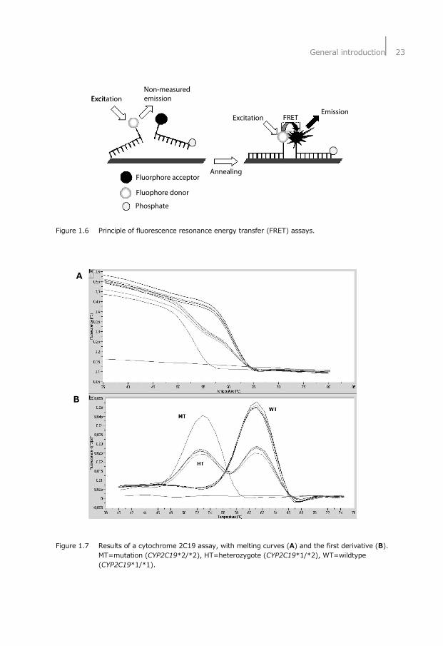
Polymorphisms The human DNA comes with faults (mutations) that can influence the function of a protein or enzyme. Mutations are random mistakes and they are called polymorphisms whenever the DNA mutation occurs in more than one percent of the population. A polymorphism can affect the function of a protein in a positive or negative manner, depending on where it is positioned or what its effect on the length or amino acid build-up of the final protein is. For example, a polymorphism that causes a so-called stop codon in the coding region of the protein will produce a truncated protein that almost always has a severely altered function or no functionality left at all. Whereas a polymorphism in the promoter region of the protein, for instance, can cause a heightened rate of production of the protein involved. Polymorphisms and, more specific single nucleotide polymorphisms (SNP), can be investigated using different PCR methods or detection techniques (e.g. restriction fragment length polymorphism (RFLP) using restriction enzymes, or real-time PCR assays using fluorescence resonance energy transfer (FRET) or hydrolysis probes). In the studies presented in this thesis SNPs were detected using melting point analysis with fluorescent hybridization probes using the FRET principle. This detection method uses the properties of a donor fluorophore that transfers its energy to an acceptor that has to be in close proximity (no more than three base pairs apart) in order to accept the energy and emit at a different wavelength when returning to its ground state. The FRET principle is visually explained in Figure 1.6 and an example of the melting curve results obtained when running a FRET assay on a LightCycler® are shown in Figure 1.7. In the upper part of Figure 1.7 the lessening of the fluorescence signal as the temperature of the samples was raised is clearly visible. This rise in temperature (the so-called melting) will cause the probes to be melted off their complementary piece of DNA. Moreover, because the distance of the two fluorophores in that situation will become more than the required maximum of three base pairs, FRET is no longer possible and a reduction of signal is apparent (Figure 1.6). As the probes that spans the SNP is complementary, either to the wild type DNA or to the SNP one wishes to examine, more heat is needed to melt it off when the fit is perfect. So when both alleles (heterozygote) are present, one part of the probes will melt off at a lower temperature than the other. When applying the first derivative to these melting curves, the bottom part of Figure 1.7 is produced and the different melting temperatures will be more clearly visible as separate peaks. In the shown example the used probe spanning the SNP was complementary to the wild type, therefore producing a peak for the mutation at a lower temperature.
General introduction⏐23
Figure 1.6 Principle of fluorescence resonance energy transfer (FRET) assays. A B Figure 1.7 Results of a cytochrome 2C19 assay, with melting curves (A) and the first derivative (B).
MT=mutation (CYP2C19*2/*2), HT=heterozygote (CYP2C19*1/*2), WT=wildtype (CYP2C19*1/*1).
Excitation
ExcitationEmission
Non-measuredemission
FRET
AnnealingFluorphore acceptor
Fluophore donor
Phosphate
Excitation
ExcitationEmission
Non-measuredemission
FRET
AnnealingFluorphore acceptor
Fluophore donor
Phosphate
24⏐Chapter 1
Scope and aims of the study The aims of the studies presented in this thesis were to investigate several polymorphisms and assess their possible role in the cause and course of interstitial lung disease (ILD). ILD, especially drug-induced ILD (DI-ILD), can occur as a cause of drug(s) or drug-drug interactions. The CYP enzyme family plays an important role in the metabolization of all sorts of ingested or inhaled xenobiotic substances. In Chapter 2 the possible role of cytochrome P450 (CYP) enzymes in DI-ILD is reviewed. Chapter 3 describes a simple and uncomplicated method to isolate DNA from easy obtainable and patient friendly dried blood spot and/or buccal swab samples prior to real-time polymerase chain reactions (PCR). The aim of Chapter 4 was to establish whether allelic variation in specific CYP polymorphic genes, namely CYP2D6, CYP2C9, and CYP2C19, contributes to variability in drug response and unexpected toxicity. Therefore, a case-control study was conducted. The cases consisted of patients with DI-ILD. Two control groups were used: one group of healthy volunteers and one group of patients with idiopathic pulmonary fibrosis (IPF). In Chapter 5 the role of CYP polymorphisms is further illustrated in the case-report describing the therapeutic failure of venlafaxine in a case lacking CYP2D6 activity. In Chapter 6 it was hypothesized that in patients treated with coumarins a serious complication i.e. diffuse alveolar hemorrhage (DAH) may be associated with vitamin K epoxide reductase complex 1 (VKORC1) and CYP (CYP2C9 and CYP2C19) variant alleles. Clinical information of patients using coumarins with at least one episode of DAH was gathered retrospectively during a seven year period. The aim of Chapter 7 was to evaluate the relationship between the presence of tumor necrosis factor (TNF) polymorphisms, human leukocyte antigen (HLA)-DRB1*03 linkage and the prognosis of sarcoidosis. In a retrospective case-control study TNF-α G-308A, TNF-α G-238A and LTA were genotyped in 625 sarcoidosis patients. These patients were classified into patients with persistent disease and patients with non-persistent disease using chest X-ray appearances and lung function parameters after at least two years of follow-up. The aim of Chapter 8 was to assess the association of butyrophilin-like 2 (BTNL2) G16071A with the course of pulmonary sarcoidosis and verify association with disease predisposition. In addition, the linkage between BTNL2 G16071A and certain HLA-DRB1/DQB1 types was investigated. In a retrospective case-control study BTNL2 G16071A, HLA-DQB1 and DRB1 were typed in 632 sarcoidosis patients. These patients were classified into 304 patients with persistent and 328 patients with non-persistent sarcoidosis using chest X-ray stages after at least two years follow-up. Finally, in Chapter 9, a summary and the implications of the findings presented in this thesis are argued and directions for future research are briefly discussed.
General introduction⏐25
References 1. Crystal RG, Gadek JE, Ferrans VJ, Fulmer JD, Line BR, Hunninghake GW. Interstitial
lung disease: current concepts of pathogenesis, staging and therapy. Am J Med. 1981;70:542-68.
2. Travis WD, Hunninghake G, King TE, Jr., Lynch DA, Colby TV, Galvin JR, Brown KK, Chung MP, Cordier JF, du Bois RM, Flaherty KR, Franks TJ, Hansell DM, Hartman TE, Kazerooni EA, Kim DS, Kitaichi M, Koyama T, Martinez FJ, Nagai S, Midthun DE, Muller NL, Nicholson AG, Raghu G, Selman M, Wells A. Idiopathic nonspecific interstitial pneumonia: report of an American Thoracic Society project. Am J Respir Crit Care Med. 2008;177:1338-47.
3. Meyer KC. Bronchoalveolar lavage as a diagnostic tool. Semin Respir Crit Care Med. 2007;28:546-60.
4. Drent M, Mansour K, Linssen C. Bronchoalveolar lavage in sarcoidosis. Semin Respir Crit Care Med. 2007;28:486-95.
5. Demedts M, Behr J, Buhl R, Costabel U, Dekhuijzen R, Jansen HM, MacNee W, Thomeer M, Wallaert B, Laurent F, Nicholson AG, Verbeken EK, Verschakelen J, Flower CD, Capron F, Petruzzelli S, De Vuyst P, van den Bosch JM, Rodriguez-Becerra E, Corvasce G, Lankhorst I, Sardina M, Montanari M. High-dose acetylcysteine in idiopathic pulmonary fibrosis. N Engl J Med. 2005;353:2229-42.
6. Baughman RP, du Bois RM, Lynch JP, Wells AU. Diffuse Lung Disease A Practical Approach: Hodder Arnold London;2004.
7. Camus P, Fanton A, Bonniaud P, Camus C, Foucher P. Interstitial lung disease induced by drugs and radiation. Respiration. 2004;71:301-26.
8. Wijnen PA, Drent M, Nelemans PJ, Kuijpers PM, Koek GH, Neef C, Haenen GR, Bekers O. Role of cytochrome P450 polymorphisms in the development of pulmonary drug toxicity: a case-control study in the Netherlands. Drug Saf. 2008;31:1125-34.
9. Foucher P, Biour M, Blayac JP, Godard P, Sgro C, Kuhn M, Vergnon JM, Vervloet D, Pfitzenmeyer P, Ollagnier M, Mayaud C, Camus P. Drugs that may injure the respiratory system. Eur Respir J. 1997;10:265-79.
10. Bressler R. Grapefruit juice and drug interactions. Exploring mechanisms of this interaction and potential toxicity for certain drugs. Geriatrics. 2006;61:12-8.
11. Camus P, Kudoh S, Ebina M. Interstitial lung disease associated with drug therapy. Br J Cancer. 2004;91 Suppl 2:S18-23.
12. Drent M, Singh S, Gorgels AP, Hansell DM, Bekers O, Nicholson AG, van Suylen RJ, du Bois RM. Drug-induced pneumonitis and heart failure simultaneously associated with venlafaxine. Am J Respir Crit Care Med. 2003;167:958-61.
13. Nemery B, Bast A, Behr J, Borm PJ, Bourke SJ, Camus PH, De Vuyst P, Jansen HM, Kinnula VL, Lison D, Pelkonen O, Saltini C. Interstitial lung disease induced by exogenous agents: factors governing susceptibility. Eur Respir J Suppl. 2001;32: 30s-42s.
14. Rahman I, Skwarska E, Henry M, Davis M, O'Connor CM, FitzGerald MX, Greening A, MacNee W. Systemic and pulmonary oxidative stress in idiopathic pulmonary fibrosis. Free Radic Biol Med. 1999;27:60-8.
15. Balkisson R, Murray D, Hoffstein V. Alveolar damage due to inhalation of amitrole-containing herbicide. Chest. 1992;101:1174-6.
16. Jinn Y, Akizuki N, Ohkouchi M, Inase N, Ichioka M, Marumo F. Acute lung injury after inhalation of water-proofing spray while smoking a cigarette. Respiration. 1998;65:486-8.
17. Kayser K, Plodziszewska M, Waitr E, Slodkowska J, Altiner M, Gabius HJ. Diffuse pulmonary hemosiderosis after exposure to pesticides. A case report. Respiration. 1998;65:214-8.
26⏐Chapter 1
18. Spahr JE, Maul JS, Rodgers GM. Superwarfarin poisoning: a report of two cases and review of the literature. Am J Hematol. 2007;82:656-60.
19. De Lassence A, Fleury-Feith J, Escudier E, Beaune J, Bernaudin JF, Cordonnier C. Alveolar hemorrhage. Diagnostic criteria and results in 194 immunocompromised hosts. Am J Respir Crit Care Med. 1995;151:157-63.
20. Baughman RP, Lower EE, du Bois RM. Sarcoidosis. Lancet. 2003;361:1111-8. 21. Kataria YP, Holter JF. Sarcoidosis: A Model of Granulomatous Inflammation of
Unknown Etiology Associated with a Hyperactive Immune System. Methods. 1996; 9:268-94.
22. Design of a case control etiologic study of sarcoidosis (ACCESS). ACCESS Research Group. J Clin Epidemiol. 1999;52:1173-86.
23. Rossman MD, Kreider ME. Lesson learned from ACCESS (A Case Controlled Etiologic Study of Sarcoidosis). Proc Am Thorac Soc. 2007;4:453-6.
24. Judson MA, Baughman RP, Thompson BW, Teirstein AS, Terrin ML, Rossman MD, Yeager H, Jr., McLennan G, Bresnitz EA, DePalo L, Hunninghake G, Iannuzzi MC, Johns CJ, Moller DR, Newman LS, Rabin DL, Rose C, Rybicki BA, Weinberger SE, Knatterud GL, Cherniak R. Two year prognosis of sarcoidosis: the ACCESS experience. Sarcoidosis Vasc Diffuse Lung Dis. 2003;20:204-11.
25. Rybicki BA, Maliarik MJ, Major M, Popovich J, Jr., Iannuzzi MC. Epidemiology, demographics, and genetics of sarcoidosis. Semin Respir Infect. 1998;13:166-73.
26. Baughman RP, Winget DB, Bowen EH, Lower EE. Predicting respiratory failure in sarcoidosis patients. Sarcoidosis Vasc Diffuse Lung Dis. 1997;14:154-8.
27. Arcasoy SM, Christie JD, Pochettino A, Rosengard BR, Blumenthal NP, Bavaria JE, Kotloff RM. Characteristics and outcomes of patients with sarcoidosis listed for lung transplantation. Chest. 2001;120:873-80.
28. DeRemee RA. The roentgenographic staging of sarcoidosis. Historic and contemporary perspectives. Chest. 1983;83:128-33.
29. Moller DR. Cells and cytokines involved in the pathogenesis of sarcoidosis. Sarcoidosis Vasc Diffuse Lung Dis. 1999;16:24-31.
30. Castro-Giner F, Kogevinas M, Machler M, de Cid R, Van Steen K, Imboden M, Schindler C, Berger W, Gonzalez JR, Franklin KA, Janson C, Jarvis D, Omenaas E, Burney P, Rochat T, Estivill X, Anto JM, Wjst M, Probst-Hensch NM. TNFA -308G>A in two international population-based cohorts and risk of asthma. Eur Respir J. 2008;32:350-61.
31. Grunewald J, Eklund A, Olerup O. Human leukocyte antigen class I alleles and the disease course in sarcoidosis patients. Am J Respir Crit Care Med. 2004;169: 696-702.
32. Grutters JC, Sato H, Pantelidis P, Lagan AL, McGrath DS, Lammers JW, van den Bosch JM, Wells AU, du Bois RM, Welsh KI. Increased frequency of the uncommon tumor necrosis factor -857T allele in British and Dutch patients with sarcoidosis. Am J Respir Crit Care Med. 2002;165:1119-24.
33. Hajeer AH, Hutchinson IV. Influence of TNFalpha gene polymorphisms on TNFalpha production and disease. Hum Immunol. 2001;62:1191-9.
34. Morohashi K, Takada T, Omori K, Suzuki E, Gejyo F. Vascular endothelial growth factor gene polymorphisms in Japanese patients with sarcoidosis. Chest. 2003;123: 1520-6.
35. Sato H, Williams HR, Spagnolo P, Abdallah A, Ahmad T, Orchard TR, Copley SJ, Desai SR, Wells AU, du Bois RM, Welsh KI. CARD15/NOD2 polymorphisms are associated with severe pulmonary sarcoidosis. Eur Respir J. 2010;35:324-30.
36. Zorzetto M, Bombieri C, Ferrarotti I, Medaglia S, Agostini C, Tinelli C, Malerba G, Carrabino N, Beretta A, Casali L, Pozzi E, Pignatti PF, Semenzato G, Cuccia MC, Luisetti M. Complement receptor 1 gene polymorphisms in sarcoidosis. Am J Respir Cell Mol Biol. 2002;27:17-23.


![[PPT]Chapter 18 Oxidative phosphorylation and … · Web viewTitle Chapter 18 Oxidative phosphorylation and photophosphorylation Last modified by h Document presentation format 全屏显示(4:3)](https://img.pdfslide.tips/doc/110x75/5aee63477f8b9ac62b8c00c8/pptchapter-18-oxidative-phosphorylation-and-viewtitle-chapter-18-oxidative.jpg)

![Antidepressant and Antiaging Effects of Açaí (Euterpe ...€¦ · pathophysiology of unipolar and bipolar depression [16]. In parallel to oxidative stress, immunoinflammatory mecha-nisms](https://img.pdfslide.tips/doc/110x75/5f35ec8467a8e700807ec0ea/antidepressant-and-antiaging-effects-of-aa-euterpe-pathophysiology-of-unipolar.jpg)