Embed Size (px)
DESCRIPTION
hg
Citation preview
Original Article
Effect of deacetylation conditions on antimicrobial activity of
chitosans prepared from carapace of black tiger shrimp
(Penaeus monodon)
Tipparat Hongpattarakere* and Oraphan Riyaphan
Department of Industrial Biotechnology, Faculty of Agro-Industry,
Prince of Songkla University, Hat Yai, Songkhla 90112 Thailand.
Received 29 December 2006; Accepted 2 April 2008
Abstract
Chitosan was prepared from black tiger shrimp carapace by deacetylation process performed in 50% NaOH at 100oC
under vacuum, nitrogen and regular atmospheres. Each condition was maintained for 0.5, 1.0 and 2.0 h. Chitosan obtained
from 1.0 h of deacetylation under regular atmosphere showed the lowest minimum inhibitory concentration (MIC) value of
625 ppm against Escherichia coli and Staphylococcus aureus, while Candida albicans was inhibited at MIC value of 312.5
ppm due to its higher degree of deacetylation (% DD) and lower molecular weight (MW) compared to chitosan deacetylated
under vacuum and nitrogen atmospheres. Chitosan hydrolysates obtained from both chemical (H2O
2 in the presence of ferric
ions) and enzymatic (lysozyme) hydrolysis were not as inhibitory as the native one, except that from chemical hydrolysis,
which showed the elevation of the antifungal activity against C. albicans when longer hydrolysis was performed. However,
antibacterial activity against E. coli decreased when the MW of hydrolyzed chitosan decreased.
Keywords: chitosan, antimicrobials, deacetylation, black tiger shrimp, Penaeus monodon
Songklanakarin J. Sci. Technol.
30 (Suppl.1), 1-9, April 2008
1. Introduction
Chitosan, the deacetylated derivative of chitin, is a
copolymer made up of 2-amino-2-deoxy-D-glucose (GlcN)
and 2-acetamido-2-deoxy-D-glucose units. It is a versatile
biopolymer exhibiting various unique biological properties
hence its wide application in food, biomedical and chemical
industries (Shahidi et al., 1999). Suzuki et al. (1986 cited by
No et al., 2002) and Tokoro et al. (1988 cited by No et al.,
2002) reported antitumor activity of hexa-N-acetylchito-
hexaose and chitohexaose. Hypocholesterolemic activity in
rat of chitosan hydrolysates has also been studied (Sugano et
al., 1992 cited by No et al., 2002). Recently most studies on
chitosan have focused on its strong antimicrobial activity and
use of chitosan as a food preservative. Antifungal activity of
chitosan was reported against growth of Aspergillus niger and
aflatoxin production of Aspergillus parasiticus (Fang et al.,
1994). Spoilage yeast such as Candida sp., Saccharomyces
cerevisiae, Zygosaccharomyces bailii, Saccharomycodes
ludwigii and Rhodococcus sp. were inactivated by chitosan
hydrochloride and its hydrolysates (Rhoades and Roller,
2000). Additionally, chitosan has shown antibacterial activity
against many Gram-positive and Gram-negative bacteria
including food-borne pathogens (Helander et al., 2001;
Knowles and Roller, 2001; No et al., 2002; Tsai and Hwang,
2004; Vishu Kumar et al., 2004). Antibacterial activity of
chitosan was shown against Streptococcus mutans and
Micrococcus luteus at minimum inhibitory concentrations
(MIC) of 0.8 % v/v, as well as Staphylococcus aureus,
Staphylococcus epidermis and Bacillus subtilis at MIC of
0.6% v/v (Jeon et al., 2001). Lactic acid bacteria, including
Lactobacillus bulgaricus, Lactobacillus casei, Lactobacillus
fermentum and Streptococcus faecalis were completely*Corresponding author.
Email address: [email protected]
http://www.sjst.psu.ac.th
Hongpattarakere & Riyaphan / Songklanakarin J. Sci. Technol. 30 (Suppl.1), 1-9, 20082
inhibited by chitosan at MIC of 0.03 % v/v. Chitosan exhib-
ited stronger inhibition against Gram-positive bacteria than
Gram-negative bacteria such as Escherichia coli, Escherichia
coli O157:H7, Salmonella typhi, Pseudomonas aeruginosa,
Vibrio sp., Shigella dysenteriae and Salmonella typhimu-
rium, which were inhibited at MICs 100-10,000 ppm (Chen
et al., 1998; Tsai and Su, 1999; Rhoades and Roller, 2000;
Tsai et al., 2000; Jeon et al., 2001). However, growth of
certain Gram-negative bacteria such as Erwinia sp.,
Klebsiella pneumoniae and S. enteritidis PT4 could not be
inhibited by chitosan at concentration as high as 5000 ppm
(Chen et al., 1998; Roller and Covill, 2000).
Apart from microbial strains, antimicrobial activity of
chitosan varies widely, depending on % DD, molecular
weight (MW), pH, temperature and the presence of interfer-
ing substances such as proteins, fats and other antimicrobials
(Rhoades and Roller, 2000; Tsai et al., 2000; Jeon et al.,
2001; Knowles and Roller, 2001; No et al., 2002; Zheng and
Zhu, 2003). This study has investigated antimicrobial activity
of native chitosan prepared from black tiger shrimp carapace
and chemically, and enzymatically-hydrolyzed chitosan.
2. Materials and Methods
2.1 Materials
Frozen shrimp heads were supplied by Piti Seafood
Co., Ltd., Songkhla (Thailand). Muller Hinton Broth (for
bacteria) and Potato Dextrose Broth (for yeast) were obtained
from Difco (Detroit, MI, USA). A sterile 96 well microplate
was purchased from Nunc Brand Products (Denmark). An
Ubbelohde capillary viscometer was purchased from
Canon, No.200 M548 (USA). Commercial grade 70%, 80%
and 90% DD chitosan were obtained from Katokichi (Japan).
Lysozyme (EC3.2.1.17) from chicken egg white (catalog no.
6876) was supplied by Sigma (St. Louis, MO, USA). Stan-
dard solution of n/400 potassium polyvinylsulfate (PVSK)
was obtained from Wako Pure Chemical Industries, Ltd.
(Tokyo, Japan). All chemicals (reagent grade) were supplied
by Lab-Scan Ltd. (Ireland).
A microplate reader was obtained from ELx808 Bio-
TEX instrument INC. (Belgium). High performance liquid
chromatography (HPLC) system including column, detector,
recorder and other accessories were manufactured by Waters
Co. (Massachusetts, USA). Pullulans standard was supplied
by Polymer Laboratories Ltd. (Shropshire, UK).
2.2 Microorganisms
Pathogenic Escherichia coli isolated from a patient
was obtained from culture collection of Prince of Songkla
Hospital, Prince of Songkla University, Hat Yai campus.
Staphylococcus aureus, and Candida albicans were from the
culture collection of the Microbiology Laboratory, Faculty
of Agro-Industry, Prince of Songkla University, Hat Yai
campus.
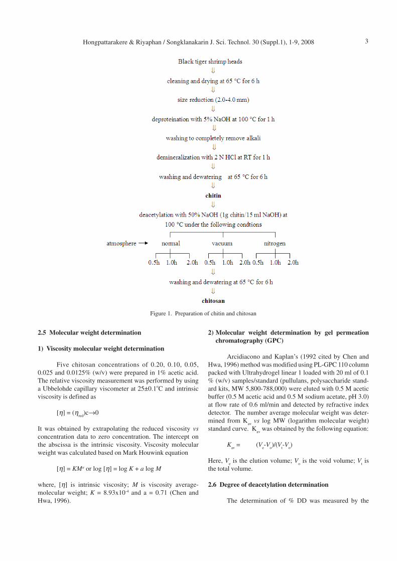
2.3 Preparation of chitin and chitosan
As shown in Figure 1, black tiger shrimp heads were
cleaned and washed with water several times. They were
then dried at temperature of 65oC for 6 hours, and ground
into 2-4 mm in size. The dried shrimp head powder was kept
in brown bottles and stored at room temperature.
Preparation of chitin began with deproteination
process performed by alkaline treatments with 5 % (w/v)
sodium hydroxide solution and heating at 100oC for 1 hour.
The alkaline-treated materials were then washed with water
to completely remove the alkali. Demineralization process
was carried out thereafter by soaking the alkaline- treated
materials in 2.0 N of hydrochloric acid for 1 hour at room
temperature, and then washed with water to reach neutral pH.
The final product called “chitin” was obtained and was kept
in a brown bottle, which was stored at room temperature
(Benjakul and Sophanodora, 1990).
Chitosan was prepared under various conditions of
deacetylation processes, which were performed in 50%
(w/v) NaOH (1 g chitin/15 ml NaOH) at 100oC under
normal, vacuum and nitrogen atmospheres, each of which
was conducted for 0.5, 1.0 and 2.0 hours. Chitosan obtained
from each condition was further investigated for its anti-
microbial activity, molecular weight and degree of deacetyl-
ation.
2.4 Antimicrobial Activity assay
Pathogenic Escherichia coli, Staphylococcus aureus
and Candida albicans were used as the test microorganisms.
The bacteria were cultured in Muller Hinton Broth (MHB),
while yeast was cultured in Potato Dextrose Broth (PDB)
and incubated at 37oC for 24 hours. Then, the active cultures
were inoculated into 10 ml of MHB for bacteria and PDB for
yeast and incubated at 37oC for 15 hours. Microorganisms
were diluted with MHB/PDB to obtain bacterial/yeast count
of 5-10x105 CFU/ml.
Antimicrobial activity of prepared chitosan was
assayed by microdilution method, performed using a sterile
96 well-microplate (Rhoades and Roller, 2000). Chitosan
solutions was prepared in 1% acetic acid at a concentration
of 20 mg/ml (20,000 ppm) before being applied to broth and
each solution was diluted (serial two-fold dilutions) with
MHB or PDB to give final chitosan concentrations of 2500,
1250, 625, 312.5, 156.25 and 78.125 ppm (2-fold dilutions).
One hundred microliters of each chitosan concentration and
100 ml of microbial suspension (prepared above) were added
to obtain final chitosan concentrations of 1250, 625, 312.5,
156.25, 78.125 and 39.06 ppm. The plate was then incubated
at 37oC for 48 h. Growth of microorganisms was determined
by measuring the absorption at 595 nm at 0, 24 and 48 h
using the microplate reader. Antimicrobial activity of chitosan
was recorded in terms of MIC, which was defined as the
lowest concentration of chitosan required to completely
inhibit microbial growth after incubation at 37oC for 24 hours.
Hongpattarakere & Riyaphan / Songklanakarin J. Sci. Technol. 30 (Suppl.1), 1-9, 2008 3
2.5 Molecular weight determination
1) Viscosity molecular weight determination
Five chitosan concentrations of 0.20, 0.10, 0.05,
0.025 and 0.0125% (w/v) were prepared in 1% acetic acid.
The relative viscosity measurement was performed by using
a Ubbelohde capillary viscometer at 25±0.1oC and intrinsic
viscosity is defined as
[η] = (ηred
)c→0
It was obtained by extrapolating the reduced viscosity vs
concentration data to zero concentration. The intercept on
the abscissa is the intrinsic viscosity. Viscosity molecular
weight was calculated based on Mark Houwink equation
[η] = KMa or log [η] = log K + a log M
where, [η] is intrinsic viscosity; M is viscosity average-
molecular weight; K = 8.93x10⁻⁴ and a = 0.71 (Chen and
Hwa, 1996).
2) Molecular weight determination by gel permeation
chromatography (GPC)
Arcidiacono and Kaplan’s (1992 cited by Chen and
Hwa, 1996) method was modified using PL-GPC 110 column
packed with Ultrahydrogel linear 1 loaded with 20 ml of 0.1
% (w/v) samples/standard (pullulans, polysaccharide stand-
ard kits, MW 5,800-788,000) were eluted with 0.5 M acetic
buffer (0.5 M acetic acid and 0.5 M sodium acetate, pH 3.0)
at flow rate of 0.6 ml/min and detected by refractive index
detector. The number average molecular weight was deter-
mined from Kav
vs log MW (logarithm molecular weight)
standard curve. Kav
was obtained by the following equation:
Kav
= (Ve-V
o)/(V
t-V
o)
Here, Ve is the elution volume; V
o is the void volume; V
t is
the total volume.
2.6 Degree of deacetylation determination
The determination of % DD was measured by the
Figure 1. Preparation of chitin and chitosan
Hongpattarakere & Riyaphan / Songklanakarin J. Sci. Technol. 30 (Suppl.1), 1-9, 20084
colloid titration procedure using toluidine blue as an indica-
tor reported by Toei and Kohara (1976) and Chen and Hwa
(1996). Chitosan 0.5 g was dissolved in 100 ml of 5% acetic
acid. One gram of chitosan/ acetic acid solution was mixed
with 30 ml distilled water. After adding 2 to 3 drops of 0.1%
toluidine blue (indicator), the solution was titrated with n/
400 potassium polyvinylsulfate (PVSK) which had been
calibrated with cetylpyridium chloride monohydrate. The
degree of deacetylation was calculated as following:
% DD = [X/161/(X/161+Y/203)] × 100
Here, X = 1/400 × 1/1000 × f x 161 × V
Y = 0.5 × 1/100 - X
V: Titrated volume (ml) of n/400 PVSK; f: Factor of n/400
PVSK solution (0.995).
2.7 Preparation of hydrolyzed chitosan
The selected chitosans were degraded via oxidative-
reductive reaction and hydrolysis with lysozyme from
chicken egg white with an activity of 58,100 units/mg of
protein. (Rhoades and Roller, 2000)
In the oxidative-reductive reaction, twenty gram of
chitosan was dissolved in one liter of 1% acetic acid. Aliquots
(8.5 ml) of chitosan solution were mixed with 1.0 ml of 10
mM aqueous FeCl3. Hydrogen peroxide (1 M) was added to
obtain final concentrations of 0.0, 5.0, 10.0 and 25.0 mM,
and the volume of each reaction mixture was adjusted to
10.0 ml with distilled water. After 18 hours of incubation at
room temperature, the viscosity and molecular weight of
each mixture was measured by using a Ubbelohde capillary
viscometer at 25oC. Degraded chitosan solutions were stored
at 4oC for no more than 24 hours before antimicrobial activity
was tested.
Lysozyme from chicken egg white was dissolved in
0.1 M potassium chloride and added to a solution containing
5.0 g of chitosan per liter in 1% acetic acid/0.2 M acetic
acid - sodium acetate buffer (pH 4.9) so that the final enzyme
concentration was 0.05% (wt/v). The reaction mixture was
stirred at room temperature, and samples taken periodically
for 0, 5, 10, 30 min and 24 hours to determine viscosity
molecular weight and antimicrobial activity. Samples taken
at 0, 5, 10, 30 min and 24 hours were boiled for 30 min to
inactivate the enzyme and were store at 4oC for no more than
24 hours for antimicrobial activity was tested.
3. Results and Discussion
3.1 Effects of deacetylation conditions on % yield, char-
acteristics and antimicrobial activities of chitosans
prepared from black tiger shrimp
Production yield of chitin and chitosan obtained from
black tiger shrimp head were only 0.91% and 0.67% based
on initial fresh weight of raw material (Table 1). The major
component of shrimp head was lost as shrimp gut, which
was accounted to 58.4%, and only 13.3% of dried shrimp
carapace was obtained from the initial fresh weight. After
grinding and sizing, only 34.4% of ground carapace was
obtained due to the low efficiency of grinding machine and
sizing through 2.0-4.0 mm sieves. Only 19.9% of chitin was
obtained from ground carapace, which was higher than that
obtained from crab shell (10 % yield of chitin), but lower
than that from squid and crayfish, which yielded 40 and
32%, respectively (Tolaimate et al., 2000).
Chitosan prepared by deacetylation processes, in
which shrimp chitin was heated in the presence of alkaline
under normal atmosphere, vacuum and nitrogen flux for 0.5,
1 and 2 hours, exhibited different characteristics in term of
molecular weight and degree of deacetylation (% DD).
Chitosan obtained from deacetylation under normal atmost-
phere condition for 1 and 2 hours exhibited % DD of 74.80%
and 74.19%, respectively, whereas chitosan obtained from
deacetylation under vacuum condition for 1 and 2 hours
exhibited % DD of 65.41% and 68.28%, respectively. De-
acetylation under nitrogen flux was not successful, because
major component remained as chitin with % DD of 14.72,
13.03, and 13.88 at 0.5, 1.0 and 2 hours of deacetylation and
the chitosan product was partially insoluble in 1% acetic
acid. Deacetylation under normal atmosphere was low in
an early stage of the process due to the presence of oxygen.
However, the oxygen effect was minimized due to a forma-
tion of water vapor in the headspace of the closed system.
The replacement of water vapor led to a reduction in oxygen
content, and therefore facilitated the process of deacetyl-
ation under normal atmospheric condition. Degree of
deacetylation of 35.67% was accomplished after 30 min,
whereas that of chitosan prepared under vacuum condition
was 63.91% (Table 2), indicating the presence of oxygen in
Table 1. Yield percentage of chitin and chitosan obtained
from black tiger shrimp head
Preparation steps Wet weight (kg) % Yield
Fresh shrimp head 100 100a
Shrimp carapace
(after gut removal) 41.6±1.77 41.6a
Drying 13.3±0.92 13.3a
Grinding and sizing 4.58±0.23 4.58a
Demineralization and washing
(chitin) 0.91±0.04 0.91a
19.9b
Deacetylation (chitosan) 0.67±0.07 0.67a
14.6b
73.6c
a % yield based on initial raw materialsb % yield based on dried and ground shrimp carapace c % yield based on chitin weight before deacetylation
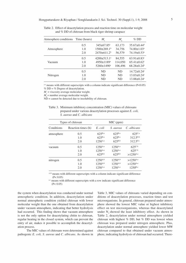
Hongpattarakere & Riyaphan / Songklanakarin J. Sci. Technol. 30 (Suppl.1), 1-9, 2008 5
the system when deacetylation was conducted under normal
atmospheric condition. In addition, deacetylation under
normal atmospheric condition yielded chitosan with lower
molecular weight than the one obtained from deacetylation
under vacuum atmosphere, indicating that better hydrolysis
had occurred. This finding shows that vacuum atmosphere
is not the only option for deacetylating chitin to chitosan,
regular heating in the closed system, which can prevent the
entry of air, makes it possible to accomplish the deacetyl-
ation process.
The MIC values of chitosans were determined against
pathogenic E. coli, S. aureus and C. albicans. As shown in
Table 3, MIC values of chitosans varied depending on con-
ditions of deacetylation processes, reaction times and test
microorganisms. In general, chitosan prepared under atmos-
phere showed the lowest MIC value or highest inhibitory
effect on test microorganisms, whereas that deacetylated
under N2
showed the least inhibitory effect. As shown in
Table 2, deacetylation under normal atmosphere yielded
chitosan with highest % DD, but % DD was lowest when
chitosan was prepared under nitrogen atmosphere. Plus,
deacetylation under normal atmosphere yielded lower MW
chitosan compared to that obtained under vacuum atmos-
phere, indicating hydrolysis of chitosan had occurred. There-
Table 2. Effect of deacetylation process and reaction time on molecular weight
and % DD of chitosan from black tiger shrimp carapace
Atmosphere conditions Time (hours) Mv
Mn
% DD
0.5 345±67.85c 63,173 35.67±0.44d
Atmosphere 1.0 1500±269.1b 34,756 74.80±1.03a
2.0 2470±611.2b 56,579 74.19±0.53a
0.5 4200±513.1a 84,555 63.91±0.83c
Vacuum 1.0 4950±1189a 114,050 65.41±0.62c
2.0 5260±1490a 106,496 68.28±0.24b
0.5 ND ND 14.72±0.24e
Nitrogen 1.0 ND ND 13.03±0.24e
2.0 ND ND 13.88±0.24e
a-e means with different superscripts with a column indicate significant difference (P<0.05)
% DD = % Degree of deacetylation.
Mv = viscosity average molecular weight.
Mn = number average molecular weight.
ND = cannot be detected due to insolubility of chitosan.
Table 3. Minimum inhibitory concentration (MIC) values of chitosans
prepared under various deacetylation processes against E. coli,
S. aureus and C. albicans
Types of chitosan MIC (ppm)
Conditions Reaction times (h) E. coli S. aureus C. albicans
atmosphere 0.5 625B,a 625B,a 625C,a
1.0 625B,a 625B,a 312.5D,b
2.0 1250A,a 625B,b 312.5D,c
vacuum 0.5 1250A,a 1250A,a 625C,b
1.0 1250A,a 1250A,a 625C,b
2.0 625B,b 625B,b >1250A,a
nitrogen 0.5 1250A,b 1250A,b >1250A,a
1.0 1250A,b 1250A,b >1250A,a
2.0 1250A,a 1250A,a 1250B,a
A-D means with different superscripts with a column indicate significant difference
(P< 0.05)a-c means with different superscripts with a row indicate significant difference
(P< 0.05)
Hongpattarakere & Riyaphan / Songklanakarin J. Sci. Technol. 30 (Suppl.1), 1-9, 20086
fore, antimicrobial activity of chitosan was dependent on the
deacetylation methods used. However, there is not enough
evidence to specify which parameter (between % DD and
MW) has the major contribution. Variation of deacetylation
process yielded chitosan with significant differences in %
DD as well as variation of the molecular weight. As Tsigos
et al. (2000) reported, chitosan with high the degree of
deacetylation had more inhibitory activity against micro-
organisms than chitosan with low the degree of deacetyl-
ation. Similarly, Simpson et al. (1997) reported that chitosan
with a degree of deacetylation of 92.5% was more effective
than chitosan with a degree of deacetylation of 85%. On the
contrary, Ikinci et al. (2002) reported that change in degree
of deacetylation (73, 84 and 95%) did not have any effect on
antimicrobial activity of chitosan (similar inhibition zone)
against Porphyromonas gingivalis.
Chitosan obtained from 1 hour deacetylation under
atmospheric condition exhibited highest antifungal activity,
but inhibited pathogenic E. coli and S. aureus at the same
level with the one from 0.5 h deacetylation. Under atmos-
pheric condition, chitosan deacetylated for 1 hour demon-
strated higher inhibitory activity against test microorganisms
than those deacetylated for 0.5 and 2.0 hours. Among all test
microorganisms, C. albicans appeared to be most susceptible
to chitosan from black tiger shrimp carapace, compared to
pathogenic E. coli and S. aureus. Generally, spoilage yeasts
were more sensitive to chitosan than Gram-positive bacteria,
which were in turn more sensitive than Gram-negative
bacteria. (Sagoo et al., 2002). Nikaido (1996) reported that
as a polymeric macromolecule, chitosan is unable to pass
the outer membrane of Gram-negative bacteria, since this
membrane functions as an efficient outer permeability barrier
against macromolecules. Moreover, the antimicrobial effect
of chitosan is reported to be dependent on its MW (Zheng
and Zhu, 2003). Various investigators have reported the
close relationships between molecular weight and antimicro-
bial activity of chitosan. Jeon and Kim (1998) found that the
highest MW oligomers (MW 500-10,000 Da) had the
strongest bactericidal and fungicidal activities against most
pathogens tested. Jeon et al. (2001) reported that MW (10-1
kDa) of chitosan oligomers was critical for microorganism
inhibition and efficacy increased with MW increased
although the native one (MW 685 kDa) was the most
inhibitory. Zheng and Zhu (2003) reported that the effect of
Table 4. Antimicrobial activity of native and hydrolyzed chitosan prepared
by chemical hydrolysis catalyzed with H2O
2 at different concen-
trations against E. coli, S. aureus and C. albicans in 1% acetic
acid
MIC (ppm)mM H
2O
2MW (kDa)
E. coli S. aureus C. albicans
0 3260A 625B,a 625A,a 312.5A,b
5 157 625B,a 625A,a 156.25B,b
10 3.17C 625B,a 625A,a 78.13C,b
25 0.24D 1250A,a 625A,b 78.13C,c
A-Dmeans with different superscripts with a column indicate significant difference
(P< 0.05)a-cmeans with different superscripts with a row indicate significant difference
(P< 0.05)
Table 5. Antimicrobial activity of native and hydrolyzed chitosan prepared by
enzymatic hydrolysis catalyzed with lysozyme against E. coli, S. aureus
and C. albicans in 0.2 M acetate buffer
MIC (ppm)Time for hydrolysis MW (kDa)
E. coli S. aureus C. albicans
0 min 438A 625A,a >1250A,b >1250A,b
5 min 586A 625A,a >1250A,b >1250A,b
10 min 473A 625A,a >1250A,b >1250A,b
30 min 543A 625A,a >1250A,b >1250A,b
24 hours 501A 625A,a >1250A,b >1250A,b
A means with different superscripts with a column indicate significant difference (P<0.05)a-b means with different superscripts with a row indicate significant difference (P<0.05)
Hongpattarakere & Riyaphan / Songklanakarin J. Sci. Technol. 30 (Suppl.1), 1-9, 2008 7
chitosan with MW below 300 kDa on Staphylococcus aureus
was strengthened as the MW increased, whereas the anti-
microbial effect on Escherichia coli increased as the MW
was decreased. Thus, two possible antimicrobial mechanisms
were proposed that the chitosan of higher MW forms a film
which may inhibit nutrient adsorptions, but that with a lower
MW enters the microbial cell more easily to disturb the
metabolism of the cell.
3.2 Antimicrobial activity of hydrolyzed chitosan
The hydrolyzed chitosan prepared by chemical re-
action led to a significant reduction of molecular weight. The
treatment of chitosan with H2O
2 also resulted in a change in
the chemical structure, as the degradation of chitosan by
H2O
2 is a random process and can be completely degraded
to its constituent monomers. The changes, such as formation
of carboxyl groups and deamination, increased with the
decrease of molecular weight. Nordtveit et al. (1994)
prepared chitosan hydrolysates using H2O
2, in the presence
of Fe (III) to generate hydroxyl radicals, which cleaved the
chitosan molecule by nucleophilic attack. As shown in Table
4, the molecular weight of chitosan decreased as hydrogen
peroxide concentration increased. The molecular weight of
chitosan obtained at H2O
2 concentration of 0, 5, 10 and 25
mM were 3260, 157, 3.17 and 0.24 kDa, respectively. Anti-
microbial activities of chitosan hydrolysates (3.17-157 kDa)
against both pathogenic E. coli and S. aureus were shown at
MIC of 625, 625 ppm, respectively, which were not differ-
ent from native chitosan (3260 kDa). However, the low
molecular weight chitosan obtained at H2O
2 concentration of
10 and 25 mM showed lowest MIC of 78.125 ppm against
C. albicans. The results are in agreement with Hirano and
Nagao (1989) which found that low molecular weight chito-
san in agar system inhibited a range of phytopathogenic
fungi more effectively than high molecular weight chitosan.
However, hydrolyzed chitosan with lower MW had no in-
hibitory activity against pathogenic E. coli and S. aureus;
particular, the one with MW of 0.24 kDa had less inhibitory
effect against pathogenic E. coli. This phenomenon was
different from the finding of Jeon et al. (2001), which
indicated that native chitosan (685 kDa) was more effective
on Gram-positive and Gram-negative bacteria than chitosan
oligomers, whose MW was between 1-10 kDa. No et al.
(2002) was also reported the same result that native chitosan
showed higher antibacterial activities than chitosan oligomers.
For enzymatic degradation of chitosan, several re-
searchers have studied the preparation and physiological
activities of chitosan oligomers with the aid of lysozyme.
Nordtveit et al. (1996) reported lysozyme from chicken egg
used for chitosan degradation has been shown to be most
efficacious when the chitosan is only partially deacetylated.
Rhoades and Roller (2000) also found that a degraded form
of chitosan was successfully produced by using lysozyme-
overnight treatment of chitosan (viscosities >800 s) with
lysozyme resulted in degraded forms of chitosan with visco-
sities of 140 s. However, the hydrolyzed chitosan prepared
by using lysozyme in this study did not show significant
difference in the molecular weight compared to the native
one. As shown in Table 5, hydrolysis of native chitosan with
lysozyme for 0, 5, 10, 30 minutes and 24 hours had the
molecular weight 438, 586, 473, 543 and 501 kDa, respec-
tively. These results disagree with the results of other
authors, which may be due to differences of chitosan in terms
of chemical structure and complexity. Antimicrobial activity
of hydrolyzed chitosan (473-583 kDa) against pathogenic
E. coli was not different from the native chitosan (438 kDa),
whereas the inhibition against S. aureus and C. albicans was
lower than the native one. The chitosan molecular weight
and antimicrobial activity from the enzymatic study were
different from the previous result due to the limitation of
chitosan solubility in the buffer (instead of 1% acetic acid),
which was used to facilitate the lysozyme activity.
4. Conclusions
Deacetylation of chitin obtained from black tiger
shrimp carapace under atmosphere yielded chitosans exhibit-
ing highest antimicrobial activity, due to higher % DD
chitosan, whereas chitosans obtained under nitrogen atmos-
phere showed the least inhibition against all test microorgan-
isms. Degree of deacetylation exhibited significant effect on
the antimicrobial activity of chitosan as well as its MW.
Chitosan hydrolyzed by chemical reaction can improve anti-
fungal activity greatly against C. albicans as the MW de-
creases, but had antibacterial activity that decreased with
the MW. However, hydrolyzed chitosan prepared by using
lysozyme did not show siginificant effect on the molecular
weight. The native chitosan and the hydrolyzed chitosan
were equally inhibitory against pathogenic E. coli, but the
degraded ones were not effective on S. aureus and C.
albicans at a concentration of 1250 ppm, the highest con-
centration tested. This is maybe caused by performing the
antimicrobial assay of the chitosan in a buffer system, which
may limit the solubility of chitosan.
Acknowledgements
Our great appreciation would be expressed to Prince
of Songkla University Funding Program (2003-2005) for
providing research funding and Professor Yasuhisa Asano,
Toyama Prefectural University, for supplying certain chemi-
cals, which are available only in Japan.
References
Arcidacono, S. and Kaplan, D. L. 1992. Molecular weight
distribution of chitosan isolated from Mucor rouxii
under different culture and processing conditions.
Biotech. Bioeng. 39: 281-286. Cited by Chen, R. H.
and Hwa, H. D. 1996. Effect of molecular weight of
chitosan with the same degree of deacetylation on the
Hongpattarakere & Riyaphan / Songklanakarin J. Sci. Technol. 30 (Suppl.1), 1-9, 20088
thermal, mechanical and permeability properties of
the prepared membrane. Carbohydr. Polym. 29: 353-
358.
Benjakul, S. and Sophanodora, P. 1990. Production of chito-
san from banana prawn shell. Songklanakarin J. Sci.
Technol. 12: 439-443.
Chen, C. S., Liau, W. Y. and Tsai, G. J. 1998. Antibacterial
effects of N-sulfonated chitosan and N-sulfobenzoyl
chitosan and applications to oyster preservation. J.
Food Prot. 61: 1124-1128.
Chen, R. H. and Hwa, H. D. 1996. Effect of molecular weight
of chitosan with the same degree of deacetylation on
the thermal, mechanical and permeability properties
of the prepared membrane. Carbohydr. Polym. 29:
353-358.
Fang, S. W., Li, C. F. and Shih, Y. C. 1994. Antifungal activity
of chitosan and its preservative effect on low-sugar
candied kumquat. J. Food Prot. 56: 136-140.
Helander, I. M., Nurmiaho-Lassila, E. L., Ahvenainen, R.,
Rhoades, J. and Roller, S. 2001. Chitosan disrupts the
barrier properties of the outer membrane of Gram-
negative bacteria. Int. J. Food Microbiol. 71: 235-
244.
Hirano, S. and Nagao, N. 1989. Effecfs of chitosan, pectic
acid, lysozyme and chitinase on the growth of the
several phytopathogens. Agric. Biol. Chem. 53: 3065-
3066.
Ikinci, G., Senel, S., Akιncιbay, H., Kas¸ S., Ercis¸ S., Wilson,
C. G. and Hιncal, A. A. 2002. Effect of chitosan on a
periodontal pathogen Porphyromonas gingivalis. Int.
J. Pharm. 235: 121-127.
Jeon, Y. J., Park, P. J. and Kim, S. K. 2001. Antimicrobial
effect of chitooligosaccha -rides produced by bio-re-
actor. Carbohydr. Polym. 44: 71-76.
Knowles, J. and Roller, S. 2001. Efficacy of chitosan, car-
vacrol, and a hydrogen peroxide-based biocide against
foodborne microorganisms in suspension and adhered
to stainless steel. J. Food Prot. 1542-1548.
Nikaido, H. 1996. outer membrane. 2nd ed. In: Neidhardt,
F.C. (Ed.), 1996. Escherichia coli and Salmonella:
Cellular and Molecular Biology, vol. 1, American
Society for Microbiology, Washington, D.C., pp 29-
47.
Nordtveit, R. J., Varum, K. M. and Smidsrod, O. 1994. Deg-
radation of fully water- soluble, partially N-acetylated
chitosan with lysozyme. Carbohydr. Polym. 23: 253-
260.
No, K. H., Park, Y. N., Lee, H. S. and Meyers, P. S. 2002.
Antibacteria activity of chitosans and oligomers with
different molecular weights. Int. J. Food Microbiol.
74 : 65-72.
Rhoades, J. and Roller, R. 2000. Antimicrobial action of
degraded and native chitosan against spoilage organ-
isms in laboratory media and food. Appl. Environ.
Microbiol. 66: 80-86.
Roller, S. and Covill, N. 2000. The antimicrobial properties
of chitosan in mayonnaise and mayonnaise-based
shrimp salads. J. Food Prot. 63: 202-209.
Sagoo, S., Board, R. and Sibel, R. 2002. Chitosan inhibits
growth of spoilage microorganisms in chilled pork
products. Food Microbiol. 19: 175-182.
Shahidi, F., Arachchi, J. K. V. and Jeon, Y. J. 1999. Food
applications of chitin and chitosans. Trends Food Sci.
Technol. 10: 37-51.
Simpson, B.K., Gagn!e, N., Ashie, I.N.A. and Noroozi, E.
1997. Utilization of chitosan for preservation of raw
shrimp (Pandalus borealis). Food Biotechnol. 11: 25-
44.
Sugano, M., Yoshida, K., Hashimoto, M., Enomoto, K. and
Hirano, S. 1992. Hypocholesterolemic activity of
partially hydrolyzed chitosan in rats. In: Brine, C.J.,
Sandford, P.A., Zikakis, J.P. (Eds.). Advances in
Chitin and Chitosan. Elsevier, London, pp. 472-478.
Cited by No, K. H., Park, Y. N., Lee, H. S. and
Meyers, P. S. 2002. Antibacteria activity of chitosans
and oligomers with different molecular weights. Int.
J. Food Microbiol. 74 : 65-72.
Suzuki, K., Mikami, T., Okawa, Y., Tokoro, A., Suzuki, S. and
Suzuki, M. 1986. Antitumor effect of hexa-N-acetyl-
chitohexaose and chitohexaose. Carbohydr. Res. 151:
403-408. Cited by No, K. H., Park, Y. N., Lee, H. S.
and Meyers, P. S. 2002. Antibacteria activity of
chitosans and oligomers with different molecular
weights. Int. J. Food Microbiol. 74 : 65-72.
Toei, K. and Kohara, T. 1976. A conductometric method for
colloid titrations. Analytica. Chimica. Acta. 83: 59-
65.
Tokoro, A., Tatewaki, N., Suzuki, K., Mikami, T., Suzuki, S.
and Suzuki, M. 1988. Growth-inhibitory effect of
hexa-N-acetylchitohexaose and chitohexaose against
Meth-A solid tumor. Chem. Pharm. Bull. 36: 784-790.
Cited by No, K. H., Park, Y. N., Lee, H. S. and
Meyers, P. S. 2002. Antibacteria activity of chitosans
and oligomers with different molecular weights. Int.
J. Food Microbiol. 74 : 65-72.
Tolaimate, A., Desbrieres, J., Rhazi, M., Alagui, A., Vincen-
don, M. and Vottero, P. 2000. On the influence of
deacetylation process on the physiochemical charac-
teristics of chitosan from squid chitin. Polym. 41:
2463-2469.
Tsai , G. J. and Hwang, S. P. 2004. In vitro and in vivo anti-
bacterial activity of shrimp chitosan against some
intestinal bacteria. Fish. Sci. 70: 675.
Tsai, G. J. and Su, W. H. 1999. Antibacterial activity of
shrimp chitosan against Escherichia coli. J. Food
Prot. 62: 239-243.
Tsai, G. J., Wu, Z. Y. and Su, W. H. 2000. Antibacterial activ-
ity of a chitooligosaccha- ride mixture prepared by
cellulase digestion of shrimp chiosan and its applica-
tion to milk preservation. J. Food Prot. 747-752.
Hongpattarakere & Riyaphan / Songklanakarin J. Sci. Technol. 30 (Suppl.1), 1-9, 2008 9
Tsigos, I., Martinou, A., Kafetzopoulos, D. and Bouriotis, V.
2000. Chitin deacetylases: new, versatile tools in Bio-
technology. Tibtech. 18: 305-312.
Vishu Kumar, A. B. Varadaraj, M. C., Lalitha, R. G. and
Tharanathan, R. N. 2004. Low molecular weight
chitosans: preparation with the aid of papain and
characterization. Biochim. Biophys. Acta 1670: 137-
146.
Wang, G. H. 1992. Inhibition and inactivation of five species
of foodborne pathogens by chitosan. J. Food Prot. 55:
916-919.
Zheng, L.-Y. and Zhu, J.-F. 2003. Study on antimicrobial
activity of chitosan with different molecular weights.
Carbohydr. Polym. 54: 527-530.