Embed Size (px)
Citation preview
2000, 38(12):4586. J. Clin. Microbiol.
Kiyofumi Ohkusu Simple Biochemical TestsOrientation Medium in Conjunction withEvaluation of the Use of CHROMagar Routine Urine, Pus, and Stool Cultures:Identification of Gram-Negative Bacilli in Cost-Effective and Rapid Presumptive
http://jcm.asm.org/content/38/12/4586Updated information and services can be found at:
These include:
REFERENCEShttp://jcm.asm.org/content/38/12/4586#ref-list-1at:
This article cites 18 articles, 16 of which can be accessed free
CONTENT ALERTS more»articles cite this article),
Receive: RSS Feeds, eTOCs, free email alerts (when new
http://journals.asm.org/site/misc/reprints.xhtmlInformation about commercial reprint orders: http://journals.asm.org/site/subscriptions/To subscribe to to another ASM Journal go to:
on March 13, 2014 by guest
http://jcm.asm
.org/D
ownloaded from
on M
arch 13, 2014 by guesthttp://jcm
.asm.org/
Dow
nloaded from
JOURNAL OF CLINICAL MICROBIOLOGY,0095-1137/00/$04.0010
Dec. 2000, p.4586–4592 Vol. 38, No. 12
Copyright © 2000, American Society for Microbiology. All Rights Reserved.
Cost-Effective and Rapid Presumptive Identification of Gram-NegativeBacilli in Routine Urine, Pus, and Stool Cultures: Evaluation of the
Use of CHROMagar Orientation Medium in Conjunction withSimple Biochemical Tests
KIYOFUMI OHKUSU*
Department of Clinical Laboratory, Chiba Children’s Hospital, Chiba, 266-0007, Japan
Received 5 June 2000/Returned for modification 6 August 2000/Accepted 16 September 2000
The algorithm for a new identification system was designed on the basis of colony color and morphology onCHROMagar Orientation medium in conjunction with simple biochemical tests such as indole (IND), lysinedecarboxylase (LDC), and ornithine decarboxylase (ODC) utilization tests with gram-negative bacilli isolatedfrom urine samples as well as pus, stool, and other clinical specimens by the following colony characteristics,biochemical reactions, and serological results: pinkish to red, IND positive (IND1), Escherichia coli; metallicblue, IND1, LDC1, and ODC negative (ODC2), Klebsiella oxytoca; IND1, LDC2, and ODC1, Citrobacterdiversus; IND1 or IND2, LDC2, and ODC2, Citrobacter freundii; IND2, LDC1, and ODC1, Enterobacteraerogenes; IND2, LDC2, and ODC1, Enterobacter cloacae; IND2, LDC1, and ODC2, Klebsiella pneumoniae;diffuse brown and IND1, Morganella morganii; IND2, Proteus mirabilis; aqua blue, Serratia marcescens; bluishgreen and IND1, Proteus vulgaris; transparent yellow-green, serology positive, Pseudomonas aeruginosa; clearand serology positive, Salmonella sp.; other colors and reactions, the organism was identified by the fullidentification methods. The accuracy and cost-effectiveness of this new system were prospectively evaluated.During an 8-month period, a total of 345 specimens yielded one or more gram-negative bacilli. A total of 472gram-negative bacillus isolates were detected on CHROMagar Orientation medium. For 466 of the isolates(98.7%), no discrepancies in the results were obtained on the basis of the identification algorithm. The cost ofidentification of gram-negative bacilli during this period was reduced by about 70%. The results of this trialfor the differentiation of the most commonly encountered gram-negative pathogens in clinical specimens withthe new algorithm were favourable in that it permitted reliable detection and presumptive identification. Inaddition, this rapid identification system not only significantly reduced costs but it also improved the dailywork flow within the clinical microbiology laboratory.
The media most widely used for the isolation and differen-tiation of coliform gram-negative bacteria and other entericpathogens from clinical specimens are MacConkey agar in theUnited States and modified Drigalski agar in Japan, but theirability to differentiate these types of organisms is minimalbecause they depend only on determination of lactose utiliza-tion. Several proprietary chromogenic mixtures, which havebeen available for some years, allow the presumptive iden-tification of pathogenic organisms on the basis of colonialmorphology and distinctive color patterns (9, 11, 12, 17).CHROMagar Orientation medium is a chromogenic culturemedium that is usually used for the isolation and enumerationof urinary tract pathogens. It has several advantages, such as agreater ability to differentiate gram-negative bacilli, and facil-itates the detection as well as presumptive identification ofgram-negative bacilli (12). Several previous studies have shownthat it can serve as a primary isolation and reliable differenti-ation medium for urinary tract pathogens, including gram-negative bacilli, enterococci, and staphylococci (9, 12, 17).However, its suitability for use with clinical specimens otherthan urine samples has not been assessed.
In the present study, I sought to evaluate the accuracy of therapid identification system on the basis of colony colors, mor-
phology on CHROMagar Orientation medium, and the resultsof additional simple biochemical tests such as indole, lysinedecarboxylase, and ornithine decarboxylase utilization testswith gram-negative bacilli isolated from various clinical speci-mens. The objectives of the study were (i) to evaluate theusefulness of CHROMagar Orientation medium for the detec-tion and presumptive identification of gram-negative bacilliwhen they were present in mixed cultures of clinical specimensafter direct plating not only from urine but also from woundswab, stool, and other specimens, and (ii) to establish a cost-effective and rapid identification approach with CHROMagarOrientation medium that would aid in the optimal use ofidentification kits.
(This study was presented at the 100th General Meeting ofthe American Society for Microbiology, Los Angeles, Calif., 21to 25 May 2000.)
MATERIALS AND METHODS
Study design. The algorithm for the rapid identification system (Table 1) wasdesigned on the basis of colony color and morphology on CHROMagar Orien-tation medium in conjunction with simple biochemical tests such as indole, lysinedecarboxylase, and ornithine decarboxylase utilization tests according to themanufacturer’s instructions and as described by Merlino et al. (12). I prospec-tively evaluated the accuracy of the identification system with gram-negativebacilli from various clinical specimens between February 1999 and September1999 in the microbiology laboratory of Chiba Children’s Hospital, a 200-bedtertiary-care hospital. All gram-negative bacilli observed on CHROMagar Ori-entation medium were identified by use of the algorithm, and the results werecompared to those obtained with the Crystal E/NF system (Becton DickinsonMicrobiology Systems [BBL], Cockeysville, Md.) and, if required, the API 20Esystem (bioMerieux, Marcy l’Etoile, France).
* Mailing address: Department of Clinical Laboratory, Chiba Chil-dren’s Hospital, Heta-cho, Midori-ku, Chiba, 266-0007, Japan. Phone:81-43-292-2111. Fax: 81-43-292-3815. E-mail: [email protected].
4586
on March 13, 2014 by guest
http://jcm.asm
.org/D
ownloaded from
Media (i) CHROMagar Orientation medium. CHROMagar Orientation me-dium was purchased as prepared plates from BBL.
(ii) Quality control. The following American Type Culture Collection (ATCC)strains were used for quality control of the medium and to assess color stability;Acinetobacter baumannii ATCC 19606, Aeromonas caviae ATCC 15468, Aero-monas hydrophila ATCC 7966, Citrobacter freundii ATCC 8090, Enterobacteraerogenes ATCC 13048, Enterobacter cloacae ATCC 13047, Escherichia coliATCC 35218, Klebsiella oxytoca ATCC 49131, Klebsiella pneumoniae ATCC27736, Morganella morganii ATCC 25830, Plesiomonas shigelloides ATCC 51903,Proteus mirabilis ATCC 7002, Proteus vulgaris ATCC 6380, Pseudomonas aerugi-nosa ATCC 27853, Serratia marcescens ATCC 8100, Stenotrophomonas malto-philia ATCC 13637, and Yersinia enterocolitica ATCC 23715.
Adjunctive simple biochemical tests. An indole spot test was performed withcolonies on CHROMagar Orientation medium and/or colonies from a pureculture on modified heart infusion slant agar with DMACA Indole Dropperreagent (BBL).
Lysine decarboxylase and ornithine decarboxylase tests were carried out withOIML medium (Eiken Chemical Co., Ltd., Tokyo, Japan); they were combinedin one tube to save time and materials. Colonies on CHROMagar Orientationmedium were inoculated onto OIML medium by stabbing. After 18 to 24 h ofincubation at 35°C, lysine decarboxylase and ornithine decarboxylase utilizationwas noted. Lysine decarboxylase utilization was determined by the presence of apurple color throughout the lower portion of the tube, and ornithine decarbox-ylase utilization was determined by the presence of a green or blue color in theupper portion.
The oxidase test was performed with an oxidase test stick (Eiken ChemicalCo.) according to the manufacturer’s instructions.
Salmonella O serogroup and Vi antigen were detected by agglutination withSalmonella antisera (Denka Seiken Co., Ltd., Tokyo, Japan), and serovar iden-tification was confirmed by the Public Health Laboratory of Chiba Prefecture.
Serotyping of Pseudomonas aeruginosa was performed with Pseudomonas an-tisera (Denka Seiken).
Bacteriological procedures by specimen type. (i) Urine. Urine samples wereinoculated onto modified Drigalski agar plates (Nippon Becton Dickinson Co.,Ltd., Tokyo, Japan) with a calibrated 5-ml loop. After centrifugation at 3,000 rpm(2,010 3 g) for 10 min, Gram staining of the urine sediments was performed toevaluate whether a pyogenic reaction was present, and if gram-negative bacilliwere also observed, these sediments were inoculated onto CHROMagar Orien-tation medium. The plates were incubated at 35°C for 18 to 24 h. In mostlaboratories, Gram staining of urine is not routinely performed with urinarysediment, and the culture plate would also not be inoculated, depending onwhether gram-negative bacilli were observed in the direct smear. However, I hadseveral reasons for routinely Gram staining urine sediments. Although Gram-staining examination of urine is slightly labor intensive, it is still a low-costmethod for the estimation of bacteriuria and the presence of neutrophils. Mostof the urine samples from pediatric patients in my hospital were collected in anadhesive bag placed on the perineum, meaning that they were often contami-
nated with multiple organisms normally present in the fecal flora. Accordingly,urine microscopy may help with determination of whether organisms in the urineare the possible causal bacteria or whether contamination is to blame, and itwould also determine whether organism identification and susceptibility testingneed to be carried out. Only in rare instances, when more fastidious organismssuch as Haemophilus influenzae are suspected, is an enriched medium such aschocolate agar required to determine the cause of infection.
(ii) Pus, otorrheic, sputum, exudate, and other specimens. Pus, otorrheic,sputum, exudate, and other specimens were also examined for the presence ofgram-negative bacilli by Gram staining of smears. If gram-negative bacilli wereobserved, each sample was streaked onto CHROMagar Orientation medium, inaddition to the appropriate agar plates.
(iii) Stool specimens. Stool specimens were inoculated in parallel ontoCHROMagar Orientation medium and appropriate agar plates such as modifiedDrigalski agar, Columbia-colistin-nalidixic acid (CNA) agar with 5% sheepblood, and CHROMagar Candida for surveillance for normal resident flora.Routinely, my laboratory also attempts to isolate Salmonella, Shigella, Esche-richia coli O157, Yersinia, and Campylobacter strains from all stool specimenssubmitted for culture because of gastrointestinal indications. Routine surveil-lance for fecal flora is not performed by many laboratories and would not be partof established laboratory protocols. In my hospital, as the number of stoolspecimens submitted for surveillance for normal resident flora is greater than thenumber submitted for surveillance for gastrointestinal pathogens, it was deemednecessary to try to evaluate the performance of CHROMagar Orientation me-dium for recognition of normal resident flora as well as enteric pathogens.
RESULTSAccuracy of the algorithm. During the 8-month study period,
a total of 345 specimens yielded one or more gram-negativebacilli (Table 2), for a total of 472 gram-negative bacilli isolateson CHROMagar Orientation medium. More than one gram-negative bacillus species was detected in 80 (43.5%) of 184stool specimens, 6 (6.0%) of 100 urine specimens, 5 (29.4%) of17 pus specimens, and 11 (25.0%) of 44 other specimens. Thecolony characteristics of these isolates on this medium and theidentification results are given in Table 3. Four hundred sixty-six (98.7%) of the total of 472 isolates were correctly identifiedby the identification system. For the remaining six isolates, oneisolate of Citrobacter amalonaticus was misidentified asCitrobacter diversus, one isolate of Enterobacter agglomeranswas misidentified as Citrobacter freundii, one isolate of Proteuspenneri was misidentified as Proteus mirabilis and three isolates
TABLE 1. Algorithm for presumptive identification of gram-negative bacilli isolated from clinical specimens
Color reaction on CHROMagarOrientation medium
Results of differential reactionsa
Expected isolate(s)Requirement forconfirmation ofidentificationa,bIND LDC ODC Serology
Pinkish to red 1 Escherichia coli IND negative
Metallic blue (with or without halo) 1 1 2 Klebsiella oxytoca1 2 1 Citrobacter diversus6 2 2 Citrobacter freundii2 1 1 Enterobacter aerogenes2 2 1 Enterobacter cloacae2 1 2 Klebsiella pneumoniae
Diffuse brown 1 Morganella morganii2 Proteus mirabilis
Transparent yellow-green 1 Pseudomonas aeruginosa Serology negative
Aqua blue Serratia marcescens
Bluish green 1 Proteus vulgaris IND negative
Clear (whitish) 2 1 Salmonella sp. is suspicious All isolates
Other colors All isolates
a IND, spot indole test; LDC, lysine decarboxylase; ODC, ornithine decarboxylase.b Crystal E/NF system; if required, the API 20E system.
VOL. 38, 2000 COST-EFFECTIVE IDENTIFICATION OF GRAM-NEGATIVE BACILLI 4587
on March 13, 2014 by guest
http://jcm.asm
.org/D
ownloaded from
of Providenciae were misidentified as Morganella morganii be-cause the algorithm did not include these species.
The accuracy of the algorithm by the observed colors of thecolonies on the CHROMagar Orientation medium is shown inTable 4. All 218 isolates of pinkish to red colonies were cor-rectly identified as Escherichia coli in conjunction with theirindole positivity. I found that characteristic transparent yellow-green colonies (n 5 42) were also correctly identified asPseudomonas aeruginosa in conjunction with their serologicalresults. All aqua blue colonies (n 5 19) were correctly identi-fied as Serratia marcescens. This color deepened to a dark blueafter 1 to 2 h at room temperature, during the period ofexamination, a finding in accordance with the results of anearlier study (12). This characteristic appearance was ex-tremely useful in the identification of this organism. Clear towhite undifferentiated colonies (n 5 18) were correctly iden-tified as Salmonella sp. (n 5 15), Stenotrophomonas maltophilia(n 5 2), or Escherichia coli (n 5 1) by the Crystal E/NF systemaccording to the identification algorithm.
Cost-effectiveness of the approach. Cost comparison resultswere based on the commercial cost of each system (Table 5),but the cost of the labor associated with testing was not in-cluded. Only 24 identification kits were needed during the8-month period. An overall reduction of about 70% in the costof identification of gram-negative bacilli was achieved duringthis period.
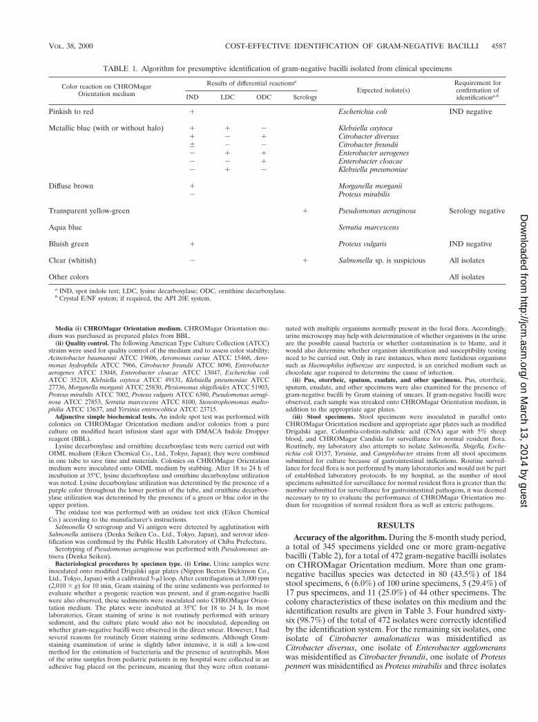
Evaluation of usefulness of CHROMagar Orientation me-dium. (i) Recognition of gram-negative bacilli. Figure 1 dem-onstrates representative color reactions for selected organismson the CHROMagar Orientation medium. Escherichia coli wasthe predominant isolate in the study, with 218 of 220 isolates(99.1%) producing a pinkish-to-red color (Fig. 1, no. 1).Twelve lactose-negative isolates and three mucoid-colony iso-
lates also demonstrated the same color. Of the two remainingEscherichia coli isolates, one was colorless (o-nitrophenyl-b-D-galactopyranoside [ONPG] negative), and the other had a blueinterior with a pink halo (both ONPG and b-glucoside posi-tive), which was different from the color of Klebsiella-Enter-obacter-Citrobacter (K-E-C) group (metallic blue).
The K-E-C group isolates regularly produced a metallic bluecolor with or without a purple to pink halo (Fig. 1, no. 2 to 7).Although the K-E-C group may be difficult, if not impossible,to identify by colony color and morphology alone, these iso-lates were distinguished as coliforms from other gram-negativebacilli and could easily be differentiated by the use of simpleadditional tests, such as indole, lysine decarboxylase, or orni-thine decarboxylase utilization tests, by following the identifi-cation chart.
The Proteus, Morganella, and Providencia (P-M-P) groupisolates demonstrated diffuse brown colonies as a result oftryptophan deaminase production (Fig. 1, no. 8 to 10). Proteusvulgaris isolates (n 5 2) had bluish green colonies with a slightbrown background (Fig. 1, no. 13). Acinetobacter baumannii(n 5 4) consisted of nontransparent convex colonies (Fig. 1,no. 14), making these isolates easily distinguishable from theother species described above.
(ii) Recognition of enteric pathogens. During the study pe-riod, a total of 15 Salmonella isolates were isolated from freshstool samples, and no other enteric organisms were recovered.The 15 isolates were lactose negative and H2S positive, yieldingclear colonies on CHROMagar Orientation medium (Fig. 1,no. 16). They were distinguishable from the common species instool by their colony color. One isolate was detected withCHROMagar Orientation medium only, and for three isolatesthe number of colonies on this medium was greater than thenumber on Salmonella-shigella agar, which is usually used for
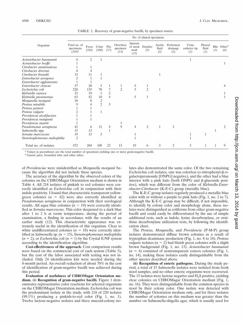
TABLE 2. Recovery of gram-negative bacilli, by specimen source
Organism
No. of clinical specimens
Total no. ofspecimens
(345)a
Feces(184)
Urine(100)
Pus(17)
Otorrheicspecimen
(13)
Sputumor nasal
swab(13)
Exudate(5)
Asciticfluid(2)
Peritonealdrainage
(2)
Urinecatheter tip
(1)
Pleuralfluid(1)
Bile(1)
Otherb
(6)
Acinetobacter baumannii 4 2 1 1Acinetobacter lwoffii 1 1Citrobacter amalonaticus 1 1Citrobacter diversus 4 4Citrobacter freundii 13 11 1 1Enterobacter aerogenes 2 1 1Enterobacter agglomerans 1 1Enterobacter cloacae 37 20 5 1 1 5 1 4Escherichia coli 220 135 70 7 2 3 2 1Klebsiella oxytoca 23 19 1 2 1Klebsiella pneumoniae 63 42 10 4 5 1 1Morganella morganii 10 8 2Proteus mirabilis 9 4 2 3Proteus penneri 1 1Proteus vulgaris 2 1 1Providencia alcalifaciens 1 1Providencia rustigianii 1 1Providencia stuartii 1 1Pseudomonas aeruginosa 42 6 12 4 9 4 2 1 2 2Salmonella spp. 15 13 1 1Serratia marcescens 19 13 3 1 1 1Stenotrophomonas maltophilia 2 1 1
Total no. of isolates 472 284 109 22 13 19 6 3 2 3 1 1 9
a Values in parentheses are the total number of specimens yielding one or more gram-negative bacilli.b Gastric juice, bronchial tube and other tubes.
4588 OHKUSU J. CLIN. MICROBIOL.
on March 13, 2014 by guest
http://jcm.asm
.org/D
ownloaded from
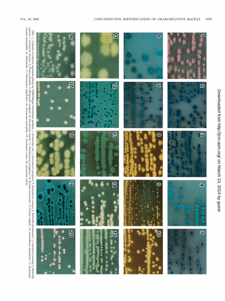
FIG
.1.
Colonies
ofselected
organisms
platedon
CH
RO
Magar
Orientation
medium
.1,Escherichia
coli;2,Enterobacter
aerogenes;3,Enterobacter
cloacae;4,Klebsiella
pneumoniae;5,C
itrobacterfreundii;6,K
lebsiellaoxytoca;7,C
itrobacterdiversus;8,P
roteusm
irabilis;9,Morganella
morganii;10,P
rovidenciastuartii;11,P
seudomonas
aeruginosa;12,Serratiam
arcescens;13,Proteus
vulgaris;14,Acinetobacter
baumannii;15,Stenotroph-
omonas
maltophilia;16,Salm
onella;17,Plesiom
onasshigelloides;18,A
eromonas
hydrophila;19,Aerom
onascaviae;20,A
eromonas
sobria.
VOL. 38, 2000 COST-EFFECTIVE IDENTIFICATION OF GRAM-NEGATIVE BACILLI 4589
on March 13, 2014 by guest
http://jcm.asm
.org/D
ownloaded from
isolation. It is probable that the peptone and yeast extract inthe formulation provided additional enrichment, and the ab-sence of inhibitory effects may also contribute to its efficiency.I found that this medium allows the detection of Salmonellaeven in mixed cultures and even when the colony counts are
low. Additionally, this medium was useful for the differentia-tion of Salmonella (clear) from other H2S-producing memberssuch as Citrobacter freundii (metallic blue) and Proteus (brown)and permitted the accurate retrieval of colonies to elicit otherbiochemical and serological reactions.
(iii) Other advantages. The CHROMagar Orientation me-dium prevented the swarming of Proteus isolates and limitedthe spread of mucoid Escherichia coli, Klebsiella pneumoniae,and Klebsiella oxytoca isolates, which may yield confluentgrowth on plates, as was also demonstrated in previous studies(9, 12, 17).
DISCUSSION
Laboratories must perform accurate and cost-effective iden-tification of clinical isolates of gram-negative bacilli (14, 15,18). In addition, rapid bacterial identification and susceptibilitytesting in the microbiology laboratory can have a demonstrableclinical impact as well as provide significant cost savings, as wasdemonstrated by Doern et al. (3) in a large prospective study.One of the more recent advances in the rapid presumptiveidentification of pathogenic organisms is the use of differentcolony colors that are produced by the reactions of genus- orspecies-specific enzymes with chromogenic substrates (1, 2,
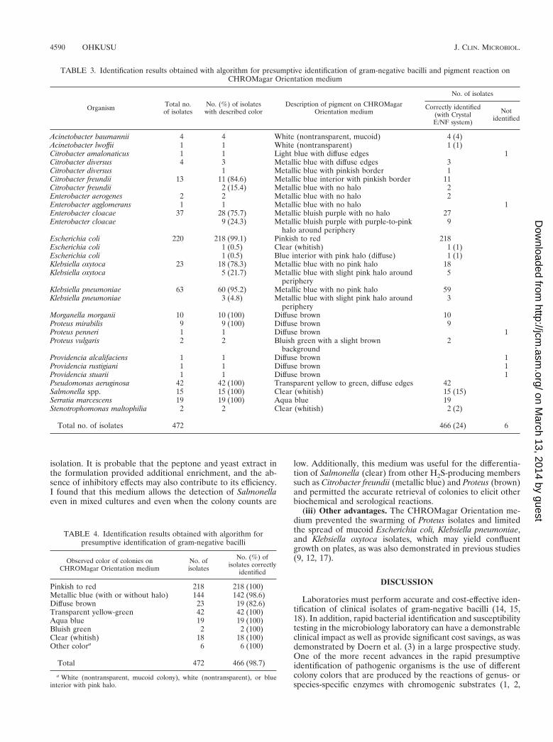
TABLE 3. Identification results obtained with algorithm for presumptive identification of gram-negative bacilli and pigment reaction onCHROMagar Orientation medium
Organism Total no.of isolates
No. (%) of isolateswith described color
Description of pigment on CHROMagarOrientation medium
No. of isolates
Correctly identified(with CrystalE/NF system)
Notidentified
Acinetobacter baumannii 4 4 White (nontransparent, mucoid) 4 (4)Acinetobacter lwoffii 1 1 White (nontransparent) 1 (1)Citrobacter amalonaticus 1 1 Light blue with diffuse edges 1Citrobacter diversus 4 3 Metallic blue with diffuse edges 3Citrobacter diversus 1 Metallic blue with pinkish border 1Citrobacter freundii 13 11 (84.6) Metallic blue interior with pinkish border 11Citrobacter freundii 2 (15.4) Metallic blue with no halo 2Enterobacter aerogenes 2 2 Metallic blue with no halo 2Enterobacter agglomerans 1 1 Metallic blue with no halo 1Enterobacter cloacae 37 28 (75.7) Metallic bluish purple with no halo 27Enterobacter cloacae 9 (24.3) Metallic bluish purple with purple-to-pink
halo around periphery9
Escherichia coli 220 218 (99.1) Pinkish to red 218Escherichia coli 1 (0.5) Clear (whitish) 1 (1)Escherichia coli 1 (0.5) Blue interior with pink halo (diffuse) 1 (1)Klebsiella oxytoca 23 18 (78.3) Metallic blue with no pink halo 18Klebsiella oxytoca 5 (21.7) Metallic blue with slight pink halo around
periphery5
Klebsiella pneumoniae 63 60 (95.2) Metallic blue with no pink halo 59Klebsiella pneumoniae 3 (4.8) Metallic blue with slight pink halo around
periphery3
Morganella morganii 10 10 (100) Diffuse brown 10Proteus mirabilis 9 9 (100) Diffuse brown 9Proteus penneri 1 1 Diffuse brown 1Proteus vulgaris 2 2 Bluish green with a slight brown
background2
Providencia alcalifaciens 1 1 Diffuse brown 1Providencia rustigiani 1 1 Diffuse brown 1Providencia stuarii 1 1 Diffuse brown 1Pseudomonas aeruginosa 42 42 (100) Transparent yellow to green, diffuse edges 42Salmonella spp. 15 15 (100) Clear (whitish) 15 (15)Serratia marcescens 19 19 (100) Aqua blue 19Stenotrophomonas maltophilia 2 2 Clear (whitish) 2 (2)
Total no. of isolates 472 466 (24) 6
TABLE 4. Identification results obtained with algorithm forpresumptive identification of gram-negative bacilli
Observed color of colonies onCHROMagar Orientation medium
No. ofisolates
No. (%) ofisolates correctly
identified
Pinkish to red 218 218 (100)Metallic blue (with or without halo) 144 142 (98.6)Diffuse brown 23 19 (82.6)Transparent yellow-green 42 42 (100)Aqua blue 19 19 (100)Bluish green 2 2 (100)Clear (whitish) 18 18 (100)Other colora 6 6 (100)
Total 472 466 (98.7)
a White (nontransparent, mucoid colony), white (nontransparent), or blueinterior with pink halo.
4590 OHKUSU J. CLIN. MICROBIOL.
on March 13, 2014 by guest
http://jcm.asm
.org/D
ownloaded from

4–13, 16, 17). In this regard, the study by Merlino et al. (12)was attractive, as it described the major advantages ofCHROMagar Orientation medium and showed that it had agreater ability to differentiate the common species of gram-negative bacilli. The purpose of the present study not only wasto determine the usefulness of this medium but also was toevaluate the accuracy and cost-effectiveness of the algorithmfor the rapid identification of gram-negative bacilli in mixedcultures of various clinical specimens. Additionally, in thisevaluation, I considered that use of this medium might reducethe number of identification kits needed but not the number ofculture media used, resulting in more accurate identificationswhen the results obtained with the medium were used in con-junction with growth characteristics on other media, and mightsubstantially lower the work load in the laboratory.
The overall accuracy of the present approach to the identi-fication of gram-negative bacilli was 98.7% (466 of 472 iso-lates). The result obtained with the proposed algorithm foridentification showed an excellent correlation with the resultsobtained with the commercial identification panel, confirmingits usefulness for primary isolation and differentiation of clin-ical gram-negative bacilli from various specimens such asurine, wound swab, otorrheic, and stool specimens for surveil-lance for normal resident flora and other organisms. In addi-tion, I estimate that the use of this identification system willresult in material savings equivalent to about $3,850 per year inmy laboratory, in addition to reducing labor and enabling rapidreporting of clinically relevant laboratory results, without a lossof sensitivity. I believe that this protocol could easily beadopted by most clinical laboratories.
Several other studies have recommended the use ofCHROMagar Orientation medium as a single medium forreliable detection, enumeration, and presumptive identifica-tion of urinary tract pathogens (9, 17). My study revealed thatabout 64% (70 of 109) of the gram-negative bacilli from urinespecimens were Escherichia coli, a proportion similar to thatfound in prior studies (9, 17). In addition, all 109 isolates fromthe 100 urine samples containing more than one organismwere accurately identified without discrepancy when the algo-rithm was used. Thus, this algorithm appears to be an excellentand time-saving method for the rapid identification of thecausative pathogens of urinary tract infections.
Of the six isolates misidentified in this study, two (Citrobacteramalonaticus and Enterobacter agglomerans) were not includedin the flow diagram, and their frequency of isolation in theroutine laboratory may be very low. As for the remaining fourisolates, one of Proteus penneri and three of the genus Provi-dencia were not correctly identified because other simple testsother than the indole reaction were not performed in this trial.Members of the P-M-P group were easily distinguished on
primary plates because of their brown colonies on a diffusebeige pigment background. Proteus mirabilis and Morganellamorganii, important pathogens in urinary tract infections, maybe differentiated by the indole spot test. I reasoned that as thefrequencies of isolation of Proteus penneri and Providencia spp.in the routine laboratory are probably low, they need not beincluded in the algorithm. However, because of the results ofthe present study, I intend to change the logic for the screeningof these organisms and include the ornithine decarboxylasetest, that is, to use the ornithine decarboxylase test when in-dole-negative and ornithine-positive isolates are identified asProteus mirabilis and when both indole- and ornithine-negativeisolates are identified as Proteus penneri, as well as when bothindole- and ornithine-positive isolates are identified as Mor-ganella morganii and indole-positive and ornithine-negativeisolates are presumptively identified as Providencia species. Ifdefinitive identification is still required, the Crystal E/NF sys-tem could be used. This logic may allow the more accurateidentification of the P-M-P group of organisms.
As pointed out by Merlino et al. (12), the use of CHROMa-gar Orientation medium by laboratory workers afflicted withvarious types of color blindness may lead to difficulties indistinguishing differences in color and colony appearance. Inthe present study, I created a colony color table to help over-come this problem. Practice is also required to correctly inter-pret the colors of colonies of major organisms before thismedium is used. Furthermore, it is extremely important thatwhen questionable colony colors and morphology are encoun-tered in the interpretation of culture results, the color logicchart should not be applied and the colony should be investi-gated by the full identification methods available.
In summary, the approach to presumptive identification ofgram-negative bacilli on the basis of both colony color andmorphology on CHROMagar Orientation medium and ad-junctive simple biochemical tests described here provides areliable routine method for the primary isolation and differ-entiation of isolates from specimens from various body sites.An additional important advantage of this medium is the easyrecognition of bacterial growth and the simultaneous detectionof multiple organisms so that the identity of the organisms andthe results of antibiotic susceptibility tests can be more accu-rately reported. Furthermore, the new rapid identification sys-tem contributes significantly to reducing the number of iden-tification kits needed and to streamlining the daily work flow inthe clinical laboratory.
ACKNOWLEDGMENT
I am grateful to Shigeru Nakayama for the photography.
TABLE 5. Comparison of cost of the Crystal E/NF identification system and new rapid identification systema
Item (cost [$] per test)Crystal E/NF system New rapid identification system
No. of tests Total cost ($) No. of tests Total cost ($)
Crystal E/NF system (7.69) 472 3,630 24 184CHROMagar Orientation medium (2.21) 345 763OIML agarb (1.04) 144 150Modified heart infusion slant agarc (0.02) 472 11 224 5
Total 472 3,641 472 1,102
a During an 8-month period.b Ornithine decarboxylase and lysine decarboxylase production test.c Indole production test.
VOL. 38, 2000 COST-EFFECTIVE IDENTIFICATION OF GRAM-NEGATIVE BACILLI 4591
on March 13, 2014 by guest
http://jcm.asm
.org/D
ownloaded from
REFERENCES
1. Dalet, F., and T. Segovia. 1995. Evaluation of a new agar in Uricult-for rapiddetection of Escherichia coli in urine. J. Clin. Microbiol. 33:1395–1398.
2. Delisle, G. J., and A. Ley. 1989. Rapid detection of Escherichia coli in urinesamples by a new chromogenic b-glucuronidase assay. J. Clin. Microbiol.27:778–779.
3. Doern, G., R. Vautour, M. Gaudet, and B. Levy. 1994. Clinical impact ofrapid in vitro susceptibility testing and bacterial idenification. J. Clin. Mi-crobiol. 32:1757–1762.
4. Dusch, H., and M. Altwegg. 1993. Comparison of Rambach agar, SM-IDmedium, and Hektoen Enteric agar for primary isolation of non-typhi sal-monellae from stool samples. J. Clin. Microbiol. 31:410–412.
5. Edberg, S. C., and C. M. Kontnick. 1986. Comparison of b-glucuronidase-based substrate systems for identification of Escherichia coli. J. Clin. Micro-biol. 24:368–371.
6. Gaillot, O., P. D. Camillo, P. Berche, R. Courcol, and C. Savage. 1999.Comparison of CHROMagar Salmonella medium and Hektoen enteric agarfor isolation of salmonellae from stool samples. J. Clin. Microbiol. 37:762–765.
7. Geiss, H. K. 1990. Comparison of two test kits for rapid identification of E.coli by a beta-glucuronidase assay. Eur. J. Clin. Microbiol. Infect. Dis. 9:151–152.
8. Heizmann, W., P. C. Doller, B. Gutbrod, and H. Werner. 1988. Rapididentification of Escherichia coli by Fluorocult media and positive indolereaction. J. Clin. Microbiol. 26:2682–2684.
9. Hengstler, K. A., R. Hammann, and A. Fahr. 1997. Evaluation of BBLCHROMagar Orientation medium for detection and presumptive identifi-cation of urinary tract pathogens. J. Clin. Microbiol. 35:2773–2777.
10. Larinkari, U., and M. Rantio. 1995. Evaluation of a new dipslide with a
selective medium for the rapid detection of beta-glucuronidase-positiveEscherichia coli. Eur. J. Clin. Microbiol. Infect. Dis. 14:606–609.
11. Mazoyer, M. A., S. Orenga, F. Doleans, and J. Freney. 1995. Evaluation ofCPS ID2 medium for detection of urinary tract bacterial isolates in speci-mens from a rehabilitation center. J. Clin. Microbiol. 33:1025–1027.
12. Merlino, J., S. Siarakas, G. J. Robertson, G. R. Funnell, T. Gottlieb, and R.Bradbury. 1996. Evaluation of CHROMagar Orientation for differentiationand presumptive identification of gram-negative bacilli and Enterococcusspecies. J. Clin. Microbiol. 34:1788–1793.
13. Odds, F. C., and R. Bernaerts. 1994. CHROMagar Candida, a new differ-ential isolation medium for presumptive identification of clinically importantCandida species. J. Clin. Microbiol. 32:1923–1929.
14. Pattyn, S. R., J. P. Sion, and J. Verhoeven. 1990. Evaluation of the LOGICsystem for the rapid identification of members of the family Enterobacteri-aceae in the clinical microbiology laboratory. J. Clin. Microbiol. 28:1449–1450.
15. Perry, J. D., M. Ford, N. Hjersing, and F. K. Gould. 1988. Rapid conven-tional scheme for biochemical identification of antibiotic resistant Enterobac-teriaceae isolates from urine. J. Clin. Pathol. 41:1010–1012.
16. Pfaller, M. A., A. Houston, and S. Coffmann. 1996. Application ofCHROMagar Candida for rapid screening of clinical specimens for Candidaalbicans, Candida tropicalis, Candida krusei, and Candida (Torulopsis) gla-brata. J. Clin. Microbiol. 34:58–61.
17. Samra, Z., M. Heifetz, J. Talmor, E. Bain, and J. Bahar. 1998. Evaluation ofuse of a new chromogenic agar in detection of urinary tract pathogens.J. Clin. Microbiol. 36:990–994.
18. Yong, D.C.T., J. S. Thompson, and A. Prytula. 1985. Rapid microbiochemi-cal method for presumptive identification of gastroenteritis-associated mem-bers of the family Enterobacteriaceae. J. Clin. Microbiol. 21:914–918.
4592 OHKUSU J. CLIN. MICROBIOL.
on March 13, 2014 by guest
http://jcm.asm
.org/D
ownloaded from


![Chrom journal8 fr[1]](https://img.pdfslide.tips/doc/110x75/55700931d8b42ac0178b4776/chrom-journal8-fr1.jpg)