Embed Size (px)
Citation preview
Journal of Gastroenterology and Hepatology
(2002)
17,
772–778
Blackwell Science, LtdOxford, UKJGHJournal of Gastroenterology and Hepatology0815-93192002 Blackwell Science Asia Pty Ltd
172806
Positivity of AFP-L3, DCP and HCC featuresH Okuda et al.
10.1046/j.0815-9319.2002.02806.xOriginal ArticleBEES SGML
Correspondence: Dr Hiroaki Okuda, Institute of Gastroenterology, Tokyo Women’s Medical University, 8-1 Kawada-cho,Shinjuku-ku, Tokyo 162-8666, Japan. Email: [email protected]
Accepted for publication 20 March 2002.
CLINICAL STUDIES ON HEPATOCELLULAR CARCINOMA
Clinicopathologic features of patients with hepatocellular carcinoma seropositive for
αααα
-fetoprotein-L3 and seronegative for des-
γγγγ
-carboxy prothrombin in comparison with those seropositive for
des-
γγγγ
-carboxy prothrombin alone
HIROAKI OKUDA,* TOSHIMI NAKANISHI,* KAZUKO TAKATSU,* AKIKO SAITO,* NAOAKI HAYASHI,* MASAKAZU YAMAMOTO,* KEN TAKASAKI*
AND MASAYUKI NAKANO
†
*
Institute of Gastroenterology, Tokyo Women’s Medical University, Tokyo and
†
Division of Clinical Pathology, National Chiba Hospital, Chiba, Japan
Abstract
Background
:
There has been no study of the clinicopathologic features of patients with hepatocellularcarcinoma (HCC) who are seropositive for lectin-reactive
α
-fetoprotein (AFP-L3) alone, or seropositivefor AFP-L3 and seronegative for des-
γ
-carboxy prothrombin (DCP) in comparison with those who areseropositive for DCP alone. Thus, the present comparative study was performed.
Methods
:
The clinicopathologic features of HCC patients with either one or two tumors who under-went a hepatectomy (
n
=
88) were compared among the following five groups according to the seropos-itivity of AFP, AFP-L3 and DCP: (i) group A, seropositive for AFP above 100 ng/mL, AFP-L3 above15% and DCP above 100 mAU/mL; (ii) group B, seropositive for AFP-L3 and seronegative for DCPbelow 40 mAU/mL; (iii) group C, seronegative for AFP below 20 ng/mL, AFP-L3 below 15% and serop-ositive for DCP; (iv) group D, seropositive for AFP and seronegative for AFP-L3 and DCP; and (v)group E, seronegative for AFP, AFP-L3 and DCP.
Results
:
Group B patients showed a higher incidence of infiltrative-type HCC with an irregular mar-gin (
P
< 0.05) and a higher frequency of poorly differentiated HCC (
P
< 0.01) compared with group Cpatients. Group A patients had larger tumors and more massive-type tumors than group B patients.Our HCC cases showed that advanced clinicopathologic features were demonstrated in the order ofgroup B, group C and group D. Group A and B patients and group D and E patients showed similarcharacteristics.
Conclusions
:
Hepatocellular carcinoma patients who were seropositive for AFP-L3 and seronegativefor DCP demonstrated clinicopathologic features of more advanced HCC compared with those whowere seropositive for DCP alone.© 2002 Blackwell Publishing Asia Pty Ltd
Key words
:
clinicopathologic features, des-
γ
-carboxy prothrombin, hepatocellular carcinoma, lectin-reactive
α
-fetoprotein, seropositivity.
INTRODUCTION
Hepatocellular carcinoma (HCC) is one of the mostcommon malignancies in the world and has anextremely poor prognosis.
1
Not only imaging studies,but also tumor markers, including
α
-fetoprotein(AFP)
2–4
and des-
γ
-carboxy prothrombin (DCP),
5–11
are important and valuable diagnostic tools for HCC.
Since the recent development of the revised enzymeimmunoassay for DCP (Eitest PIVKA-II; Eisai, Tokyo,Japan), the ability of diagnosing HCC with this markerhas increased.
12–16
Although AFP (with a cut-off value of 20 ng/mL) hasa somewhat higher sensitivity to HCC than DCP, itsspecificity to HCC is much lower than that of DCP.
α
-Fetoprotein occasionally becomes seropositive in
Positivity of AFP-L3, DCP and HCC features
773
non-HCC liver diseases, requiring the measurement oflectin-reactive AFP fractions (AFP-L3).
17,18
It has been reported that a high serum AFP-L3 con-centration in HCC patients is an unfavorable prognosticfactor and that the prognosis is extremely poor for HCCpatients who are seropositive for AFP-L3 during post-treatment follow up compared with those who areseronegative for AFP-L3 prior to initial treatment.
17,18
However, there have been no studies of the clinico-pathologic features of HCC patients who are seroposi-tive for AFP-L3 alone (there are very few such patientsbecause most patients positive for AFP-L3 are alsoseropositive for AFP), or those who are seropositive forAFP-L3 and seronegative for DCP. We have reportedthat HCC patients who are seropositive for DCP alonehave clinicopathologic features of more advanced HCCthan those who are seropositive for AFP alone.
19
Therefore, in the present study, we investigated theclinicopathologic features of HCC patients who wereseropositive for AFP-L3 and seronegative for DCP incomparison with those who were seropositive for DCPalone. We also studied the features of patients who wereseropositive for AFP, AFP-L3 and DCP in order to ana-lyze the clinicopathologic features of HCC patients whoare seropositive for AFP-L3. This study should some-what clarify the relationship between the characteristicsand progression of HCC and the seropositivity of AFP,AFP-L3 and DCP.
METHODS
Patients
Eighty-eight patients who had either one or two HCCand who underwent a hepatectomy during the periodfrom January 1997 to December 2000 were studied.There were 59 males and 29 females, and the patients’ages ranged from 42 to 77 years (mean (
±
SD) age63.5
±
7.4 years). Hepatitis B surface antigen (HBsAg)was positive in 18 patients (20.5%) and 57 patients(64.8%) were positive for both antibodies to hepatitis Cvirus (anti-HCV) and HCV RNA, all of which weremeasured by enzyme immunoassay (EIA). Among the13 patients who were negative for both HBsAg and anti-HCV, six had a history of excessive alcohol consumptionexceeding 120 g/day for more than 10 years (Table 1).
Informed consent was obtained from each patient.The study protocol conformed to the 1975 Declarationof Helsinki and was approved by the local scientific eth-ics committee.
The diagnosis of HCC was made histologically byexamination of resected surgical samples in all 88 cases.Sixty-nine cases (78.4%) had solitary HCC and 19(21.6%) had two HCC macroscopically. A diagnosis ofnon-tumorous liver was made histologically and serumlevels of AFP, AFP-L3 and DCP were measured beforeresection in all HCC patients. Forty-eight patients(54.5%) had cirrhosis and 34 (38.6%) had chronic hep-atitis as accompanying lesions. In the remaining sixpatients, the non-tumorous liver showed alcoholic fibro-sis in two patients, alcoholic steatohepatitis in twopatients and a nearly normal liver in the remaining two
patients. The characteristics of the HCC patients aregiven in Table 1.
Twenty-eight cases (31.8%) had tumors smaller than2 cm in their greatest dimension, 21 (23.9%) werebetween 2 and 3 cm, 22 (25.0%) were between 3 and5 cm and 17 (19.3%) were larger than 5 cm. The HCCnodules were graded histologically into one of three cat-egories: well differentiated, moderately differentiated orpoorly differentiated, according to the criteria proposedby the Liver Cancer Study Group of Japan.
20,21
Whenthere were two nodules, the greatest dimension and his-tologic differentiation of the larger nodule was used,and when the tumor consisted of two or more grades ofhistologic differentiation, the most dominant grade wasaccepted. Thus, 16 cases (18.2%) had well-differenti-ated HCC, 57 cases (64.8%) had moderately differen-tiated HCC and 15 cases (17.0%) had poorlydifferentiated HCC.
Assay of serum levels of AFP, AFP-L3, and DCP
The serum levels of AFP were determined by EIA andthe cut-off value was set at 20 ng/mL. Serum DCP lev-els were measured by the newly developed revised EIAkit for DCP with increased sensitivity (Eitest PIVKA-IIkit; Eisai). The cut-off value was 40 mAU/mL accordingto previous studies.
12–16
Lectin-reactive AFP fractions (fucosylated fraction ofAFP, AFP-L3) were measured by lectin-affinity electro-phoresis coupled with antibody-affinity blotting
22
usingthe AFP differentiation kit L (Wako Pure ChemicalIndustries, Osaka, Japan).
17,18
The AFP-L3 level wascalculated as follows:
A cut-off level of 15% was used on the basis of previousreports.
17,18
,
22
A positive AFP-L3 level was defined asan AFP-L3 level greater than the cut-off level of 15%.
Comparison of the clinicopathologic features of HCC according to the seropositivity of AFP, AFP-l3 and DCP
The clinicopathologic features of these HCC patients(
n
=
88) were compared according to the seropositivity
AFP-L3 total AFP %[ ] [ ] ¥ ( )100
Table 1
Characteristics of patients with hepatocellularcarcinoma
Median (range) age (years) 63.6 (42–77)No. males/females 59/29No. HBsAg positive (%) 18 (20.5)No. anti-HCV positive (%) 57 (64.8)No. negative for both HBsAg and anti-HCV
(%)13 (14.8)
No. with complication of cirrhosis (%) 48 (54.5)
HBsAg, hepatitis B surface antigen; anti-HCV, antibody tohepatitis C virus.
774
H Okuda
et al.
of AFP, AFP-L3 and DCP. The HCC cases weredivided into the following five groups: (i) group A,seropositive for AFP above 100 ng/mL, AFP-L3 above15% and DCP above 100 mAU/mL (
n
=
20); (ii) groupB, seropositive for AFP-L3 above 15% and seronegativefor DCP below 40 mAU/mL (
n
=
15; AFP below 100 ng/mL in six cases (8–68 ng/mL, almost equivalent to thosewho were seropositive for AFP-L3 alone) and AFPabove 100 ng/mL in nine cases (250–7597 ng/mL)); (iii)group C, seronegative for AFP below 20 ng/mL andAFP-L3 below 15% and seropositive for DCP above100 mAU/mL (
n
=
17); (iv) group D, seropositive forAFP above 100 ng/mL and seronegative for AFP-L3below 15% and DCP below 40 mAU/mL (
n
=
15); and(v) group E, seronegative for AFP below 20 ng/mL,AFP-L3 below 15% and DCP below 40 mAU/mL(
n
=
21). Seven patients who did not belong to any ofthese five groups were excluded from the study.
Hepatocellular carcinoma patients with serum AFPvalues between 20 and 100 ng/mL or with serum DCPvalues between 40 and 100 mAU/mL (
n
=
22) were alsoexcluded from the study because these AFP and DCPvalues were regarded as equivocal. This was because ofa relatively low specificity of AFP and DCP when theircut-off values were 20 ng/mL and 40 mAU/mL, respec-tively, as mentioned previously.
12,16
,
19
Therefore, 29patients, of the 117 patients who were initially enrolledin the study, were subsequently excluded. Thus, fea-tures including macroscopic appearance, greatestdimension, histologic differentiation of HCC and prog-nosis were compared among the remaining 88 patientswho were assigned to one of the five groups.
The macroscopic features of HCC were classifiedinto tumors with an indistinct margin or a vaguely nod-ular type (
n
=
12), those with a distinct margin or nod-ular type (
n
=
55), those with an irregular margin orinfiltrative type (
n
=
10) and those with massive-typeHCC (
n
=
11) according to the classification of primaryliver cancer by the Liver Cancer Study Group ofJapan.
19–21
A vaguely nodular-type HCC with indistinctmargin shows an indistinct border between cancerousand non-cancerous tissue and is very often a solitarytumor of 2 cm or less in its greatest dimension (smallliver cancer) with well-differentiated HCC. It isregarded as HCC in its early stage.
19–21
An infiltrative-type HCC with an irregular margin shows a yellowish–white color that is clearly demarcated from the sur-rounding non-tumor liver. The margin between thetumor and non-tumor portions is irregular because ofthe infiltrative growth of the tumor. Histologically, thistype of tumor mostly shows either a poorly differenti-ated HCC or sclerosing-type HCC.
20,21
A massive-typeHCC consists of a large tumor that occupies one ormore segments of the liver. The margin between thetumor and non-tumor portions is indistinct and irreg-ular and intrahepatic metastases are almost alwaysobserved in this type of HCC.
20,21
There were no casesof diffuse-type HCC.
20,21
The same comparisons were made among these fivegroups in relation to greatest dimension, histologicdifferentiation of HCC and also clinicopathologic vari-ables, including age, sex, nodular size of HCC, inci-dence of microscopic findings of portal vein invasion
and intrahepatic metastases.
20,21
When there were twotumors, the findings of the larger tumor were accepted.The survival rates and disease-free survival ratesafter hepatectomy (as of 31 August 2001) were alsocompared. The mean (
±
SD) duration of follow upwas 15.6
±
11.0, 25.6
±
15.5, 23.8
±
13.4, 25.1
±
10.3and 33.4
±
10.6 months for groups A, B, C, D and E,respectively.
Comparisons were made between patients in group Band patients in the other groups (particularly group Cpatients).
Statistical analysis
Results were analyzed with the chi-squared test and theStudent’s
t
-test.
P
< 0.05 was considered significant.
RESULTS
Differences in the clinicopathologic features of HCC according to the seropositivity of AFP, AFP-L3 and DCP
All 12 cases of HCC with an indistinct margin belongedto either group D (AFP single positive;
n
=
5) or groupE (AFP, AFP-L3, DCP negative;
n
=
7). Group Bpatients (AFP-L3 positive, DCP negative) showed alower incidence of indistinct margin compared withgroup D (
P
< 0.05) and group E (
P
< 0.01) patients.Group B cases had a higher incidence of infiltrative-typeHCC with an irregular margin than those in group C(DCP single positive;
P
< 0.05), group D (
P
< 0.01) andgroup E (
P
< 0.01) cases, thus showing characteristicsof more advanced HCC. Group A (AFP, AFP-L3, DCPpositive) showed a higher frequency of massive-typeHCC compared with group B (
P
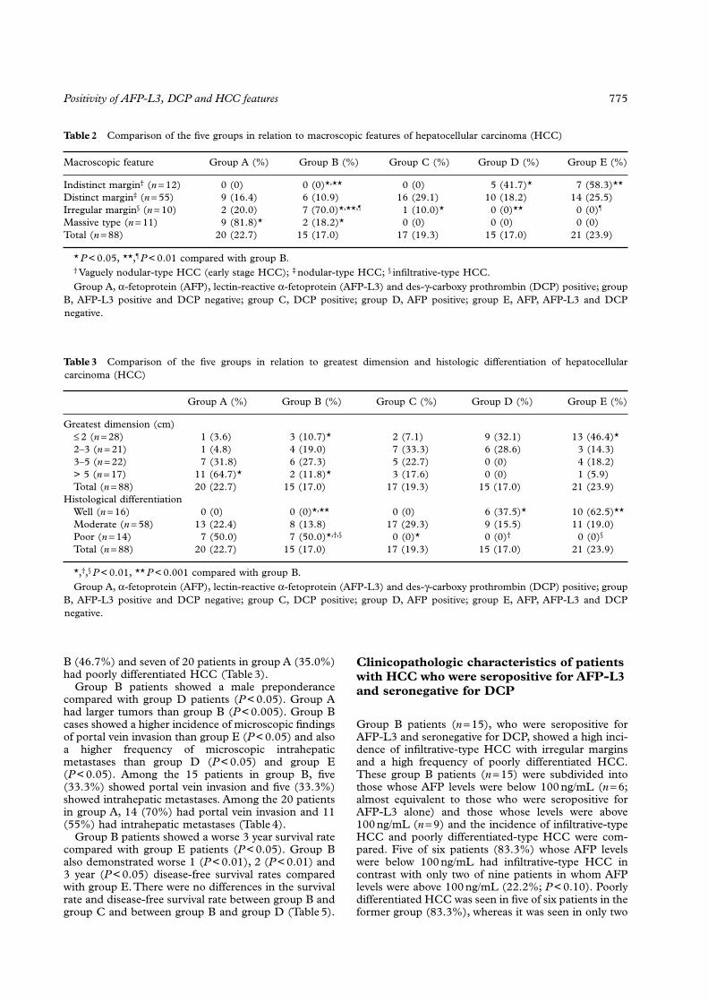
< 0.05). Seven of the15 patients in group B (46.7%) showed infiltrative-typeHCC with an irregular margin and nine of 20 patientsin group A (45.0%) showed massive-type HCC(Table 2).
Group B showed a lower incidence of small tumorsless than 2 cm in their greatest dimension comparedwith group E (
P
< 0.01). Group A patients had a higherfrequency of large tumors more than 5 cm than that ingroup B (
P
< 0.01). Eleven of 20 patients in group A(55.0%) showed tumors larger than 5 cm (Table 3).
All 16 cases of well-differentiated HCC belonged toeither group D (
n
=
6) or group E (
n
=
10). None of thegroup B patients had well-differentiated HCC andgroup B patients showed a lower incidence of well-differentiated HCC compared with both group D(
P
< 0.01) and group E (
P
< 0.001) patients. In con-trast, group B had a higher frequency of poorly differ-entiated HCC than group C (
P
< 0.01), group D(
P
< 0.01) and group E (
P
< 0.01), thus showing char-acteristics of more advanced HCC. All 14 cases ofpoorly differentiated HCC belonged to either group B(
n
=
7) or group A (
n
=
7). Seven of 15 patients in group
Positivity of AFP-L3, DCP and HCC features
775
B (46.7%) and seven of 20 patients in group A (35.0%)had poorly differentiated HCC (Table 3).
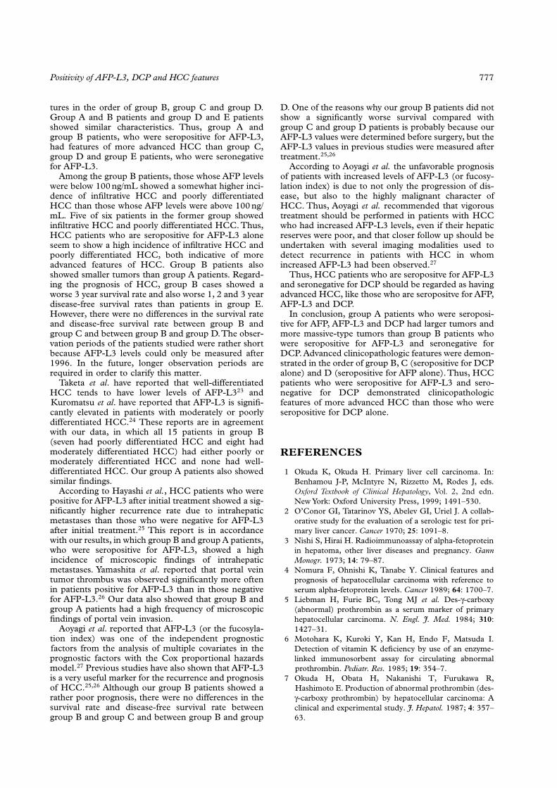
Group B patients showed a male preponderancecompared with group D patients (
P
< 0.05). Group Ahad larger tumors than group B (
P < 0.005). Group Bcases showed a higher incidence of microscopic findingsof portal vein invasion than group E (P < 0.05) and alsoa higher frequency of microscopic intrahepaticmetastases than group D (P < 0.05) and group E(P < 0.05). Among the 15 patients in group B, five(33.3%) showed portal vein invasion and five (33.3%)showed intrahepatic metastases. Among the 20 patientsin group A, 14 (70%) had portal vein invasion and 11(55%) had intrahepatic metastases (Table 4).
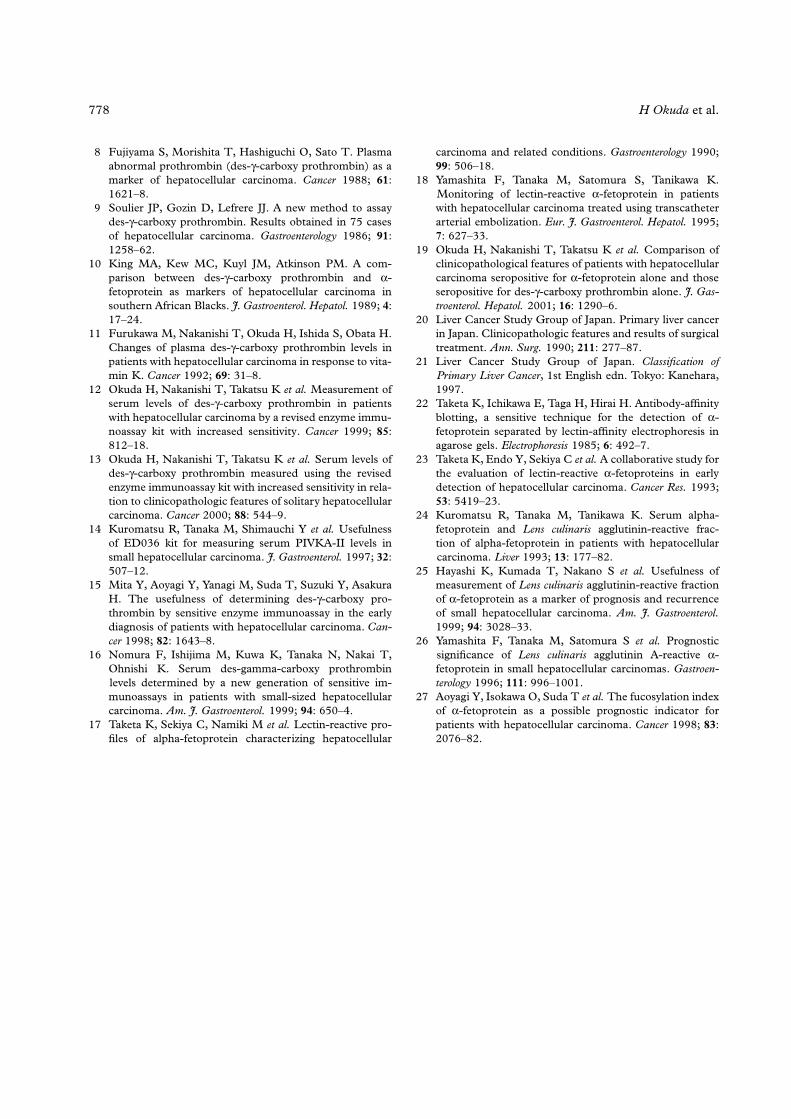
Group B patients showed a worse 3 year survival ratecompared with group E patients (P < 0.05). Group Balso demonstrated worse 1 (P < 0.01), 2 (P < 0.01) and3 year (P < 0.05) disease-free survival rates comparedwith group E. There were no differences in the survivalrate and disease-free survival rate between group B andgroup C and between group B and group D (Table 5).
Clinicopathologic characteristics of patients with HCC who were seropositive for AFP-L3 and seronegative for DCP
Group B patients (n = 15), who were seropositive forAFP-L3 and seronegative for DCP, showed a high inci-dence of infiltrative-type HCC with irregular marginsand a high frequency of poorly differentiated HCC.These group B patients (n = 15) were subdivided intothose whose AFP levels were below 100 ng/mL (n = 6;almost equivalent to those who were seropositive forAFP-L3 alone) and those whose levels were above100 ng/mL (n = 9) and the incidence of infiltrative-typeHCC and poorly differentiated-type HCC were com-pared. Five of six patients (83.3%) whose AFP levelswere below 100 ng/mL had infiltrative-type HCC incontrast with only two of nine patients in whom AFPlevels were above 100 ng/mL (22.2%; P < 0.10). Poorlydifferentiated HCC was seen in five of six patients in theformer group (83.3%), whereas it was seen in only two
Table 2 Comparison of the five groups in relation to macroscopic features of hepatocellular carcinoma (HCC)
Macroscopic feature Group A (%) Group B (%) Group C (%) Group D (%) Group E (%)
Indistinct margin† (n = 12) 0 (0) 0 (0)*,** 0 (0) 5 (41.7)* 7 (58.3)**Distinct margin‡ (n = 55) 9 (16.4) 6 (10.9) 16 (29.1) 10 (18.2) 14 (25.5)Irregular margin§ (n = 10) 2 (20.0) 7 (70.0)*,**,¶ 1 (10.0)* 0 (0)** 0 (0)¶
Massive type (n = 11) 9 (81.8)* 2 (18.2)* 0 (0) 0 (0) 0 (0)Total (n = 88) 20 (22.7) 15 (17.0) 17 (19.3) 15 (17.0) 21 (23.9)
* P < 0.05, **,¶ P < 0.01 compared with group B.† Vaguely nodular-type HCC (early stage HCC); ‡ nodular-type HCC; § infiltrative-type HCC.Group A, α-fetoprotein (AFP), lectin-reactive α-fetoprotein (AFP-L3) and des-γ-carboxy prothrombin (DCP) positive; group
B, AFP-L3 positive and DCP negative; group C, DCP positive; group D, AFP positive; group E, AFP, AFP-L3 and DCPnegative.
Table 3 Comparison of the five groups in relation to greatest dimension and histologic differentiation of hepatocellularcarcinoma (HCC)
Group A (%) Group B (%) Group C (%) Group D (%) Group E (%)
Greatest dimension (cm)≤ 2 (n = 28) 1 (3.6) 3 (10.7)* 2 (7.1) 9 (32.1) 13 (46.4)*2–3 (n = 21) 1 (4.8) 4 (19.0) 7 (33.3) 6 (28.6) 3 (14.3)3–5 (n = 22) 7 (31.8) 6 (27.3) 5 (22.7) 0 (0) 4 (18.2)> 5 (n = 17) 11 (64.7)* 2 (11.8)* 3 (17.6) 0 (0) 1 (5.9)Total (n = 88) 20 (22.7) 15 (17.0) 17 (19.3) 15 (17.0) 21 (23.9)
Histological differentiationWell (n = 16) 0 (0) 0 (0)*,** 0 (0) 6 (37.5)* 10 (62.5)**Moderate (n = 58) 13 (22.4) 8 (13.8) 17 (29.3) 9 (15.5) 11 (19.0)Poor (n = 14) 7 (50.0) 7 (50.0)*,†,§ 0 (0)* 0 (0)† 0 (0)§
Total (n = 88) 20 (22.7) 15 (17.0) 17 (19.3) 15 (17.0) 21 (23.9)
*,†,§ P < 0.01, ** P < 0.001 compared with group B.Group A, α-fetoprotein (AFP), lectin-reactive α-fetoprotein (AFP-L3) and des-γ-carboxy prothrombin (DCP) positive; group
B, AFP-L3 positive and DCP negative; group C, DCP positive; group D, AFP positive; group E, AFP, AFP-L3 and DCPnegative.
776 H Okuda et al.
of nine patients in the latter group (22.2%; P < 0.10).Eight of 15 patients (53.3%) had two HCC nodules inthis group. One showed sclerosing-type HCC and twoshowed sarcomatous changes, findings that were notobserved in the other four groups. Six of 15 cases(40.0%) were positive for HBsAg.
DISCUSSION
The present study demonstrated that HCC patientswho were seropositive for AFP-L3 and seronegative forDCP showed a higher incidence of infiltrative HCCwith an irregular margin and a higher frequency ofpoorly differentiated HCC than those in HCC patientswho were seropositive for DCP alone. Thus, the formergroup showed clinicopathologic features of moreadvanced HCC than those in the latter group.
There have been no studies of the clinicopathologicfeatures of HCC patients who are seropositive for AFP-L3 alone or those who are seropositive for AFP-L3 andseronegative for DCP in comparison with those who areseropositive for DCP alone. Thus, the present compar-ative study was undertaken.
We divided our surgically resected HCC patients,who had either one or two tumors, into five groupsaccording to the seropositivity of AFP, AFP-L3 andDCP. The present study showed that group B cases,who were seropositive for AFP-L3 and seronegative forDCP, showed a higher frequency of infiltrative-typeHCC with an irregular margin and a higher incidence ofpoorly differentiated HCC than those in group C, whowere seropositive for DCP alone, showing features ofmore advanced HCC. The reason why we did notinclude patients who were seropositive for AFP andDCP and seronegative for AFP-L3 or those who wereseropositive for AFP-L3 and DCP and seronegative forAFP (there are very few of such cases) was because it isdifficult to analyze the clinicopathologic features ofpatients who are seropositive for two of the threemarkers.
Group A patients, who were seropositive for AFP,AFP-L3 and DCP, had larger tumors and more mas-sive-type tumors than group B patients.
However, because the number of cases in each groupis rather small, a larger number of cases needs to bestudied to confirm our findings.
Our surgical HCC cases, having either one or twotumors, demonstrated advanced clinicopathologic fea-
Table 4 Comparison of clinicopathologic variables among the five groups with hepatocellular carcinoma
Group A (n = 20) Group B (n = 15) Group C (n = 17) Group D (n = 15) Group E (n = 21)
Mean (± SD) age (years) 61.4 ± 9.0 60.6 ± 8.8 65.5 ± 7.0 65.4 ± 5.7 64.9 ± 5.2No. males/females 15/5 13/2* 15/2 6/9* 10/11Mean (±SD) nodular size
(mm)78.9 ± 49.1** 36.3 ± 19.3** 35.5 ± 15.4 21.3 ± 4.5 24.1 ± 17.6
Portal vein invasion (%) 14 (70.0) 5 (33.3)* 4 (23.5) 1 (16.7) 0 (0)*Intrahepatic metastases (%) 11 (55.0) 5 (33.3)*,§ 2 (11.8) 0 (0)* 0 (0)§
*,§ P < 0.05, ** P < 0.005 compared with group B.Group A, α-fetoprotein (AFP), lectin-reactive α-fetoprotein (AFP-L3) and des-γ-carboxy prothrombin (DCP) positive; group
B, AFP-L3 positive and DCP negative; group C, DCP positive; group D, AFP positive; group E, AFP, AFP-L3 and DCPnegative.
Table 5 Comparison of survival rates and disease-free survival rates among the five groups with hepatocellular carcinoma
Group A (n = 20) Group B (n = 15) Group C (n = 17) Group D (n = 15) Group E (n = 21)
Survival rates (%)1 year 10/19 (52.6) 11/14 (78.6) 14/16 (87.5) 13/13 (100) 21/21 (100)2 year 3/12 (25.0) 8/13 (61.5) 8/12 (66.7) 9/9 (100) 17/18 (94.4)3 year 1/10 (10.0) 4/10 (40.0)* 4/9 (44.4) 2/3 (66.7) 10/11 (90.9)*
Disease-free survival rates (%)1 year 3/19 (15.8) 6/14 (42.9)** 9/16 (56.3) 9/13 (69.2) 19/21 (90.5)**2 year 1/12 (8.3) 4/13 (30.8)** 5/12 (41.7) 4/9 (44.4) 15/18 (83.3)**3 year 0/10 (0) 1/10 (10.0)* 2/9 (22.2) 0/3 (0) 8/11 (72.7)*
* P < 0.05, ** P < 0.01 compared with group B.Group A, α-fetoprotein (AFP), lectin-reactive α-fetoprotein (AFP-L3) and des-γ-carboxy prothrombin (DCP) positive; group
B, AFP-L3 positive and DCP negative; group C, DCP positive; group D, AFP positive; group E, AFP, AFP-L3 and DCPnegative.
Positivity of AFP-L3, DCP and HCC features 777
tures in the order of group B, group C and group D.Group A and B patients and group D and E patientsshowed similar characteristics. Thus, group A andgroup B patients, who were seropositive for AFP-L3,had features of more advanced HCC than group C,group D and group E patients, who were seronegativefor AFP-L3.
Among the group B patients, those whose AFP levelswere below 100 ng/mL showed a somewhat higher inci-dence of infiltrative HCC and poorly differentiatedHCC than those whose AFP levels were above 100 ng/mL. Five of six patients in the former group showedinfiltrative HCC and poorly differentiated HCC. Thus,HCC patients who are seropositive for AFP-L3 aloneseem to show a high incidence of infiltrative HCC andpoorly differentiated HCC, both indicative of moreadvanced features of HCC. Group B patients alsoshowed smaller tumors than group A patients. Regard-ing the prognosis of HCC, group B cases showed aworse 3 year survival rate and also worse 1, 2 and 3 yeardisease-free survival rates than patients in group E.However, there were no differences in the survival rateand disease-free survival rate between group B andgroup C and between group B and group D. The obser-vation periods of the patients studied were rather shortbecause AFP-L3 levels could only be measured after1996. In the future, longer observation periods arerequired in order to clarify this matter.
Taketa et al. have reported that well-differentiatedHCC tends to have lower levels of AFP-L323 andKuromatsu et al. have reported that AFP-L3 is signifi-cantly elevated in patients with moderately or poorlydifferentiated HCC.24 These reports are in agreementwith our data, in which all 15 patients in group B(seven had poorly differentiated HCC and eight hadmoderately differentiated HCC) had either poorly ormoderately differentiated HCC and none had well-differentiated HCC. Our group A patients also showedsimilar findings.
According to Hayashi et al., HCC patients who werepositive for AFP-L3 after initial treatment showed a sig-nificantly higher recurrence rate due to intrahepaticmetastases than those who were negative for AFP-L3after initial treatment.25 This report is in accordancewith our results, in which group B and group A patients,who were seropositive for AFP-L3, showed a highincidence of microscopic findings of intrahepaticmetastases. Yamashita et al. reported that portal veintumor thrombus was observed significantly more oftenin patients positive for AFP-L3 than in those negativefor AFP-L3.26 Our data also showed that group B andgroup A patients had a high frequency of microscopicfindings of portal vein invasion.
Aoyagi et al. reported that AFP-L3 (or the fucosyla-tion index) was one of the independent prognosticfactors from the analysis of multiple covariates in theprognostic factors with the Cox proportional hazardsmodel.27 Previous studies have also shown that AFP-L3is a very useful marker for the recurrence and prognosisof HCC.25,26 Although our group B patients showed arather poor prognosis, there were no differences in thesurvival rate and disease-free survival rate betweengroup B and group C and between group B and group
D. One of the reasons why our group B patients did notshow a significantly worse survival compared withgroup C and group D patients is probably because ourAFP-L3 values were determined before surgery, but theAFP-L3 values in previous studies were measured aftertreatment.25,26
According to Aoyagi et al. the unfavorable prognosisof patients with increased levels of AFP-L3 (or fucosy-lation index) is due to not only the progression of dis-ease, but also to the highly malignant character ofHCC. Thus, Aoyagi et al. recommended that vigoroustreatment should be performed in patients with HCCwho had increased AFP-L3 levels, even if their hepaticreserves were poor, and that closer follow up should beundertaken with several imaging modalities used todetect recurrence in patients with HCC in whomincreased AFP-L3 had been observed.27
Thus, HCC patients who are seropositve for AFP-L3and seronegative for DCP should be regarded as havingadvanced HCC, like those who are seropositve for AFP,AFP-L3 and DCP.
In conclusion, group A patients who were seroposi-tive for AFP, AFP-L3 and DCP had larger tumors andmore massive-type tumors than group B patients whowere seropositive for AFP-L3 and seronegative forDCP. Advanced clinicopathologic features were demon-strated in the order of group B, C (seropositive for DCPalone) and D (seropositive for AFP alone). Thus, HCCpatients who were seropositive for AFP-L3 and sero-negative for DCP demonstrated clinicopathologicfeatures of more advanced HCC than those who wereseropositive for DCP alone.
REFERENCES
1 Okuda K, Okuda H. Primary liver cell carcinoma. In:Benhamou J-P, McIntyre N, Rizzetto M, Rodes J, eds.Oxford Textbook of Clinical Hepatology, Vol. 2, 2nd edn.New York: Oxford University Press, 1999; 1491–530.
2 O’Conor GI, Tatarinov YS, Abelev GI, Uriel J. A collab-orative study for the evaluation of a serologic test for pri-mary liver cancer. Cancer 1970; 25: 1091–8.
3 Nishi S, Hirai H. Radioimmunoassay of alpha-fetoproteinin hepatoma, other liver diseases and pregnancy. GannMonogr. 1973; 14: 79–87.
4 Nomura F, Ohnishi K, Tanabe Y. Clinical features andprognosis of hepatocellular carcinoma with reference toserum alpha-fetoprotein levels. Cancer 1989; 64: 1700–7.
5 Liebman H, Furie BC, Tong MJ et al. Des-γ-carboxy(abnormal) prothrombin as a serum marker of primaryhepatocellular carcinoma. N. Engl. J. Med. 1984; 310:1427–31.
6 Motohara K, Kuroki Y, Kan H, Endo F, Matsuda I.Detection of vitamin K deficiency by use of an enzyme-linked immunosorbent assay for circulating abnormalprothrombin. Pediatr. Res. 1985; 19: 354–7.
7 Okuda H, Obata H, Nakanishi T, Furukawa R,Hashimoto E. Production of abnormal prothrombin (des-γ-carboxy prothrombin) by hepatocellular carcinoma: Aclinical and experimental study. J. Hepatol. 1987; 4: 357–63.
778 H Okuda et al.
8 Fujiyama S, Morishita T, Hashiguchi O, Sato T. Plasmaabnormal prothrombin (des-γ-carboxy prothrombin) as amarker of hepatocellular carcinoma. Cancer 1988; 61:1621–8.
9 Soulier JP, Gozin D, Lefrere JJ. A new method to assaydes-γ-carboxy prothrombin. Results obtained in 75 casesof hepatocellular carcinoma. Gastroenterology 1986; 91:1258–62.
10 King MA, Kew MC, Kuyl JM, Atkinson PM. A com-parison between des-γ-carboxy prothrombin and α-fetoprotein as markers of hepatocellular carcinoma insouthern African Blacks. J. Gastroenterol. Hepatol. 1989; 4:17–24.
11 Furukawa M, Nakanishi T, Okuda H, Ishida S, Obata H.Changes of plasma des-γ-carboxy prothrombin levels inpatients with hepatocellular carcinoma in response to vita-min K. Cancer 1992; 69: 31–8.
12 Okuda H, Nakanishi T, Takatsu K et al. Measurement ofserum levels of des-γ-carboxy prothrombin in patientswith hepatocellular carcinoma by a revised enzyme immu-noassay kit with increased sensitivity. Cancer 1999; 85:812–18.
13 Okuda H, Nakanishi T, Takatsu K et al. Serum levels ofdes-γ-carboxy prothrombin measured using the revisedenzyme immunoassay kit with increased sensitivity in rela-tion to clinicopathologic features of solitary hepatocellularcarcinoma. Cancer 2000; 88: 544–9.
14 Kuromatsu R, Tanaka M, Shimauchi Y et al. Usefulnessof ED036 kit for measuring serum PIVKA-II levels insmall hepatocellular carcinoma. J. Gastroenterol. 1997; 32:507–12.
15 Mita Y, Aoyagi Y, Yanagi M, Suda T, Suzuki Y, AsakuraH. The usefulness of determining des-γ-carboxy pro-thrombin by sensitive enzyme immunoassay in the earlydiagnosis of patients with hepatocellular carcinoma. Can-cer 1998; 82: 1643–8.
16 Nomura F, Ishijima M, Kuwa K, Tanaka N, Nakai T,Ohnishi K. Serum des-gamma-carboxy prothrombinlevels determined by a new generation of sensitive im-munoassays in patients with small-sized hepatocellularcarcinoma. Am. J. Gastroenterol. 1999; 94: 650–4.
17 Taketa K, Sekiya C, Namiki M et al. Lectin-reactive pro-files of alpha-fetoprotein characterizing hepatocellular
carcinoma and related conditions. Gastroenterology 1990;99: 506–18.
18 Yamashita F, Tanaka M, Satomura S, Tanikawa K.Monitoring of lectin-reactive α-fetoprotein in patientswith hepatocellular carcinoma treated using transcatheterarterial embolization. Eur. J. Gastroenterol. Hepatol. 1995;7: 627–33.
19 Okuda H, Nakanishi T, Takatsu K et al. Comparison ofclinicopathological features of patients with hepatocellularcarcinoma seropositive for α-fetoprotein alone and thoseseropositive for des-γ-carboxy prothrombin alone. J. Gas-troenterol. Hepatol. 2001; 16: 1290–6.
20 Liver Cancer Study Group of Japan. Primary liver cancerin Japan. Clinicopathologic features and results of surgicaltreatment. Ann. Surg. 1990; 211: 277–87.
21 Liver Cancer Study Group of Japan. Classification ofPrimary Liver Cancer, 1st English edn. Tokyo: Kanehara,1997.
22 Taketa K, Ichikawa E, Taga H, Hirai H. Antibody-affinityblotting, a sensitive technique for the detection of α-fetoprotein separated by lectin-affinity electrophoresis inagarose gels. Electrophoresis 1985; 6: 492–7.
23 Taketa K, Endo Y, Sekiya C et al. A collaborative study forthe evaluation of lectin-reactive α-fetoproteins in earlydetection of hepatocellular carcinoma. Cancer Res. 1993;53: 5419–23.
24 Kuromatsu R, Tanaka M, Tanikawa K. Serum alpha-fetoprotein and Lens culinaris agglutinin-reactive frac-tion of alpha-fetoprotein in patients with hepatocellularcarcinoma. Liver 1993; 13: 177–82.
25 Hayashi K, Kumada T, Nakano S et al. Usefulness ofmeasurement of Lens culinaris agglutinin-reactive fractionof α-fetoprotein as a marker of prognosis and recurrenceof small hepatocellular carcinoma. Am. J. Gastroenterol.1999; 94: 3028–33.
26 Yamashita F, Tanaka M, Satomura S et al. Prognosticsignificance of Lens culinaris agglutinin A-reactive α-fetoprotein in small hepatocellular carcinomas. Gastroen-terology 1996; 111: 996–1001.
27 Aoyagi Y, Isokawa O, Suda T et al. The fucosylation indexof α-fetoprotein as a possible prognostic indicator forpatients with hepatocellular carcinoma. Cancer 1998; 83:2076–82.


![Leprosy presenting as remitting seronegative symmetrical ......years [3]. Transmission, still not fully understood, ap-pears to occur by skin-to-skin contact or nasal secre-tions/aerosols](https://img.pdfslide.tips/doc/110x75/60e0847ba925be044c1e3f5d/leprosy-presenting-as-remitting-seronegative-symmetrical-years-3-transmission.jpg)