Embed Size (px)
Citation preview
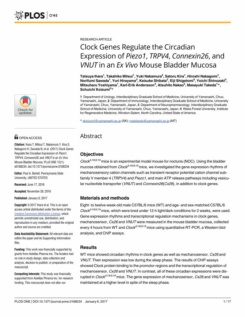
RESEARCH ARTICLE
Clock Genes Regulate the Circadian
Expression of Piezo1, TRPV4, Connexin26, and
VNUT in an Ex Vivo Mouse Bladder Mucosa
Tatsuya Ihara1, Takahiko Mitsui1, Yuki Nakamura2, Satoru Kira1, Hiroshi Nakagomi1,
Norifumi Sawada1, Yuri Hirayama3, Keisuke Shibata3, Eiji Shigetomi3, Yoichi Shinozaki3,
Mitsuharu Yoshiyama1, Karl-Erik Andersson4, Atsuhito Nakao2, Masayuki Takeda1*,
Schuichi Koizumi3*
1 Department of Urology, Interdisciplinary Graduate School of Medicine, University of Yamanashi, Chuo,
Yamanashi, Japan, 2 Department of Immunology, Interdisciplinary Graduate School of Medicine, University
of Yamanashi, Chuo, Yamanashi, Japan, 3 Department of Neuropharmacology, Interdisciplinary Graduate
School of Medicine, University of Yamanashi, Chuo, Yamanashi, Japan, 4 Wake Forest University, Institute
for Regenerative Medicine, Winston-Salem, North Carolina, United State of America
* [email protected] (SK); [email protected] (MT)
Abstract
Objectives
ClockΔ19/Δ19 mice is an experimental model mouse for nocturia (NOC). Using the bladder
mucosa obtained from ClockΔ19/Δ19 mice, we investigated the gene expression rhythms of
mechanosensory cation channels such as transient receptor potential cation channel sub-
family V member 4 (TRPV4) and Piezo1, and main ATP release pathways including vesicu-
lar nucleotide transporter (VNUT) and Connexin26(Cx26), in addition to clock genes.
Materials and methods
Eight- to twelve-week-old male C57BL/6 mice (WT) and age- and sex-matched C57BL/6
ClockΔ19/Δ19 mice, which were bred under 12-h light/dark conditions for 2 weeks, were used.
Gene expression rhythms and transcriptional regulation mechanisms in clock genes,
mechanosensor, Cx26 and VNUT were measured in the mouse bladder mucosa, collected
every 4 hours from WT and ClockΔ19/Δ19 mice using quantitative RT-PCR, a Western blot
analysis, and ChIP assays.
Results
WT mice showed circadian rhythms in clock genes as well as mechanosensor, Cx26 and
VNUT. Their expression was low during the sleep phase. The results of ChIP assays
showed Clock protein binding to the promotor regions and the transcriptional regulation of
mechanosensor, Cx26 and VNUT. In contrast, all of these circadian expressions were dis-
rupted in ClockΔ19/Δ19 mice. The gene expression of mechanosensor, Cx26 and VNUT was
maintained at a higher level in spite of the sleep phase.
PLOS ONE | DOI:10.1371/journal.pone.0168234 January 6, 2017 1 / 17
a1111111111
a1111111111
a1111111111
a1111111111
a1111111111
OPENACCESS
Citation: Ihara T, Mitsui T, Nakamura Y, Kira S,
Nakagomi H, Sawada N, et al. (2017) Clock Genes
Regulate the Circadian Expression of Piezo1,
TRPV4, Connexin26, and VNUT in an Ex Vivo
Mouse Bladder Mucosa. PLoS ONE 12(1):
e0168234. doi:10.1371/journal.pone.0168234
Editor: Paul A. Bartell, Pennsylvania State
University, UNITED STATES
Received: June 17, 2016
Accepted: November 28, 2016
Published: January 6, 2017
Copyright: © 2017 Ihara et al. This is an open
access article distributed under the terms of the
Creative Commons Attribution License, which
permits unrestricted use, distribution, and
reproduction in any medium, provided the original
author and source are credited.
Data Availability Statement: All relevant data are
within the paper and its Supporting Information
files.
Funding: This work was financially supported by
grants from Astellas Pharma Inc. The funders had
no role in study design, data collection and
analysis, decision to publish, or preparation of the
manuscript.
Competing Interests: This study was financially
supported from Astellas Phrama Inc. for research
funding. This manuscript does not alter our
Conclusions
Mechanosensor, Cx26 and VNUT expressed with circadian rhythm in the mouse bladder
mucosa. The disruption of circadian rhythms in these genes, induced by the abnormalities in
clock genes, may be factors contributing to NOC because of hypersensitivity to bladder wall
extension.
Introduction
The products of clock genes act as transcription factors, are expressed in most cells and organs.
The products of clock genes produce oscillations in sleep-awake rhythms and the gene expres-
sion of various metabolic enzymes, channels, and receptors with circadian rhythms. Among
more than 10 types of clock genes that have been identified to date, Clock and Bmal1 protein
dimers bind to E-box enhancer elements, which exist in the promoter sequence of target genes
such as Per, Cry, and Clock, and clock-controlled genes, and then activate the transcription of
these genes. The translational products of Per and Cry inhibit Clock and Bmal1 protein dimers.
As a result, the transcription of these genes is down-regulated. These translational cycles are
known as the ‘core loop’. Other feedback loops are known as ‘sub loops’, which are created by
other clock genes: e.g. the products of Dbp and E4bp4 bind to the D-box site while those of
RORα and Rev-erbα bind to the RORE site. Exact circadian gene expression is driven by the
formation of a large number of complex feedback loops under the control of the master clock
in the suprachiasmatic nucleus (SCN) [1].
Nocturia (NOC) is exceedingly common general complain. It is defined as the waking up at
night one or more times to void [2], which is reported that nearly 90% of the elderly men are
suffering from NOC [3]. With the prevalence of NOC, it raise the various risk such as sleeping
disorder, mental health, bone fracture by fall and reduce the life span [4–7]. Many diseases
cause NOC, and the palliative treatments depending on the causes, such as α1-blockers for
benign prostatic hyperplasia, anti-cholinergic drugs or β3-agonists for an overactive bladder,
desmopressin for nocturnal polyuria, anti-hypertensive drugs and diuretic medications for
high blood pressure patients, are provided in clinical settings [8]. However, because the patho-
physiologies of NOC are multifactorial and remain unclear in a large number of patients, these
treatments are often not so effective and become refractory. So far, there was no ideal animal
in order to examine the complicated pathophysiology of NOC.
We previously reported that Clock mutant mice showed the phenotype of NOC [9]. Fur-
thermore, other groups found that that urine products and lower urinary tract function are
regulated by clock genes [10, 11]. These findings provided us with a concern regarding the
relationship between abnormalities in clock genes and lower urinary tract symptoms (LUTS).
On the other hand, the bladder mucosa senses bladder extension and transmits signals to affer-
ent nerves by releasing neurotransmitters, including adenosine triphosphate (ATP) [12–15].
Bladder extension is sensed by mechanosensor such as transient receptor potential cation
channel subfamily V member 4 (TRPV4) and Piezo1 via intracellular Ca2+ influx [16–18],
which triggers ATP release from the bladder urothelium. Although many pathways are
involved in the release of ATP, we identified the vesicular nucleotide transporter (VNUT), that
mediates exocytosis [19] and conductive release connexin- or pannexin-hemichannels that
mediate diffusible ATP release in the mucosa [20–25], particularly Connexin26 (Cx26) [26], as
a main ATP release pathway in the bladder mucosa.
The Circadian Gene Expression in the Mouse Bladder Mucosa
PLOS ONE | DOI:10.1371/journal.pone.0168234 January 6, 2017 2 / 17
adherence to PLOS ONE policies on sharing data
and materials.
Abbreviations: ATP, adenosine triphosphate; ChIP,
chromatin immunoprecipitation; Clock mutant,
ClockΔ19/Δ19; CNS, central nervous system; Cx26,
Connexin26; LUTS, lower urinary tract symptoms;
NOC, nocturia; RT-PCR, reverse transcriptase
polymerase chain reaction; SE, standard error;
SCN, suprachiasmatic nucleus; TRPV4, transient
receptor potential cation channel subfamily V
member 4; VNUT, vesicular nucleotide transporter;
ZT, zeitgeber time.
Based on these findings, we hypothesize that the expression of mechanosensor such as
Piezo1 and TRPV4, and main ATP pathway such as Cx26 and VNUT, is regulated by clock
genes in the bladder mucosa, and these create a circadian rhythm for the sensation of bladder
fullness. Moreover, abnormalities in clock genes enforce hypersensitive to mechanosensory
stimuli upon the bladder mucosa during sleep because of a disruption in the circadian rhythms
of mechanosensor, Cx26 and VNUT.
In the present study, we investigated whether the gene expression of mechanosensor, Cx26and VNUT show circadian rhythms in the bladder mucosa, and also if they are regulated by
clock genes.
Materials and Methods
Animals
Eight- to twelve-week-old male C57BL/6 mice (WT; SLC, Shizuoka, Japan) and age- and sex-
matched C57BL/6 Clock mutant mice (ClockΔ19/Δ19; Jackson Laboratories, Bar Harbor, ME,
USA) were used in the following experiments. For genotyping of ClockΔ19/Δ19 mice, polymerase
chain reaction was performed using the following primers: WT forward,5’GGTCAAGGGCTACAGGTA-3’;common,5’TGGGGTAAAAAGACCTCTTGCC-3’;mutant forward,5’AGCACCTTCCTTTGCAGTTCG-3’; mutant reverse,5’TGTGCTCAGACAGAATAAGTA-3’.Mice were sacrificed by cervical dislocation after the anesthesia using sevoflurane to
minimize the animal suffering. ClockΔ19/Δ19 mice have an A to T mutation in the 5’ splice site
of intron 19, and, as a consequence, an in-frame deletion of the entire exon 19 (ClockΔ19/Δ19).
The product of ClockΔ19/Δ19 results in the loss of normal transcriptional activity as a transcrip-
tion factor [27].
All experiments were performed using these mice, which were bred under 12-h light/dark
conditions for 2 weeks. The light period started from 6 AM, which is zeitgeber time (ZT) 0,
and the dark period started from 6 PM, which is ZT 12.
All procedures were conducted in accordance with the Guiding Principles in the Care and
Use of Animals in the Field of the Physiological Society of Japan, all experiment were approved
by the Institutional Animal Care and Use Committee of the University of Yamanashi (Chuo,
Yamanashi, Japan).
Collection method for the mouse bladder mucosa
Mice were sacrificed every 4 hr from ZT0, and bladders were removed and everted. The inner
surface (mucosa) of the bladder was scraped with a knife in PBS (S1 Fig). Suspensions includ-
ing the mouse bladder mucosa were centrifuged at 1,000 rpm for 5 min, and cell pellets were
collected. All samples were lysed in Buffer RLT (Qiagen, Germany) to extract mRNA for the
quantitative real-time reverse transcription polymerase chain reaction (RT-PCR) and in
M-PER Mammalian Protein Extraction Reagent (Thermo Fisher Scientific, USA) to extract
protein for a Western Blotting analysis.
Histological examination of mouse bladder tissues
Frozen tissues embedded in OCT compound (Sakura Finetek Japan, Tokyo, Japan) were cut
into 7-μm-thick sections. Staining with standard hematoxylin and eosin (H&E) and Masson’s
trichrome was performed in order to compare morphological changes before or after scraping
of the bladder mucosa.
The Circadian Gene Expression in the Mouse Bladder Mucosa
PLOS ONE | DOI:10.1371/journal.pone.0168234 January 6, 2017 3 / 17
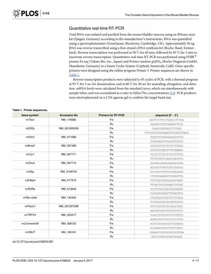
Quantitative real-time RT-PCR
Total RNA was isolated and purified from the mouse bladder mucosa using an RNeasy mini
kit (Qiagen, Germany) according to the manufacturer’s instructions. RNA was quantified
using a spectrophotometer (GeneQuant, Biochrom, Cambridge, UK). Approximately 50 ng
RNA was reverse transcribed using a first-strand cDNA synthesis kit (Roche, Basel, Switzer-
land). Reverse transcription was performed at 50˚C for 60 min, followed by 85˚C for 5 min to
inactivate reverse transcriptase. Quantitative real-time RT-PCR was performed using SYBR1
premix Ex taq (Takara Bio, Inc., Japan) and Primer random p(dN)6 (Roche Diagnosis GmbH,
Mannheim, Germany) in a Smart Cycler System (Cepheid, Sunnyvale, Calif). Gene-specific
primers were designed using the online program Primer 3. Primer sequences are shown in
Table 1.
Reverse transcription products were subjected to 45 cycles of PCR, with a thermal program
at 95˚C for 5 sec for denaturation, and at 60˚C for 20 sec for annealing, elongation, and detec-
tion. mRNA levels were calculated from the standard curve, which ran simultaneously with
sample tubes, and was normalized as a ratio to Eif2a/Tbcc concentrations [12]. PCR products
were electrophoresed on a 2.5% agarose gel to confirm the target band size.
Table 1. Primer sequences.
Gene symbol Accession No Primers for RT-PCR sequence (5’!3’)
mTbcc NM_178385 Fw GACTCCTTCCTGAACCTCTGG
Rv GGAGGCCATTCAAAACTTCA
mEif2a NM_001005509 Fw CAACGTGGCAGCCTTACA
Rv TTTCATGTCATAAAGTTGTAGGTTAGG
mPer2 NM_011066 Fw TGGTTTCTGGGAAGATCCTG
Rv CCACAAACTTGGCATCACTG
mBmal1 NM_007489 Fw CGCCGCTCTTTCTTCTGTAG
Rv GGTGGCCAGCTTTTCAAATA
mCry1 NM_007771 Fw TCCCCTCCCCTTTCTCTTTA
Rv TTCTTGTCCCAAGGGATCTG
mClock NM_007715 Fw GGTAACGCGAGAAAGATGGA
Rv AGCATCTGACTGTGCAGTGG
mDbp NM_0169745 Fw GCCCACTTGGTACAGAAGGA
Rv CTGCAGAAAGGTGCAACTCA
mE4bp4 NM_017373 Fw TTCTGATGGGGAAGACGAAC
Rv TTCACTTCCGGAACCTTCAC
mRORα NM_013646 Fw TCCTTCACCAACGGAGAGAC
Rv CCAGGTGGGATTTGGATATG
mRev-erbα NM_145434 Fw CTGCAGGCTGATTCTTCACA
Rv TCTTGGGGTGGCTATACTGC
mPiezo1 NM_001037298 Fw ATCCTGCTGTATGGGCTGAC
Rv AAGGGTAGCGTGTGTGTTCC
mTRPV4 NM_022017 Fw TCACCTTCGTGCTCCTGTTG
Rv AGATGTGCTTGCTCTCCTTG
mConnexin26 NM_008125 Fw ACTCCACCAGCATTGGAAAG
Rv ACAAAATCGGCTTGCTCATC
mVNUT NM_183161 Fw CAGAGTCATCACGGTGCGTAA
Rv GACCCAGACACAGGGCAAA
doi:10.1371/journal.pone.0168234.t001
The Circadian Gene Expression in the Mouse Bladder Mucosa
PLOS ONE | DOI:10.1371/journal.pone.0168234 January 6, 2017 4 / 17
Western blotting analysis
The protein concentration of each lysed sample by M-PER Mammalian Protein Extraction
Reagent (Thermo Fisher Scientific, USA) was measured using Pierce 660nM Protein Assay
Reagent (Thermo Fisher Scientific, USA). Samples were diluted to the reference concentration,
which was the lowest concentrated sample. Diluted lysates were then subjected to sodium
dodecyl sulfate-polyacrylamide gel electrophoresis (SDS-PAGE) on 7.5% gels using a Power
station 1000VC system (Atto, USA) at 20 mA for 90 min. Proteins were transferred to polyvi-
nylidene fluoride (PVDF) membranes using a Power Pac (Bio-Rad, USA) at 70 V for 120 min.
The transferred membrane was cut into several strips according to the product size of target
protein. The stripped membranes were blocked with 2% ECL prime Blocking Agent (GE Life
Sciences, Japan) at room temperature (RT) for 1 h and then washed 3 times with PBS indepen-
dently. The each membrane was incubated with the following first antibodies diluted with
Can Get Signal1 solution 1 (Toyobo, Japan) at 4˚C overnight: a rabbit anti-β-Actin antibody
(Santa Cruz; 1:5000), rabbit anti-Clock antibody (Cell Signaling Technology; 1: 800), rabbit
anti-Bmal1 antibody (Abcam; 1:800), rabbit anti-Piezo1 antibody (Novus; 1:500), rabbit anti-
TRPV4 antibody (Santa Cruz; 1:800), rabbit anti-Connexin26 antibody (Invitrogen; 1:100),
and rabbit anti-VNUT antibody (Millipore; 1:500). After washing 3 times, membranes were
incubated with horseradish peroxidase-conjugated anti-rabbit antibodies (1:6000; Amersham
Pharmacia Biotech Inc., Piscataway, NJ, USA) diluted with Can Get Signal1 solution 2
(Toyobo, Japan) at RT for 1 h. The proteins were visualized as bands by chemiluminescence
ECL select Western Blotting Detection Reagent (GE Life Sciences, Japan).
Chromatin immunoprecipitation assay (ChIP)
The ability of Clock to bind to the promoter regions of Piezo1, TRPV4, Cx26, and VNUT in the
mouse bladder mucosa was analyzed at different time points using ChIP assays. ChIP assays
were performed using a Simple ChIP1 plus enzymatic chromatin IP kit (Cell Signaling tech-
nology, USA) according to the manufacturer’s instructions. The mouse bladder mucosa was
obtained at ZT0 and ZT8 or ZT4 and ZT12 depending on the peak and nadir time in each
gene. Samples were cross-linked with formaldehyde solution (Sigma-Aldrich, USA) and lysed,
and chromatin was fragmented by partial digestion with Micrococcal Nuclease in order to
obtain chromatin fragments of 1 to 5 nucleosomes. ChIP assays were performed with ChIP-
Grade Protein G Agarose Beads, using a negative control rabbit IgG and rabbit anti-Clock
antibody (Cell Signaling Technology; 1: 100). After the reversal of protein-DNA cross-links,
DNA was purified. The amounts of Clock bound to target DNA were quantified in 50 cycles of
real-time PCR using primers and Taq Man probes using Taq Man1 Fast Advanced Master
Mix (Applied Biosystems, USA), with a thermal program of 95˚C for 15 sec for denaturation
and 60˚C for 60 sec for annealing, elongation, and detection in a Smart Cycler System (Cepheid,
Sunnyvale Calif). The ratio of a specific DNA fragment in each immunoprecipitated sample (IP
sample) to that fragment in DNA before immunoprecipitation (input DNA) was calculated
from each cycling threshold cycle of PCR reaction (C[T]) using the equation described in the
manufacturer’s instructions as Percent Input = 2%×2C[T] input DNA–C[T] IP sample. The primers and
Taq Man probes used were listed in Table 2.
Statistical analyses
Experimental values were expressed as means ± standard error (SE). The significance of differ-
ences between two groups was analyzed using Mann-Whitney’s U-test. A one-way ANOVA
was used to compare differences among the time points in each group. A two-way ANOVA
The Circadian Gene Expression in the Mouse Bladder Mucosa
PLOS ONE | DOI:10.1371/journal.pone.0168234 January 6, 2017 5 / 17
with Bonferroni’s test was used to compare differences at each time point between 2 groups. A
P value of less than 0.05 was considered significant.
Results
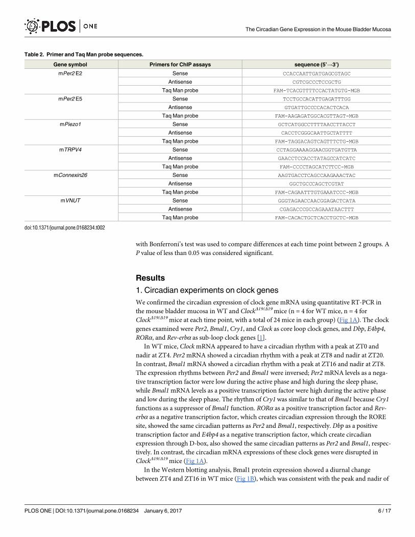
1. Circadian experiments on clock genes
We confirmed the circadian expression of clock gene mRNA using quantitative RT-PCR in
the mouse bladder mucosa in WT and ClockΔ19/Δ19 mice (n = 4 for WT mice, n = 4 for
ClockΔ19/Δ19 mice at each time point, with a total of 24 mice in each group) (Fig 1A). The clock
genes examined were Per2, Bmal1, Cry1, and Clock as core loop clock genes, and Dbp, E4bp4,
RORα, and Rev-erbα as sub-loop clock genes [1].
In WT mice, Clock mRNA appeared to have a circadian rhythm with a peak at ZT0 and
nadir at ZT4. Per2 mRNA showed a circadian rhythm with a peak at ZT8 and nadir at ZT20.
In contrast, Bmal1 mRNA showed a circadian rhythm with a peak at ZT16 and nadir at ZT8.
The expression rhythms between Per2 and Bmal1 were inversed; Per2 mRNA levels as a nega-
tive transcription factor were low during the active phase and high during the sleep phase,
while Bmal1 mRNA levels as a positive transcription factor were high during the active phase
and low during the sleep phase. The rhythm of Cry1 was similar to that of Bmal1 because Cry1functions as a suppressor of Bmal1 function. RORα as a positive transcription factor and Rev-erbα as a negative transcription factor, which creates circadian expression through the RORE
site, showed the same circadian patterns as Per2 and Bmal1, respectively. Dbp as a positive
transcription factor and E4bp4 as a negative transcription factor, which create circadian
expression through D-box, also showed the same circadian patterns as Per2 and Bmal1, respec-
tively. In contrast, the circadian mRNA expressions of these clock genes were disrupted in
ClockΔ19/Δ19 mice (Fig 1A).
In the Western blotting analysis, Bmal1 protein expression showed a diurnal change
between ZT4 and ZT16 in WT mice (Fig 1B), which was consistent with the peak and nadir of
Table 2. Primer and Taq Man probe sequences.
Gene symbol Primers for ChIP assays sequence (5’!3’)
mPer2 E2 Sense CCACCAATTGATGAGCGTAGC
Antisense CGTCGCCCTCCGCTG
Taq Man probe FAM-TCACGTTTTCCACTATGTG-MGB
mPer2 E5 Sense TCCTGCCACATTGAGATTTGG
Antisense GTGATTGCCCCACACTCACA
Taq Man probe FAM-AAGAGATGGCACGTTAGT-MGB
mPiezo1 Sense GCTCATGGCCTTTTAACCTTACCT
Antisense CACCTCGGGCAATTGCTATTTT
Taq Man probe FAM-TAGGACAGTCAGTTTCTG-MGB
mTRPV4 Sense CCTAGGAAAAGGAACGGTGATGTTA
Antisense GAACCTCCACCTATAGCCATCATC
Taq Man probe FAM-CCCCTAGCATCTTCC-MGB
mConnexin26 Sense AAGTGACCTCAGCCAAGAAACTAC
Antisense GGCTGCCCAGCTCGTAT
Taq Man probe FAM-CAGAATTTGTGAAATCCC-MGB
mVNUT Sense GGGTAGAACCAACGGAGACTCATA
Antisense CGAGACCCGCCAGAAATAACTTT
Taq Man probe FAM-CACACTGCTCACCTGCTC-MGB
doi:10.1371/journal.pone.0168234.t002
The Circadian Gene Expression in the Mouse Bladder Mucosa
PLOS ONE | DOI:10.1371/journal.pone.0168234 January 6, 2017 6 / 17
The Circadian Gene Expression in the Mouse Bladder Mucosa
PLOS ONE | DOI:10.1371/journal.pone.0168234 January 6, 2017 7 / 17
Bmal1 mRNA expression. In the case of the Clock protein, a change in band density was not
observed between ZT4 and ZT16 (Fig 1B), which was consistent with Clock mRNA expression.
In contrast, ClockΔ19/Δ19 mice showed the loss of a diurnal change in the Bmal1 protein, which
was consistent with Bmal1 mRNA expression in ClockΔ19/Δ19 mice (Fig 1B).
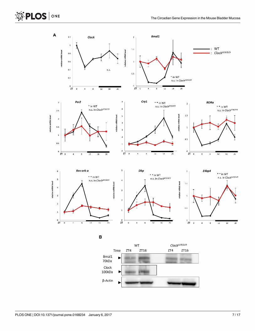
2. Circadian experiments on mechanosensor, Cx26 and VNUT
Subsequent to the circadian rhythms of clock genes, the circadian mRNA expression of
mechanosensor such as Piezo1 and TRPV4, and main ATP pathway including Cx26 and
VNUT was examined in the mouse bladder mucosa (n = 4 for WT mice, n = 4 for ClockΔ19/Δ19
mice at each time point, with a total of 24 mice in each group).
In WT mice, mRNA in Piezo1, TRPV4, Cx26, and VNUT showed circadian changes in
expression (Fig 2). Peaks were observed at ZT12 at the beginning of the active phase, and
nadirs were at ZT4 at the middle of the sleep phase. The peaks and nadirs of these genes were
slightly earlier than those of Per2 and Bmal1 in Fig 1A. In contrast, the mRNA expression of
Piezo1, TRPV4, Cx26, and VNUT lost circadian rhythms and was observed at the same level
between ZT0 and ZT20 in ClockΔ19/Δ19 mice (Fig 2). The absolute mRNA levels of these genes
were compared using unnormalized data (S2 Fig). The mRNA abundances between WT and
ClockΔ19/Δ19 mice during the sleep phase (ZT4 and ZT8) were significantly higher in ClockΔ19/Δ19
mice than in WT mice. In contrast, the absolute mRNA levels of these genes during the active
phase (ZT12) increased in WT mice. Significant differences were observed in TRPV4 and
VNUT between WT mice and ClockΔ19/Δ19 mice.
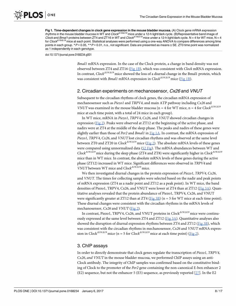
We then investigated diurnal changes in the protein expression of Piezo1, TRPV4, Cx26,
and VNUT. The times for collecting samples were selected based on the nadir and peak points
of mRNA expression (ZT4 as a nadir point and ZT12 as a peak point). In WT mice, the band
densities of Piezo1, TRPV4, Cx26, and VNUT were lower at ZT4 than at ZT12 (Fig 3A). Quan-
titative analyses revealed that the protein abundance of Piezo1, TRPV4, Cx26, and VNUT
were significantly greater at ZT12 than at ZT4 (Fig 3B) (n = 5 for WT mice at each time point).
These diurnal changes were consistent with the circadian rhythms in the mRNA levels of
mechanosensor, Cx26 and VNUT (Fig 2).
In contrast, Piezo1, TRPV4, Cx26, and VNUT proteins in ClockΔ19/Δ19 mice were continu-
ously expressed at the same level between ZT4 and ZT12 (Fig 3A). Quantitative analyses also
showed the disruption of diurnal expression rhythms between ZT4 and ZT12 (Fig 3B), which
was consistent with the circadian rhythms in mechanosensor, Cx26 and VNUT mRNA expres-
sion in ClockΔ19/Δ19 mice (n = 5 for ClockΔ19/Δ19 mice at each time point) (Fig 2).
3. ChIP assays
In order to directly demonstrate that clock genes regulate the transcription of Piezo1, TRPV4,
Cx26, and VNUT in the mouse bladder mucosa, we performed ChIP assays using an anti-
Clock antibody. The integrity of ChIP samples was confirmed based on the constitutive bind-
ing of Clock to the promoter of the Per2 gene containing the non-canonical E-box enhancer 2
(E2) sequence, but not the enhancer 5 (E5) sequence, as previously reported [27]. In the E2
Fig 1. Time-dependent changes in clock gene expression in the mouse bladder mucosa. (A) Clock gene mRNA expression
rhythms in the mouse bladder mucosa in WT and ClockΔ19/Δ19 mice under a 12-h light/dark cycle. (B)Representative band image of
Clock and Bmal1 proteins between ZT4 and ZT16 in WT and ClockΔ19/Δ19 mice under a 12-h light/dark cycle. N = 4 for WT mice, N = 4
for ClockΔ19/Δ19 mice at each point. Statistical analyses were performed using a one-way ANOVA to compare differences among time
points in each group. *P < 0.05, **P < 0.01, n.s., not significant. Data are presented as means ± SE. ZT0 time point was normalized
as 1 independently in each genotype.
doi:10.1371/journal.pone.0168234.g001
The Circadian Gene Expression in the Mouse Bladder Mucosa
PLOS ONE | DOI:10.1371/journal.pone.0168234 January 6, 2017 8 / 17
region, significant Clock binding was observed at ZT8, but not at ZT0, which was consistent
with the expression rhythms of Per2 mRNA. In contrast, no Clock binding was noted in the E5
region at either time point (n = 3 for the Control group and anti-Clock Ab group at each time
point) (S3 Fig).
Several E-box-like elements, to which the Clock/Bmal1 complex may theoretically bind, are
present in the promoter regions of Piezo1, TRPV4, Cx26, and VNUT (Fig 4B, 4D, 4F and 4H)
[27, 28]. The amount of precipitated chromatin fragments obtained using the anti-Clock anti-
body was significantly higher at ZT4 and ZT12 in Piezo1, Cx26, and VNUT than that using
control IgG. As for TRPV4, this significant difference was only observed at ZT12 (n = 9 for the
Control group and anti-Clock Ab group at each time point), (Fig 4A, 4C, 4E and 4G). A com-
parison of differences in precipitated chromatin fragments using the anti-Clock antibody
between ZT4 and ZT12 revealed significant changes in Cx26 and VNUT (p = 0.0007 and
0.0031, respectively, by Mann-Whitney’s U-test). Furthermore, Piezo1 and TRPV4 showed
slightly more Clock binding at ZT12 than at ZT4 (p = 0.070 and 0.102 by Mann-Whitney’s U-
test). These differences in the sensitivities of Clock binding to the E-box-like element appear to
Fig 2. Time-dependent changes in mechanosensor,Cx26 and VNUT mRNA expression in the mouse bladder mucosa. Mechanosensor,Cx26
and VNUT mRNA expression rhythms in the mouse bladder mucosa in WT and ClockΔ19/Δ19 mice under a 12-h light/dark cycle. N = 4 for WT mice,
N = 4 for ClockΔ19/Δ19 mice at each point. Statistical analyses were performed using a one-way ANOVA to compare differences among time points in
each group. *P < 0.05, **P < 0.01, n.s., not significant. Data are presented as means ± SE. ZT0 was normalized as 1 independently in each genotype.
Using S2 Fig, a two-way ANOVA and Bonferroni’s test was used to compare differences of absolute mRNA level between WT and ClockΔ19/Δ19 mice at
each time point (S2 Fig). # P < 0.05, ## P < 0.01.
doi:10.1371/journal.pone.0168234.g002
The Circadian Gene Expression in the Mouse Bladder Mucosa
PLOS ONE | DOI:10.1371/journal.pone.0168234 January 6, 2017 9 / 17
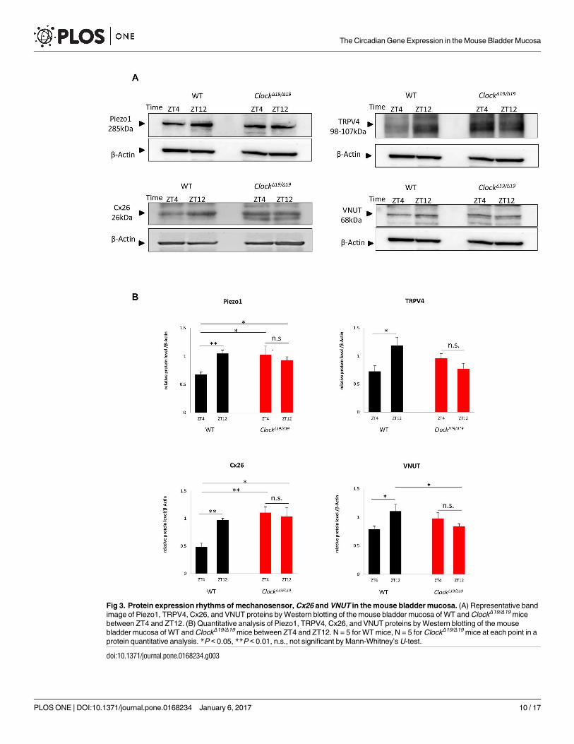
Fig 3. Protein expression rhythms of mechanosensor, Cx26 and VNUT in the mouse bladder mucosa. (A) Representative band
image of Piezo1, TRPV4, Cx26, and VNUT proteins by Western blotting of the mouse bladder mucosa of WT and ClockΔ19/Δ19 mice
between ZT4 and ZT12. (B) Quantitative analysis of Piezo1, TRPV4, Cx26, and VNUT proteins by Western blotting of the mouse
bladder mucosa of WT and ClockΔ19/Δ19 mice between ZT4 and ZT12. N = 5 for WT mice, N = 5 for ClockΔ19/Δ19 mice at each point in a
protein quantitative analysis. *P < 0.05, **P < 0.01, n.s., not significant by Mann-Whitney’s U-test.
doi:10.1371/journal.pone.0168234.g003
The Circadian Gene Expression in the Mouse Bladder Mucosa
PLOS ONE | DOI:10.1371/journal.pone.0168234 January 6, 2017 10 / 17
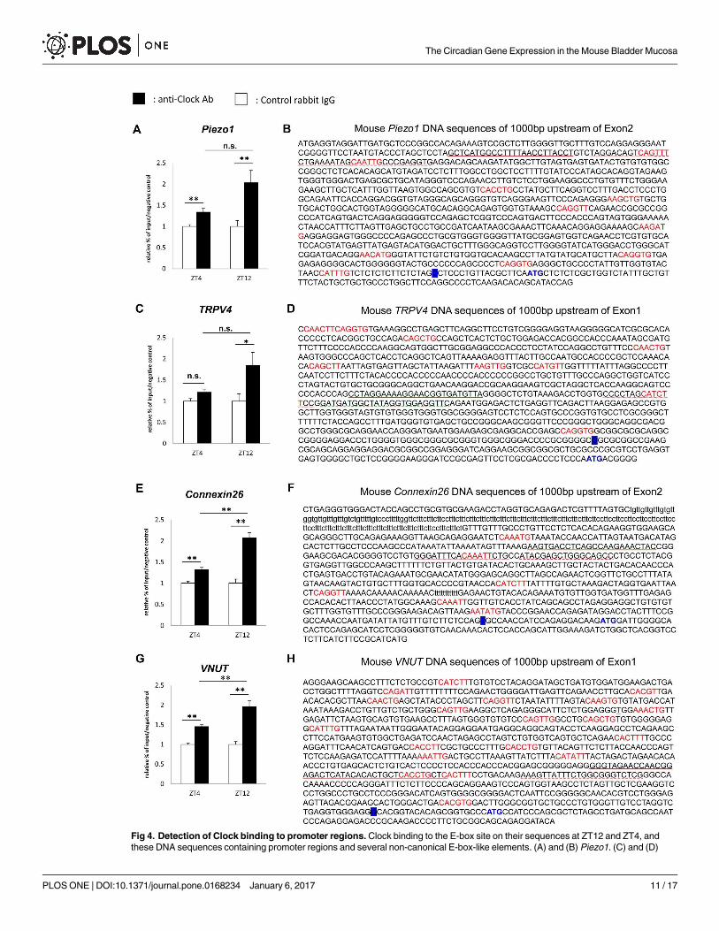
Fig 4. Detection of Clock binding to promoter regions. Clock binding to the E-box site on their sequences at ZT12 and ZT4, and
these DNA sequences containing promoter regions and several non-canonical E-box-like elements. (A) and (B) Piezo1. (C) and (D)
The Circadian Gene Expression in the Mouse Bladder Mucosa
PLOS ONE | DOI:10.1371/journal.pone.0168234 January 6, 2017 11 / 17
be consistent with the peaks and nadirs in the mRNA of Piezo1, TRPV4, Cx26, and VNUT(Fig 2B).
Discussion
We herein demonstrated that clock genes exist and create typical circadian expression profiles
in the bladder mucosa derived from WT mice. In addition, the expression rhythms of mechan-
osensor (Piezo1 and TRPV4), Cx26 and VNUT, which are involved in the sensation of bladder
fullness, also showed circadian rhythms associated with the circadian expression of clock
genes. These products in mechanosensor, Cx26 and VNUT increased during the active phase
and decreased during the sleep phase in WT mice. ChIP assays showed the transcriptional reg-
ulation of these genes by Clock binding at their promoter sequences. On the other hand,
ClockΔ19/Δ19 mice, a model mouse for NOC [9], showed disrupted circadian expression not
only in clock genes, but also in mechanosensor, Cx26 and VNUT. These results suggest that
the sensation of bladder fullness may change in a time-dependent manner in WT mice. In con-
trast, ClockΔ19/Δ19 mice may constantly sense bladder fullness between the sleep and active
phases due to the loss of the circadian expression of mechanosensor, Cx26 and VNUT.
Treatments for NOC are often ineffective clinically, and unknown causes have been sug-
gested to influence the incidence of NOC. We previously demonstrated that ClockΔ19/Δ19 mice
showed the NOC phenotype, and abnormalities in clock genes may be one of the causes of
NOC [9]. In order to investigate etiologies regarding a disruption in the circadian rhythm of
voiding behavior in ClockΔ19/Δ19 mice, we focused on the gene expression rhythms of clock
genes, mechanosensor, Cx26 and VNUT in the mouse bladder mucosa, in which mechanosen-
sor, Cx26 and VNUT were reported to sense bladder wall extension and transmit signals of
bladder fullness to the CNS [16–18, 20–26].
In WT mice, core loop and sub loop clock genes showed typical mRNA expression patterns
in the mouse bladder mucosa, as reported previously (Fig 1) [1]. The circadian expression of
all clock genes was abrogated in ClockΔ19/Δ19 mice. These results demonstrated that clock genes
exist in the mouse bladder mucosa and regulate exact circadian gene expression in WT mice.
In addition to clock genes, mechanosensor, Cx26 and VNUT mRNA are also expressed based
on circadian rhythms in WT mice. The peaks observed in the mRNA expression of mechano-
sensor, Cx26 and VNUT were consistent with the active phase, whereas the nadirs were con-
sistent with the sleep phase (Fig 2). In contrast, the circadian expression of all clock genes,
mechanosensor, Cx26 and VNUT observed in WT mice was disrupted in ClockΔ19/Δ19 mice
(Figs 1 and 2). Furthermore, the absolute mRNA of mechanosensor, Cx26 and VNUT in the
mouse bladder mucosa of ClockΔ19/Δ19 mice were maintained at significantly higher levels than
those in WT mice during the sleep phase (Fig 2 and S2 Fig). Taken all these results together,
ClockΔ19/Δ19 mice sense the sensation of bladder fullness more than WT mice during the sleep
phase, resulting in the NOC phenotype in ClockΔ19/Δ19 mice [9].
We performed a Western blot analysis to confirm the relationship between circadian
mRNA expression levels and the abundance of these products. Protein expression of clock
TRPV4. (E) and (F) Cx26. (G) and (H) VNUT. ‘‘CANNTG” or ‘‘CANNTT” or their reverse sequences are indicated in red. The
transcription start site is labeled in blue. The start codon is indicated in blue. The underlined part indicates primers and probe sequence
sites for the ChIP assay. The straight line indicates the sense primer, the broken line the antisense primer, and the wave line the Taq
Man probe. The ChIP sample was obtained in 3 experiments. PCR was performed 3 times for each gene. Data are presented as a
relative value to that of the input at each time point, and as means ± SE. (n = 9 for the Control group and anti-Clock Ab group at each
time point). *P < 0.05, **P < 0.01, n.s., not significant by Mann-Whitney’s U-test. Each graph was described with the normalized value
by the input DNA in each time point. The P values of the precipitated chromatin fragments using an anti-Clock antibody between ZT4
and ZT12 were 0.070 in Piezo1 and 0.102 in TRPV4.
doi:10.1371/journal.pone.0168234.g004
The Circadian Gene Expression in the Mouse Bladder Mucosa
PLOS ONE | DOI:10.1371/journal.pone.0168234 January 6, 2017 12 / 17
genes, mechanosensor, Cx26 and VNUT was detected between 2 time points: ZT4 and ZT16,
the nadir and peak times for the mRNA expression of clock gene proteins, and ZT4 and ZT12
for that of mechanosensor, Cx26 and VNUT.
In clock genes, Bmal1 protein expression was associated with its mRNA expression. Protein
levels were higher at ZT16 than at ZT4 in WT mice, but were almost equal between ZT4 and
ZT16 in ClockΔ19/Δ19 mice (Fig 1B). The protein abundance of mechanosensor, Cx26 and
VNUT was significantly higher at ZT12 than at ZT4 in WT mice. In contrast, ClockΔ19/Δ19 mice
lost these differences between ZT4 and ZT12, namely, protein abundance was maintained at a
constant level between the active and sleep phases (Fig 3). Differences in time-dependent pro-
tein expression changes in clock genes, mechanosensor, Cx26 and VNUT correlated with circa-
dian mRNA expression in WT and ClockΔ19/Δ19 mice (Figs 1–3).
These results indicate that proteins also show circadian expression according to the circa-
dian mRNA expression of mechanosensor, Cx26 and VNUT. Moreover, these results indicate
that the sensation of bladder fullness may be stronger in ClockΔ19/Δ19 mice than in WT mice
due to the higher expression levels of mechanosensor, Cx26 and VNUT in the sleep phase
because of the lack of a negative transcription feedback loop.
Although only two time points of comparisons in protein expression may be insufficient
and hard to assert enough discussion, we focused on the timing of the expression in clock
genes between mRNA and protein. The timing of the expression of clock proteins in other
organs was approximately 6 hrs later than that of mRNA expression in peripheral tissues such
as the mouse liver [29]. In contrast, mRNA and protein rhythms were almost simultaneous in
the CNS [30]. A number of mechanisms responsible for circadian gene regulation have been
reported such as the indirect function of clock proteins as co-factors [10], RNA methylation
cycles [31], protein anti-oxidant cycles [32], and protein ubiquitination cycles [33]. The inter-
ventions of these transcription factors, which make translational pathway to be complicated,
may delay the timing of protein expression after transcription. In this view, the dynamics of
gene expression include various processes, which differ in each cell and gene [34–36]. Specific
genes endowed with important functions appear to be translated immediately after mRNA
transcription [37]. The timing of protein expression after the transcription of mechanosensor,
Cx26 and VNUT mRNAs also seemed to be simultaneous in the bladder mucosa. In addition,
receptors in the bladder, the functions of which change with circadian rhythms, were previ-
ously reported to act as regulators of circadian rhythms in the local area [38]. Possibly, circa-
dian gene expression processes of mechanosensor, Cx26 and VNUT under the regulation of
clock genes may dominate substantial role to create circadian function of bladder, although
another factors that contribute to maintain circadian rhythm could exists in the bladder.
In order to elucidate the molecular mechanisms underlying the circadian expression of
mechanosensor, Cx26 and VNUT in WT mice and their abrogation in ClockΔ19/Δ19 mice, we
performed ChIP assays using an anti-Clock antibody on the mouse bladder mucosa. The results
obtained demonstrated that the circadian expression of mechanosensor, Cx26 and VNUT were
regulated by Clock binding to the promoter region in the mouse mucosa (Fig 4), and were con-
sistent with circadian mRNA expression rhythms in Piezo1, TRPV4, VNUT, and Cx26 (Fig 2).
The sensitivity of mPer2 E2 to Clock binding, which was detected by ChIP, was weaker at
ZT0 than at ZT12 (S3 Fig). Furthermore, this sensitivity was significantly stronger at ZT12
than at ZT4 for Cx26 and VNUT (Fig 4E and 4G). Piezo1 and TRPV4 also showed slightly
stronger Clock binding at ZT12 than at ZT4 (Fig 4A and 4C). The differences observed in sen-
sitivity to Clock binding may mediate one of the reasons for differences in the timing of the
peaks and nadirs among each gene.
The pathophysiology of NOC is multifactorial and complex and its etiology currently
remains unclear in a large number of elderly patients. Based on the results of the present study
The Circadian Gene Expression in the Mouse Bladder Mucosa
PLOS ONE | DOI:10.1371/journal.pone.0168234 January 6, 2017 13 / 17
using ClockΔ19/Δ19 mice, we advocate a new concept that abnormalities in clock genes may be
one of the causes of NOC based on hypersensitivity to the sensation of bladder fullness during
the sleep phase.
For the limitations in the present study, we used the bladder mucosa, which is including
not only the epithelial cell layer but also the cells constituting lamina propria. These compo-
nents except for bladder urothelium may affect the differences of the result: in the timing of
the peak and nadir of the gene expression, in the timing of protein expression after transcrip-
tion, and the sensitivity of Clock bindings at different time points. These phenomena may
limit our discussion of the underlying mechanisms of the sensation of bladder fullness, the
function of bladder urothelium involved in NOC, based on the results of the present study. In
order to examine the relationship between molecular expression rhythms and functional circa-
dian rhythms in mechanosensor, Cx26 and VNUT, further studies are needed to investigate
whether the functions of only the bladder urothelium, which senses urine storage, show the
circadian rhythm according to the circadian expressions in mechanosensor, Cx26 and VNUTunder the conditions excluding the effects from another components cells of the bladder and
CNS.
Furthermore, the sensitivity of Clock protein binding on the promoter regions of Cx26 or
VNUT was different between dark and light phase in ChIP experiment. However, the expres-
sion in Clock protein abundance did not seem to correlated to diurnal change. It seemed to be
difficult to discuss about the mechanism of the differences of Clock protein binding in ChIP
experiment. Although several possibilities could be raised such as behaviors of Bmal1 protein,
DNA accessibility, or the differences of Clock protein abundances [39, 40], we must await fur-
ther extensive studies to clarify these such as ChIP experiment under Bmal1 knockdown con-
dition, and Western blot experiments with finely separated time courses using ClockΔ19/Δ19
mice etc.
Conclusions
We obtained three novel insights into NOC based on the results of the present study. The
expression of mechanosensor, Cx26 and VNUT is regulated by clock genes in the bladder
mucosa. The sensation of bladder fullness may have a circadian rhythm due to the expression
rhythms of mechanosensor, Cx26 and VNUT in the bladder mucosa, which were sensitive
during the active phase and insensitive during the sleep phase. The disruption of circadian
rhythms in these channels may be one of the factors contributing to NOC. Our results provide
a novel aspect of NOC as well as a deeper understanding of and new therapeutic concepts for
NOC.
Supporting Information
S1 Fig. Scraping of the mouse bladder mucosa from the whole bladder. (A) Hematoxylin-
Eosin stain (H-E) before scraping. (B) Masson-Trichrome stain (M-T) before scraping. (C)
H-E after scraping. (D) M-T after scraping. Only the mouse bladder mucosa was removed
from the lamina propria. L: lumen, the arrowhead indicates the bladder mucosa. Left panel:
×40 magnification, middle panel: ×100 magnification, Right panel: ×400 magnification.
(TIF)
S2 Fig. Comparisons of mRNA abundance in the gene expression rhythm of mechanosen-
sor, Cx26 and VNUT in the mouse bladder mucosa. The absolute mRNA level of mechano-
sensor,Cx26 and VNUT in the mouse bladder mucosa in WT and ClockΔ19/Δ19 mice under a
12-h light/dark cycle. N = 4 for WT mice, N = 4 for ClockΔ19/Δ19 mice at each point. Statistical
The Circadian Gene Expression in the Mouse Bladder Mucosa
PLOS ONE | DOI:10.1371/journal.pone.0168234 January 6, 2017 14 / 17
analyses were performed using a two-way ANOVA and Bonferroni’s test in order to compare
differences of absolute mRNA level between WT and ClockΔ19/Δ19 mice at each time point.
# P< 0.05, ## P< 0.01.
(TIF)
S3 Fig. Detection of Clock binding to the promoter region of mPer2. Each graph was
described with the normalized value by the input DNA in each time point. Data are presented
as a relative value of that of the control at each time point, and as means ± SE. (n = 3 for the
Control group and anti-Clock Ab group at each time point). �P< 0.05, Mann-Whitney’s U-
test, n.s., not significant.
(TIF)
Acknowledgments
We thank Ms. Sachiko Hirose and Ms. Mie Kanda for their technical assistance.
Author Contributions
Conceptualization: MT S Koizumi S Kira HN.
Data curation: TI TM YN S Kira HN YH KS ES YS AN MT S Koizumi.
Formal analysis: TI TM YN S Kira HN NS YH KS ES YS MY KA AN MT S Koizumi.
Funding acquisition: MT S Koizumi.
Investigation: TI TM YN MY KA AN MT S Koizumi.
Methodology: TI MT S Kira AN TM YN S Koizumi HN NS YH KS ES YS MY.
Project administration: YN MT S Koizumi.
Resources: YN S Kira HN NS YH KS ES YS AN.
Software: YN S Kira HN NS YH KS ES YS.
Supervision: TM YN MY KA AN MT S Koizumi.
Validation: TI TM YN KA AN MT S Koizumi.
Visualization: TI YN S Kira HN NS YH KS ES YS.
Writing – original draft: TI MT KA MT S Koizumi.
Writing – review & editing: MT YN KA MY AN MT S Koizumi.
References1. Okamura H, Doi M, Fustin JM, Yamaguchi Y, Matsuo M. Mammalian circadian clock system: Molecular
mechanisms for pharmaceutical and medical sciences. Adv Drug Deliv Rev. 2010; 62(9–10):876–84.
doi: 10.1016/j.addr.2010.06.004 PMID: 20620185
2. Abrams P, Cardozo L, Fall M, Griffiths D, Rosier P, Ulmsten U, et al. The standardisation of terminology
of lower urinary tract function: report from the Standardisation Sub-committee of the International Conti-
nence Society. Neurourol Urodyn. 2002; 21(2):167–78. Epub 2002/02/22. PMID: 11857671
3. Bosch JL, Weiss JP. The prevalence and causes of nocturia. J Urol. 2013; 189(1 Suppl):S86–92. Epub
2012/12/19. doi: 10.1016/j.juro.2012.11.033 PMID: 23234639
4. Asplund R. Mortality in the elderly in relation to nocturnal micturition. BJU Int. 1999; 84(3):297–301.
Epub 1999/09/01. PMID: 10468725
The Circadian Gene Expression in the Mouse Bladder Mucosa
PLOS ONE | DOI:10.1371/journal.pone.0168234 January 6, 2017 15 / 17
5. Asplund R, Marnetoft SU, Selander J, Akerstrom B. Nocturia in relation to somatic health, mental health
and pain in adult men and women. BJU Int. 2005; 95(6):816–9. Epub 2005/03/30. doi: 10.1111/j.1464-
410X.2005.05407.x PMID: 15794789
6. Bing MH, Moller LA, Jennum P, Mortensen S, Skovgaard LT, Lose G. Prevalence and bother of noc-
turia, and causes of sleep interruption in a Danish population of men and women aged 60–80 years.
BJU Int. 2006; 98(3):599–604. Epub 2006/07/11. doi: 10.1111/j.1464-410X.2006.06390.x PMID:
16827903
7. Asplund R. Hip fractures, nocturia, and nocturnal polyuria in the elderly. Arch Gerontol Geriatr. 2006; 43
(3):319–26. Epub 2006/02/07. doi: 10.1016/j.archger.2005.12.002 PMID: 16457897
8. Oelke M, Adler E, Marschall-Kehrel D, Herrmann TR, Berges R. Nocturia: state of the art and critical
analysis of current assessment and treatment strategies. World J Urol. 2014; 32(5):1109–17. Epub
2014/09/14. doi: 10.1007/s00345-014-1396-0 PMID: 25216925
9. Ihara T, Mitsui T, Nakamura Y, Kira S, Miyamoto T, Nakagomi H, et al. The Clock mutant mouse is a
novel experimental model for nocturia and nocturnal polyuria. Neurourol Urodyn. 2016. Epub 2016/06/
28.
10. Negoro H, Kanematsu A, Doi M, Suadicani SO, Matsuo M, Imamura M, et al. Involvement of urinary
bladder Connexin43 and the circadian clock in coordination of diurnal micturition rhythm. Nat Commun.
2012; 3:809. Epub 2012/05/03. PubMed Central PMCID: PMCPmc3541943. doi: 10.1038/
ncomms1812 PMID: 22549838
11. Stow LR, Gumz ML. The circadian clock in the kidney. J Am Soc Nephrol. 2011; 22(4):598–604. Epub
2011/03/26. doi: 10.1681/ASN.2010080803 PMID: 21436284
12. Birder LA. More than just a barrier: urothelium as a drug target for urinary bladder pain. Am J Physiol
Renal Physiol. 2005; 289(3):F489–95. Epub 2005/08/12. doi: 10.1152/ajprenal.00467.2004 PMID:
16093424
13. de Groat WC. The urothelium in overactive bladder: passive bystander or active participant? Urology.
2004; 64(6 Suppl 1):7–11. Epub 2004/12/29.
14. Gevaert T, Vriens J, Segal A, Everaerts W, Roskams T, Talavera K, et al. Deletion of the transient
receptor potential cation channel TRPV4 impairs murine bladder voiding. J Clin Invest. 2007; 117
(11):3453–62. Epub 2007/10/20. PubMed Central PMCID: PMCPmc2030459. doi: 10.1172/JCI31766
PMID: 17948126
15. Wang EC, Lee JM, Ruiz WG, Balestreire EM, von Bodungen M, Barrick S, et al. ATP and purinergic
receptor-dependent membrane traffic in bladder umbrella cells. J Clin Invest. 2005; 115(9):2412–22.
Epub 2005/08/20. PubMed Central PMCID: PMCPmc1187935. doi: 10.1172/JCI24086 PMID:
16110327
16. Miyamoto T, Mochizuki T, Nakagomi H, Kira S, Watanabe M, Takayama Y, et al. Functional role for
Piezo1 in stretch-evoked Ca(2)(+) influx and ATP release in urothelial cell cultures. J Biol Chem. 2014;
289(23):16565–75. Epub 2014/04/25. PubMed Central PMCID: PMCPmc4047422. doi: 10.1074/jbc.
M113.528638 PMID: 24759099
17. Mochizuki T, Sokabe T, Araki I, Fujishita K, Shibasaki K, Uchida K, et al. The TRPV4 cation channel
mediates stretch-evoked Ca2+ influx and ATP release in primary urothelial cell cultures. J Biol Chem.
2009; 284(32):21257–64. Epub 2009/06/18. PubMed Central PMCID: PMCPmc2755849. doi: 10.1074/
jbc.M109.020206 PMID: 19531473
18. Coste B, Mathur J, Schmidt M, Earley TJ, Ranade S, Petrus MJ, et al. Piezo1 and Piezo2 are essential
components of distinct mechanically activated cation channels. Science. 2010; 330(6000):55–60. Epub
2010/09/04. PubMed Central PMCID: PMCPmc3062430. doi: 10.1126/science.1193270 PMID:
20813920
19. Nakagomi H, Yoshiyama M, Mochizuki T, Miyamoto T, Komatsu R, Imura Y, et al. Urothelial ATP exocy-
tosis: regulation of bladder compliance in the urine storage phase. Sci Rep. 2016; 6:29761. Epub 2016/
07/15. PubMed Central PMCID: PMCPMC4944198. doi: 10.1038/srep29761 PMID: 27412485
20. Koizumi S. Synchronization of Ca2+ oscillations: involvement of ATP release in astrocytes. Febs j.
2010; 277(2):286–92. Epub 2009/11/10.
21. Sawada K, Echigo N, Juge N, Miyaji T, Otsuka M, Omote H, et al. Identification of a vesicular nucleotide
transporter. Proc Natl Acad Sci U S A. 2008; 105(15):5683–6. Epub 2008/04/01. PubMed Central
PMCID: PMCPmc2311367. doi: 10.1073/pnas.0800141105 PMID: 18375752
22. Bjaelde RG, Arnadottir SS, Overgaard MT, Leipziger J, Praetorius HA. Renal epithelial cells can release
ATP by vesicular fusion. Front Physiol. 2013; 4:238. Epub 2013/09/26. PubMed Central PMCID:
PMCPmc3776935. doi: 10.3389/fphys.2013.00238 PMID: 24065923
23. Lohman AW, Billaud M, Isakson BE. Mechanisms of ATP release and signalling in the blood vessel
wall. Cardiovasc Res. 2012; 95(3):269–80. Epub 2012/06/09. PubMed Central PMCID:
PMCPmc3400358. doi: 10.1093/cvr/cvs187 PMID: 22678409
The Circadian Gene Expression in the Mouse Bladder Mucosa
PLOS ONE | DOI:10.1371/journal.pone.0168234 January 6, 2017 16 / 17

24. Lazarowski ER. Vesicular and conductive mechanisms of nucleotide release. Purinergic Signal. 2012; 8
(3):359–73. Epub 2012/04/25. PubMed Central PMCID: PMCPmc3360093. doi: 10.1007/s11302-012-
9304-9 PMID: 22528679
25. Knight GE, Bodin P, De Groat WC, Burnstock G. ATP is released from guinea pig ureter epithelium on
distension. Am J Physiol Renal Physiol. 2002; 282(2):F281–8. Epub 2002/01/15. doi: 10.1152/ajprenal.
00293.2000 PMID: 11788442
26. Haefliger JA, Tissieres P, Tawadros T, Formenton A, Beny JL, Nicod P, et al. Connexins 43 and 26 are
differentially increased after rat bladder outlet obstruction. Exp Cell Res. 2002; 274(2):216–25. Epub
2002/03/20. doi: 10.1006/excr.2001.5465 PMID: 11900482
27. Nakamura Y, Nakano N, Ishimaru K, Hara M, Ikegami T, Tahara Y, et al. Circadian regulation of allergic
reactions by the mast cell clock in mice. J Allergy Clin Immunol. 2014; 133(2):568–75. Epub 2013/09/
26. doi: 10.1016/j.jaci.2013.07.040 PMID: 24060274
28. Hardin PE. Transcription regulation within the circadian clock: the E-box and beyond. J Biol Rhythms.
2004; 19(5):348–60. Epub 2004/11/10. doi: 10.1177/0748730404268052 PMID: 15534316
29. Lee C, Etchegaray JP, Cagampang FR, Loudon AS, Reppert SM. Posttranslational mechanisms regu-
late the mammalian circadian clock. Cell. 2001; 107(7):855–67. Epub 2002/01/10. PMID: 11779462
30. Tamaru T, Isojima Y, Yamada T, Okada M, Nagai K, Takamatsu K. Light and glutamate-induced degra-
dation of the circadian oscillating protein BMAL1 during the mammalian clock resetting. J Neurosci.
2000; 20(20):7525–30. Epub 2000/10/12. PMID: 11027210
31. Fustin JM, Doi M, Yamaguchi Y, Hida H, Nishimura S, Yoshida M, et al. RNA-methylation-dependent
RNA processing controls the speed of the circadian clock. Cell. 2013; 155(4):793–806. Epub 2013/11/
12. doi: 10.1016/j.cell.2013.10.026 PMID: 24209618
32. Edgar RS, Green EW, Zhao Y, van Ooijen G, Olmedo M, Qin X, et al. Peroxiredoxins are conserved
markers of circadian rhythms. Nature. 2012; 485(7399):459–64. Epub 2012/05/25. PubMed Central
PMCID: PMCPmc3398137. doi: 10.1038/nature11088 PMID: 22622569
33. Hirano A, Yumimoto K, Tsunematsu R, Matsumoto M, Oyama M, Kozuka-Hata H, et al. FBXL21 regu-
lates oscillation of the circadian clock through ubiquitination and stabilization of cryptochromes. Cell.
2013; 152(5):1106–18. Epub 2013/03/05. doi: 10.1016/j.cell.2013.01.054 PMID: 23452856
34. Panda S, Antoch MP, Miller BH, Su AI, Schook AB, Straume M, et al. Coordinated transcription of key
pathways in the mouse by the circadian clock. Cell. 2002; 109(3):307–20. Epub 2002/05/23. PMID:
12015981
35. Ko CH, Takahashi JS. Molecular components of the mammalian circadian clock. Hum Mol Genet.
2006; 15 Spec No 2:R271–7. Epub 2006/09/22. doi: 10.1093/hmg/ddl207 PMID: 16987893
36. Storch KF, Lipan O, Leykin I, Viswanathan N, Davis FC, Wong WH, et al. Extensive and divergent circa-
dian gene expression in liver and heart. Nature. 2002; 417(6884):78–83. Epub 2002/04/23. doi: 10.
1038/nature744 PMID: 11967526
37. Schwanhausser B, Busse D, Li N, Dittmar G, Schuchhardt J, Wolf J, et al. Global quantification of mam-
malian gene expression control. Nature. 2011; 473(7347):337–42. Epub 2011/05/20. doi: 10.1038/
nature10098 PMID: 21593866
38. Wu C, Sui G, Archer SN, Sassone-Corsi P, Aitken K, Bagli D, et al. Local receptors as novel regulators
for peripheral clock expression. FASEB J. 2014; 28(11):4610–6. Epub 2014/08/26. PubMed Central
PMCID: PMCPmc4200324. doi: 10.1096/fj.13-243295 PMID: 25145629
39. Tamaru T, Isojima Y, van der Horst GT, Takei K, Nagai K, Takamatsu K. Nucleocytoplasmic shuttling
and phosphorylation of BMAL1 are regulated by circadian clock in cultured fibroblasts. Genes Cells.
2003; 8(12):973–83. Epub 2004/01/31. PMID: 14750952
40. Kondratov RV, Chernov MV, Kondratova AA, Gorbacheva VY, Gudkov AV, Antoch MP. BMAL1-depen-
dent circadian oscillation of nuclear CLOCK: posttranslational events induced by dimerization of tran-
scriptional activators of the mammalian clock system. Genes Dev. 2003; 17(15):1921–32. Epub 2003/
08/05. PubMed Central PMCID: PMCPMC196247. doi: 10.1101/gad.1099503 PMID: 12897057
The Circadian Gene Expression in the Mouse Bladder Mucosa
PLOS ONE | DOI:10.1371/journal.pone.0168234 January 6, 2017 17 / 17

![GENETICS Copyright © 2021 Extensive tissue-specific ... · (11)] or can even induce certain circadian clock-related pathologies, such as delayed sleep phase disorder (12). However,](https://img.pdfslide.tips/doc/110x75/61052a54eccb1d35d45e7b9f/genetics-copyright-2021-extensive-tissue-specific-11-or-can-even-induce.jpg)


![6LHPHQV ±SR]LRP F] ühome.agh.edu.pl/flaga_st/m/Kurs-czesc-01.pdf · 2017-04-23 · 0.625 Hzclock: 0.5 clock: e ofclock memory byte 00.0 (clock 10Hz) (clock 5Hz) (clock 2. (cl (clock](https://img.pdfslide.tips/doc/110x75/5e792c30ca7263576743b389/6lhphqv-srlrp-f-homeagheduplflagastmkurs-czesc-01pdf-2017-04-23.jpg)