Embed Size (px)
Citation preview
Coherent and Incoherent Interactions between CofacialΠ‑Conjugated Oligomer Dimers in Macrocycle TemplatesSu Liu,† Daniela Schmitz,‡ Stefan-S. Jester,‡ Nicholas J. Borys,† Sigurd Hoger,*,‡ and John M. Lupton*,†,§
†Department of Physics & Astronomy, University of Utah, Salt Lake City, Utah 84112, United States‡Kekule-Institut fur Organische Chemie und Biochemie der Universitat Bonn, 53121 Bonn, Germany§Institut fur Experimentelle und Angewandte Physik, Universitat Regensburg, 93053 Regensburg, Germany
*S Supporting Information
ABSTRACT: The interactions between two π-conjugatedoligomers templated in molecular scaffolds are revealed as afunction of separation and orientation, providing models ofintermolecular interactions in bulk organic semiconductormaterials. For a variety of dimer geometries (acyclic andmacrocyclic) of the same model oligomer, no change influorescence spectra, fluorescence dynamics, or low-temper-ature single-molecule emission characteristics is observed. Asmall red-shift and slowing of fluorescence in the most closelyspaced macrocyclic dimer structure is thought to arise bothdue to an intramolecular solvatochromic shift as well as fromweak intramolecular aggregate formation. No corresponding effect is observed in bulk films of the acyclic model oligomer,implying the absence of intermolecular aggregate or excimer formation due to random relative dipole orientations. The largesteffect of intramolecular geometry of the model dimer structures is seen in transient fluorescence depolarization, where an openring geometry leads to rapid depolarization, compared to the corresponding macrocycle, due to the presence of a range ofmolecular transition dipole moment orientations. Self-assembled monolayers of the molecules on HOPG investigated byscanning-tunneling microscopy further illustrate the conformational variability of the open dimers in contrast to the fixedconformation of the closed dimers.
■ INTRODUCTIONOne of the greatest puzzles in organic electronics relates to theemergence of bulk-like properties from individual molecularbuilding blocks.1−3 The electronic characteristics of individualmolecules are reasonably well understood on a microscopiclevel,3−8 and sophisticated empirical models exist to describethe function of bulk materials in devices, such as the creation,migration, and recombination of charges and excitons.1,9−14
However, few attempts have been made to go beyond the levelof an individual molecule to begin to construct the bulkmaterial from the bottom up.15−21 With present advances in thesynthesis of complex organic functional materials by means ofstructural templating, for example, through rigid macrocyclicscaffolds,22−24 routes are emerging to precisely control therelative spacing and orientation between individual conjugatedsegments to assess how cooperative and collective effectsemerge on the way from the individual unit to the bulk.1,25
Interactions between individual units may occur either in theweak or in the strong coupling regime.4,16,19−21,26 In theformer, fluorescence resonance energy transfer (FRET) canarise,4,26 which, given suitable orientations of the transitiondipole moments, can be revealed by means of fluorescencedepolarization spectroscopy.22,23 In the latter regime, acoherent delocalization between excited states arises whichmodifies both the transition spectrum and lifetime.16,20,25,27,28
Depending on the relative dipole orientation of thechromophores, oscillator strength may add up or subtract forthe lowest-energy transition, leading to a rise or a decrease ofradiative rate.7,21 Such coherent interactions will generallymodify the spectra, whereas incoherent interactions will leavethe spectra unchanged.21,26 While there have been severalstudies on controlled interactions between individual con-jugated units within a molecule,4,12,16,25 most commonly inoligoacene derivatives, and attempts have been made to tuneinteractions between building blocks within the bulk,13,28 it ismuch harder to create molecular frameworks in which thecoupling of larger units such as the electronically activesegments of a conjugated polymer chain can be investigated.Here, we focus on a material system based on oligo(phenylene-ethynylene-butadiynylenes) of defined oligomerization degree,which closely resembles a typical conjugated unit, thechromophore, as is formed in a polymer chain. By tuning thespacing between intramolecularly connected parallel units, and
Special Issue: B: Paul F. Barbara Memorial Issue
Received: February 27, 2012Revised: April 25, 2012Published: April 30, 2012
Article
pubs.acs.org/JPCB
© 2012 American Chemical Society 4197 dx.doi.org/10.1021/jp301903u | J. Phys. Chem. B 2013, 117, 4197−4203
their relative orientation, coherent coupling and incoherentenergy transfer can be differentiated.
■ RESULTS AND DISCUSSION
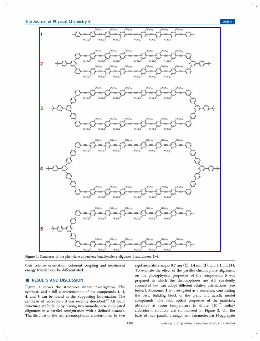
Figure 1 shows the structures under investigation. Thesynthesis and a full characterization of the compounds 1, 2,4, and 5 can be found in the Supporting Information. Thesynthesis of macrocycle 3 was recently described.24 All cyclicstructures are built up by placing two monodisperse conjugatedoligomers in a parallel configuration with a defined distance.The distance of the two chromophores is determined by two
rigid aromatic clamps: 0.7 nm (2), 1.4 nm (3), and 2.1 nm (4).To evaluate the effect of the parallel chromophore alignmenton the photophysical properties of the compounds, 5 wasprepared in which the chromophores are still covalentlyconnected but can adopt different relative orientations (seebelow). Monomer 1 is investigated as a reference, constitutingthe basic building block of the cyclic and acyclic modelcompounds. The basic optical properties of the materials,obtained at room temperature in dilute (10−7 molar)chloroform solution, are summarized in Figure 2. On thebasis of their parallel arrangement, intramolecular H-aggregate
Figure 1. Structures of the phenylene-ethynylene-butadiynylene oligomer 1 and dimers 2−5.
The Journal of Physical Chemistry B Article
dx.doi.org/10.1021/jp301903u | J. Phys. Chem. B 2013, 117, 4197−42034198
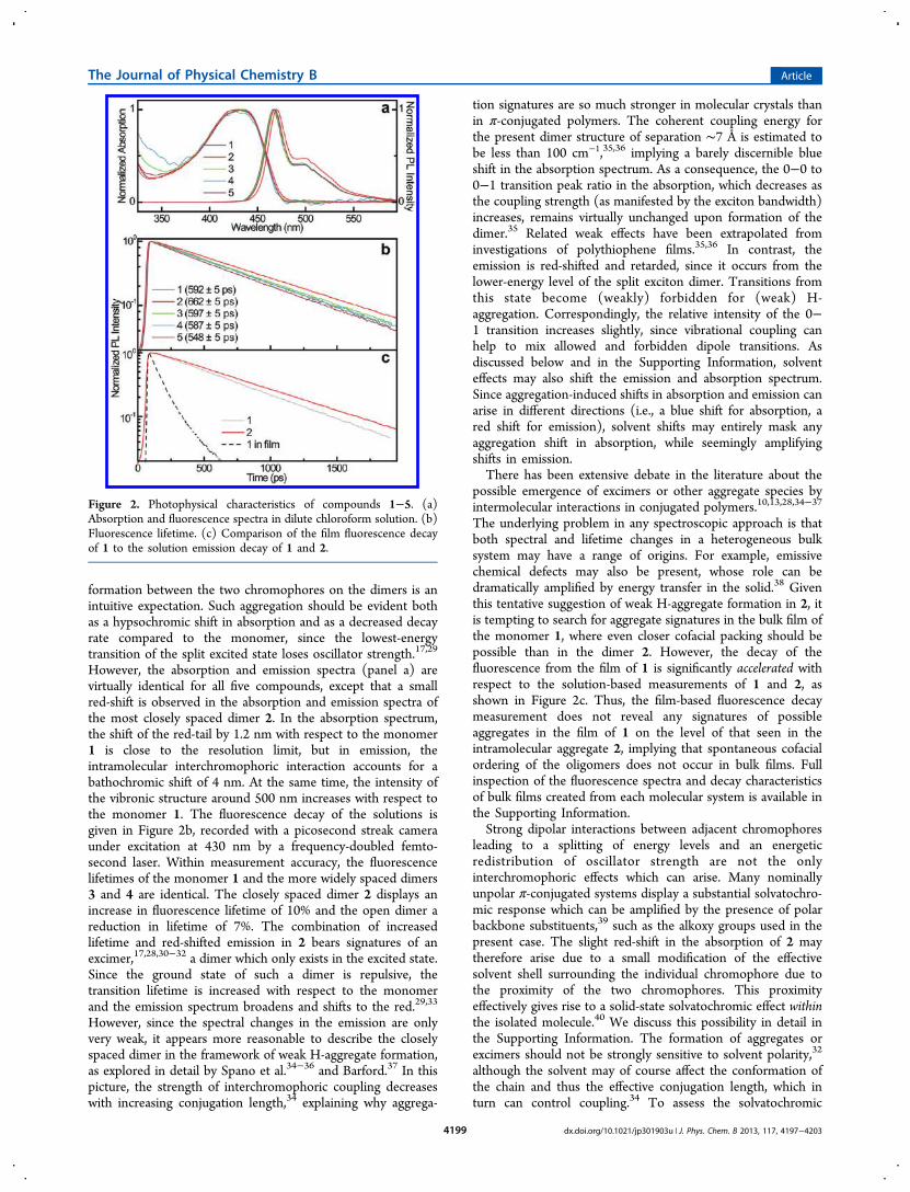
formation between the two chromophores on the dimers is anintuitive expectation. Such aggregation should be evident bothas a hypsochromic shift in absorption and as a decreased decayrate compared to the monomer, since the lowest-energytransition of the split excited state loses oscillator strength.17,29
However, the absorption and emission spectra (panel a) arevirtually identical for all five compounds, except that a smallred-shift is observed in the absorption and emission spectra ofthe most closely spaced dimer 2. In the absorption spectrum,the shift of the red-tail by 1.2 nm with respect to the monomer1 is close to the resolution limit, but in emission, theintramolecular interchromophoric interaction accounts for abathochromic shift of 4 nm. At the same time, the intensity ofthe vibronic structure around 500 nm increases with respect tothe monomer 1. The fluorescence decay of the solutions isgiven in Figure 2b, recorded with a picosecond streak cameraunder excitation at 430 nm by a frequency-doubled femto-second laser. Within measurement accuracy, the fluorescencelifetimes of the monomer 1 and the more widely spaced dimers3 and 4 are identical. The closely spaced dimer 2 displays anincrease in fluorescence lifetime of 10% and the open dimer areduction in lifetime of 7%. The combination of increasedlifetime and red-shifted emission in 2 bears signatures of anexcimer,17,28,30−32 a dimer which only exists in the excited state.Since the ground state of such a dimer is repulsive, thetransition lifetime is increased with respect to the monomerand the emission spectrum broadens and shifts to the red.29,33
However, since the spectral changes in the emission are onlyvery weak, it appears more reasonable to describe the closelyspaced dimer in the framework of weak H-aggregate formation,as explored in detail by Spano et al.34−36 and Barford.37 In thispicture, the strength of interchromophoric coupling decreaseswith increasing conjugation length,34 explaining why aggrega-
tion signatures are so much stronger in molecular crystals thanin π-conjugated polymers. The coherent coupling energy forthe present dimer structure of separation ∼7 Å is estimated tobe less than 100 cm−1,35,36 implying a barely discernible blueshift in the absorption spectrum. As a consequence, the 0−0 to0−1 transition peak ratio in the absorption, which decreases asthe coupling strength (as manifested by the exciton bandwidth)increases, remains virtually unchanged upon formation of thedimer.35 Related weak effects have been extrapolated frominvestigations of polythiophene films.35,36 In contrast, theemission is red-shifted and retarded, since it occurs from thelower-energy level of the split exciton dimer. Transitions fromthis state become (weakly) forbidden for (weak) H-aggregation. Correspondingly, the relative intensity of the 0−1 transition increases slightly, since vibrational coupling canhelp to mix allowed and forbidden dipole transitions. Asdiscussed below and in the Supporting Information, solventeffects may also shift the emission and absorption spectrum.Since aggregation-induced shifts in absorption and emission canarise in different directions (i.e., a blue shift for absorption, ared shift for emission), solvent shifts may entirely mask anyaggregation shift in absorption, while seemingly amplifyingshifts in emission.There has been extensive debate in the literature about the
possible emergence of excimers or other aggregate species byintermolecular interactions in conjugated polymers.10,13,28,34−37
The underlying problem in any spectroscopic approach is thatboth spectral and lifetime changes in a heterogeneous bulksystem may have a range of origins. For example, emissivechemical defects may also be present, whose role can bedramatically amplified by energy transfer in the solid.38 Giventhis tentative suggestion of weak H-aggregate formation in 2, itis tempting to search for aggregate signatures in the bulk film ofthe monomer 1, where even closer cofacial packing should bepossible than in the dimer 2. However, the decay of thefluorescence from the film of 1 is significantly accelerated withrespect to the solution-based measurements of 1 and 2, asshown in Figure 2c. Thus, the film-based fluorescence decaymeasurement does not reveal any signatures of possibleaggregates in the film of 1 on the level of that seen in theintramolecular aggregate 2, implying that spontaneous cofacialordering of the oligomers does not occur in bulk films. Fullinspection of the fluorescence spectra and decay characteristicsof bulk films created from each molecular system is available inthe Supporting Information.Strong dipolar interactions between adjacent chromophores
leading to a splitting of energy levels and an energeticredistribution of oscillator strength are not the onlyinterchromophoric effects which can arise. Many nominallyunpolar π-conjugated systems display a substantial solvatochro-mic response which can be amplified by the presence of polarbackbone substituents,39 such as the alkoxy groups used in thepresent case. The slight red-shift in the absorption of 2 maytherefore arise due to a small modification of the effectivesolvent shell surrounding the individual chromophore due tothe proximity of the two chromophores. This proximityeffectively gives rise to a solid-state solvatochromic effect withinthe isolated molecule.40 We discuss this possibility in detail inthe Supporting Information. The formation of aggregates orexcimers should not be strongly sensitive to solvent polarity,32
although the solvent may of course affect the conformation ofthe chain and thus the effective conjugation length, which inturn can control coupling.34 To assess the solvatochromic
Figure 2. Photophysical characteristics of compounds 1−5. (a)Absorption and fluorescence spectra in dilute chloroform solution. (b)Fluorescence lifetime. (c) Comparison of the film fluorescence decayof 1 to the solution emission decay of 1 and 2.
The Journal of Physical Chemistry B Article
dx.doi.org/10.1021/jp301903u | J. Phys. Chem. B 2013, 117, 4197−42034199
effect, we performed time-resolved spectroscopy in foursolvents with different polarities, and found that the moleculesdemonstrate obvious solvatochromism. In all cases, 2 shows themost red-shifted spectra and the slowest PL decay compared tothe other compounds. We conclude that both aggregation andintramolecular solvatochromism are most likely responsible forthe observed distinctive difference of 2 with respect to the othercompounds.The slight acceleration in fluorescence decay of the open
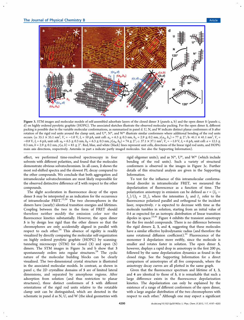
dimer 5 may be interpreted as an indication for the occurrenceof intramolecular FRET.12,26 The two chromophores in thedimers have (nearly) identical transition energies and lifetimes.Coupling between the two in the form of FRET shouldtherefore neither modify the emission color nor thefluorescence kinetics substantially. However, the open dimer5 is by design less rigid than the other dimers so that thechromophores are only accidentally aligned in parallel withrespect to each other.24 This absence of rigidity is readilyvisualized by directly comparing the molecular self-organizationon highly ordered pyrolytic graphite (HOPG) by scanning-tunneling microscopy (STM) for closed (3) and open (5)dimers. The STM images in Figure 3a and b show that 3spontaneously orders into regular structures.24 The cyclicnature of the molecular building blocks can be clearlyvisualized. The two-dimensional crystal structure is illustratedin the associated molecular models. In contrast, as shown inpanel c, the 2D crystalline domains of 5 are of limited lateraldimensions, and separated by amorphous regions. Afteradsorption from solution (and thus restriction to planarstructures), three distinct conformers of 5 with differentorientations of the rigid rod units relative to the rotatableclamp unit can be distinguished, which are denoted in theschematic in panel d as N, U, and W (the ideal geometries with
rigid oligomer units); and as N*, U*, and W* (which includebending of the rod units). Such a variety of structuralconformers is observed in the images in Figure 3c. Furtherdetails of this structural analysis are given in the SupportingInformation.To test for the influence of this intramolecular conforma-
tional disorder in intramolecular FRET, we measured thedepolarization of fluorescence as a function of time. Thepolarization anisotropy in emission can be defined as r = (I∥ −I⊥)/(I∥ + 2I⊥), where the intensities I∥ and I⊥ refer to thefluorescence polarized parallel and orthogonal to the incidentlaser, respectively. r is expected to decrease with time as themolecule tumbles in solution, starting from an initial value of0.4 as expected for an isotropic distribution of linear transitiondipoles in space.23,41 Figure 4 exhibits the transient anisotropyfor the five model compounds. There is no difference betweenthe rigid dimers 2, 3, and 4, suggesting that these moleculeshave a similar effective hydrodynamic radius (and therefore thesame rotational diffusion coefficient).22 Fluorescence of themonomer 1 depolarizes more swiftly, since the molecule issmaller and rotates faster in solution. The open dimer 5,however, displays a rapid drop in anisotropy in the first 200 ps,followed by the same depolarization dynamics as found in theclosed rings. See the Supporting Information for a directcomparison of anisotropies of all five compounds, where theanisotropy decay curves are all plotted in the same graph.Given that the fluorescence spectrum and lifetime of 1, 3,
and 4 are identical to those of 5, it is remarkable that such alarge difference exists in the fluorescence depolarizationkinetics. The depolarization can only be explained by theexistence of a range of different conformers of the open dimer,with a large angular distribution of the two chromophores withrespect to each other.5 Although one may expect a significant
Figure 3. STM images and molecular models of self-assembled adsorbate layers of the closed dimer 3 (panels a, b) and the open dimer 5 (panels c,d) on highly ordered pyrolytic graphite (HOPG). The associated sketches illustrate the observed molecular packing. For the open dimer 5, differentpacking is possible due to the variable molecular conformations, as summarized in panel d. U, N, and W indicate distinct planar conformers of 5 afterrotation of the rigid rod units around the clamp unit, and U*, N*, and W* illustrate similar conformers where additional bending of the rod unitsoccurs. (a: 35.1 × 35.1 nm2, Vs = −1.0 V, It = 10 pA, unit cell: aA = 6.3 ± 0.3 nm, bA = 2.9 ± 0.2 nm, γ(aA, bA) = 77 ± 2°; b: 41.1 × 41.1 nm2, Vs =−0.8 V, It = 6 pA, unit cell: aB = 6.5 ± 0.3 nm, bB = 6.3 ± 0.3 nm, γ(aB, bB) = 74 ± 2°; c: 37.1 × 37.1 nm2, Vs = −1.0 V, It = 6 pA, unit cell: a = 12.5 ±0.3 nm, b = 2.9 ± 0.2 nm, γ(a, b) = 83 ± 2°. Red, blue, and white (black) lines represent unit cells, directions of the linear rigid rod units, and HOPGmain axis directions, respectively. Asterisks in part a indicate partly imaged molecules. See also the Supporting Information).
The Journal of Physical Chemistry B Article
dx.doi.org/10.1021/jp301903u | J. Phys. Chem. B 2013, 117, 4197−42034200
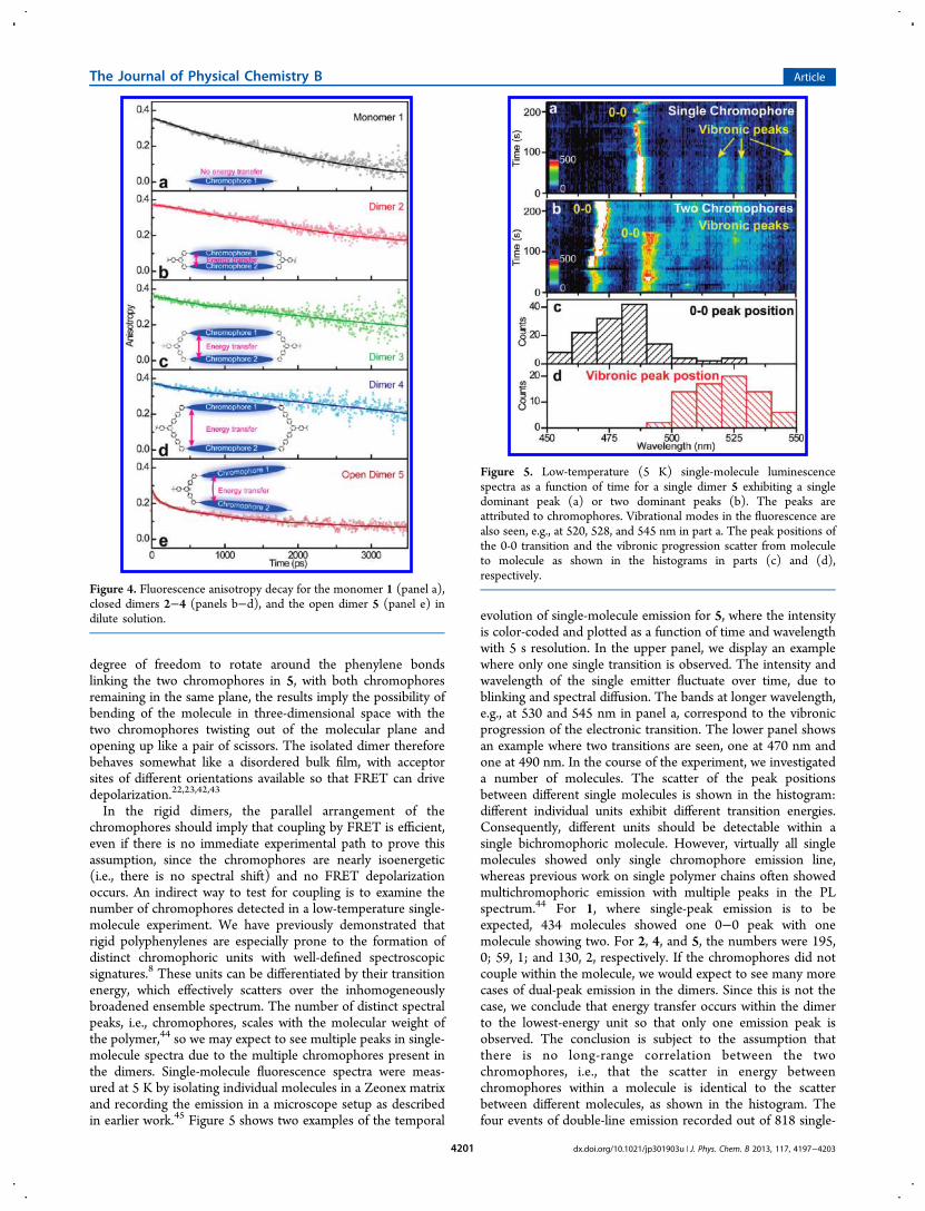
degree of freedom to rotate around the phenylene bondslinking the two chromophores in 5, with both chromophoresremaining in the same plane, the results imply the possibility ofbending of the molecule in three-dimensional space with thetwo chromophores twisting out of the molecular plane andopening up like a pair of scissors. The isolated dimer thereforebehaves somewhat like a disordered bulk film, with acceptorsites of different orientations available so that FRET can drivedepolarization.22,23,42,43
In the rigid dimers, the parallel arrangement of thechromophores should imply that coupling by FRET is efficient,even if there is no immediate experimental path to prove thisassumption, since the chromophores are nearly isoenergetic(i.e., there is no spectral shift) and no FRET depolarizationoccurs. An indirect way to test for coupling is to examine thenumber of chromophores detected in a low-temperature single-molecule experiment. We have previously demonstrated thatrigid polyphenylenes are especially prone to the formation ofdistinct chromophoric units with well-defined spectroscopicsignatures.8 These units can be differentiated by their transitionenergy, which effectively scatters over the inhomogeneouslybroadened ensemble spectrum. The number of distinct spectralpeaks, i.e., chromophores, scales with the molecular weight ofthe polymer,44 so we may expect to see multiple peaks in single-molecule spectra due to the multiple chromophores present inthe dimers. Single-molecule fluorescence spectra were meas-ured at 5 K by isolating individual molecules in a Zeonex matrixand recording the emission in a microscope setup as describedin earlier work.45 Figure 5 shows two examples of the temporal
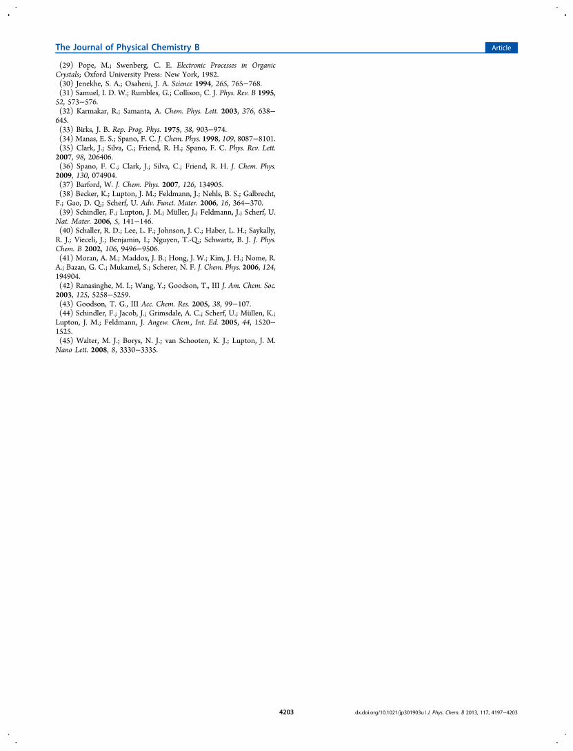
evolution of single-molecule emission for 5, where the intensityis color-coded and plotted as a function of time and wavelengthwith 5 s resolution. In the upper panel, we display an examplewhere only one single transition is observed. The intensity andwavelength of the single emitter fluctuate over time, due toblinking and spectral diffusion. The bands at longer wavelength,e.g., at 530 and 545 nm in panel a, correspond to the vibronicprogression of the electronic transition. The lower panel showsan example where two transitions are seen, one at 470 nm andone at 490 nm. In the course of the experiment, we investigateda number of molecules. The scatter of the peak positionsbetween different single molecules is shown in the histogram:different individual units exhibit different transition energies.Consequently, different units should be detectable within asingle bichromophoric molecule. However, virtually all singlemolecules showed only single chromophore emission line,whereas previous work on single polymer chains often showedmultichromophoric emission with multiple peaks in the PLspectrum.44 For 1, where single-peak emission is to beexpected, 434 molecules showed one 0−0 peak with onemolecule showing two. For 2, 4, and 5, the numbers were 195,0; 59, 1; and 130, 2, respectively. If the chromophores did notcouple within the molecule, we would expect to see many morecases of dual-peak emission in the dimers. Since this is not thecase, we conclude that energy transfer occurs within the dimerto the lowest-energy unit so that only one emission peak isobserved. The conclusion is subject to the assumption thatthere is no long-range correlation between the twochromophores, i.e., that the scatter in energy betweenchromophores within a molecule is identical to the scatterbetween different molecules, as shown in the histogram. Thefour events of double-line emission recorded out of 818 single-
Figure 4. Fluorescence anisotropy decay for the monomer 1 (panel a),closed dimers 2−4 (panels b−d), and the open dimer 5 (panel e) indilute solution.
Figure 5. Low-temperature (5 K) single-molecule luminescencespectra as a function of time for a single dimer 5 exhibiting a singledominant peak (a) or two dominant peaks (b). The peaks areattributed to chromophores. Vibrational modes in the fluorescence arealso seen, e.g., at 520, 528, and 545 nm in part a. The peak positions ofthe 0-0 transition and the vibronic progression scatter from moleculeto molecule as shown in the histograms in parts (c) and (d),respectively.
The Journal of Physical Chemistry B Article
dx.doi.org/10.1021/jp301903u | J. Phys. Chem. B 2013, 117, 4197−42034201

molecule spectra may signify a rare situation of FRET blockage,e.g., due to particularly weak dipolar coupling betweenchromophores with weak vibrational coupling and correspond-ingly narrow spectral resonances and an absence of spectraloverlap between donor and acceptor; or else could arise due totwo molecules being present within the diffraction-limited focalspot of the microscope. Interestingly, no difference is seenbetween compounds 1, 2, 4, and 5 on the single-molecule levelin terms of the spectrum, blinking, brightness, and spectraldynamics, implying that efficient incoherent interchromophorecoupling to a lowest-energy state also occurs in the mostdistantly spaced aggregate 4.
■ CONCLUSIONS
In summary, we have performed a range of optical andstructural studies on open and closed π-conjugated bichromo-phoric systems as model systems for interchromophoricinteractions in organic electronic materials. The spectralproperties and emission lifetime are remarkably resilientunder structural variations. Introduction of intramoleculardisorder by removal of one of the macrocycle clamps revealsinterchromophoric intramolecular FRET in terms of anaccelerated decay of polarization anisotropy. The occurrenceof FRET in the open dimers implies that it must also arise inthe closed dimers, even though it can only be inferredindirectly, for example, by the low-temperature single-moleculeemission characteristics. Interchromophoric interactions, andespecially light-harvesting phenomena, are usually visualized bymeans of a change in fluorescence spectrum, lifetime, orpolarization anisotropy. The systematic approach presentedhere, by constructing conformationally controlled dimers fromindividual building blocks, illustrates that interchromophoricinteractions may even arise when no immediate observable isavailable, since neither lifetime, spectrum, nor polarizationchange in the parallel closed dimers. Realizing the presence ofsuch interactions is crucial for understanding limitations inquantum efficiency of the material, since interchromophoriccoupling can promote migration of excitation energy toquenching species. At the same time, large aggregates ofparallel chromophores in macrocyclic templates could poseexcellent systems for fluorescence quenching-based sensing,with superior interaction cross sections when compared toconventional linear polymers.
■ ASSOCIATED CONTENT
*S Supporting InformationPhotoluminescence of bulk films, solvatochromism, detaileddiscussion of fluorescence depolarization, experimental meth-ods, scanning-tunneling microscopy results, and details ofsynthesis. This material is available free of charge via theInternet at http://pubs.acs.org.
■ AUTHOR INFORMATION
Corresponding Author*Fax: +49-228-735662 (S.H.); +49-941-943-4226 (J.M.L.).Phone: +49-228-736127 (S.H.); +49-941-943-2080 (J.M.L.).E-mail: [email protected] (S.H.); [email protected] (J.M.L.).
NotesThe authors declare no competing financial interest.
■ ACKNOWLEDGMENTSCollaborative funding by the Volkswagen Foundation is greatlyappreciated. We acknowledge financial support by the DFG(especially through the SFB 624) and the Fonds derChemischen Industrie. J.M.L. is indebted to the David &Lucile Packard Foundation for providing a fellowship.
■ REFERENCES(1) Lupton, J. M.; Samuel, I. D. W.; Beavington, R.; Burn, P. L.;Bassler, H. Adv. Mater. 2001, 13, 258−261.(2) Becker, K.; Lagoudakis, P. G.; Gaefke, G.; Hoger, S.; Lupton, J.M. Angew. Chem., Int. Ed. 2007, 46, 3450−3455.(3) Becker, K.; Da Como, E.; Feldmann, J.; Scheliga, F.; Csanyi, E.T.; Tretiak, S.; Lupton, J. M. J. Phys. Chem. B 2008, 112, 4859−4864.(4) Hofkens, J.; Cotlet, M.; Vosch, T.; Tinnefeld, P.; Weston, K. D.;Ego, C.; Grimsdale, A.; Mullen, K.; Beljonne, D.; Bredas, J. L.; Jordens,S.; Schweitzer, G.; Sauer, M.; De Schryver , F. Proc. Natl. Acad. Sci.U.S.A. 2003, 100, 13146−13151.(5) Hofmann, C.; Ketelaars, M.; Matsushita, M.; Michel, H.; Aartsma,T. J.; Kohler, J. Phys. Rev. Lett. 2003, 90, 013004.(6) Lupton, J. M.; Samuel, I. D. W.; Burn, P. L.; Mukamel, S. J. Chem.Phys. 2002, 116, 455−459.(7) Hernando, J.; van der Schaaf, M.; van Dijk, E. M. H. P.; Sauer,M.; García-Parajo, M. F.; van Hulst, N. F. J. Phys. Chem. A 2003, 107,43−52.(8) Lupton, J. M. Adv. Mater. 2010, 22, 1689−1721.(9) Oelkrug, D.; Tompert, A.; Gierschner, J.; Egelhaaf, H.-J.; Hanack,M.; Hohloch, M.; Steinhuber, E. J. Phys. Chem. B 1998, 102, 1902−1907.(10) Nguyen, T.-Q.; Doan, V.; Schwartz, B. J. J. Chem. Phys. 1999,110, 4068−4078.(11) Cornil, J.; Beljonne, D.; Calbert, J.-P.; Bredas, J. L. Adv. Mater.2001, 13, 1053−1067.(12) Beljonne, D.; Pourtois, G.; Silva, C.; Hennebicq, E.; Herz, L. M.;Friend, R. H.; Scholes, G. D.; Setayesh, S.; Mullen, K.; Bredas, J. L.Proc. Natl. Acad. Sci. U.S.A. 2002, 99, 10982−10987.(13) Schwartz, B. J. Annu. Rev. Phys. Chem. 2003, 54, 141−172.(14) Spano, F. C. Annu. Rev. Phys. Chem. 2006, 57, 217−243.(15) Sundstrom, V.; Pullerits, T.; van Grondelle, R. J. Phys. Chem. B1999, 103, 2327−2346.(16) Tretiak, S.; Zhang, W. M.; Chernyak, V.; Mukamel, S. Proc. Natl.Acad. Sci. U.S.A. 1999, 96, 13003−13008.(17) Bazan, G. C.; Oldham, W. J., Jr.; Lachicotte, R. J.; Tretiak, S.;Chernyak, V.; Mukamel, S. J. Am. Chem. Soc. 1998, 120, 9188−9204.(18) Wang, S.; Bazan, G. C.; Tretiak, S.; Mukamel, S. J. Am. Chem.Soc. 2000, 122, 1289−1297.(19) Varnavski, O. P.; Ostrowski, J. C.; Sukhomlinova, L.; Twieg, R.J.; Bazan, G. C.; Goodson, T., III J. Am. Chem. Soc. 2002, 124, 1736−1743.(20) Hernando, J.; Hoogenboom, J. P.; van Dijk, E. M. H. P.; García-Lopez, J. J.; Crego-Calama, M.; Reinhoudt, D. N.; van Hulst, N. F.;García-Parajo, M. F. Phys. Rev. Lett. 2004, 93, 236404.(21) Lippitz, M.; Hubner, C. G.; Christ, T.; Eichner, H.; Bordat, P.;Herrmann, A.; Mullen, K.; Basche, T. Phys. Rev. Lett. 2004, 92, 103001.(22) Becker, K.; Fritzsche, M.; Hoger, S.; Lupton, J. M. J. Phys. Chem.B 2008, 112, 4849−4853.(23) Mossinger, D.; Chaudhuri, D.; Kudernac, T.; Lei, S.; De Feyter,S.; Lupton, J. M.; Hoger, S. J. Am. Chem. Soc. 2010, 132, 1410−1423.(24) Jester, S.-S.; Schmitz, D.; Eberhagen, F.; Hoger, S. Chem.Commun. 2011, 47, 8838−8840.(25) Bartholomew, G. P.; Bazan, G. C. Acc. Chem. Res. 2001, 34, 30−39.(26) Scholes, G. D. Annu. Rev. Phys. Chem. 2003, 54, 57−87.(27) Kuroda, D. G.; Singh, C. P.; Peng, Z. H.; Kleiman, V. D. Science2009, 326, 263−267.(28) Gierschner, J.; Ehni, M.; Egelhaaf, H.-J.; Medina, B. M.;Beljonne, D.; Benmansour, H.; Bazan, G. C. J. Chem. Phys. 2005, 123,144914.
The Journal of Physical Chemistry B Article
dx.doi.org/10.1021/jp301903u | J. Phys. Chem. B 2013, 117, 4197−42034202
(29) Pope, M.; Swenberg, C. E. Electronic Processes in OrganicCrystals; Oxford University Press: New York, 1982.(30) Jenekhe, S. A.; Osaheni, J. A. Science 1994, 265, 765−768.(31) Samuel, I. D. W.; Rumbles, G.; Collison, C. J. Phys. Rev. B 1995,52, 573−576.(32) Karmakar, R.; Samanta, A. Chem. Phys. Lett. 2003, 376, 638−645.(33) Birks, J. B. Rep. Prog. Phys. 1975, 38, 903−974.(34) Manas, E. S.; Spano, F. C. J. Chem. Phys. 1998, 109, 8087−8101.(35) Clark, J.; Silva, C.; Friend, R. H.; Spano, F. C. Phys. Rev. Lett.2007, 98, 206406.(36) Spano, F. C.; Clark, J.; Silva, C.; Friend, R. H. J. Chem. Phys.2009, 130, 074904.(37) Barford, W. J. Chem. Phys. 2007, 126, 134905.(38) Becker, K.; Lupton, J. M.; Feldmann, J.; Nehls, B. S.; Galbrecht,F.; Gao, D. Q.; Scherf, U. Adv. Funct. Mater. 2006, 16, 364−370.(39) Schindler, F.; Lupton, J. M.; Muller, J.; Feldmann, J.; Scherf, U.Nat. Mater. 2006, 5, 141−146.(40) Schaller, R. D.; Lee, L. F.; Johnson, J. C.; Haber, L. H.; Saykally,R. J.; Vieceli, J.; Benjamin, I.; Nguyen, T.-Q.; Schwartz, B. J. J. Phys.Chem. B 2002, 106, 9496−9506.(41) Moran, A. M.; Maddox, J. B.; Hong, J. W.; Kim, J. H.; Nome, R.A.; Bazan, G. C.; Mukamel, S.; Scherer, N. F. J. Chem. Phys. 2006, 124,194904.(42) Ranasinghe, M. I.; Wang, Y.; Goodson, T., III J. Am. Chem. Soc.2003, 125, 5258−5259.(43) Goodson, T. G., III Acc. Chem. Res. 2005, 38, 99−107.(44) Schindler, F.; Jacob, J.; Grimsdale, A. C.; Scherf, U.; Mullen, K.;Lupton, J. M.; Feldmann, J. Angew. Chem., Int. Ed. 2005, 44, 1520−1525.(45) Walter, M. J.; Borys, N. J.; van Schooten, K. J.; Lupton, J. M.Nano Lett. 2008, 8, 3330−3335.
The Journal of Physical Chemistry B Article
dx.doi.org/10.1021/jp301903u | J. Phys. Chem. B 2013, 117, 4197−42034203


![High-order TRAIL oligomer formation in TRAIL-coated … · sTRAIL) anchored on their surface were generated as previously described [37,42]. Briefly, after generating the lipid nanoparticles,](https://img.pdfslide.tips/doc/110x75/5b7aa5527f8b9a22238c8cec/high-order-trail-oligomer-formation-in-trail-coated-strail-anchored-on-their.jpg)
![γN arXiv:1604.01555v1 [nucl-th] 6 Apr 2016arXiv:1604.01555v1 [nucl-th] 6 Apr 2016 On thenear-threshold incoherent φphotoproduction on thedeuteron: any traceof a resonance? Alvin](https://img.pdfslide.tips/doc/110x75/5ffe209af462ba2aac5bf74f/n-arxiv160401555v1-nucl-th-6-apr-2016-arxiv160401555v1-nucl-th-6-apr-2016.jpg)