Embed Size (px)
Citation preview
1
Combination of Ibrutinib and ABT-199 in Diffuse Large B-Cell Lymphoma and Follicular Lymphoma
Hsu-Ping Kuo1, Scott A Ezell1, Karl J Schweighofer1, Leo WK Cheung1, Sidney Hsieh1,
Mutiah Apatira1, Mint Sirisawad1, Karl Eckert1, Ssucheng J Hsu1, Chun-Te Chen1,
Darrin M Beaupre1, Matthias Versele2, and Betty Y Chang1
1Research Department, Pharmacyclics LLC, an AbbVie Company, Sunnyvale, CA, USA 2Janssen Research and Development, Beerse, Belgium
Running title (limit 60 characters, currently 37): Ibrutinib and ABT-199 in DLBCL and
FL
Word count (limit 5000, includes abstract + manuscript + figure legends): 4917
Abstract word count (limit: 250): 179
Figures (limit 6): 6
Key words: ibrutinib, ABT-199, diffuse large B-cell lymphoma, follicular lymphoma,
combination therapy
Financial support: This study was supported by funding from Pharmacyclics, LLC, an
AbbVie Company.
Corresponding author: Betty Y Chang, 995 E. Arques Avenue, Sunnyvale, CA 94085;
408-215-3358 (phone and fax); [email protected]
Conflict of interest disclosure: HPK, MS, KE: employment and patents with
Pharmacyclics, an AbbVie Company; stock from AbbVie; SAE: employment and travel
accommodations with Pharmacyclics, an AbbVie Company; employment with
AstraZeneca and Amgen; KJS: employment, travel accommodations, and research
funding from Pharmacyclics, an AbbVie Company; employment and stock from AbbVie;
on March 21, 2020. © 2017 American Association for Cancer Research. mct.aacrjournals.org Downloaded from
Author manuscripts have been peer reviewed and accepted for publication but have not yet been edited. Author Manuscript Published OnlineFirst on April 20, 2017; DOI: 10.1158/1535-7163.MCT-16-0555
2
LWKC: employment and patent with Pharmacyclics, an AbbVie Company; employment
and stock from Eli Lilly & Co, stock from AbbVie; SH: employment with Pharmacyclics,
an AbbVie Company; MA: employment with Pharmacyclics, an AbbVie Company, and
Kaiser Permanente, stock from AbbVie; JH: employment, stock, and patents with
Pharmacyclics, an AbbVie Company; and AbbVie, employment and patents with
Astellas; CTC: employment with Pharmacyclics, an AbbVie Company; patents with MD
Anderson Cancer Center; DB: employment, leadership, research funding, patents, and
travel accommodations from Pharmacyclics, an AbbVie Company; stock from AbbVie;
MV: employment and patents with Janssen, stock from Johnson and Johnson; BYC:
employment and patents with Pharmacyclics, an AbbVie Company; stock from AbbVie.
on March 21, 2020. © 2017 American Association for Cancer Research. mct.aacrjournals.org Downloaded from
Author manuscripts have been peer reviewed and accepted for publication but have not yet been edited. Author Manuscript Published OnlineFirst on April 20, 2017; DOI: 10.1158/1535-7163.MCT-16-0555
3
ABSTRACT
Diffuse large B-cell lymphoma (DLBCL) and follicular lymphoma (FL) are the most
prevalent B-lymphocyte neoplasms in which abnormal activation of the Bruton’s tyrosine
kinase (BTK)–mediated B-cell receptor (BCR) signaling pathway contributes to
pathogenesis. Ibrutinib is an oral covalent BTK inhibitor that has shown some efficacy in
both indications. To improve ibrutinib efficacy through combination therapy, we first
investigated differential gene expression in parental and ibrutinib-resistant cell lines to
better understand the mechanisms of resistance. Ibrutinib-resistant TMD8 cells had
higher BCL2 gene expression and increased sensitivity to ABT-199, a BCL-2 inhibitor.
Consistently, clinical samples from ABC-DLBCL patients who experienced poorer
response to ibrutinib had higher BCL2 gene expression. We further demonstrated
synergistic growth suppression by ibrutinib and ABT-199 in multiple ABC-DLBCL, GCB-
DLBCL, and FL lymphoma cell lines. The combination of both drugs also reduced
colony formation, increased apoptosis, and inhibited tumor growth in a TMD8 xenograft
model. A synergistic combination effect was also found in ibrutinib-resistant cells
generated by either genetic mutation or drug treatment. Together, these findings
suggest a potential clinical benefit from ibrutinib and ABT-199 combination therapy.
on March 21, 2020. © 2017 American Association for Cancer Research. mct.aacrjournals.org Downloaded from
Author manuscripts have been peer reviewed and accepted for publication but have not yet been edited. Author Manuscript Published OnlineFirst on April 20, 2017; DOI: 10.1158/1535-7163.MCT-16-0555
4
INTRODUCTION
Diffuse large B-cell lymphoma (DLBCL) is the most common subtype of non-Hodgkin's
lymphoma (NHL), accounting for roughly 30% of newly diagnosed cases in the United
States. DLBCL is a heterogeneous lymphoma consisting of activated B cell (ABC) and
germinal center B cell–like (GCB) subtypes that have different gene expression profiles,
oncogenic aberrations, and clinical outcomes (1, 2). Compared with the GCB subtype,
ABC-DLBCL has a significantly lower survival rate after multiagent chemotherapy (3)
and is characterized by chronically active B-cell receptor (BCR) signaling (4), which is
required for cell survival. Therefore, components of the BCR signaling pathway are
emerging as attractive therapeutic targets in the ABC subtype of DLBCL.
Bruton's tyrosine kinase (BTK), pivotal to BCR signaling, is covalently bound by ibrutinib
with high affinity. Ibrutinib is a first-in-class, once-daily, oral inhibitor of BTK that is
approved by the US FDA for the treatment of patients with chronic lymphocytic leukemia
(CLL), including those with deletion 17p, patients with mantle cell lymphoma (MCL) who
have received at least one prior therapy, and those with Waldenström’s
macroglobulinemia. With respect to DLBCL, a phase I/II clinical trial showed an overall
response rate of 37% in the ABC subtype (5) with single-agent ibrutinib therapy.
Tumor responses to single-agent kinase inhibitor therapies are often limited by the cell’s
ability to bypass the target via alternative pathways or acquired mutations in the target
or its pathway (6, 7). It has been shown that a small number of CLL patients acquire
resistance to ibrutinib through mutations in BTK and its substrate phospholipase C
gamma 2 (PLCG2) following prolonged treatment (8, 9). In addition to acquisition of
these mutations, other mechanisms of resistance, such as upregulation of potentially
on March 21, 2020. © 2017 American Association for Cancer Research. mct.aacrjournals.org Downloaded from
Author manuscripts have been peer reviewed and accepted for publication but have not yet been edited. Author Manuscript Published OnlineFirst on April 20, 2017; DOI: 10.1158/1535-7163.MCT-16-0555
5
druggable survival pathways or clonal evolution of other genetic alterations, have been
reported for ABC-DLBCL (10) and CLL (11). Such mechanisms may be overcome by
combinations of targeted agents that block pathways that cooperate in resistance.
Through screening of parental and acquired ibrutinib-resistant cell lines, we have
identified and report here that a B-cell lymphoma 2 (BCL-2) inhibitor, ABT-199,
synergizes with ibrutinib in vitro and in vivo and is able to overcome the ibrutinib-
resistant phenotype in tumor cells overexpressing BCL-2.
on March 21, 2020. © 2017 American Association for Cancer Research. mct.aacrjournals.org Downloaded from
Author manuscripts have been peer reviewed and accepted for publication but have not yet been edited. Author Manuscript Published OnlineFirst on April 20, 2017; DOI: 10.1158/1535-7163.MCT-16-0555
6
MATERIALS AND METHODS
Cell culture
The TMD8 and HBL1 cell lines were gifts from Dr. Daniel Krappmann in 2011 (German
Research Center for Environmental Health, Neuherberg, Germany). The OCI-LY10 cell
line was a gift from Dr. Richard Davis in 2010 (MD Anderson Cancer Center, Houston,
TX). The WSU-DLCL-2, RL, SU-DHL-4, DoHH2, and WSU-FSCCL cell lines were
purchased from ATCC or DSMZ in 2014. CellCheck service by IDEXX (Westbrook, ME)
was used to provide cell line authentication. Cell lines were grown to log phase at 37oC
in the presence of 5% CO2. TMD8 and HBL1 cells were cultured in RPMI 1640 medium
(Life Technologies) with 10% FBS (Atlanta Biologicals), 1 mM sodium pyruvate (Life
Technologies), and 1% Pen/Strep (Life Technologies). OCI-LY10 cells were cultured in
IMDM medium (Life Technologies) with 20% heparinized normal human plasma
(Equitech-Bio), 55 µM 2-mercaptoethanol (Life Technologies), and 1% Pen/Strep. WSU-
DLCL-2, RL, SU-DHL-4, DoHH2, and WSU-FSCCL cells were cultured in RPMI 1640
medium with 10% FBS, and 1% Pen/Strep. Ibrutinib-resistant HBL1, TMD8, and DoHH2
cells were generated by in vitro culture of the parental cell lines for prolonged periods of
time with progressively increasing concentrations of ibrutinib.
Generation of BTK-WT and BTK-C481S cell lines
Custom BTK- plasmid constructs expressing either the wild-type BTK (BTK-WT) or BTK
genes containing a single mutation C481S (BTK-C481S) were obtained from
GeneCopoeia with the Lv201 lentiviral backbone. Plasmids were amplified, purified, and
sequenced by System Biosciences. Plasmids (5.0 µg) were then used to transfect 293T
on March 21, 2020. © 2017 American Association for Cancer Research. mct.aacrjournals.org Downloaded from
Author manuscripts have been peer reviewed and accepted for publication but have not yet been edited. Author Manuscript Published OnlineFirst on April 20, 2017; DOI: 10.1158/1535-7163.MCT-16-0555
7
cells in a 10-cm dish using a lentiviral packaging kit (GeneCopoeia) following the
manufacturer’s instructions. Media were removed 16 hours after transfection, and fresh
media were added to the dish and incubated for 24 hours. Virus-containing
supernatants were harvested and filtered (0.2-µm filter) and used for TMD8
transduction.
TMD8 cells were plated in 6-well plates in 2-mL medium at the concentrations of 1 × 10⁶
cells/mL. Virus supernatants (500 µL) were added to wells followed by DOTAP
Liposomal Transfection Reagent (final concentration 10 µg/mL; Sigma). Plates were
centrifuged for 1 hour at 2,000 rpm and kept in culture at 37°C overnight. Supernatants
were removed and cells were resuspended in fresh medium and incubated for 2 days.
Cells were selected using 0.2 µg/mL puromycin to generate stable cell lines.
OCI-LY10 (BTK-C481S) was generated by introducing mutant HIS6/Strep-tagged BTK
(C481S) under control of the EF1α promotor into the OCI-LY10 cell line using lentiviral
transduction. Transduced cells were selected using blasticidin as an antibiotic
resistance marker.
Cell viability assays
CellTiter-Glo® (Promega) luminescent cell viability assay was performed according to
the manufacturer’s instructions. Briefly, cells were seeded at 8,000 to 25,000 cells/well
in a 96-well plate in the presence of single drugs or drug combinations for 3 or 5 days.
The number of viable cells in culture was determined by quantification of ATP present,
which was proportional to luminescent signal detected. Combination index (CI), a drug
on March 21, 2020. © 2017 American Association for Cancer Research. mct.aacrjournals.org Downloaded from
Author manuscripts have been peer reviewed and accepted for publication but have not yet been edited. Author Manuscript Published OnlineFirst on April 20, 2017; DOI: 10.1158/1535-7163.MCT-16-0555
8
interactivity measurement, was calculated with CalcuSyn (Biosoft). Chalice Analyzer
(Horizon CombinatoRx) was used to calculate the Loewe excess values, which were
commonly used to indicate the excess percent inhibition. Excess percent inhibition was
calculated by deducting the expected percent inhibition values of various combinations
from the experimental percent inhibition values. These data allowed us to generate the
isobolograms and synergy scores. In general, synergy scores >1 and CI <1 indicate a
synergistic combination effect (12).
qRT-PCR assays
The TaqMan® Fast Cells-to-CT™ Kit (Life Technologies) was used to extract total RNA
and reverse transcribe RNA to cDNA according to the manufacturer’s specifications.
Four microliters of cDNA from the RT reaction was used to set up TaqMan® quantitative
reverse transcription polymerase chain reaction (qRT-PCR) on a QuantStudio™ 7 Flex
Real-Time PCR System (Life Technologies). The TaqMan® Gene Expression Assays
used for this study include BCL2 (Hs00608023_m1), BAX (Hs00180269_m1), MCL1
(Hs01050896_m1), BTK (Hs00975865_m1), MAP3K7 (Hs00177373_m1), IRAK4
(Hs00211610_m1), GAPDH (Hs02758991_g1), and ACTB (Hs01060665_g1).
Western blot
Cells were washed twice with ice-cold phosphate-buffered saline and lysed with RIPA
buffer (R0278, Sigma-Aldrich) supplemented with 1× protease/phosphatase inhibitor.
Cell lysates were subjected to SDS-PAGE separation and subsequently transferred
on March 21, 2020. © 2017 American Association for Cancer Research. mct.aacrjournals.org Downloaded from
Author manuscripts have been peer reviewed and accepted for publication but have not yet been edited. Author Manuscript Published OnlineFirst on April 20, 2017; DOI: 10.1158/1535-7163.MCT-16-0555
9
onto a polyvinylidene difluoride membrane (IPFL00010, Millipore). The membranes
were incubated with Odyssey Blocking Buffer (927-40000, LI-COR Biosciences) for 1
hour and probed overnight at 4°C with rabbit anti-BCL-2 (ab182858, Abcam), mouse
anti- β -actin (3700S, Cell Signaling). After washing with 0.1% Tween-TBS, the
membranes were incubated with IRDye®800CW- or IRDye®680RD-conjugated
secondary antibodies for 1 hour in the dark and detected using the Odyssey Imaging
System (LI-COR).
siRNA transfection
Accell human SMARTpool siRNAs targeting MAP3K7 (E-003790-00-0005) or IRAK4 (E-
003302-00-0005) were purchased from Dharmacon. TMD8 cells (1 × 10⁶/mL) were
incubated with the Accell delivery medium (1% FBS) containing 1 µM of siRNAs at 37°C
according to the manufacturer's instructions. After overnight incubation, fresh delivery
medium (1% FBS) was added to each well. Cells were harvested 3 days after
transfection and used for qRT-PCR.
Xenograft study
All animal studies were completed under the Institutional Animal Care and Use
Committee (IACUC)–approved protocols for animal welfare. CB17 SCID mice (Charles
River Laboratories) were subcutaneously inoculated with 1 × 10⁷ TMD8 cells in a
suspension containing Matrigel (Corning). When tumors reached approximately 100
mm3 (16 days after tumor inoculation), mice were randomly assigned and treated once
on March 21, 2020. © 2017 American Association for Cancer Research. mct.aacrjournals.org Downloaded from
Author manuscripts have been peer reviewed and accepted for publication but have not yet been edited. Author Manuscript Published OnlineFirst on April 20, 2017; DOI: 10.1158/1535-7163.MCT-16-0555
10
daily with ibrutinib (12 mg/kg), ABT-199 (40 mg/kg), or the combination of both by oral
gavage, with 10 mice per group. Tumor volume was measured twice a week and
calculated as tumor volume = (length × width2) × 0.5.
Apoptosis assays
The ApoDETECT™ Annexin V-FITC Kit (Life Technologies) was used to quantify the
apoptotic cell population according to the manufacturer’s specifications. Briefly, cells
were washed with ice-cold phosphate-buffered saline and resuspended in 1× binding
buffer at a concentration of 5 × 10⁵ cells/mL. Annexin V-FITC (10 µL) was added to 190
µL of cell suspension and incubated at room temperature for 10 minutes. After being
washed with 1× binding buffer, cells were resuspended in 190 µL of binding buffer with
10 µL of 20 µg/mL propidium iodide (PI) and analyzed by flow cytometry.
Colony formation assays
HBL1 cells (1,000 cells per well) were suspended in 0.9% methylcellulose (MethocultTM
H4100, STEMCELL Technologies) containing culture medium with vehicle, ibrutinib,
ABT-199, or an ABT-199/ibrutinib combination, and 0.3 mL of the mixture was plated in
each well of 24-well culture plates. The colonies were counted on day 7.
on March 21, 2020. © 2017 American Association for Cancer Research. mct.aacrjournals.org Downloaded from
Author manuscripts have been peer reviewed and accepted for publication but have not yet been edited. Author Manuscript Published OnlineFirst on April 20, 2017; DOI: 10.1158/1535-7163.MCT-16-0555
11
Microarray data analyses and statistics
The GeneChip® Human Transcriptome Array 2.0 (HTA 2.0, Affymetrix) was used to
analyze gene expression in TMD8 parental and ibrutinib-resistant cell lines and the work
was done at Open Medicine Institute. A heatmap of apoptosis-related gene expression
was generated using Transcriptome Analysis Console v2.0 (Affymetrix).
Gene expression of formalin-fixed paraffin-embedded (FFPE) specimens from the
phase II PCYC-1106 trial (NCT01325701) was analyzed using the GeneChip® Human
Genome U133 Plus 2.0 Array (Affymetrix), and data were normalized using the robust
multiarray average (RMA) algorithm. Subtypes of DLBCL were identified based on the
classification algorithm (3). For the analysis restricted to ABC-DLBCL subtype, only the
samples having a gene expression profiling call of ABC-DLBCL were used and
normalized separately. A test for differential expression of genes between ABC-DLBCL
responders (complete response [CR] + partial response [PR]) and nonresponders
(stable disease [SD] + progressive disease [PD]) to ibrutinib was performed using the
rank product statistic (RankProd R package). For the ABC-DLBCL versus GCB-DLBCL
comparison plot and heatmap, all subtypes were normalized together. The data were
plotted in linear scale.
The data discussed in this publication have been deposited in NCBI's Gene Expression
Omnibus (GEO) and are accessible through GEO Series accession number GSE93986
(https://www.ncbi.nlm.nih.gov/geo/query/acc.cgi?acc=GSE93986).
on March 21, 2020. © 2017 American Association for Cancer Research. mct.aacrjournals.org Downloaded from
Author manuscripts have been peer reviewed and accepted for publication but have not yet been edited. Author Manuscript Published OnlineFirst on April 20, 2017; DOI: 10.1158/1535-7163.MCT-16-0555
12
RESULTS
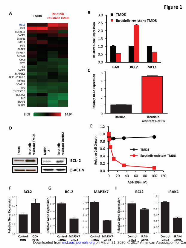
Ibrutinib-resistant TMD8 cells had higher BCL2 gene expression and were more
sensitive to ABT-199
Ibrutinib-resistant TMD8 and DoHH2 cells were generated by culturing the parental cell
lines in vitro with progressively increasing concentrations of ibrutinib. The EC50s of
ibrutinib-resistant TMD8 and DoHH2 cells were 1,061 nM and 210 nM compared with
13 nM and 4 nM for TMD8 and DoHH2 parental cells. BCL2, among other apoptosis-
related genes, showed increased expression in ibrutinib-resistant TMD8 cells compared
with TMD8 parental cells in our microarray data analysis (Figure 1A). In addition, BCL2
is the only gene in the BCL-2 family with an anti-apoptotic function that was significantly
increased (P < 0.05, Supplemental Table 1). The upregulation of BCL2 gene expression
was confirmed by qRT-PCR (Figure 1B). An increase in BCL2 gene expression was
also observed in ibrutinib-resistant DoHH2 cells (Figure 1C). Consistent with its
increased gene expression, a higher level of BCL-2 protein was detected in ibrutinib-
resistant cells (Figure 1D). In addition to increased BCL2 expression, our microarray
analyses showed reduced levels of MCL1 and BCL2A1 in ibrutinib-resistant cells.
Reduction of both MCL1 and BCL2A1 may contribute to higher ABT-199 sensitivity in
the ibrutinib-resistant cells (13-15). Indeed, ibrutinib-resistant TMD8 cells were much
more sensitive to ABT-199 than TMD8 parental cells (Figure 1E).
The identification of MYD88 L265P mutations in ABC-DLBCL suggests the importance
of Toll-like receptor (TLR) signaling in this malignancy (16). ABC-DLBCL cells that are
most sensitive to ibrutinib harbor both CD79A/B and MYD88 L265P mutations with
on March 21, 2020. © 2017 American Association for Cancer Research. mct.aacrjournals.org Downloaded from
Author manuscripts have been peer reviewed and accepted for publication but have not yet been edited. Author Manuscript Published OnlineFirst on April 20, 2017; DOI: 10.1158/1535-7163.MCT-16-0555
13
chronically active BCR signaling in addition to TLR pathways (5). Similar to chronically
active BCR signaling, TLR signaling also contributes to the activation of the NF-κB
pathway (17), which is involved in the transcriptional regulation of BCL2 (18).
Stimulation with a TLR9 agonist (ODN 2216) resulted in a 20-fold increase in the EC50
of ibrutinib in TMD8 cells. Intriguingly, we observed a 1.42-fold increase in MAP3K7 (P
= 0.028) and a 1.83-fold increase in IRAK4 (P = 0.004) in ibrutinib-resistant TMD8 cells.
We additionally observed a 6.69-fold reduction in the negative regulator of TLR
signaling, IRAK3 (P = 0.052), in ibrutinib-resistant TMD8 cells. To investigate the role of
TLR signaling in regulating BCL2 gene expression, we stimulated TMD8 cells with a
TLR9 agonist, CpG ODN 2216, and detected an increased BCL2 level compared with
the control ODN treated cells (Figure 1F). Consistently, knockdown of MAP3K7 or
IRAK4 using siRNA reduced BCL2 gene expression (Figures 1G and 1H). Therefore,
we postulate that resistant cells have upregulated TLR pathways, which lead to an
increase in BCL2 gene expression. However, we believe that further mechanistic
characterization of this interesting question is beyond the scope of this manuscript.
In addition to BCL2, we identified several genes with 3–10-fold increases in expression
in the ibrutinib-resistant TMD8 cells (P < 0.05, Supplemental Table 2) and another
subset of genes with 3–10-fold reductions in expression (P < 0.05, Supplemental Table
3).
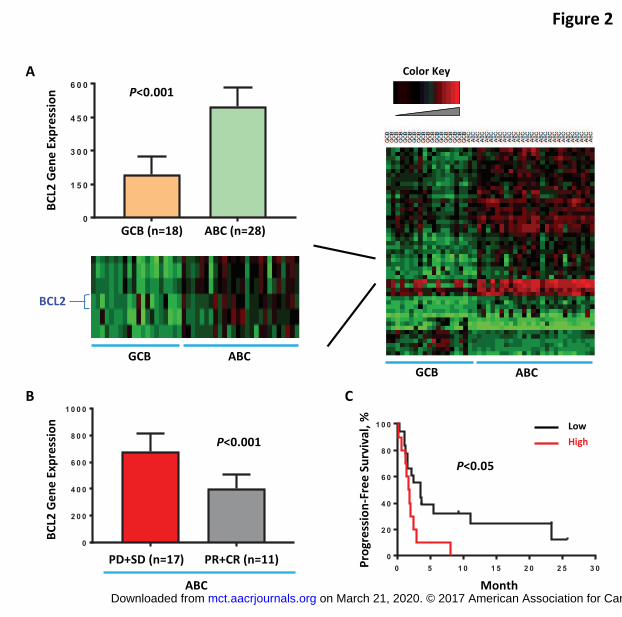
Higher BCL2 gene expression was observed in tumors from ABC-DLBCL patients
with poorer responses to ibrutinib
on March 21, 2020. © 2017 American Association for Cancer Research. mct.aacrjournals.org Downloaded from
Author manuscripts have been peer reviewed and accepted for publication but have not yet been edited. Author Manuscript Published OnlineFirst on April 20, 2017; DOI: 10.1158/1535-7163.MCT-16-0555
14
Gene expression was analyzed in clinical pretreatment FFPE specimens from the
PCYC-1106 study, a phase II trial testing single-agent ibrutinib in patients with DLBCL.
BCL2 was found to be differentially expressed in ABC-DLBCL and GCB-DLBCL patient
samples, with ABC-DLBCL samples having higher BCL2 gene expression than those
from patients with GCB-DLBCL (Figure 2A). Three of 18 (17%) GCB-DLBCL versus 20
of 28 (71%) ABC-DLBCL patients had BCL2 expression higher than the median level.
Interestingly, within the ABC subtype (n=28), patients who experienced objective
response to ibrutinib (CR + PR) had lower BCL2 gene expression (Figure 2B). In
addition, ABC-DLBCL patients with lower BCL2 gene expression had a longer median
progression-free survival (PFS) after ibrutinib therapy (Figure 2C).
Ibrutinib and ABT-199 synergistically inhibited cell growth of ABC-DLBCL, GCB-
DLBCL, and FL cells
Previous work (19) identified ABT-199 as a compound with potential synergistic effects
when combined with ibrutinib in ABC-DLBCL cells. Consistent results were observed
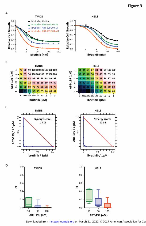
when combining these two compounds in the TMD8 and HBL1 cell lines (Figure 3A). To
illustrate the synergistic effects of this combination, we normalized viability data relative
to the effect of ABT-199 as a single agent. In these plots, the dose response of ABT-
199 combined with ibrutinib shifted toward a lower EC50 compared with the ibrutinib-only
dose response, indicating that the combination of these agents induced greater toxicity
than either agent alone and suggesting that addition of ABT-199 is able to overcome
BCL-2–associated ibrutinib resistance. Chalice Analyzer was used to analyze the drug
dose matrix and obtain the percentage of growth suppression for each of the
on March 21, 2020. © 2017 American Association for Cancer Research. mct.aacrjournals.org Downloaded from
Author manuscripts have been peer reviewed and accepted for publication but have not yet been edited. Author Manuscript Published OnlineFirst on April 20, 2017; DOI: 10.1158/1535-7163.MCT-16-0555
15
combinations (Figure 3B). The isobologram shows how much less drug is required
when it is used in combination compared with the single-agent doses needed to achieve
a desired effect. Synergy between ABT-199 and ibrutinib was confirmed using
isobologram analyses, synergy scores, and the CI obtained for each of the cell lines
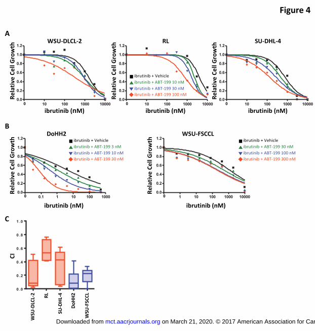
tested (Figures 3C and 3D). The combination of ibrutinib and ABT-199 was also
confirmed to have synergistic growth suppressive effects in GCB-DLBCL (WSU-DLCL-
2, RL, and SU-DHL-4) and FL (DoHH2 and WSU-FSCCL) cell lines, as shown by our
cell viability results revealing a dose response shift toward a lower EC50 (Figures 4A and
4B) and by our CI analyses (Figure 4C).
Ibrutinib and ABT-199 synergistically suppressed cell growth in ibrutinib-
resistant ABC-DLBCL and FL cells
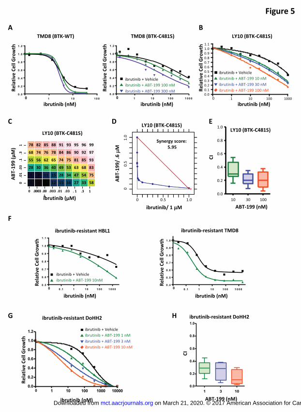
Given the synergy of ibrutinib and ABT-199 in parental ABC-DLBCL cells, we explored
the effects of this combination in ibrutinib-resistant cells generated by genetic mutation
(TMD8 [BTK-C481S] and OCI-LY10 [BTK-C481S]) or drug treatment (ibrutinib-resistant
HBL1 and TMD8). Relative gene expression of total BTK in TMD8 (BTK-WT) and in
TMD8 (BTK-C481S) was confirmed by qRT-PCR (Supplemental Figure 1A). ABT-199
increased the sensitivity of both cell lines to ibrutinib treatment (Figure 5A). We
observed that the effect of ABT-199 on ibrutinib sensitivity may be treatment time-
dependent as evidenced by the reduced effect of ABT-199 after a 3-day treatment in
TMD8 (BTK-WT) cells compared with a 5-day treatment in the parental TMD8 cells
(Figure 3A). We further generated OCI-LY10 (BTK-C481S) cells, which were confirmed
to display approximately 300-fold resistance to ibrutinib compared with parental OCI-
on March 21, 2020. © 2017 American Association for Cancer Research. mct.aacrjournals.org Downloaded from
Author manuscripts have been peer reviewed and accepted for publication but have not yet been edited. Author Manuscript Published OnlineFirst on April 20, 2017; DOI: 10.1158/1535-7163.MCT-16-0555
16
LY10 cells (Supplemental Figure 1B). Consistently, ABT-199 sensitized OCI-LY10
(BTK-C481S) cells to ibrutinib (Figures 5B and 5C). A strong synergistic toxicity of
ibrutinib and ABT-199 was confirmed by isobologram analysis of viability data (Figure
5D), as well as the CI obtained (Figure 5E). Consistent results were obtained in
ibrutinib-resistant HBL1 and TMD8 cells (Figure 5F). In addition to ibrutinib-resistant
ABC-DLBCL cells, the combination of ABT-199 and ibrutinib enhanced the sensitivity of
ibrutinib-resistant DoHH2 cells to ibrutinib (Figure 5G), and synergy between these two
compounds was demonstrated by the CI obtained (Figure 5H).
Combining ibrutinib and ABT-199 increased apoptosis, inhibited colony
formation, and suppressed tumor growth in ABC-DLBCL cells
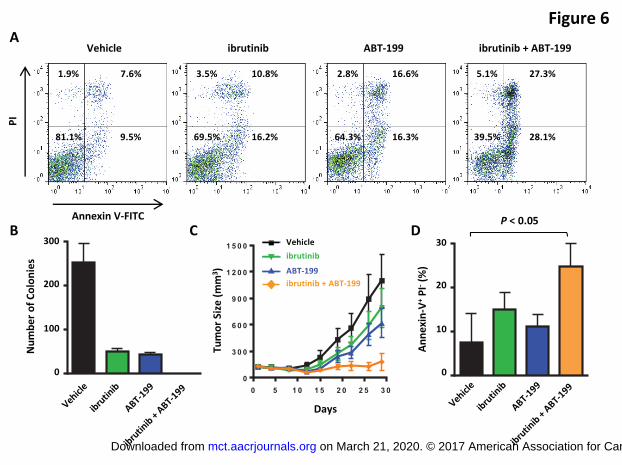
In addition to the effects on cell growth, treatment of TMD8 cells with a combination of
ibrutinib and ABT-199 resulted in increased cellular apoptosis (Figure 6A). We also
evaluated the effect of this combination on the clonogenicity of HBL1 cells. While single-
agent ibrutinib and ABT-199 significantly reduced the colony number, the combination
of both compounds completely abrogated colony formation in the methylcellulose
medium (Figure 6B).
We next investigated the effect of this drug combination in a xenograft model of ABC-
DLBCL. As a single agent, 12 mg/kg of ibrutinib or 40 mg/kg of ABT-199 only partially
suppressed TMD8 tumor growth, whereas the combination of these agents produced
full growth inhibition (Figure 6C). Notably, the apoptotic cell population significantly
on March 21, 2020. © 2017 American Association for Cancer Research. mct.aacrjournals.org Downloaded from
Author manuscripts have been peer reviewed and accepted for publication but have not yet been edited. Author Manuscript Published OnlineFirst on April 20, 2017; DOI: 10.1158/1535-7163.MCT-16-0555
17
increased in the drug combination–treated tumors (P < 0.05, unpaired t test) (Figure
6D), consistent with our in vitro observation (Figure 6A).
DISCUSSION
Although ibrutinib shows significant promise in ABC-DLBCL, it has limited efficacy as a
single agent (5). Fortunately, potential mechanisms by which to bypass this limitation
are quickly being discovered (9, 20). One approach to overcoming these obstacles is
the use of combination therapy. BCL-2 inhibition in combination with other targeted
therapies has demonstrated efficacy in several hematologic malignancies (21, 22).
Consistent with previous high-throughput analyses (19), we showed that ibrutinib and
ABT-199 synergistically suppressed cell growth, reduced colony formation, increased
apoptotic cell death, and inhibited tumor growth in an ABC-DLBCL mouse model. The
combination effect of ibrutinib and ABT-199 is not limited to ABC-DLBCL. We also
identified synergy between these two agents in both GCB-DLBCL and FL. Similar
efforts have been utilized and promising results have been obtained for this combination
strategy in other B-cell malignancies, including CLL (23), MCL (24, 25), and
Waldenström’s macroglobulinemia (26). Surprisingly, we also observed synergy
between ibrutinib and ABT-199 in ibrutinib-resistant cell lines, both in cells selected in
vitro for ibrutinib resistance and in those carrying a C481S-mutated form of BTK.
Deregulation of the antiapoptotic protein BCL-2 has been associated with resistance to
targeted therapy and chemotherapy in cancers (27, 28). We show in this study that
on March 21, 2020. © 2017 American Association for Cancer Research. mct.aacrjournals.org Downloaded from
Author manuscripts have been peer reviewed and accepted for publication but have not yet been edited. Author Manuscript Published OnlineFirst on April 20, 2017; DOI: 10.1158/1535-7163.MCT-16-0555
18
response to ibrutinib correlates with expression of BCL-2 in both cell lines and patient
tissues, with higher expression of BCL-2 associated with a more limited response.
Segregating ABC-DLBCL patients by BCL2 expression appears to identify a
subpopulation of patients with worse PFS, consistent with our finding that patients with
SD and PD during ibrutinib treatment have higher BCL2 expression than those with PR
and CR. These data suggest the existence of an antiapoptotic mechanism that limits the
impact of BTK inhibition.
Early studies indicated that ibrutinib interferes with the homing process of MCL cells and
increases circulating tumor cells (29). These circulating cells are more sensitive to ABT-
199 than those attached to fibroblast cells (13), providing the rationale for combining
ibrutinib and ABT-199 in MCL. The finding discussed here that ibrutinib-resistant cells
may have elevated sensitivity to ABT-199 due to compensatory upregulation of
antiapoptotic proteins such as BCL-2 provides another molecular mechanism underlying
the synergy between ibrutinib and ABT-199 in ABC-DLBCL cells. Whether there are
other disease-dependent mechanisms accounting for this combination effect requires
further investigation.
Several genes that were upregulated in ibrutinib-resistant cells according to our
microarray analyses have been previously linked to ibrutinib resistance and
development of lymphomas and leukemias. In a phase I/II clinical trial, ABC-DLBCL
patients with tumors harboring mutations in both CD79B and MYD88 had a much higher
ibrutinib response rate (4/5; 80%) than those with tumors containing WT CD79B and
mutant MYD88 (0/7; 0%) (5). Interestingly, recent work from Kim et al. showed
upregulation of CD79B expression in three distinct ibrutinib-resistant DLBCL cell lines
on March 21, 2020. © 2017 American Association for Cancer Research. mct.aacrjournals.org Downloaded from
Author manuscripts have been peer reviewed and accepted for publication but have not yet been edited. Author Manuscript Published OnlineFirst on April 20, 2017; DOI: 10.1158/1535-7163.MCT-16-0555
19
(30). In addition, this study convincingly demonstrated that overexpression of WT
CD79B induced tumor cell resistance to ibrutinib while depletion of CD79B sensitized
tumor cells to ibrutinib. Consistently, our microarray analyses revealed that CD79B,
among others, was significantly increased in ibrutinib-resistant TMD8 cells. Interestingly,
TMD8 cells are known to be heterozygous for a CD79B mutation (4). Whether the
increase in CD79B gene expression is of the WT or mutant form and whether these
different forms play distinct functions in the cells requires further investigation. In
addition to CD79B, we identified an increase in the BCR signaling-related gene PLCG2
in ibrutinib-resistant cells. This upregulation is consistent with previous findings
identifying PLCG2 mutations in clinical samples from CLL patients with ibrutinib
resistance and the role of PLCG2 in the formation of a BTK-bypass pathway (11, 31).
Several additional genes that were upregulated in ibrutinib-resistant cells are known to
be involved in the development of lymphomas or leukemias, including FOXP1 (32),
IGF1R (33), and KDM1A (34). Combining ibrutinib treatment with inhibitors targeting
these proteins may prevent the formation of ibrutinib resistance.
Collectively, these data indicate that the combination of ibrutinib with ABT-199 may be
highly effective in treating DLBCL and FL. Our data also implicate BCL-2 in resistance
to single-agent ibrutinib therapy and suggest that disruption of this compensatory
mechanism can shift the cell toward an apoptotic fate. Based on our findings in both in
vitro and in vivo models, the combination of ibrutinib with ABT-199 appears promising
and are currently being tested in clinical trials.
on March 21, 2020. © 2017 American Association for Cancer Research. mct.aacrjournals.org Downloaded from
Author manuscripts have been peer reviewed and accepted for publication but have not yet been edited. Author Manuscript Published OnlineFirst on April 20, 2017; DOI: 10.1158/1535-7163.MCT-16-0555
20
ACKNOWLEDGMENTS
We thank Kamaldeep Dhami, PhD and Kevin Kwei, PhD for providing TMD8 (BTK-WT)
and TMD8 (BTK-C481S) cells and performing drug combination studies. Brian Haas,
PhD, of Nexus GG Science, a medical writer supported by funding from Pharmacyclics,
LLC, an AbbVie Company, provided editorial assistance to the authors during
preparation of this manuscript.
on March 21, 2020. © 2017 American Association for Cancer Research. mct.aacrjournals.org Downloaded from
Author manuscripts have been peer reviewed and accepted for publication but have not yet been edited. Author Manuscript Published OnlineFirst on April 20, 2017; DOI: 10.1158/1535-7163.MCT-16-0555
21
REFERENCES
1. Alizadeh AA, Eisen MB, Davis RE, Ma C, Lossos IS, Rosenwald A, et al. Distinct
types of diffuse large B-cell lymphoma identified by gene expression profiling. Nature.
2000;403:503-11.
2. Staudt LM, Dave S. The biology of human lymphoid malignancies revealed by
gene expression profiling. Adv Immunol. 2005;87:163-208.
3. Wright G, Tan B, Rosenwald A, Hurt EH, Wiestner A, Staudt LM. A gene
expression-based method to diagnose clinically distinct subgroups of diffuse large B cell
lymphoma. Proc Natl Acad Sci U S A. 2003;100:9991-6.
4. Davis RE, Ngo VN, Lenz G, Tolar P, Young RM, Romesser PB, et al. Chronic
active B-cell-receptor signalling in diffuse large B-cell lymphoma. Nature. 2010;463:88-
92.
5. Wilson WH, Young RM, Schmitz R, Yang Y, Pittaluga S, Wright G, et al.
Targeting B cell receptor signaling with ibrutinib in diffuse large B cell lymphoma. Nat
Med. 2015;21:922-6.
6. Gazdar AF. Activating and resistance mutations of EGFR in non-small-cell lung
cancer: role in clinical response to EGFR tyrosine kinase inhibitors. Oncogene. 2009;28
Suppl 1:S24-31.
7. Nardi V, Azam M, Daley GQ. Mechanisms and implications of imatinib resistance
mutations in BCR-ABL. Curr Opin Hematol. 2004;11:35-43.
8. Chang BY, Furman RR, Zapatka M, Barrientos JC, Li D, Steggerda S, et al. Use
of tumor genomic profiling to reveal mechanisms of resistance to the BTK inhibitor
on March 21, 2020. © 2017 American Association for Cancer Research. mct.aacrjournals.org Downloaded from
Author manuscripts have been peer reviewed and accepted for publication but have not yet been edited. Author Manuscript Published OnlineFirst on April 20, 2017; DOI: 10.1158/1535-7163.MCT-16-0555
22
ibrutinib in chronic lymphocytic leukemia (CLL). J Clin Oncol. 31, 2013 (suppl; abstr
7014).
9. Woyach JA, Furman RR, Liu TM, Ozer HG, Zapatka M, Ruppert AS, et al.
Resistance mechanisms for the Bruton's tyrosine kinase inhibitor ibrutinib. N Engl J
Med. 2014;370:2286-94.
10. Zhang SQ, Smith SM, Zhang SY, Lynn Wang Y. Mechanisms of ibrutinib
resistance in chronic lymphocytic leukaemia and non-Hodgkin lymphoma. Br J
Haematol. 2015;170:445-56.
11. Burger JA, Landau DA, Taylor-Weiner A, Bozic I, Zhang H, Sarosiek K, et al.
Clonal evolution in patients with chronic lymphocytic leukaemia developing resistance to
BTK inhibition. Nat Commun. 2016;7:11589.
12. Chou TC. Theoretical basis, experimental design, and computerized simulation
of synergism and antagonism in drug combination studies. Pharmacol Rev.
2006;58:621-81.
13. Chiron D, Dousset C, Brosseau C, Touzeau C, Maiga S, Moreau P, et al.
Biological rational for sequential targeting of Bruton tyrosine kinase and Bcl-2 to
overcome CD40-induced ABT-199 resistance in mantle cell lymphoma. Oncotarget.
2015;6:8750-9.
14. Vogler M, Butterworth M, Majid A, Walewska RJ, Sun XM, Dyer MJ, et al.
Concurrent up-regulation of BCL-XL and BCL2A1 induces approximately 1000-fold
resistance to ABT-737 in chronic lymphocytic leukemia. Blood. 2009;113:4403-13.
on March 21, 2020. © 2017 American Association for Cancer Research. mct.aacrjournals.org Downloaded from
Author manuscripts have been peer reviewed and accepted for publication but have not yet been edited. Author Manuscript Published OnlineFirst on April 20, 2017; DOI: 10.1158/1535-7163.MCT-16-0555
23
15. Souers AJ, Leverson JD, Boghaert ER, Ackler SL, Catron ND, Chen J, et al.
ABT-199, a potent and selective BCL-2 inhibitor, achieves antitumor activity while
sparing platelets. Nat Med. 2013;19:202-8.
16. Ngo VN, Young RM, Schmitz R, Jhavar S, Xiao W, Lim KH, et al. Oncogenically
active MYD88 mutations in human lymphoma. Nature. 2011;470:115-9.
17. Wiestner A. The role of B-cell receptor inhibitors in the treatment of patients with
chronic lymphocytic leukemia. Haematologica. 2015;100:1495-507.
18. Catz SD, Johnson JL. Transcriptional regulation of bcl-2 by nuclear factor kappa
B and its significance in prostate cancer. Oncogene. 2001;20:7342-51.
19. Mathews Griner LA, Guha R, Shinn P, Young RM, Keller JM, Liu D, et al. High-
throughput combinatorial screening identifies drugs that cooperate with ibrutinib to kill
activated B-cell-like diffuse large B-cell lymphoma cells. Proc Natl Acad Sci U S A.
2014;111:2349-54.
20. Furman RR, Cheng S, Lu P, Setty M, Perez AR, Guo A, et al. Ibrutinib resistance
in chronic lymphocytic leukemia. N Engl J Med. 2014;370:2352-4.
21. Li L, Pongtornpipat P, Tiutan T, Kendrick SL, Park S, Persky DO, et al.
Synergistic induction of apoptosis in high-risk DLBCL by BCL2 inhibition with ABT-199
combined with pharmacologic loss of MCL1. Leukemia. 2015;29:1702-12.
22. Knorr KL, Schneider PA, Meng XW, Dai H, Smith BD, Hess AD, et al. MLN4924
induces Noxa upregulation in acute myelogenous leukemia and synergizes with Bcl-2
inhibitors. Cell Death Differ. 2015;22:2133-42.
23. Cervantes-Gomez F, Lamothe B, Woyach JA, Wierda WG, Keating MJ,
Balakrishnan K, et al. Pharmacological and Protein Profiling Suggests Venetoclax (ABT-
on March 21, 2020. © 2017 American Association for Cancer Research. mct.aacrjournals.org Downloaded from
Author manuscripts have been peer reviewed and accepted for publication but have not yet been edited. Author Manuscript Published OnlineFirst on April 20, 2017; DOI: 10.1158/1535-7163.MCT-16-0555
24
199) as Optimal Partner with Ibrutinib in Chronic Lymphocytic Leukemia. Clin Cancer
Res. 2015;21:3705-15.
24. Zhao X, Bodo J, Sun D, Durkin L, Lin J, Smith MR, et al. Combination of ibrutinib
with ABT-199: synergistic effects on proliferation inhibition and apoptosis in mantle cell
lymphoma cells through perturbation of BTK, AKT and BCL2 pathways. Br J Haematol.
2015;168:765-8.
25. Li Y, Bouchlaka MN, Wolff J, Grindle KM, Lu L, Qian S, et al. FBXO10 deficiency
and BTK activation upregulate BCL2 expression in mantle cell lymphoma. Oncogene.
2016;35:6223-34.
26. Cao Y, Yang G, Hunter ZR, Liu X, Xu L, Chen J, et al. The BCL2 antagonist
ABT-199 triggers apoptosis, and augments ibrutinib and idelalisib mediated cytotoxicity
in CXCR4 Wild-type and CXCR4 WHIM mutated Waldenstrom macroglobulinaemia
cells. Br J Haematol. 2015;170:134-8.
27. Dupont T, Yang SN, Patel J, Hatzi K, Malik A, Tam W, et al. Selective targeting
of BCL6 induces oncogene addiction switching to BCL2 in B-cell lymphoma.
Oncotarget. 2016;7:3520-32.
28. Scarfo L, Ghia P. Reprogramming cell death: BCL2 family inhibition in
hematological malignancies. Immunol Lett. 2013;155:36-9.
29. Chang BY, Francesco M, De Rooij MF, Magadala P, Steggerda SM, Huang MM,
et al. Egress of CD19(+)CD5(+) cells into peripheral blood following treatment with the
Bruton tyrosine kinase inhibitor ibrutinib in mantle cell lymphoma patients. Blood.
2013;122:2412-24.
on March 21, 2020. © 2017 American Association for Cancer Research. mct.aacrjournals.org Downloaded from
Author manuscripts have been peer reviewed and accepted for publication but have not yet been edited. Author Manuscript Published OnlineFirst on April 20, 2017; DOI: 10.1158/1535-7163.MCT-16-0555
25
30. Kim JH, Kim WS, Ryu K, Kim SJ, Park C. CD79B limits response of diffuse large
B cell lymphoma to ibrutinib. Leuk Lymphoma. 2016;57:1413-22.
31. Liu TM, Woyach JA, Zhong Y, Lozanski A, Lozanski G, Dong S, et al.
Hypermorphic mutation of phospholipase C, gamma2 acquired in ibrutinib-resistant CLL
confers BTK independency upon B-cell receptor activation. Blood. 2015;126:61-8.
32. Barrans SL, Fenton JA, Banham A, Owen RG, Jack AS. Strong expression of
FOXP1 identifies a distinct subset of diffuse large B-cell lymphoma (DLBCL) patients
with poor outcome. Blood. 2004;104:2933-5.
33. Yaktapour N, Ubelhart R, Schuler J, Aumann K, Dierks C, Burger M, et al.
Insulin-like growth factor-1 receptor (IGF1R) as a novel target in chronic lymphocytic
leukemia. Blood. 2013;122:1621-33.
34. Harris WJ, Huang X, Lynch JT, Spencer GJ, Hitchin JR, Li Y, et al. The histone
demethylase KDM1A sustains the oncogenic potential of MLL-AF9 leukemia stem cells.
Cancer Cell. 2012;21:473-87.
on March 21, 2020. © 2017 American Association for Cancer Research. mct.aacrjournals.org Downloaded from
Author manuscripts have been peer reviewed and accepted for publication but have not yet been edited. Author Manuscript Published OnlineFirst on April 20, 2017; DOI: 10.1158/1535-7163.MCT-16-0555
26
FIGURE LEGENDS
Figure 1. Ibrutinib-resistant TMD8 cells had higher BCL2 gene expression and were
more sensitive to ABT-199. (A) Heatmap presentation of normalized log2-transformed
apoptosis-related gene expression profiles in TMD8 parental versus ibrutinib-resistant
TMD8 cells (red, high; green, low). (B) BCL2 gene expression increased in ibrutinib-
resistant TMD8 cells. Gene expression levels of BAX, BCL2, and MCL1 were
determined by qRT-PCR, with GAPDH and ACTB used as reference genes. All data are
presented as fold change over TMD8 parental cells. (C) BCL2 gene expression
increased in ibrutinib-resistant DoHH2 cells. BCL2 gene expression was determined by
qRT-PCR, with GAPDH used as a reference gene. Data are presented as fold change
over DoHH2 parental cells. (D) BCL-2 protein level in parental and ibrutinib-resistant
TMD8 or DoHH2 cells was determined by immunoblot analysis. (E) Ibrutinib-resistant
TMD8 cells were more sensitive to ABT-199 than TMD8 parental cells. Cells were
treated with ABT-199 for 3 days, and the drug effect on cell growth was determined by
CellTiter-Glo luminescent cell viability assay. (F) TMD8 cells were stimulated with 1 μM
of the TLR9 agonist ODN 2216 for 3 days, and the gene expression level of BCL2 was
determined by qRT-PCR, and GAPDH was used as the reference gene. Data are
presented as fold change over control ODN treated cells. (G) TMD8 cells were
transfected with nontargeting control siRNA or siRNAs targeting MAP3K7 and gene
expression levels of BCL2 and MAP3K7 were determined by qRT-PCR, and GAPDH
was used as the reference gene. Data are presented as fold change over control siRNA
transfected cells. (H) TMD8 cells were transfected with nontargeting control siRNA or
siRNAs targeting IRAK4 and gene expression levels of BCL2 and IRAK4 were
on March 21, 2020. © 2017 American Association for Cancer Research. mct.aacrjournals.org Downloaded from
Author manuscripts have been peer reviewed and accepted for publication but have not yet been edited. Author Manuscript Published OnlineFirst on April 20, 2017; DOI: 10.1158/1535-7163.MCT-16-0555
27
determined by qRT-PCR, and GAPDH was used as the reference gene. Data are
presented as fold change over control siRNA transfected cells.
on March 21, 2020. © 2017 American Association for Cancer Research. mct.aacrjournals.org Downloaded from
Author manuscripts have been peer reviewed and accepted for publication but have not yet been edited. Author Manuscript Published OnlineFirst on April 20, 2017; DOI: 10.1158/1535-7163.MCT-16-0555
28
Figure 2. Higher BCL-2 gene expression was observed in tumors from patients with
poorer response to ibrutinib. (A) Differential BCL2 gene expression was observed in
tumors from ABC-DLBCL and GCB-DLBCL patients. (B) Higher BCL2 gene expression
was detected in tumors from ABC-DLBCL patients with poorer response (PD+SD).
BCL2 gene expression levels were analyzed, and a rank-based statistic (RankProd)
was used to determine the significance (P < 0.001). (C) Kaplan-Meier survival curves of
progression-free survival for patients with low BCL2 (black) and high BCL2 (red) gene
expression. ABC-DLBCL patients with higher BCL2 gene expression had significantly
worse survival than those with lower BCL2 gene expression (P < 0.05, log-rank test).
on March 21, 2020. © 2017 American Association for Cancer Research. mct.aacrjournals.org Downloaded from
Author manuscripts have been peer reviewed and accepted for publication but have not yet been edited. Author Manuscript Published OnlineFirst on April 20, 2017; DOI: 10.1158/1535-7163.MCT-16-0555
29
Figure 3. Ibrutinib and ABT-199 synergistically suppressed cell growth in ABC-DLBCL
cells. (A) TMD8 and HBL1 cells were treated with the indicated concentrations of
ibrutinib combined with ABT-199 (10, 30, and 100 nM) or vehicle for 5 days, and drug
effect on cell growth was determined using CellTiter-Glo® Luminescent Cell Viability
Assay. (B) Drug dose matrix data in TMD8 and HBL1 cells. The numbers indicate the
percentage of growth inhibition of cells treated with the corresponding compound
combination relative to vehicle control–treated cells. The data are visualized over the
matrix using a color scale. (C) Isobologram analyses and synergy scores of the data in
(B) indicate synergy for the combination of ibrutinib and ABT-199. (D) CI of ibrutinib and
ABT-199 at indicated concentrations in TMD8 and HBL1 cells. Boxes represent median
values with the first and third quartiles. Whiskers represent the maximum and minimum
values.
on March 21, 2020. © 2017 American Association for Cancer Research. mct.aacrjournals.org Downloaded from
Author manuscripts have been peer reviewed and accepted for publication but have not yet been edited. Author Manuscript Published OnlineFirst on April 20, 2017; DOI: 10.1158/1535-7163.MCT-16-0555

30
Figure 4. Ibrutinib and ABT-199 synergistically suppressed cell growth in GCB-DLBCL
and FL cells. (A) GCB-DLBCL cells (WSU-DLCL-2, RL, and SU-DHL-4) were treated
with the indicated concentrations of ibrutinib combined with ABT-199 (10, 30, and 100
nM) or vehicle for 3 days, and drug effect on cell growth was determined using CellTiter-
Glo Luminescent Cell Viability Assay. (B) FL cells (DoHH2 and WSU-FSCCL) were
treated with indicated concentrations of ibrutinib combined with ABT-199 or vehicle for 3
days, and drug effect on cell growth was determined using CellTiter-Glo Luminescent
Cell Viability Assay. (C) CI of the ibrutinib and ABT-199 combination in GCB-DLBCL
and FL cells. The CIs of different concentrations of ibrutinib combined with ABT-199 at
100 nM (WSU-DLCL-2, RL, and SU-DHL-4), 30 nM (DoHH2), and 300 nM (WSU-
FSCCL) are shown.
on March 21, 2020. © 2017 American Association for Cancer Research. mct.aacrjournals.org Downloaded from
Author manuscripts have been peer reviewed and accepted for publication but have not yet been edited. Author Manuscript Published OnlineFirst on April 20, 2017; DOI: 10.1158/1535-7163.MCT-16-0555
31
Figure 5. Ibrutinib and ABT-199 synergistically suppressed cell growth in ibrutinib-
resistant ABC-DLBCL and FL cells. (A) TMD8 (BTK-WT) and TMD8 (BTK-C481S) cells
were treated with indicated concentrations of ibrutinib combined with ABT-199 (100 and
300 nM) or vehicle for 3 days, and drug effect on cell growth was determined using
CellTiter-Glo Luminescent Cell Viability Assay. (B) OCI-LY10 (BTK-C481S) cells were
treated with indicated concentrations of ibrutinib combined with ABT-199 (10, 30, and
100 nM) or vehicle for 5 days, and drug effect on cell growth was determined using
CellTiter-Glo Luminescent Cell Viability Assay. (C) Drug dose matrix data from OCI-
LY10 (BTK-C481S) cells. (D) Isobologram analysis and synergy score of the data
shown in (B). (E) CI of ibrutinib and ABT-199 at the indicated concentrations in OCI-
LY10 (BTK-C481S) cells. (F) Ibrutinib-resistant HBL1 and TMD8 cells were treated with
indicated concentrations of ibrutinib combined with ABT-199 (10 nM) or vehicle for 3
days, and drug effect on cell growth was determined using CellTiter-Glo Luminescent
Cell Viability Assay. (G) Ibrutinib-resistant DoHH2 cells were treated with the indicated
concentrations of ibrutinib combined with ABT-199 (1, 3, and 10 nM) or vehicle for 3
days, and drug effect on cell growth was determined using CellTiter-Glo Luminescent
Cell Viability Assay. (H) CIs of ibrutinib and ABT-199 at the indicated concentrations in
ibrutinib-resistant DoHH2 cells.
on March 21, 2020. © 2017 American Association for Cancer Research. mct.aacrjournals.org Downloaded from
Author manuscripts have been peer reviewed and accepted for publication but have not yet been edited. Author Manuscript Published OnlineFirst on April 20, 2017; DOI: 10.1158/1535-7163.MCT-16-0555
32
Figure 6. Combination of ibrutinib and ABT-199 increased the apoptotic cell population,
inhibited colony formation, and suppressed tumor growth. (A) TMD8 cells were treated
for 1 day with ibrutinib (100 nM), ABT-199 (1 µM), or the combination and analyzed for
annexin V binding as well as for PI uptake. The percentage of annexin V-positive, PI-
positive or both annexin V- and PI-positive cells is indicated. (B) HBL1 cells were plated
in 0.9% MethoCult (1,000 cells/well) with vehicle, ibrutinib (10 nM), ABT-199 (50 nM), or
the combination, and colony formation was scored after 7 days. Graph represents
quantifications of 3 wells expressed as the mean ± SD (C) TMD8 tumor cells were
implanted into CB17 SCID mice, and the indicated drugs were orally administered daily
when the tumors reached 100 mm3. Tumors were measured twice a week. Graph
represents quantifications of 10 mice expressed as the mean ± SE. (D) The apoptotic
cell population (annexin V-positive and PI-negative) of TMD8 tumor cells from CB17
SCID mice treated with indicated drugs were analyzed by flow cytometry. Data are
expressed as the mean ± SE.
on March 21, 2020. © 2017 American Association for Cancer Research. mct.aacrjournals.org Downloaded from
Author manuscripts have been peer reviewed and accepted for publication but have not yet been edited. Author Manuscript Published OnlineFirst on April 20, 2017; DOI: 10.1158/1535-7163.MCT-16-0555
0.1 1 10 100 1000
0.0
0.2
0.4
0.6
0.8
1.0
1.2
0.1 1 10 100 1000
0.0
0.2
0.4
0.6
0.8
1.0
1.2
on March 21, 2020. © 2017 American Association for Cancer Research. mct.aacrjournals.org Downloaded from
Author manuscripts have been peer reviewed and accepted for publication but have not yet been edited. Author Manuscript Published OnlineFirst on April 20, 2017; DOI: 10.1158/1535-7163.MCT-16-0555
0.0
0.2
0.4
0.6
0.8
1.0
0.1 1 10 100 1000
0.0
0.1
0.2
0.3
0.4
0.5
0.6
0.7
0.8
0.9
1.0
1.1
on March 21, 2020. © 2017 American Association for Cancer Research. mct.aacrjournals.org Downloaded from
Author manuscripts have been peer reviewed and accepted for publication but have not yet been edited. Author Manuscript Published OnlineFirst on April 20, 2017; DOI: 10.1158/1535-7163.MCT-16-0555
Published OnlineFirst April 20, 2017.Mol Cancer Ther Hsu-Ping Kuo, Scott A. Ezell, Karl J. Schweighofer, et al. Lymphoma and Follicular LymphomaCombination of Ibrutinib and ABT-199 in Diffuse Large B-Cell
Updated version
10.1158/1535-7163.MCT-16-0555doi:
Access the most recent version of this article at:
Material
Supplementary
http://mct.aacrjournals.org/content/suppl/2017/04/20/1535-7163.MCT-16-0555.DC1
Access the most recent supplemental material at:
Manuscript
Authoredited. Author manuscripts have been peer reviewed and accepted for publication but have not yet been
E-mail alerts related to this article or journal.Sign up to receive free email-alerts
Subscriptions
Reprints and
To order reprints of this article or to subscribe to the journal, contact the AACR Publications
Permissions
Rightslink site. Click on "Request Permissions" which will take you to the Copyright Clearance Center's (CCC)
.http://mct.aacrjournals.org/content/early/2017/04/20/1535-7163.MCT-16-0555To request permission to re-use all or part of this article, use this link
on March 21, 2020. © 2017 American Association for Cancer Research. mct.aacrjournals.org Downloaded from
Author manuscripts have been peer reviewed and accepted for publication but have not yet been edited. Author Manuscript Published OnlineFirst on April 20, 2017; DOI: 10.1158/1535-7163.MCT-16-0555