Embed Size (px)
Citation preview
The Protein Composition of the Digestive Fluidfrom the Venus Flytrap Sheds Light on PreyDigestion Mechanisms*□S
Waltraud X. Schulze‡a, Kristian W. Sanggaard§a, Ines Kreuzer¶, Anders D. Knudsen§,Felix Bemm�, Ida B. Thøgersen§, Andrea Brautigam‡‡, Line R. Thomsen§,Simon Schliesky‡‡, Thomas F. Dyrlund§, Maria Escalante-Perez¶, Dirk Becker¶,Jorg Schultz�, Henrik Karring§§, Andreas Weber‡‡, Peter Højrup¶¶,Rainer Hedrich¶��**, and Jan J. Enghild§**
The Venus flytrap (Dionaea muscipula) is one of the mostwell-known carnivorous plants because of its unique abil-ity to capture small animals, usually insects or spiders,through a unique snap-trapping mechanism. The animalsare subsequently killed and digested so that the plantscan assimilate nutrients, as they grow in mineral-deficientsoils. We deep sequenced the cDNA from Dionaea trapsto obtain transcript libraries, which were used in the massspectrometry-based identification of the proteins se-creted during digestion. The identified proteins consistedof peroxidases, nucleases, phosphatases, phospho-lipases, a glucanase, chitinases, and proteolytic enzymes,including four cysteine proteases, two aspartic proteases,and a serine carboxypeptidase. The majority of the mostabundant proteins were categorized as pathogenesis-re-lated proteins, suggesting that the plant’s digestive sys-tem evolved from defense-related processes. This in-depth characterization of a highly specialized secretedfluid from a carnivorous plant provides new informationabout the plant’s prey digestion mechanism and the evo-lutionary processes driving its defense pathways and nu-trient acquisition. Molecular & Cellular Proteomics 11:10.1074/mcp.M112.021006, 1306–1319, 2012.
Carnivorous plants capture, digest, and “eat” animals usingfour different types of trapping strategies: (i) flypaper or ad-hesive traps (e.g. Drosera, also known as sundews, and Pin-guicula, also known as butterworts), (ii) sucking bladder traps(e.g. Utricularia, also known as bladderworts), (iii) pitfall traps(e.g. Nepenthes), and (iv) snap traps (e.g. Dionaea muscipula,also known as the Venus flytrap). These plants fascinatedCharles Darwin. The Venus flytrap, in particular, attracted hisattention, and he described the plant as “one of the mostwonderful in the world” (1). The snap trap most likely evolvedfrom the adhesive trap, because its ability to capture largerprey than the adhesive traps gives it an evolutionary advan-tage (2).
The trapping motion of Dionaea muscipula is among thefastest movements in the plant kingdom, and its mechanismhas been described in detail, starting with Charles Darwin’swork from �150 years ago (3–6). The plant’s leaves employturgor pressure and hydrodynamic flow to close the trap (3).The closing is initiated by the mechanical stimulation of triggerhairs, eliciting an action potential to close the trap, whichseals the fate of the animal inside (1). Then “touch” hormonessuch as 12-oxophytodienoic acid, which is a precursor of thephytohormone jasmonic acid, probably induce the secretionof digestive fluid (7). Touch hormones are likely to be releasedin response to the continuous mechanical stimulation of thetrigger hairs by the prey as it struggles to escape (7). The trapmay also be closed artificially by direct electrical stimulationor by the application of the bacterial phytotoxine coronatine(5, 7, 8).
The largest classes of Venus flytrap prey are spiders andflies. Highly active fliers, such as bees and wasps, are rarelycaught (9). The trapped animal faces a slow death, and ex-periments with ants demonstrate that the prey are alive andcapable of stimulating the trigger hairs up to 8 h after beingcaught (10). The nutrients obtained from the digestion of thedifferent prey are important for the Venus flytrap. Amongcarnivorous plants in their natural habitats, the Venus flytrapappears to be the most dependent on the nitrogen obtained
From the ‡Max Planck Institut fur Molekulare Pflanzenphysiologie,Am Muhlenberg 1, 14476 Potsdam, Germany; §Department of Mo-lecular Biology and Genetics, Aarhus University, Gustav Wiedsvej10C, 8000 Aarhus C, Denmark; ¶Department of Molecular PlantPhysiology & Biophysics, Universitat Wurzburg, Julius-von-Sachs-Platz 2, 97082 Wurzburg, Germany; �Department of Bioinformatics,Biozentrum, Am Hubland, Universitat Wurzburg, D-97074 Wuerzburg,Germany; ‡‡Department of Plant Biochemistry, Heinrich-Heine-Uni-versitaet Duesseldorf, Universitaetsstrasse 1, 40225 Duesseldorf,Germany; §§University of Southern Denmark, Institute of ChemicalEngineering, Biotechnology and Environmental Technology, NielsBohrs Alle 1, 5230 Odense M, Denmark; ¶¶Department of Biochem-istry and Molecular Biology, University of Southern Denmark, Cam-pusvej 55, 5230 Odense M, Denmark; ��Zoology Department, Collegeof Science, King Saud University, P.O. Box 2455, Riyadh 11451,Saudi Arabia
Received June 5, 2012, and in revised form, July 26, 2012Published, MCP Papers in Press, August 12, 2012, DOI 10.1074/
mcp.M112.021006
Research© 2012 by The American Society for Biochemistry and Molecular Biology, Inc.This paper is available on line at http://www.mcponline.org
1306 Molecular & Cellular Proteomics 11.11
from its digested prey (11). The nutrients from insects andspiders give the plants a competitive advantage in their nat-ural low-nutrient soil habitats (12).
In contrast to its trapping mechanism, only a few studieshave focused on the digestion process of the Venus flytrap,and none of the involved enzymes has been purified. How-ever, the pH during Venus flytrap digestion has been studied.The pH of the digestive fluid is 4.3, and during the secretionphase the external “stomach” is further acidified to pH 3.4 (7,13). The optimum pH for protease activity in the fluid has beenanalyzed in different studies, and the resulting values rangefrom pH 3.0 to pH 7.0 (13–16). This discrepancy is likely dueto differences in the assay conditions and the substrates usedas targets during the analyses (13, 16).
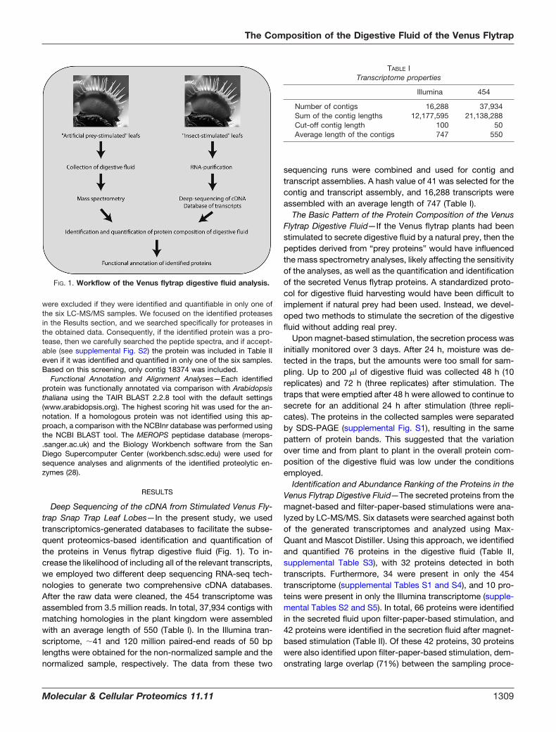
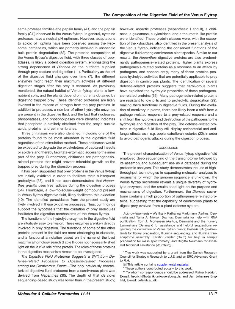
In our work, we have determined the protein composition ofthe digestive fluid of the Venus flytrap. The protein identifica-tions were based on a two-step approach involving (i) deepsequencing of the cDNA from stimulated leaves (RNA-seq)and (ii) subsequent mass spectrometric (MS)1 analyses of theproteins in the collected digestive fluids (Fig. 1). Both theRNA-seq analyses and the digestion fluid proteomics wereperformed on independent samples using complementary ap-proaches. The obtained mass spectra were searched againstthe two generated transcriptome databases, and the identi-fied proteins in the secretome were abundance-ranked basedon their intensity sums. Our results provide insights into thecomplex composition of the Venus flytrap’s digestive fluid,which has vital functions in defense and nutrient digestion.
EXPERIMENTAL PROCEDURES
Plant Material for 454 Sequencing and Sampling by Filter PaperStimulation—Dionaea muscipula plants were purchased fromCRESCO Carnivora (De Kwakel, The Netherlands) and grown in plas-tic pots at 22 °C in a 16:8 h light:dark photoperiod. Three stimulationmethods were used for the transcriptomic approach: (i) the plantswere fed with ants, and the traps were collected after 24 h; (ii) theplants were sprayed with 100 �M coronatine, and the traps wereharvested after 24 h; and (iii) the plants were stimulated by theplacement of filter paper soaked with either 30 mM urea, 30 mM chitin,or water into the trap, and trap tissue was collected 1 and 8 h afterstimulation. The material for the transcriptome analyses was har-vested as follows: traps and excised trigger hairs were frozen in liquidnitrogen. Additionally, secretory cells were isolated from the inner trapsurface by gently abrading the gland complexes using a razor blade.RNA was separately isolated from each sample, and for cDNA syn-thesis, the RNA from different tissues was pooled.
To stimulate fluid secretion for the protein analyses, the closure ofhealthy mature traps was initiated by tickling the trigger hairs withinthe trap. Because secretion does not begin without further stimulationof the trigger hairs while the trap is in the closed state, a fine piece offilter paper soaked with water was trapped in the closing snap trap,allowing for the induced movement of the trigger hairs by slightmovements of the filter paper. Secreted liquid was then carefullysampled from the closed trap using a pipette tip that was insertedbetween the closed trap lobes.
Plant Material for Illumina Sequencing and Sampling by Magnet-based Stimulation—Plants were purchased at the Lammehave nurs-ery (Ringe, Denmark) and grown in a walk-in plant growth chamber at26 °C in a 12:12 h light:dark photoperiod. All of the experiments wereperformed on healthy mature plants. For the transcriptomics analyses,the digestion process was initiated by feeding the plants yellow meal-worm beetles (Tenebrio molitor). After 3, 8, 24, 48, and 72 h, the leaveswere harvested, rinsed with water to remove the partially digestedbeetle and beetle fragments, snap frozen in liquid nitrogen, and storedat �80 °C. For each time point, two stimulated traps were harvested.
For the protein analyses, magnet-based stimulation of the leaf wasused to induce the secretion of the digestive fluid. A small stick-magnet was positioned between the trap leaves, and 100 �l of thecysteine protease inhibitor trans-epoxysuccinyl-l-leucylamido-(4-guanidino) butane (E-64; 50 �M) was added to the trap to reduce thelevel of adventitious proteolysis (14, 16). Then a larger magnet wasapplied to move the smaller magnet inside the trap, stimulating thetrigger hairs and resulting in complete closure. After 48 and/or 72 h,the secreted fluid (up to 200 �l) was aspirated using a pipette that wasforced in between the leaf lobes of the trap. The collected materialwas centrifuged, and the supernatant was used for further analyses.If not used immediately, the fluid was stored at �20 °C.
Transcriptome Sequencing and Assembly—The leaves from thestimulated traps were pooled and homogenized before RNA wasextracted using a previously described hot borate buffer protocol (17).Poly-A transcripts were enriched from 3.5 �g of total RNA, and thetranscripts were fractionated in the presence of Zn2�. Subsequently,double-stranded cDNA synthesis was performed using random prim-ers and RNase H. After end repair and purification, the fragmentswere ligated with bar-coded paired-end adapters, and fragments withinsert sizes of �150 to 250 bp were isolated from an agarose gel. Halfof the library was normalized, and the other half was amplified viaPCR and purified from a gel. The library quality was assessed usingcapillary sequencing of randomly selected clones. The high-through-put sequencing of the cDNA samples from the beetle-stimulated trapswas performed on an Illumina HiSeq 2000 instrument using a paired-end run with 2 � 50 bp. The cDNA samples isolated from the trapsfrom the other combined stimulation approach were sequenced usinga 454 GS FLX Titanium platform. The filtered reads from the twotranscriptomic datasets were assembled using Oases software (56)on top of Velvet and Mira, respectively. The minimum size for theassembly was set to either 50 or 100 bases. Several parameter sets(e.g. Burrows-Wheeler Alignment) were tested to optimize theassemblies.
Sampling Procedures for Proteomics—For the filter paper stimula-tion method, the secreted fluid was collected into three independentpools from 15 to 20 traps stimulated for 68 h. The protein wasprecipitated using ice-cold acetone. Protein pellets were resus-pended in 6 M urea and 2 M thiourea (pH 8). After the reduction ofdisulfide bonds with dithiothreitol (DTT), free cysteine residues werealkylated using iodacetamide. Proteins were predigested with Lys-Cfor 3 h before the dilution of the sample with four volumes of 10 mM
Tris-HCl (pH 8). Trypsin was added (1 �g trypsin per 50 �g protein),and the digestions were performed at room temperature for 16 h.After acidification, the peptides were desalted over a C18 matrix priorto the MS analysis.
For the magnet-based method, digestive fluid was collected 48 hafter the stimulation of 18 traps. Subsequently, three pools wereprepared using the digestive fluid from five to eight plants. From thesesamples, 35 �l were withdrawn, and the pH was adjusted to 8.5.Subsequently, DTT was added, the samples were boiled for 5 min,and iodoacetamide was then added to alkylate free cysteine residues.After 15 min of incubation, trypsin was added (1:20 ratio), and thedigestions were performed at 37 °C for 16 h.
1 The abbreviations used are: DTT, dithiothreitol; LC, liquid chro-matography; MS, mass spectrometric.
The Composition of the Digestive Fluid of the Venus Flytrap
Molecular & Cellular Proteomics 11.11 1307
Gel-free Proteomics of the Digestive Fluid (from Both SamplingProcedures)—The resulting peptides from each of the digests wereseparated using an EasyLC nanoflow HPLC system (Proxeon Biosys-tems, Odense, Denmark) connected to an LTQ-Orbitrap XL massspectrometer (Thermo Fisher Scientific) equipped with a nanoESI ionsource (Proxeon Biosystems, Odense, Denmark). The chromato-graphic separation was performed on a 15-cm fused silica emitter(100 �m i.d.) that was in-house packed with RP ReproSil-Pur C18-AQ3 �m resin (Dr. Marisch GmbH, Ammerbuch-Entringen, Germany).The peptides were eluted using an acidic acetonitrile gradient at aflow rate of 250 nl min�1, as described elsewhere (18, 19).
MS scans (300–1800 m/z) were recorded using an Orbitrap massanalyzer at a resolution of 60,000 at 400 m/z, with 1 � 106 automaticgain control target ions and a 500-ms maximum ion injection time.The MS scans were followed by data-dependent collision-induceddissociation MS/MS scans of the five most intense multiply chargedions in the mass spectrometer at a 15,000 signal threshold, 30,000automatic gain control target, 300-ms maximum ion injection time,2.5-m/z isolation width, 30-ms activation time at 35 normalized col-lision energy, and dynamic exclusion enabled for 30 s with a repeatcount of 1. Peak picking was performed using either MaxQuant 1.114(Max Planck Institute of Biochemistry, Martinsried, Germany) or Xcali-bur 2.0 (Thermo Fisher Scientific Inc., Waltham, MA). The raw datafiles of the in-solution digestion of the Venus flytrap secretion fluidfrom both sampling methods have been deposited at the Tranchedatabase (proteomecommons.org) under the following hash key:tzwFTnv4Y04ujdmGC1tVaULYKS3OQ/0i3pmQ2P0vvvdHe6�5vX09E6zW4OzKILOJJDZ9OTXzOB8N66�5czMOCv2MORA-AAAAAAAAE/A��.
Identification and Quantification of the Secretome Proteins Usingthe 454 Transcriptome—The acquired raw data files were searchedagainst the 6-frame translation of the 454 transcriptome (in total,227,604 protein entries) using MaxQuant 1.114 (20). The carbam-idomethylation of cysteine residues was set as a fixed modification,and the oxidation of methionine residues was set as a variable mod-ification. Two missed cleavages were allowed. The mass tolerance forthe first search was set to 10 ppm, and the fragment mass tolerancewas set to 0.5 Da. The peptide and protein false discovery rates wereset to 0.01, and the identified peptides were required to have aminimum length of six amino acid residues. The assignment of theidentified peptides to translated proteins was primarily based onproteotypic peptides. Peptides with more than one protein matchwere assigned to protein groups consisting of all of the proteins withtheir respective peptide matches. The retention time alignment of theprecursor ions was used to extract intensity information from thepeaks with matching m/z values from samples in which these peakswere not selected for data-dependent fragmentation.
Identification and Quantification of the Secretome Proteins Usingthe Illumina Transcriptome—For protein identification, the raw datafrom both sampling procedures were searched against the 6-frametranslation of the Illumina transcriptome (in total, 97,728 protein en-tries) using Mascot 2.3.02 (Matrix Science, London, UK) (21). Thesearches were performed with up to one missed cleavage allowed,carbamidomethyl (C) as a fixed modification, methionine oxidation asa variable modification, a peptide mass tolerance offset of 10 ppm, afragment mass tolerance of 0.5 Dalton, and an ion score cutoff at 20.Peptide identification was defined as peptides with scores aboveMascot’s homology threshold and at a significance threshold (p) of0.01. Peptide assignments to proteins were performed according tothe default Mascot settings, i.e. each redundant peptide was primarilyassigned to the highest scoring protein. The described settings re-sulted in an average false discovery rate of 3.3%. However, additionalstringent criteria for protein and peptide acceptances were applied(see below). Proteins that did not meet the quantitative thresholds and
that were not identified and quantified in at least two of the sixsamples were rejected. To extract the quantitative information, Mas-cot Distiller 2.4.2.0 (Matrix science) was applied using fraction andcorrelation thresholds of 0.7 and 0.9, respectively. The data wereparsed using MS Data Miner 1.0, which is in-house-developed soft-ware (57).
Gel-based Proteomics of the Digestive Fluid—A total of 100 �l ofdigestive fluid from the magnet-stimulated traps was lyophilized andsubsequently dissolved in SDS sample buffer containing 30 mM DTT.The proteins were resolved in 5% to 15% acrylamide gradient gels(23). Subsequently, the gel was silver-stained, and all of the visiblebands were excised and digested with trypsin (24, 25). Tryptic pep-tides were purified using a C18 stage tip (Proxeon Biosystem A/S partof Thermo Fisher Scientific Inc., Odense, Denmark) and were subse-quently analyzed via liquid chromatography (LC)-MS/MS using anEASY-nLC (Proxeon Biosystems) connected to a Q-TOF Ultima API(Micromass/Waters, Milford, MA) mass spectrometer. As describedabove, the obtained mass spectra were analyzed using Mascot;however, the significant threshold value (p value) was set at 0.05because of the lower complexity of the samples and the lower sen-sitivity of the instrument. In addition, the peptide mass tolerance wasset at 1 Dalton because of the lower accuracy of this instrument. If theidentification was based on a single peptide, then it was acceptedonly if the protein was also observed during the gel-free analysesusing the more sensitive Orbitrap mass spectrometers.
Verification of the Peptide and Protein Assignments—We manuallyverified that the peptide hits corresponding to the same transcriptcorresponded to the same reading frame. If the peptides were iden-tified from different reading frames, then we evaluated whether thedifferent reading frames were likely explained by missing regions inthe sequence of interest, which could have led to a frameshift. If thiswas the case, then the reading frames that resulted in the identifica-tion of peptides were merged in the part of the transcript that wasmissing (see “X” in the sequences). If the identification was based onone unique peptide, then the MS/MS data were manually validatedusing the following criteria: the assignment of major peaks and theoccurrence of uninterrupted y- or b-ion series of at least three aminoacid residues. The full list of identified peptides and proteins can befound in supplemental Tables S1 (454 transcriptome) and S2 (Illuminatranscriptome). The spectra for proteins with single-peptide identifi-cations can be found in supplemental Figs. S2 (454 transcriptome,in-solution analysis), S3 (Illumina transcriptome, in-solution analysis),and S4 (Illumina transcriptome, gel analysis). The majority of theproteins were identified and quantified by means of both the Mascot/Illumina transcriptome approach and the MaxQuant/454 transcrip-tome approach, which strengthens our data.
Determination of the Proteins’ Relative Abundances—To calculatethe relative abundances, we initially calculated the sum of the ionintensities from the extracted ion chromatograms for all of the iden-tified peptides in that particular analysis (i.e. the total ion intensity).Subsequently, the individual samples were normalized. Then, therelative abundance (or fraction of the total) of a particular protein(weighted by the protein’s molecular weight) was derived using thefollowing formula: (MS intensity sum for the peptides belonging to aspecific protein/total ion intensity of the sample)/molecular weight ofthe transcript. Subsequently, the average value of the relative abun-dances was calculated based on samples from the same stimulationmethod when the protein was present. The abundance ranking per-formed here was similar to the emPAI calculation (26); however, webased our rankings on summed ion intensities rather than peptidecounts, which is analogous to the iBAQ quantification (27). Transcriptmolecular weight was used for protein size normalization. If only oneor two peptides were identified and quantifiable for a single protein,then those peptides were used for quantitation in any case. Proteins
The Composition of the Digestive Fluid of the Venus Flytrap
1308 Molecular & Cellular Proteomics 11.11
were excluded if they were identified and quantifiable in only one ofthe six LC-MS/MS samples. We focused on the identified proteasesin the Results section, and we searched specifically for proteases inthe obtained data. Consequently, if the identified protein was a pro-tease, then we carefully searched the peptide spectra, and if accept-able (see supplemental Fig. S2) the protein was included in Table IIeven if it was identified and quantified in only one of the six samples.Based on this screening, only contig 18374 was included.
Functional Annotation and Alignment Analyses—Each identifiedprotein was functionally annotated via comparison with Arabidopsisthaliana using the TAIR BLAST 2.2.8 tool with the default settings(www.arabidopsis.org). The highest scoring hit was used for the an-notation. If a homologous protein was not identified using this ap-proach, a comparison with the NCBInr database was performed usingthe NCBI BLAST tool. The MEROPS peptidase database (merops-.sanger.ac.uk) and the Biology Workbench software from the SanDiego Supercomputer Center (workbench.sdsc.edu) were used forsequence analyses and alignments of the identified proteolytic en-zymes (28).
RESULTS
Deep Sequencing of the cDNA from Stimulated Venus Fly-trap Snap Trap Leaf Lobes—In the present study, we usedtranscriptomics-generated databases to facilitate the subse-quent proteomics-based identification and quantification ofthe proteins in Venus flytrap digestive fluid (Fig. 1). To in-crease the likelihood of including all of the relevant transcripts,we employed two different deep sequencing RNA-seq tech-nologies to generate two comprehensive cDNA databases.After the raw data were cleaned, the 454 transcriptome wasassembled from 3.5 million reads. In total, 37,934 contigs withmatching homologies in the plant kingdom were assembledwith an average length of 550 (Table I). In the Illumina tran-scriptome, �41 and 120 million paired-end reads of 50 bplengths were obtained for the non-normalized sample and thenormalized sample, respectively. The data from these two
sequencing runs were combined and used for contig andtranscript assemblies. A hash value of 41 was selected for thecontig and transcript assembly, and 16,288 transcripts wereassembled with an average length of 747 (Table I).
The Basic Pattern of the Protein Composition of the VenusFlytrap Digestive Fluid—If the Venus flytrap plants had beenstimulated to secrete digestive fluid by a natural prey, then thepeptides derived from “prey proteins” would have influencedthe mass spectrometry analyses, likely affecting the sensitivityof the analyses, as well as the quantification and identificationof the secreted Venus flytrap proteins. A standardized proto-col for digestive fluid harvesting would have been difficult toimplement if natural prey had been used. Instead, we devel-oped two methods to stimulate the secretion of the digestivefluid without adding real prey.
Upon magnet-based stimulation, the secretion process wasinitially monitored over 3 days. After 24 h, moisture was de-tected in the traps, but the amounts were too small for sam-pling. Up to 200 �l of digestive fluid was collected 48 h (10replicates) and 72 h (three replicates) after stimulation. Thetraps that were emptied after 48 h were allowed to continue tosecrete for an additional 24 h after stimulation (three repli-cates). The proteins in the collected samples were separatedby SDS-PAGE (supplemental Fig. S1), resulting in the samepattern of protein bands. This suggested that the variationover time and from plant to plant in the overall protein com-position of the digestive fluid was low under the conditionsemployed.
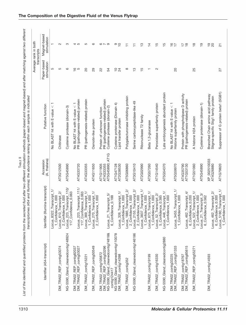
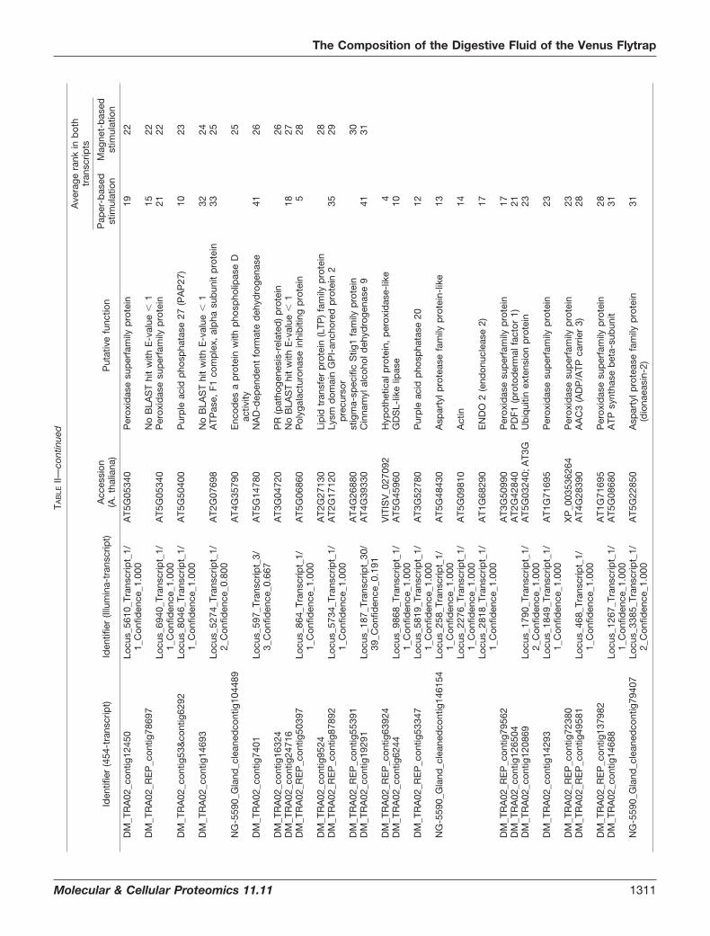
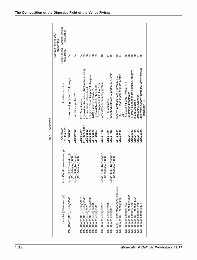
Identification and Abundance Ranking of the Proteins in theVenus Flytrap Digestive Fluid—The secreted proteins from themagnet-based and filter-paper-based stimulations were ana-lyzed by LC-MS/MS. Six datasets were searched against bothof the generated transcriptomes and analyzed using Max-Quant and Mascot Distiller. Using this approach, we identifiedand quantified 76 proteins in the digestive fluid (Table II,supplemental Table S3), with 32 proteins detected in bothtranscripts. Furthermore, 34 were present in only the 454transcriptome (supplemental Tables S1 and S4), and 10 pro-teins were present in only the Illumina transcriptome (supple-mental Tables S2 and S5). In total, 66 proteins were identifiedin the secreted fluid upon filter-paper-based stimulation, and42 proteins were identified in the secretion fluid after magnet-based stimulation (Table II). Of these 42 proteins, 30 proteinswere also identified upon filter-paper-based stimulation, dem-onstrating large overlap (71%) between the sampling proce-
FIG. 1. Workflow of the Venus flytrap digestive fluid analysis.
TABLE ITranscriptome properties
Illumina 454
Number of contigs 16,288 37,934Sum of the contig lengths 12,177,595 21,138,288Cut-off contig length 100 50Average length of the contigs 747 550
The Composition of the Digestive Fluid of the Venus Flytrap
Molecular & Cellular Proteomics 11.11 1309
TAB
LEII
List
ofth
eid
entif
ied
and
qua
ntifi
edp
rote
ins
from
the
secr
eted
fluid
afte
rtw
od
iffer
ent
stim
ulat
ion
met
hod
s(p
aper
-bas
edan
dm
agne
t-b
ased
)and
afte
rm
atch
ing
agai
nst
two
diff
eren
ttr
ansc
ripto
mes
(454
and
Illum
ina)
;th
eab
und
ance
rank
ing
with
inea
chsa
mp
leis
ind
icat
ed
Ave
rage
rank
inb
oth
tran
scrip
ts
Iden
tifie
r(4
54-t
rans
crip
t)Id
entif
ier
(Illu
min
a-tr
ansc
ript)
Acc
essi
on(A
.th
alia
na)
Put
ativ
efu
nctio
nP
aper
-bas
edst
imul
atio
nM
agne
t-b
ased
stim
ulat
ion
Locu
s_83
22_T
rans
crip
t_1/
1_C
onfid
ence
_1.0
00N
oB
LAS
Thi
tw
ithE
-val
ue�
15
1
DM
_TR
A02
_RE
P_c
ontig
5307
4Lo
cus_
610_
Tran
scrip
t_2/
2_C
onfid
ence
_1.0
00A
T3G
1250
0C
hitin
ase
52
NG
-559
0_G
land
_cle
aned
cont
ig14
8924
Locu
s_22
3_Tr
ansc
ript_
110/
117_
Con
fiden
ce_0
.063
AT5
G45
890
Cys
tein
ep
rote
ase
(dio
nain
-3)
63
DM
_TR
A02
_RE
P_c
ontig
8203
7N
oB
LAS
Thi
tw
ithE
-val
ue�
116
4D
M_T
RA
02_R
EP
_con
tig53
027
Locu
s_22
3_Tr
ansc
ript_
111/
117_
Con
fiden
ce_0
.063
AT4
G33
720
PR
(pat
hoge
nesi
s-re
late
d)
pro
tein
55
DM
_TR
A02
_con
tig15
221
Locu
s_36
9_Tr
ansc
ript_
1/1_
Con
fiden
ce_1
.000
AT4
G33
355
PR
(pat
hoge
nesi
s-re
late
d)
pro
tein
205
DM
_TR
A02
_RE
P_c
ontig
5054
9Lo
cus_
270_
Tran
scrip
t_1/
2_C
onfid
ence
_1.0
00A
T4G
1165
0O
smot
in-l
ike
pro
tein
296
DM
_TR
A02
_con
tig10
5872
AT1
G11
905
Pro
tein
ofun
know
nfu
nctio
n22
6D
M_T
RA
02_R
EP
_con
tig53
296
AT4
G33
355
PR
(pat
hoge
nesi
s-re
late
d)
pro
tein
7N
G-5
590_
Gla
nd_c
lean
edco
ntig
7481
8&co
ntig
1244
17Lo
cus_
21_T
rans
crip
t_5/
6_C
onfid
ence
_0.1
88A
T5G
4589
0;A
T1G
Cys
tein
ep
rote
ase
(dio
nain
-2)
549
NG
-559
0_G
land
_cle
aned
cont
ig11
0078
AT1
G47
128
Cys
tein
ep
rote
inas
e(D
iona
in4)
109
DM
_TR
A02
_con
tig14
398
Locu
s_32
6_Tr
ansc
ript_
1/1_
Con
fiden
ce_1
.000
AT2
G38
530
Lip
idtr
ansf
erp
rote
in24
10
DM
_TR
A02
_con
tig35
2Lo
cus_
1885
_Tra
nscr
ipt_
2/2_
Con
fiden
ce_1
.000
AT5
G06
860
Pol
ygal
actu
rona
sein
hib
iting
pro
tein
811
NG
-559
0_G
land
_cle
aned
cont
ig14
6186
Locu
s_21
55_T
rans
crip
t_2/
2_C
onfid
ence
_1.0
00A
T3G
1041
0S
erin
eca
rbox
ypep
tidas
e-lik
e49
1311
Locu
s_38
37_T
rans
crip
t_1/
1_C
onfid
ence
_1.0
00A
T2G
0299
0R
ibon
ucle
ase
T2fa
mily
1513
DM
_TR
A02
_con
tig19
199
Locu
s_67
3_Tr
ansc
ript_
1/1_
Con
fiden
ce_1
.000
AT3
G57
260
Bet
a1,
3-gl
ucan
ase
114
DM
_TR
A02
_con
tig16
330
Locu
s_52
_Tra
nscr
ipt_
2/5_
Con
fiden
ce_0
.385
AT1
G14
540
Per
oxid
ase
sup
erfa
mily
pro
tein
215
NG
-559
0_G
land
_cle
aned
cont
ig28
80Lo
cus_
448_
Tran
scrip
t_1/
1_C
onfid
ence
_1.0
00A
T5G
5437
0La
teem
bry
ogen
esis
abun
dan
tp
rote
in15
DM
_TR
A02
_con
tig33
225
No
BLA
ST
hit
with
E-v
alue
�1
16D
M_T
RA
02_R
EP
_con
tig51
333
Locu
s_34
55_T
rans
crip
t_1/
1_C
onfid
ence
_1.0
00A
T5G
5997
0H
isto
nesu
per
fam
ilyp
rote
in20
17
DM
_TR
A02
_con
tig18
767
AT4
G35
790
Pro
tein
with
pho
spho
lipas
eD
activ
ity17
DM
_TR
A02
_RE
P_c
ontig
8561
5Lo
cus_
462_
Tran
scrip
t_4/
6_C
onfid
ence
_0.2
00A
T1G
5313
0P
R(p
atho
gene
sis-
rela
ted
)p
rote
in18
DM
_TR
A02
_RE
P_c
ontig
5527
1Lo
cus_
522_
Tran
scrip
t_1/
2_C
onfid
ence
_1.0
00A
T1G
5106
0A
hist
one
H2A
pro
tein
2518
Locu
s_21
_Tra
nscr
ipt_
3/6_
Con
fiden
ce_0
.562
AT5
G45
890
Cys
tein
ep
rote
inas
e(d
iona
in-1
)19
DM
_TR
A02
_con
tig14
593
XP
_002
5100
33B
ranc
hed
Cha
inam
ino
acid
pat
hway
19Lo
cus_
462_
Tran
scrip
t_2/
6_C
onfid
ence
_0.3
00A
T4G
2688
0S
tigm
a-sp
ecifi
cS
tig1
fam
ilyp
rote
in20
Locu
s_51
80_T
rans
crip
t_1/
1_C
onfid
ence
_1.0
00A
T1G
7982
0S
upp
ress
orof
Gp
rote
inb
eta1
(SG
B1)
2721
The Composition of the Digestive Fluid of the Venus Flytrap
1310 Molecular & Cellular Proteomics 11.11
TAB
LEII—
cont
inue
d
Ave
rage
rank
inb
oth
tran
scrip
ts
Iden
tifie
r(4
54-t
rans
crip
t)Id
entif
ier
(Illu
min
a-tr
ansc
ript)
Acc
essi
on(A
.th
alia
na)
Put
ativ
efu
nctio
nP
aper
-bas
edst
imul
atio
nM
agne
t-b
ased
stim
ulat
ion
DM
_TR
A02
_con
tig12
450
Locu
s_56
10_T
rans
crip
t_1/
1_C
onfid
ence
_1.0
00A
T5G
0534
0P
erox
idas
esu
per
fam
ilyp
rote
in19
22
DM
_TR
A02
_RE
P_c
ontig
7869
7N
oB
LAS
Thi
tw
ithE
-val
ue�
115
22Lo
cus_
6940
_Tra
nscr
ipt_
1/1_
Con
fiden
ce_1
.000
AT5
G05
340
Per
oxid
ase
sup
erfa
mily
pro
tein
2122
DM
_TR
A02
_con
tig53
&co
ntig
6292
Locu
s_80
46_T
rans
crip
t_1/
1_C
onfid
ence
_1.0
00A
T5G
5040
0P
urp
leac
idp
hosp
hata
se27
(PA
P27
)10
23
DM
_TR
A02
_con
tig14
693
No
BLA
ST
hit
with
E-v
alue
�1
3224
Locu
s_52
74_T
rans
crip
t_1/
2_C
onfid
ence
_0.8
00A
T2G
0769
8A
TPas
e,F1
com
ple
x,al
pha
sub
unit
pro
tein
3325
NG
-559
0_G
land
_cle
aned
cont
ig10
4489
AT4
G35
790
Enc
odes
ap
rote
inw
ithp
hosp
holip
ase
Dac
tivity
25
DM
_TR
A02
_con
tig74
01Lo
cus_
597_
Tran
scrip
t_3/
3_C
onfid
ence
_0.6
67A
T5G
1478
0N
AD
-dep
end
ent
form
ate
deh
ydro
gena
se41
26
DM
_TR
A02
_con
tig16
324
AT3
G04
720
PR
(pat
hoge
nesi
s-re
late
d)
pro
tein
26D
M_T
RA
02_c
ontig
2471
6N
oB
LAS
Thi
tw
ithE
-val
ue�
118
27D
M_T
RA
02_R
EP
_con
tig50
397
Locu
s_86
4_Tr
ansc
ript_
1/1_
Con
fiden
ce_1
.000
AT5
G06
860
Pol
ygal
actu
rona
sein
hib
iting
pro
tein
528
DM
_TR
A02
_con
tig95
24A
T2G
2713
0Li
pid
tran
sfer
pro
tein
(LTP
)fa
mily
pro
tein
28D
M_T
RA
02_R
EP
_con
tig87
892
Locu
s_57
34_T
rans
crip
t_1/
1_C
onfid
ence
_1.0
00A
T2G
1712
0Ly
smd
omai
nG
PI-
anch
ored
pro
tein
2p
recu
rsor
3529
DM
_TR
A02
_RE
P_c
ontig
5539
1A
T4G
2688
0st
igm
a-sp
ecifi
cS
tig1
fam
ilyp
rote
in30
DM
_TR
A02
_con
tig19
291
Locu
s_18
7_Tr
ansc
ript_
30/
39_C
onfid
ence
_0.1
91A
T4G
3933
0C
inna
myl
alco
hold
ehyd
roge
nase
941
31
DM
_TR
A02
_RE
P_c
ontig
6392
4V
ITIS
V_0
2709
2H
ypot
hetic
alp
rote
in,
per
oxid
ase-
like
4D
M_T
RA
02_c
ontig
6244
Locu
s_98
68_T
rans
crip
t_1/
1_C
onfid
ence
_1.0
00A
T5G
4596
0G
DS
L-lik
elip
ase
10
DM
_TR
A02
_RE
P_c
ontig
5334
7Lo
cus_
5819
_Tra
nscr
ipt_
1/1_
Con
fiden
ce_1
.000
AT3
G52
780
Pur
ple
acid
pho
spha
tase
2012
NG
-559
0_G
land
_cle
aned
cont
ig14
6154
Locu
s_25
8_Tr
ansc
ript_
1/1_
Con
fiden
ce_1
.000
AT5
G48
430
Asp
arty
lpro
teas
efa
mily
pro
tein
-lik
e13
Locu
s_22
76_T
rans
crip
t_1/
1_C
onfid
ence
_1.0
00A
T5G
0981
0A
ctin
14
Locu
s_28
18_T
rans
crip
t_1/
1_C
onfid
ence
_1.0
00A
T1G
6829
0E
ND
O2
(end
onuc
leas
e2)
17
DM
_TR
A02
_RE
P_c
ontig
7956
2A
T3G
5099
0P
erox
idas
esu
per
fam
ilyp
rote
in17
DM
_TR
A02
_con
tig12
6504
AT2
G42
840
PD
F1(p
roto
der
mal
fact
or1)
21D
M_T
RA
02_c
ontig
1208
69Lo
cus_
1790
_Tra
nscr
ipt_
1/2_
Con
fiden
ce_1
.000
AT5
G03
240;
AT3
GU
biq
uitin
exte
nsio
np
rote
in23
DM
_TR
A02
_con
tig14
293
Locu
s_18
49_T
rans
crip
t_1/
1_C
onfid
ence
_1.0
00A
T1G
7169
5P
erox
idas
esu
per
fam
ilyp
rote
in23
DM
_TR
A02
_RE
P_c
ontig
7238
0X
P_0
0353
6264
Per
oxid
ase
sup
erfa
mily
pro
tein
23D
M_T
RA
02_R
EP
_con
tig49
581
Locu
s_46
8_Tr
ansc
ript_
1/1_
Con
fiden
ce_1
.000
AT4
G28
390
AA
C3
(AD
P/A
TPca
rrie
r3)
28
DM
_TR
A02
_RE
P_c
ontig
1379
82A
T1G
7169
5P
erox
idas
esu
per
fam
ilyp
rote
in28
DM
_TR
A02
_con
tig14
688
Locu
s_12
67_T
rans
crip
t_1/
1_C
onfid
ence
_1.0
00A
T5G
0868
0A
TPsy
ntha
seb
eta-
sub
unit
31
NG
-559
0_G
land
_cle
aned
cont
ig79
407
Locu
s_33
85_T
rans
crip
t_1/
2_C
onfid
ence
_1.0
00A
T5G
2285
0A
spar
tylp
rote
ase
fam
ilyp
rote
in(d
iona
easi
n-2)
31
The Composition of the Digestive Fluid of the Venus Flytrap
Molecular & Cellular Proteomics 11.11 1311
TAB
LEII—
cont
inue
d
Ave
rage
rank
inb
oth
tran
scrip
ts
Iden
tifie
r(4
54-t
rans
crip
t)Id
entif
ier
(Illu
min
a-tr
ansc
ript)
Acc
essi
on(A
.th
alia
na)
Put
ativ
efu
nctio
nP
aper
-bas
edst
imul
atio
nM
agne
t-b
ased
stim
ulat
ion
DM
_TR
A02
_RE
P_c
ontig
5084
0Lo
cus_
175_
Tran
scrip
t_1/
1_C
onfid
ence
_1.0
00A
T1G
7830
0G
-box
bin
din
gfa
ctor
GF1
4om
ega
34
Locu
s_52
47_T
rans
crip
t_1/
1_C
onfid
ence
_1.0
00A
T3G
1258
0H
eat
shoc
kp
rote
in70
34
DM
_TR
A02
_RE
P_c
ontig
6001
0A
T3G
5442
0A
TEP
3;ch
itina
se34
DM
_TR
A02
_RE
P_c
ontig
5654
5X
P_0
0338
0422
sup
erox
ide
dis
mut
ase
�Tric
hine
llasp
iralis
�35
DM
_TR
A02
_con
tig70
75A
T5G
0868
0A
TPsy
ntha
seb
eta-
sub
unit
36D
M_T
RA
02_R
EP
_con
tig48
999
AT5
G60
390
elon
gatio
nfa
ctor
1-al
pha
/EF-
1-al
pha
37D
M_T
RA
02_c
ontig
1260
8A
T1G
6829
0E
ND
O2
(end
onuc
leas
e2)
38D
M_T
RA
02_c
ontig
1301
AT1
G13
440
Gly
cera
ldeh
yde-
3-p
hosp
hate
deh
ydro
gena
seC
2(G
AP
C2)
39
DM
_TR
A02
_con
tig16
247
Locu
s_19
44_T
rans
crip
t_1/
1_C
onfid
ence
_1.0
00A
T4G
3753
0P
erox
idas
esu
per
fam
ilyp
rote
in40
DM
_TR
A02
_con
tig13
240
AT3
G54
420
ATE
P3;
chiti
nase
41D
M_T
RA
02_c
ontig
347
Locu
s_46
42_T
rans
crip
t_1/
1_C
onfid
ence
_1.0
00A
T5G
6713
0P
hosp
hod
iest
eras
esu
per
fam
ilyp
rote
in42
NG
-559
0_G
land
_cle
aned
cont
ig10
5066
AT1
G03
220
Asp
arty
lpro
teas
efa
mily
pro
tein
-lik
e42
DM
_TR
A02
_RE
P_c
ontig
5050
9A
T5G
0250
0H
SC
70–1
(hea
tsh
ock
cogn
ate
pro
tein
70–1
)44
DM
_TR
A02
_con
tig28
745
No
BLA
ST
hit
with
E-v
alue
�1
45D
M_T
RA
02_R
EP
_con
tig12
9322
AT5
G41
210
Glu
tath
ione
tran
sfer
ase
48D
M_T
RA
02_c
ontig
1678
0A
T2G
3646
0Fr
ucto
se-b
isp
hosp
hate
ald
olas
e,p
utat
ive
49D
M_T
RA
02_R
EP
_con
tig48
954
AT2
G44
490
Thio
gluc
osid
ase
50D
M_T
RA
02_c
ontig
1444
0A
T5G
1792
0M
ethi
onin
esy
ntha
se54
DM
_TR
A02
_con
tig18
374
AT2
G03
200
Euk
aryo
ticas
par
tylp
rote
ase
fam
ilyp
rote
in(d
iona
easi
n-1)
55
The Composition of the Digestive Fluid of the Venus Flytrap
1312 Molecular & Cellular Proteomics 11.11
dures (Table II). However, the abundance ranking displayeddifferences between the two sampling procedures, emphasiz-ing the importance of using complementary methods. Therankings based on MaxQuant and Mascot Distiller were gen-erally in good agreement (supplemental Table S3). The 32proteins identified from both transcriptome databases dis-played significant identical overlaps over long, continuousstretches. Frequently, the shorter contig from one transcriptwas completely contained within another, longer transcript(supplemental Table S6). This finding clearly supports thevalue of the complementary approach utilized in this analysisof the Venus flytrap secretome.
In order to annotate the identified proteins based on func-tion, the corresponding translated transcripts were homologysearched against the well-annotated Arabidopsis thaliana pro-teome (supplemental Tables S4 and S5). Among the mostabundant proteins in the Venus flytrap secretome were ho-mologs of proteases, chitinases, osmotin-like protein, patho-genesis-related proteins, lipid transfer proteins, peroxidases,and beta-1,3-glucanase, which all belong to families of patho-genesis-related proteins (29). The presence of defense-re-lated proteins has also been observed in the digestive fluid ofNepenthes (30), indicating that the digestive process in thesetwo plants is functionally similar, although the plants belongto two different families, namely, Droseraceae and Ne-penthaceae. Nine of the transcripts were homologous to dif-ferent proteases, showing that the proteases are one of thetwo largest protein families in the secretome. This result cor-relates with the degrading function of the fluid. The sequenceof one of the identified proteases was identical to the peptidesequence of a Venus flytrap digestive fluid cysteine proteasefrom a previous study, except for one amino acid residue (16).In that particular study, the protease was termed “dionain.”Here, we adhered to this nomenclature and named the cys-teine proteases dionain-1, dionain-2, dionain-3, and dion-ain-4. Dionain-1 is the protease that contains the previouslysequenced peptides (Table II). Nine peroxidase homologs,including one of the top-ranking proteins from the filter-paper-based sampling procedure, were identified. These data indi-
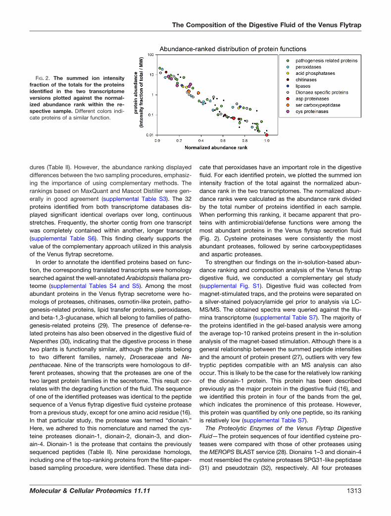
cate that peroxidases have an important role in the digestivefluid. For each identified protein, we plotted the summed ionintensity fraction of the total against the normalized abun-dance rank in the two transcriptomes. The normalized abun-dance ranks were calculated as the abundance rank dividedby the total number of proteins identified in each sample.When performing this ranking, it became apparent that pro-teins with antimicrobial/defense functions were among themost abundant proteins in the Venus flytrap secretion fluid(Fig. 2). Cysteine proteinases were consistently the mostabundant proteases, followed by serine carboxypeptidasesand aspartic proteases.
To strengthen our findings on the in-solution-based abun-dance ranking and composition analysis of the Venus flytrapdigestive fluid, we conducted a complementary gel study(supplemental Fig. S1). Digestive fluid was collected frommagnet-stimulated traps, and the proteins were separated ona silver-stained polyacrylamide gel prior to analysis via LC-MS/MS. The obtained spectra were queried against the Illu-mina transcriptome (supplemental Table S7). The majority ofthe proteins identified in the gel-based analysis were amongthe average top-10 ranked proteins present in the in-solutionanalysis of the magnet-based stimulation. Although there is ageneral relationship between the summed peptide intensitiesand the amount of protein present (27), outliers with very fewtryptic peptides compatible with an MS analysis can alsooccur. This is likely to be the case for the relatively low rankingof the dionain-1 protein. This protein has been describedpreviously as the major protein in the digestive fluid (16), andwe identified this protein in four of the bands from the gel,which indicates the prominence of this protease. However,this protein was quantified by only one peptide, so its rankingis relatively low (supplemental Table S7).
The Proteolytic Enzymes of the Venus Flytrap DigestiveFluid—The protein sequences of four identified cysteine pro-teases were compared with those of other proteases usingthe MEROPS BLAST service (28). Dionains 1–3 and dionain-4most resembled the cysteine proteases SPG31-like peptidase(31) and pseudotzain (32), respectively. All four proteases
FIG. 2. The summed ion intensityfraction of the totals for the proteinsidentified in the two transcriptomeversions plotted against the normal-ized abundance rank within the re-spective sample. Different colors indi-cate proteins of a similar function.
The Composition of the Digestive Fluid of the Venus Flytrap
Molecular & Cellular Proteomics 11.11 1313
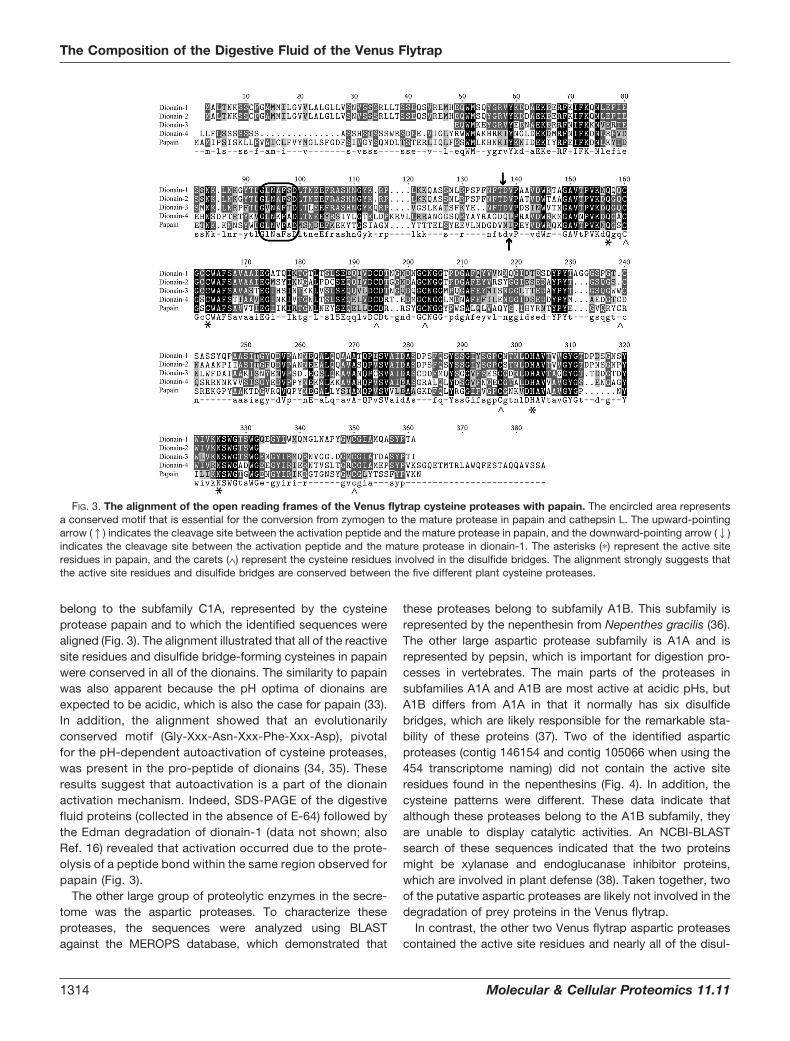
belong to the subfamily C1A, represented by the cysteineprotease papain and to which the identified sequences werealigned (Fig. 3). The alignment illustrated that all of the reactivesite residues and disulfide bridge-forming cysteines in papainwere conserved in all of the dionains. The similarity to papainwas also apparent because the pH optima of dionains areexpected to be acidic, which is also the case for papain (33).In addition, the alignment showed that an evolutionarilyconserved motif (Gly-Xxx-Asn-Xxx-Phe-Xxx-Asp), pivotalfor the pH-dependent autoactivation of cysteine proteases,was present in the pro-peptide of dionains (34, 35). Theseresults suggest that autoactivation is a part of the dionainactivation mechanism. Indeed, SDS-PAGE of the digestivefluid proteins (collected in the absence of E-64) followed bythe Edman degradation of dionain-1 (data not shown; alsoRef. 16) revealed that activation occurred due to the prote-olysis of a peptide bond within the same region observed forpapain (Fig. 3).
The other large group of proteolytic enzymes in the secre-tome was the aspartic proteases. To characterize theseproteases, the sequences were analyzed using BLASTagainst the MEROPS database, which demonstrated that
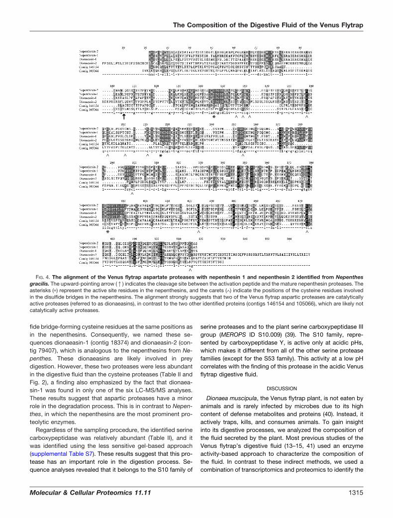
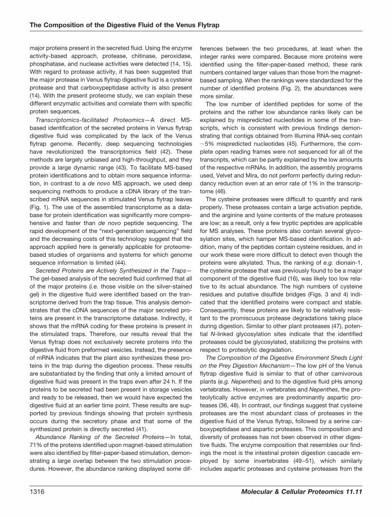
these proteases belong to subfamily A1B. This subfamily isrepresented by the nepenthesin from Nepenthes gracilis (36).The other large aspartic protease subfamily is A1A and isrepresented by pepsin, which is important for digestion pro-cesses in vertebrates. The main parts of the proteases insubfamilies A1A and A1B are most active at acidic pHs, butA1B differs from A1A in that it normally has six disulfidebridges, which are likely responsible for the remarkable sta-bility of these proteins (37). Two of the identified asparticproteases (contig 146154 and contig 105066 when using the454 transcriptome naming) did not contain the active siteresidues found in the nepenthesins (Fig. 4). In addition, thecysteine patterns were different. These data indicate thatalthough these proteases belong to the A1B subfamily, theyare unable to display catalytic activities. An NCBI-BLASTsearch of these sequences indicated that the two proteinsmight be xylanase and endoglucanase inhibitor proteins,which are involved in plant defense (38). Taken together, twoof the putative aspartic proteases are likely not involved in thedegradation of prey proteins in the Venus flytrap.
In contrast, the other two Venus flytrap aspartic proteasescontained the active site residues and nearly all of the disul-
FIG. 3. The alignment of the open reading frames of the Venus flytrap cysteine proteases with papain. The encircled area representsa conserved motif that is essential for the conversion from zymogen to the mature protease in papain and cathepsin L. The upward-pointingarrow (1) indicates the cleavage site between the activation peptide and the mature protease in papain, and the downward-pointing arrow (2)indicates the cleavage site between the activation peptide and the mature protease in dionain-1. The asterisks (�) represent the active siteresidues in papain, and the carets (∧) represent the cysteine residues involved in the disulfide bridges. The alignment strongly suggests thatthe active site residues and disulfide bridges are conserved between the five different plant cysteine proteases.
The Composition of the Digestive Fluid of the Venus Flytrap
1314 Molecular & Cellular Proteomics 11.11
fide bridge-forming cysteine residues at the same positions asin the nepenthesins. Consequently, we named these se-quences dionaeasin-1 (contig 18374) and dionaeasin-2 (con-tig 79407), which is analogous to the nepenthesins from Ne-penthes. These dionaeasins are likely involved in preydigestion. However, these two proteases were less abundantin the digestive fluid than the cysteine proteases (Table II andFig. 2), a finding also emphasized by the fact that dionaea-sin-1 was found in only one of the six LC-MS/MS analyses.These results suggest that aspartic proteases have a minorrole in the degradation process. This is in contrast to Nepen-thes, in which the nepenthesins are the most prominent pro-teolytic enzymes.
Regardless of the sampling procedure, the identified serinecarboxypeptidase was relatively abundant (Table II), and itwas identified using the less sensitive gel-based approach(supplemental Table S7). These results suggest that this pro-tease has an important role in the digestion process. Se-quence analyses revealed that it belongs to the S10 family of
serine proteases and to the plant serine carboxypeptidase IIIgroup (MEROPS ID S10.009) (39). The S10 family, repre-sented by carboxypeptidase Y, is active only at acidic pHs,which makes it different from all of the other serine proteasefamilies (except for the S53 family). This activity at a low pHcorrelates with the finding of this protease in the acidic Venusflytrap digestive fluid.
DISCUSSION
Dionaea muscipula, the Venus flytrap plant, is not eaten byanimals and is rarely infected by microbes due to its highcontent of defense metabolites and proteins (40). Instead, itactively traps, kills, and consumes animals. To gain insightinto its digestive processes, we analyzed the composition ofthe fluid secreted by the plant. Most previous studies of theVenus flytrap’s digestive fluid (13–15, 41) used an enzymeactivity-based approach to characterize the composition ofthe fluid. In contrast to these indirect methods, we used acombination of transcriptomics and proteomics to identify the
FIG. 4. The alignment of the Venus flytrap aspartate proteases with nepenthesin 1 and nepenthesin 2 identified from Nepenthesgracilis. The upward-pointing arrow (1) indicates the cleavage site between the activation peptide and the mature nepenthesin proteases. Theasterisks (�) represent the active site residues in the nepenthesins, and the carets (∧) indicate the positions of the cysteine residues involvedin the disulfide bridges in the nepenthesins. The alignment strongly suggests that two of the Venus flytrap aspartic proteases are catalyticallyactive proteases (referred to as dionaeasins), in contrast to the two other identified proteins (contigs 146154 and 105066), which are likely notcatalytically active proteases.
The Composition of the Digestive Fluid of the Venus Flytrap
Molecular & Cellular Proteomics 11.11 1315
major proteins present in the secreted fluid. Using the enzymeactivity-based approach, protease, chitinase, peroxidase,phosphatase, and nuclease activities were detected (14, 15).With regard to protease activity, it has been suggested thatthe major protease in Venus flytrap digestive fluid is a cysteineprotease and that carboxypeptidase activity is also present(14). With the present proteome study, we can explain thesedifferent enzymatic activities and correlate them with specificprotein sequences.
Transcriptomics-facilitated Proteomics—A direct MS-based identification of the secreted proteins in Venus flytrapdigestive fluid was complicated by the lack of the Venusflytrap genome. Recently, deep sequencing technologieshave revolutionized the transcriptomics field (42). Thesemethods are largely unbiased and high-throughput, and theyprovide a large dynamic range (43). To facilitate MS-basedprotein identifications and to obtain more sequence informa-tion, in contrast to a de novo MS approach, we used deepsequencing methods to produce a cDNA library of the tran-scribed mRNA sequences in stimulated Venus flytrap leaves(Fig. 1). The use of the assembled transcriptome as a data-base for protein identification was significantly more compre-hensive and faster than de novo peptide sequencing. Therapid development of the “next-generation sequencing” fieldand the decreasing costs of this technology suggest that theapproach applied here is generally applicable for proteome-based studies of organisms and systems for which genomesequence information is limited (44).
Secreted Proteins are Actively Synthesized in the Traps—The gel-based analysis of the secreted fluid confirmed that allof the major proteins (i.e. those visible on the silver-stainedgel) in the digestive fluid were identified based on the tran-scriptome derived from the trap tissue. This analysis demon-strates that the cDNA sequences of the major secreted pro-teins are present in the transcriptome database. Indirectly, itshows that the mRNA coding for these proteins is present inthe stimulated traps. Therefore, our results reveal that theVenus flytrap does not exclusively secrete proteins into thedigestive fluid from preformed vesicles. Instead, the presenceof mRNA indicates that the plant also synthesizes these pro-teins in the trap during the digestion process. These resultsare substantiated by the finding that only a limited amount ofdigestive fluid was present in the traps even after 24 h. If theproteins to be secreted had been present in storage vesiclesand ready to be released, then we would have expected thedigestive fluid at an earlier time point. These results are sup-ported by previous findings showing that protein synthesisoccurs during the secretory phase and that some of thesynthesized protein is directly secreted (41).
Abundance Ranking of the Secreted Proteins—In total,71% of the proteins identified upon magnet-based stimulationwere also identified by filter-paper-based stimulation, demon-strating a large overlap between the two stimulation proce-dures. However, the abundance ranking displayed some dif-
ferences between the two procedures, at least when theinteger ranks were compared. Because more proteins wereidentified using the filter-paper-based method, these ranknumbers contained larger values than those from the magnet-based sampling. When the rankings were standardized for thenumber of identified proteins (Fig. 2), the abundances weremore similar.
The low number of identified peptides for some of theproteins and the rather low abundance ranks likely can beexplained by mispredicted nucleotides in some of the tran-scripts, which is consistent with previous findings demon-strating that contigs obtained from Illumina RNA-seq contain�5% mispredicted nucleotides (45). Furthermore, the com-plete open reading frames were not sequenced for all of thetranscripts, which can be partly explained by the low amountsof the respective mRNAs. In addition, the assembly programsused, Velvet and Mira, do not perform perfectly during redun-dancy reduction even at an error rate of 1% in the transcrip-tome (46).
The cysteine proteases were difficult to quantify and rankproperly. These proteases contain a large activation peptide,and the arginine and lysine contents of the mature proteasesare low; as a result, only a few tryptic peptides are applicablefor MS analyses. These proteins also contain several glyco-sylation sites, which hamper MS-based identification. In ad-dition, many of the peptides contain cysteine residues, and inour work these were more difficult to detect even though theproteins were alkylated. Thus, the ranking of e.g. dionain-1,the cysteine protease that was previously found to be a majorcomponent of the digestive fluid (16), was likely too low rela-tive to its actual abundance. The high numbers of cysteineresidues and putative disulfide bridges (Figs. 3 and 4) indi-cated that the identified proteins were compact and stable.Consequently, these proteins are likely to be relatively resis-tant to the promiscuous protease degradations taking placeduring digestion. Similar to other plant proteases (47), poten-tial N-linked glycosylation sites indicate that the identifiedproteases could be glycosylated, stabilizing the proteins withrespect to proteolytic degradation.
The Composition of the Digestive Environment Sheds Lighton the Prey Digestion Mechanism—The low pH of the Venusflytrap digestive fluid is similar to that of other carnivorousplants (e.g. Nepenthes) and to the digestive fluid pHs amongvertebrates. However, in vertebrates and Nepenthes, the pro-teolytically active enzymes are predominantly aspartic pro-teases (36, 48). In contrast, our findings suggest that cysteineproteases are the most abundant class of proteases in thedigestive fluid of the Venus flytrap, followed by a serine car-boxypeptidase and aspartic proteases. This composition anddiversity of proteases has not been observed in other diges-tive fluids. The enzyme composition that resembles our find-ings the most is the intestinal protein digestion cascade em-ployed by some invertebrates (49–51), which similarlyincludes aspartic proteases and cysteine proteases from the
The Composition of the Digestive Fluid of the Venus Flytrap
1316 Molecular & Cellular Proteomics 11.11
same protease families (the pepsin family (A1) and the papainfamily (C1)) observed in the Venus flytrap. In general, cysteineproteases have a neutral pH optimum. However, adaptationsto acidic pH optima have been observed among the lyso-somal cathepsins, which are primarily involved in unspecificbulk protein degradation (52). The protease composition ofthe Venus flytrap’s digestive fluid, with three classes of pep-tidases, is likely a potent digestion system, emphasizing thestrong dependence of Dionaea on the nutrients suppliedthrough prey capture and digestion (11). Particularly as the pHof the digestive fluid changes over time (7), the differentenzymes might reach their maximum activities at differentdigestion stages after the prey is captured. As previouslymentioned, the natural habitat of Venus flytrap plants is low-nutrient soils, and the plants depend on nutrients obtained bydigesting trapped prey. These identified proteases are likelyinvolved in the release of nitrogen from the prey proteins. Inaddition to proteases, a number of other hydrolytic enzymesare present in the digestive fluid, and the fact that nucleases,phosphatases, and phospholipases were identified indicatesthat phosphate is similarly obtained from the prey’s nucleicacids, proteins, and cell membranes.
Three chitinases were also identified, including one of theproteins found to be most abundant in the digestive fluidregardless of the stimulation method. These chitinases wouldbe expected to degrade the exoskeletons of captured insectsor spiders and thereby facilitate enzymatic access to the innerpart of the prey. Furthermore, chitinases are pathogenesis-related proteins that might prevent microbial growth on thetrapped prey during the digestion process.
It has been suggested that prey proteins in the Venus flytrapare initially oxidized in order to facilitate their subsequentproteolysis (53), and it has been demonstrated that Nepen-thes gracilis uses free radicals during the digestion process(54). Plumbagin, a low-molecular-weight compound presentin Venus flytrap digestive fluid, likely facilitates this oxidation(40). The identified peroxidases from the present study arelikely involved in these oxidative processes. Thus, our findingssupport the hypothesis that the oxidation of prey moleculesfacilitates the digestion mechanisms of the Venus flytrap.
The functions of the hydrolytic enzymes in the digestive fluidare intuitively easy to envision. These enzymes are likely directlyinvolved in prey digestion. The functions of some of the otherproteins present in the fluid are more challenging to elucidate,and a functional annotation based on the name of the bestmatch in a homology search (Table II) does not necessarily shedlight on the in vivo role of the protein. The roles of these proteinsin the digestion mechanism remain to be investigated.
The Digestive Fluid Proteome Suggests a Shift from De-fense-related Processes to Digestion-related Processesamong the Carnivorous Plants—The only previously charac-terized digestive fluid proteome from a carnivorous plant wasderived from Nepenthes (30). The depth of that de novosequencing-based study was lower than in the present study;
however, aspartic proteases (nepenthesin I and II), a chiti-nase, a glucanase, a xylosidase, and a thaumatin-like proteinwere identified. These protein classes were, with the excep-tion of the xylosidase, also identified in the present analysis ofthe Venus flytrap, indicating the conserved functions of thedigestive fluid among carnivorous plant species. Similar to ourresults, the Nepenthes digestive proteins are also predomi-nantly pathogenesis-related proteins. Higher plants expresspathogenesis-related proteins as a response to an attack bypathogens, and consequently, many of these proteins pos-sess hydrolytic activities that are potentially applicable to preydigestion in carnivorous plants. The identification of severaldefense-related proteins suggests that carnivorous plantshave exploited the hydrolytic properties of these pathogene-sis-related proteins (55). Many pathogenesis-related proteinsare resistant to low pHs and to proteolytic degradation (29),making them functional in digestive fluids. During the evolu-tion of carnivory in plants, there has likely been a shift from apathogen-related response to a prey-related response and ashift from the hydrolysis and destruction of the pathogens to thehydrolysis and digestion of the prey. The defense-related pro-teins in digestive fluid likely still display antibacterial and anti-fungal effects, as in e.g. poplar extrafloral nectaries (22), in orderto avoid pathogenic attacks during the digestion process.
CONCLUSION
The present characterization of Venus flytrap digestive fluidemployed deep sequencing of the transcriptome followed byits assembly and subsequent use as a database during theproteomic analyses. This study demonstrates the use of high-throughput technologies in expanding molecular analyses toorganisms for which the genome sequence is unknown. TheVenus flytrap secretome reveals a unique diversity of hydro-lytic enzymes, and the results shed light on the purpose andmechanisms of digestion. Furthermore, the Dionaea secre-tome contains a high proportion of pathogenesis-related pro-teins, suggesting that the capability of carnivorous plants todigest prey evolved from a plant defense system.
Acknowledgments—We thank Katharina Markmann (Aarhus, Den-mark) and Tania A. Nielsen (Aarhus, Denmark) for help with RNApurification; Tom A. Mortensen (Aarhus, Denmark) and the nurseryLammehave (Denmark) for assistance and helpful suggestions re-garding the cultivation of Venus flytrap plants; Fasteris SA (Switzer-land) for library preparation, Illumina sequencing, and Illumina tran-scriptome assembly; Kerstin Zander (Golm) for help in samplepreparation for mass spectrometry; and Brigitte Neumann for excel-lent technical assistance (Wurzburg).
* This work was supported by a grant from the Danish ResearchCouncil for Strategic Research to J.J.E. and an ERC Advanced Grantto R.H.
□S This article contains supplemental material.a These authors contributed equally to this work.** To whom correspondence should be addressed: Rainer Hedrich,
E-mail: [email protected]; and Jan Johannes Eng-hild, E-mail: [email protected].
The Composition of the Digestive Fluid of the Venus Flytrap
Molecular & Cellular Proteomics 11.11 1317
REFERENCES
1. Darwin, C. (1875) Insectivorous Plants, Murray, London2. Gibson, T. C., and Waller, D. M. (2009) Evolving Darwin’s ‘most wonderful’
plant: ecological steps to a snap-trap. New Phytol. 183, 575–5873. Forterre, Y., Skotheim, J. M., Dumais, J., and Mahadevan, L. (2005) How
the Venus flytrap snaps. Nature 433, 421–4254. Markin, V. S., Volkov, A. G., and Jovanov, E. (2008) Active movements in
plants: mechanism of trap closure by Dionaea muscipula Ellis. PlantSignal Behav. 3, 778–783
5. Ueda, M., Tokunaga, T., Okada, M., Nakamura, Y., Takada, N., Suzuki, R.,and Kondo, K. (2010) Trap-closing chemical factors of the Venus flytrap(Dionaea muscipulla Ellis). Chembiochem. 11, 2378–2383
6. Williams, S. E., and Bennett, A. B. (1982) Leaf closure in the Venus flytrap:an acid growth response. Science 218, 1120–1122
7. Escalante-Perez, M., Krol, E., Stange, A., Geiger, D., Al-Rasheid, K. A.,Hause, B., Neher, E., and Hedrich, R. (2011) A special pair of phytohor-mones controls excitability, slow closure, and external stomach forma-tion in the Venus flytrap. Proc. Natl. Acad. Sci. U.S.A. 108, 15492–15497
8. Volkov, A. G., Carrell, H., Baldwin, A., and Markin, V. S. (2009) Electricalmemory in Venus flytrap. Bioelectrochemistry 75, 142–147
9. Griggs, R. F. (1935) Victims of the Venus flytrap. Science 81, 7–810. Lichtner, F. T., and Williams, S. E. (1977) Prey capture and factors control-
ling trap narrowing in Dionaea (Droseraceae). Am. J. Bot. 64, 881–88611. Schulze, W., Schulze, E. D., Schulze, I., and Oren, R. (2001) Quantification
of insect nitrogen utilization by the Venus fly trap Dionaea muscipulacatching prey with highly variable isotope signatures. J. Exp. Bot. 52,1041–1049
12. Adamec, L. (1997) Mineral nutrition of carnivorous plants: a review. Bot.Rev. 63, 273–299
13. Takahashi, K., Matsumoto, K., Nishi, W., Muramatsu, M., and Kubota, K.(2009) Comparative studies on the acid proteinase activiteis in the di-gestive fluids of NEPENTHES, CEPHALOTOUS, DIONAEA, and DROS-ERA. Carnivorous Plant Newsletter 38, 75–82
14. Robins, R. I., and Juniper, B. E. (1980) The secretory cycle of Dionaea-muscipula ellis. IV. The enzymology of the secretion. New Phytol. 86,401–412
15. Scala, J., Iott, K., Schwab, D. W., and Semersky, F. E. (1969) Digestivesecretion of Dionaea muscipula (Venus’s-flytrap). Plant Physiol. 44,367–371
16. Takahashi, K., Suzuki, T., Nishii, W., Kubota, K., Shibata, C., Isobe, T., andDohmae, N. (2011) A cysteine endopeptidase (“dionain”) is involved inthe digestive fluid of Dionaea muscipula (Venus’s fly-trap). Biosci. Bio-tech. Bioch. 75, 346–348
17. Hogslund, N., Radutoiu, S., Krusell, L., Voroshilova, V., Hannah, M. A.,Goffard, N., Sanchez, D. H., Lippold, F., Ott, T., Sato, S., Tabata, S.,Liboriussen, P., Lohmann, G. V., Schauser, L., Weiller, G. F., Udvardi,M. K., and Stougaard, J. (2009) Dissection of symbiosis and organdevelopment by integrated transcriptome analysis of lotus japonicusmutant and wild-type plants. PLoS One 4, e6556
18. Engelsberger, W. R., and Schulze, W. X. (2012) Nitrate and ammonium leadto distinct global dynamic phosphorylation patterns when resupplied tonitrogen-starved Arabidopsis seedlings. Plant J. 69, 978–995
19. Rasmussen, M. I., Refsgaard, J. C., Peng, L., Houen, G., and Hojrup, P.(2011) CrossWork: software-assisted identification of cross-linked pep-tides. J. Proteomics 74, 1871–1883
20. Cox, J., and Mann, M. (2008) MaxQuant enables high peptide identificationrates, individualized p.p.b.-range mass accuracies and proteome-wideprotein quantification. Nat. Biotechnol. 26, 1367–1372
21. Perkins, D. N., Pappin, D. J., Creasy, D. M., and Cottrell, J. S. (1999)Probability-based protein identification by searching sequence data-bases using mass spectrometry data. Electrophoresis 20, 3551–3567
22. Escalante-Perez, M., Jaborsky, M., Lautner, S., Fromm, J., Muller, T.,Dittrich, M., Kunert, M., Boland, W., Hedrich, R., and Ache, P. (2012)Poplar extrafloral nectaries: two types, two strategies of indirect de-fenses against herbivores. Plant Physiol. 159, 1176–1191
23. Bury, A. F. (1981) Analysis of protein and peptide mixtures—evaluation ofthree sodium dodecyl sulfate-polyacrylamide gel-electrophoresis buffersystems. J. Chromatogr 213, 491–500
24. Shevchenko, A., Tomas, H., Havlis, J., Olsen, J. V., and Mann, M. (2006)In-gel digestion for mass spectrometric characterization of proteins andproteomes. Nat. Protoc. 1, 2856–2860
25. Sanggaard, K. W., Karring, H., Valnickova, Z., Thogersen, I. B., and Enghild,J. J. (2005) The TSG-6 and IalphaI interaction promotes a transesterifi-cation cleaving the protein-glycosaminoglycan-protein (PGP) cross-link.J. Biol. Chem. 280, 11936–11942
26. Ishihama, Y., Oda, Y., Tabata, T., Sato, T., Nagasu, T., Rappsilber, J., andMann, M. (2005) Exponentially Modified Protein Abundance Index (em-PAI) for estimation of absolute protein amount in proteomics by thenumber of sequenced peptides per protein. Mol. Cell. Proteomics 4,1265–1272
27. Schwannhausser, B., Busse, D., Li, N., Dittmar, G., Schuchhardt, J., Wolf,J., Chen, W., and Selbach, M. (2011) Global quantification of mammaliangene expression control. Nature 473, 337–473
28. Rawlings, N. D., Barrett, A. J., and Bateman, A. (2010) MEROPS: thepeptidase database. Nucleic Acids Res. 38, D227–D233
29. van Loon, L. C., Rep, M., and Pieterse, C. M. (2006) Significance ofinducible defense-related proteins in infected plants. Annu. Rev. Phyto-pathol. 44, 135–162
30. Hatano, N., and Hamada, T. (2008) Proteome analysis of pitcher fluid of thecarnivorous plant Nepenthes alata. J. Proteome Res. 7, 809–816
31. Chen, G. H., Huang, L. T., Yap, M. N., Lee, R. H., Huang, Y. J., Cheng,M. C., and Chen, S. C. (2002) Molecular characterization of a senes-cence-associated gene encoding cysteine proteinase and its gene ex-pression during leaf senescence in sweet potato. Plant Cell Physiol. 43,984–991
32. Tranbarger, T. J., and Misra, S. (1996) Structure and expression of adevelopmentally regulated cDNA encoding a cysteine protease (pseu-dotzain) from Douglas fir. Gene 172, 221–226
33. Hoover, S. R., and Kokes, E. L. (1947) Effect of pH upon proteolysis bypapain. J. Biol. Chem. 167, 199–207
34. Vernet, T., Berti, P. J., de Montigny, C., Musil, R., Tessier, D. C., Menard, R.,Magny, M. C., Storer, A. C., and Thomas, D. Y. (1995) Processing of thepapain precursor. The ionization state of a conserved amino acid motifwithin the Pro region participates in the regulation of intramolecularprocessing. J. Biol. Chem. 270, 10838–10846
35. Collins, P. R., Stack, C. M., O’Neill, S. M., Doyle, S., Ryan, T., Brennan,G. P., Mousley, A., Stewart, M., Maule, A. G., Dalton, J. P., and Donnelly,S. (2004) Cathepsin L1, the major protease involved in liver fluke (Fas-ciola hepatica) virulence: propetide cleavage sites and autoactivation ofthe zymogen secreted from gastrodermal cells. J. Biol. Chem. 279,17038–17046
36. Athauda, S. P. B., Matsumoto, K., Rajapakshe, S., Kuribayashi, M., Kojima,M., Kubomura-Yoshida, N., Iwamatsu, A., Shibata, C., Inoue, H., andTakahashi, K. (2004) Enzymic and structural characterization of nepen-thesin, a unique member of a novel subfamily of aspartic proteinases.Biochem. J. 381, 295–306; Erratum (2004) Biochem. J. 382, 1039
37. Takahashi, K., Athauda, S. B. P., Matsumoto, K., Rajapakshe, S., Kurib-ayashi, M., Kojima, M., Kubomura-Yoshida, N., Iwamatsu, A., Shibata,C., and Inoue, H. (2005) Nepenthesin, a unique member of a novelsubfamily of aspartic proteinases: enzymatic and structural characteris-tics. Curr. Protein Pept. Sci. 6, 513–525
38. Sansen, S., De Ranter, C. J., Gebruers, K., Brijs, K., Courtin, C. M., Delcour,J. A., and Rabijns, A. (2004) Structural basis for inhibition of Aspergillusniger xylanase by triticum aestivum xylanase inhibitor-I. J. Biol. Chem.279, 36022–36028
39. Drzymala, A., and Bielawski, W. (2009) Isolation and characterization ofcarboxypeptidase III from germinating triticale grains. Acta Biochim.Biophys. Sin. 41, 69–78
40. Tokunaga, T., Takada, N., and Ueda, M. (2004) Mechanism of antifeedantactivity of plumbagin, a compound concerning the chemical defense incarnivorous plant. Tetrahedron Lett. 45, 7115–7119
41. Robins, R. J., and Juniper, B. E. (1980) The secretory cycle of Dionaea-muscipula ellis. II. Storage and synthesis of the secretory proteins. NewPhytol. 86, 297–311
42. Shendure, J., and Ji, H. (2008) Next-generation DNA sequencing. Nat.Biotechnol. 26, 1134–1145
43. Wang, Z., Gerstein, M., and Snyder, M. (2009) RNA-Seq: a revolutionarytool for transcriptomics. Nat. Rev. Genet. 10, 57–63
44. Brautigam, A., Shrestha, R. P., Whitten, D., Wilkerson, C. G., Carr, K. M.,Froehlich, J. E., and Weber, A. P. (2008) Low-coverage massively parallelpyrosequencing of cDNAs enables proteomics in non-model species:comparison of a species-specific database generated by pyrosequenc-
The Composition of the Digestive Fluid of the Venus Flytrap
1318 Molecular & Cellular Proteomics 11.11
ing with databases from related species for proteome analysis of peachloroplast envelopes. J. Biotechnol. 136, 44–53
45. Wall, C. E., Cozza, S., Riquelme, C. A., McCombie, W. R., Heimiller, J. K.,Marr, T. G., and Leinwand, L. A. (2011) Whole transcriptome analysis ofthe fasting and fed Burmese python heart: insights into extreme physi-ological cardiac adaptation. Physiol. Genomics 43, 69–76
46. Brautigam, A., Mullick, T., Schliesky, S., and Weber, A. P. (2011) Criticalassessment of assembly strategies for non-model species mRNA-Seqdata and application of next-generation sequencing to the comparison ofC(3) and C(4) species. J. Exp. Bot. 62, 3093–3102
47. Choi, K. H., and Laursen, R. A. (2000) Amino-acid sequence and glycanstructures of cysteine proteases with proline specificity from gingerrhizome Zingiber officinale. Eur. J. Biochem. 267, 1516–1526
48. Kageyama, T. (2002) Pepsinogens, progastricsins, and prochymosins:structure, function, evolution, and development. Cell Mol. Life Sci. 59,288–306
49. Williamson, A. L., Brindley, P. J., Knox, D. P., Hotez, P. J., and Loukas, A.(2003) Digestive proteases of blood-feeding nematodes. Trends Parasi-tol. 19, 417–423
50. Delcroix, M., Sajid, M., Caffrey, C. R., Lim, K. C., Dvorak, J., Hsieh, I.,Bahgat, M., Dissous, C., and McKerrow, J. H. (2006) A multienzymenetwork functions in intestinal protein digestion by a platyhelminth par-
asite. J. Biol. Chem. 281, 39316–3932951. Knox, D. (2011) Proteases in blood-feeding nematodes and their potential
as vaccine candidates. Adv. Exp. Med. Biol. 712, 155–17652. Barrett, A. J. (1992) Cellular proteolysis. An overview. Ann. N.Y. Acad. Sci.
674, 1–1553. Galek, H., Osswald, W. F., and Elstner, E. F. (1990) Oxidative protein
modification as predigestive mechanism of the carnivorous plant Di-onaea muscipula: an hypothesis based on in vitro experiments. FreeRadic. Biol. Med. 9, 427–434
54. Chia, T. F., Aung, H. H., Osipov, A. N., Goh, N. K., and Chia, L. S. (2004)Carnivorous pitcher plant uses free radicals in the digestion of prey.Redox Rep. 9, 255–261
55. Mithofer, A. (2011) Carnivorous pitcher plants: insights in an old topic.Phytochemistry 72, 1678–1682
56. Schulz, M. H., Zerbino, D. R., Vingron, M., and Birney, E. (2012) Oases:robust de novo RNA-seq assembly across the dynamic range of expres-sion levels. Bioinformatics DOI: 10.1093/bioinformatics/bts094
57. Dyrlund, T. F., Poulsen, E. T., Scavenius, C., Sanggaard, K. W., andEnghild, J. J. (2012) MS Data Miner: a web-based software tool toanalyze, compare and share mass spectrometry protein identifications.Proteomics, in press, DOI: 10.1002/pmic.201200109
The Composition of the Digestive Fluid of the Venus Flytrap
Molecular & Cellular Proteomics 11.11 1319