Embed Size (px)
Citation preview
U.P.B. Sci. Bull., Series D, Vol. 73, Iss. 2, 2011 ISSN 1454-2358
COMPARATIVE STUDY OF MECHANICAL FIXATION OF EXTREME DISTAL FEMUR FRACTURES WITH PLATES
AND CONDYLAR INTRAMEDULLARY NAILS
Dan PUTINEANU 1, Stanca COMŞA 2, Nastase-Dan CIOBOTA3, Adrian PACIOGA 4
Două modele de plăcuţe condilare şi alte două de tije intramedulare au fost testate utilizând femururi sintetice produse de firma SYNBONE AG, în vederea determinării comportamentului la solicitările mecanice similare celor in-vivo. Au fost simulate trei tipuri de fracturi supra/intercondilare, iar montajele au fost testate static şi dinamic, masurându-se deplasarea totală maximă, variaţia interstiţiului fracturii şi realizându-se o analiză statistică a celor 4 modele de implanturi.
Two models of condylar plates and two of intramedullary nails have been tested using synthetic femurs manufactured by SYNBONE AG Company, for determining the behaviour to mechanical stress similar to that in-vivo. Three types of supra/intercondylar fractures were simulated and the bone-implant constructions have been tested statically and dynamically, measuring the total maximum displacement, fracture gap changes and achieving a statistical analysis of the four models of implants
Keywords: Supra/intercondylar fractures, Condylar plates, Intramedullary nail, Static/dynamic testing.
1. Introduction
Supra- and intercondylar fractures is a particularly delicate issue in traumatology. They require a very rigorous solution because of the joint involvement, which can lead to secondary arthritis and also because of the problems related to correction of the affected limb axis (metaphyseal component). Their complexity and the treatment difficulty, makes the prognosis to remain severe, dominated by the risk of vicious callus, non-union, osteoarthritis and/or secondary joint stiffness. Distal femoral fractures occur frequently during traffic
1 MD.Prof., Floreasca Emergency Hospital, Romania; e-mail: [email protected] 2 PhD.Eng., National Institute of Research and Development for Mechatronics and Measurement Technique, Romania 3 PhD. Student Eng., National Institute of Research and Development for Mechatronics and Measurement Technique, Romania 4 Eng., National Institute of Research and Development for Mechatronics and Measurement Technique, Romania
Dan Putineanu , Stanca Comşa , Nastase-Dan Ciobota, Adrian Pacioga 60
accidents in adults, but also in household accidents at the elder persons with osteoporosis. Unicondylian femur fractures occur mainly by exaggerated valgus or varus movements of the knee [1].
Surgical treatment seeks the restoration of the original anatomy with respect of axial alignment, minimization of joint stiffness by early postoperative mobilization and reduction of surrounding soft tissue trauma.
Studies have shown that surgical internal fixation using dedicated devices (paracortical plates or intramedullary nail) confers to the fracture an increased stability compared with the external fixation methods, allowing early mobilization [2]. The purpose of the present study was the elucidation of aspects regarding stiffness and fatigue strength of four models of fracture fixation devices for supra- and intercondylar fractures, to facilitate choosing the model which gives better stability and durability.
2. Used materials and equipment
For the tests, a number of 13 synthetic femurs produced by SYNBONE AG Company were used.
The following implants models were used in the study:
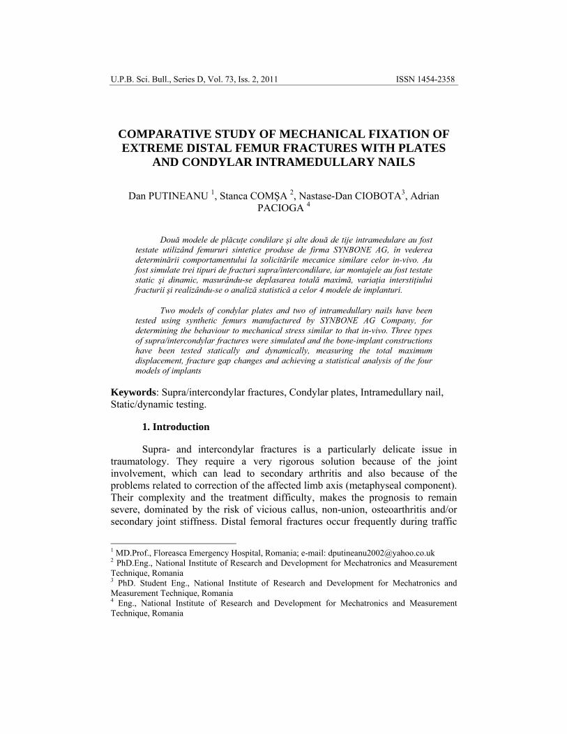
a. LISS plate b. DCS plate c. T2 nail d. SCN nail
Fig. 1. Overview of implant and fracture types
• LISS Plate (Less Invasive Stabilization System - Synthes) with five distal holes and five diaphyseal holes, length 138mm, made of titanium (see figure 1.a). Plate with angular stability (internal fixation) - uniaxial screws;
• DCS plate (Dynamic Condylar Screw - Synthes) with two distal holes and 10 diaphyseal holes, length 182mm, made of stainless steel (see figure 1.b);
• Retrograde intramedullary nail T2, with a diameter of 11mm and length 200mm, Stryker, made of titanium (see figure 1.c). Distal epiphyseal fixing screws in three planes, the most distal screw very close to the tip of the rod;
Comparative study of mechanical fixation of […] and condylar intramedullary nails 61
• Supracondylar intramedullary nail SCN (Supracondylar Nailing System - Synthes) with a diameter of 11mm and length 230mm, made of stainless steel (see figure 1.d). Distal epiphyseal uniaxial fixing screws, the most distal screw far from the tip of the rod.
The fracture model selection was done so that the fixation systems should be subjected mainly to axial loading force. By cutting the synthetic femoral models, three supra -and intercondylar femoral fractures patterns were simulated as follows (AO/OTA classification [3]):
• Simple extra-articular fracture, type 33-A1 (horizontal metaphyseal - see figure 1.a and 1.b);
• Complete simple articular fracture, simple metaphyseal fracture, type 33-C1 (in T shape, reduced displacement - see figure 1.c);
• Complete simple articular fracture, multifragmentary metaphyseal fracture, type 33-C2 (see figure 1.d).
For the static tests, a universal testing equipment H10KT HOUNSFIELD type was used, operating assisted by an external computer.
Endurance testing of the implants was performed using universal testing equipment INSTRON 8872, by which static and dynamic tests can be performed with very high travel speeds.
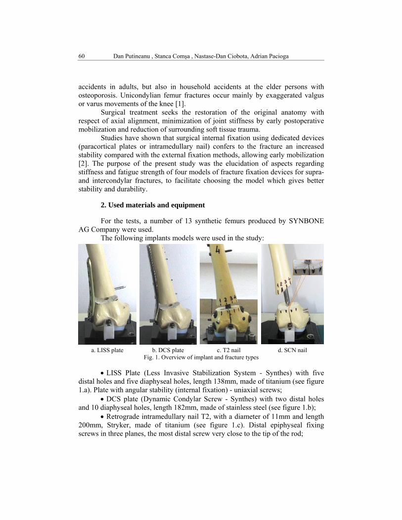
Samples were placed on the platform so that centre of the femoral head should be along the axis of testing equipment. For the loading force transmission, a low friction mechanism was mounted on the working head, which was designed to minimize forces whose direction is not coincident with the loading force (see figure 2.a).
a. Proximally b. Distally Fig. 2. Probe bearing
The support at the bottom of the sample was achieved by positioning the
femoral condyles on a bearing element of a total knee prostheses made of ultra high molecular polyethylene (UHMWPE). Its position was adjusted so that the curvature of the condyles and of the bearing should to be as congruent as possible
Dan Putineanu , Stanca Comşa , Nastase-Dan Ciobota, Adrian Pacioga 62
(see figure 2.b) in order to obtain a correct support, which does not introduce additional efforts, and which reproduces with fidelity the in-vivo conditions.
For good reproduction of the real loading, the position of the polyethylene insert on the testing machine platform was intended to be in respect with the anatomical femoral axis.
With respect to these positioning conditions, each sample was tested statically and dynamically for 100,000 cycles. In a first stage of static tests, the constructions were tested non-destructively, at two loading levels: 500N and 700N respectively, determining the total displacement of the specimens and plotting the characteristic curves of the tests. The second phase of the static tests consisted in loading assemblies with increasing force from 100N, with a 100N increment. Maximum loading force was 800N, which corresponds to a total of 8 (eight) loading levels.
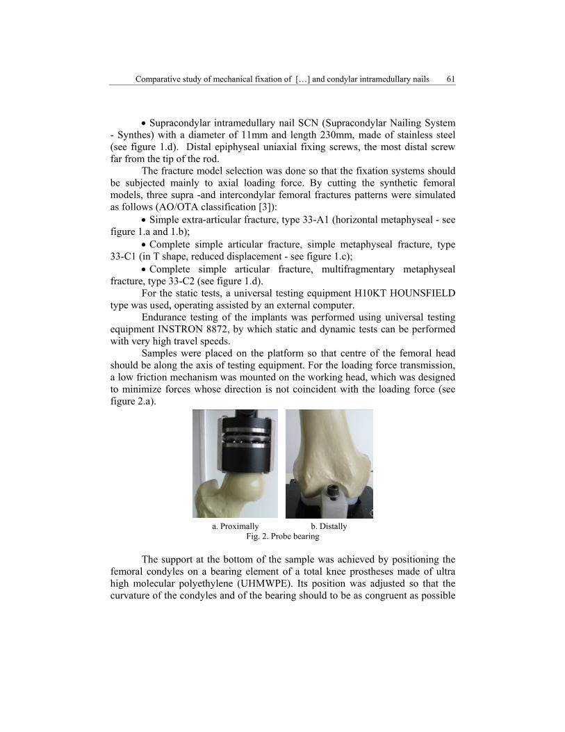
For each loading level, vertical movement of the lateral and medial extreme points were measured and recorded (see figure 3) and changes in the osteotomy gap in 4 points of the anterior region and in 4 points of the posterior region.
Fig. 3. Displacement measurement for lateral and medial extreme points
To ensure measurements repeatability, for 33-C2 fracture model, on the
osteotomy surfaces small slots were milled, to ensure correct positioning of measuring instrument tips (see detail in figure 1.d).
The differences between total displacements of the implants and changes of the osteotomy gap were analyzed, the characteristic curves of the tests were compared and the stiffness diagrams of the constructions were plotted.
Dynamic testing was carried out by controlling the applied loading force upon the femoral constructions. Maximum loading force was 500N, because the risk of fracturing the femoral models and corresponding to this value, the minimum loading force was 50N. Frequency of the cyclic loading was, f=1Hz
Comparative study of mechanical fixation of […] and condylar intramedullary nails 63
(one cycle/second), limited by the elasticity of the models used. Samples were subjected to a total of 100,000 cycles, after this number of cycles the test was stopped even if the models were not broken.
3. Obtained results
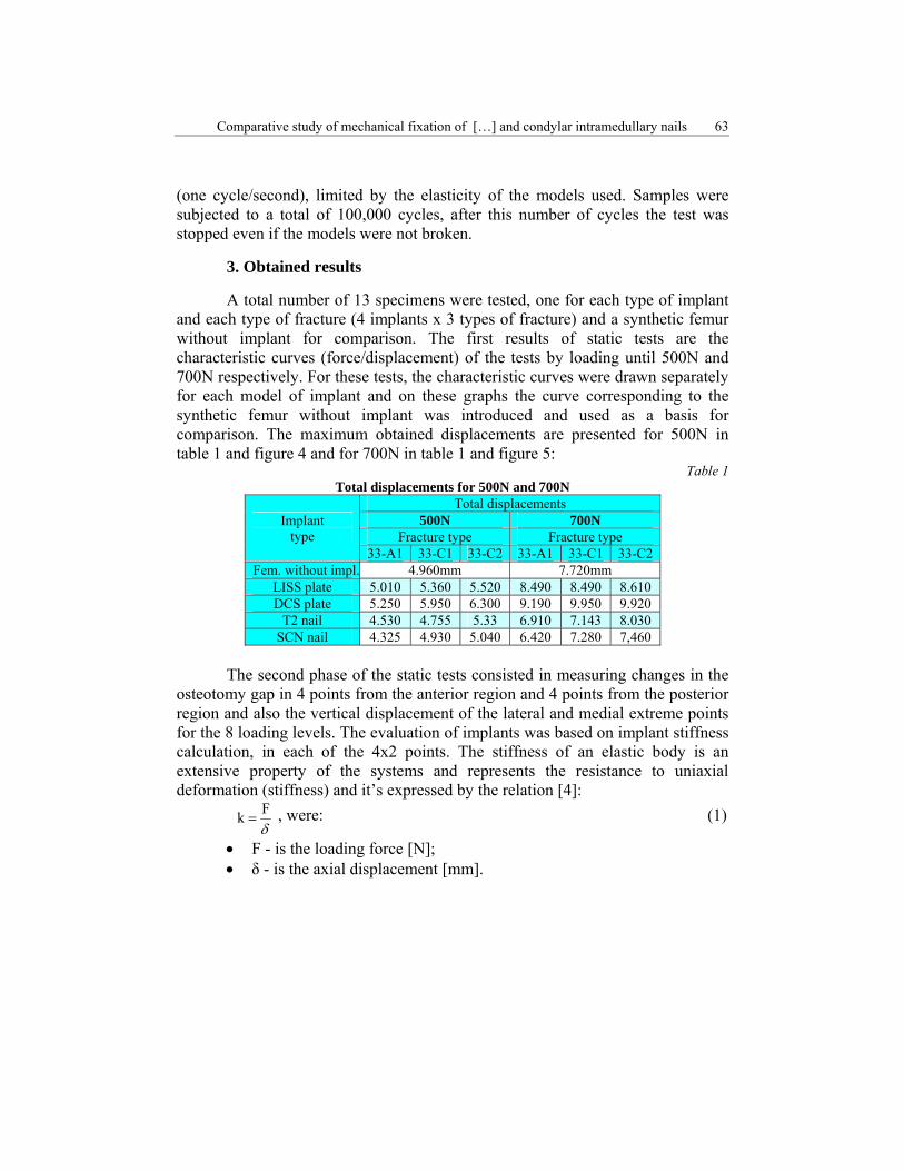
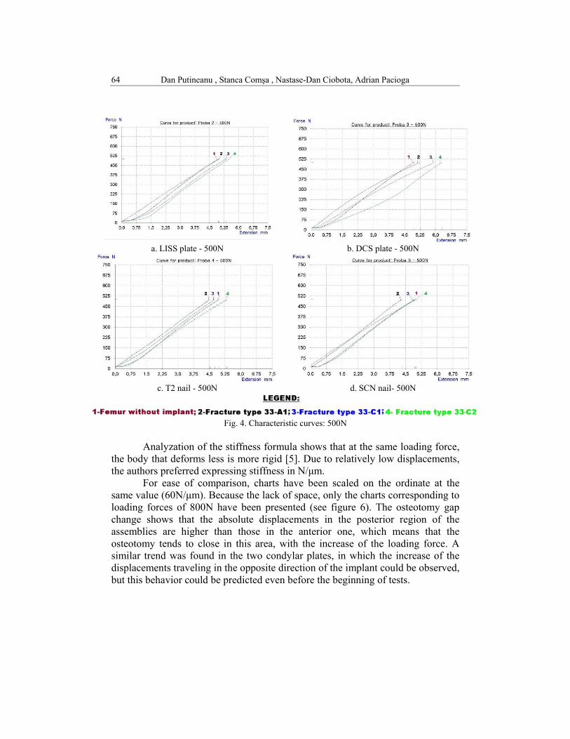
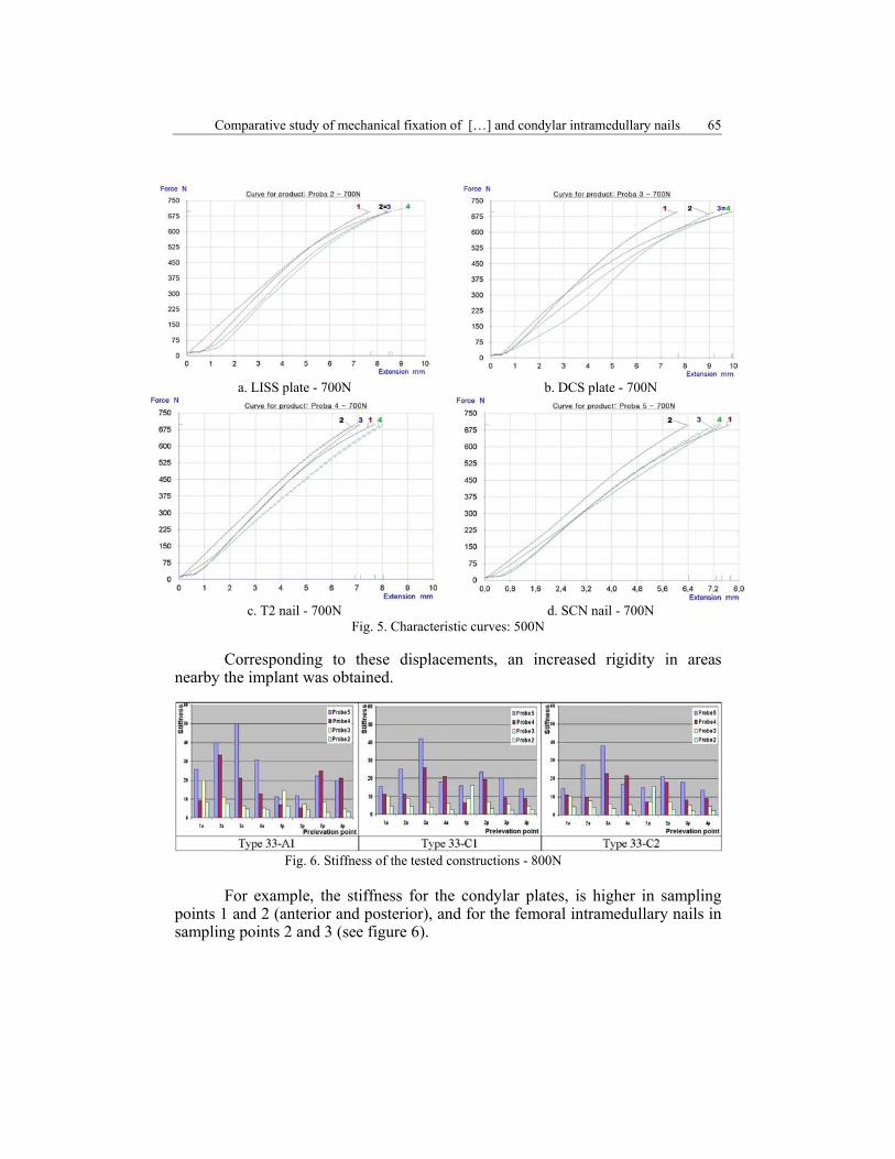
A total number of 13 specimens were tested, one for each type of implant and each type of fracture (4 implants x 3 types of fracture) and a synthetic femur without implant for comparison. The first results of static tests are the characteristic curves (force/displacement) of the tests by loading until 500N and 700N respectively. For these tests, the characteristic curves were drawn separately for each model of implant and on these graphs the curve corresponding to the synthetic femur without implant was introduced and used as a basis for comparison. The maximum obtained displacements are presented for 500N in table 1 and figure 4 and for 700N in table 1 and figure 5:
Table 1 Total displacements for 500N and 700N
Implant type
Total displacements500N 700N
Fracture type Fracture type 33-A1 33-C1 33-C2 33-A1 33-C1 33-C2
Fem. without impl. 4.960mm 7.720mm LISS plate 5.010 5.360 5.520 8.490 8.490 8.610 DCS plate 5.250 5.950 6.300 9.190 9.950 9.920
T2 nail 4.530 4.755 5.33 6.910 7.143 8.030 SCN nail 4.325 4.930 5.040 6.420 7.280 7,460
The second phase of the static tests consisted in measuring changes in the
osteotomy gap in 4 points from the anterior region and 4 points from the posterior region and also the vertical displacement of the lateral and medial extreme points for the 8 loading levels. The evaluation of implants was based on implant stiffness calculation, in each of the 4x2 points. The stiffness of an elastic body is an extensive property of the systems and represents the resistance to uniaxial deformation (stiffness) and it’s expressed by the relation [4]:
δFk = , were: (1)
• F - is the loading force [N]; • δ - is the axial displacement [mm].
Dan Putineanu , Stanca Comşa , Nastase-Dan Ciobota, Adrian Pacioga 64
a. LISS plate - 500N b. DCS plate - 500N
c. T2 nail - 500N d. SCN nail- 500N
Fig. 4. Characteristic curves: 500N
Analyzation of the stiffness formula shows that at the same loading force, the body that deforms less is more rigid [5]. Due to relatively low displacements, the authors preferred expressing stiffness in N/μm.
For ease of comparison, charts have been scaled on the ordinate at the same value (60N/μm). Because the lack of space, only the charts corresponding to loading forces of 800N have been presented (see figure 6). The osteotomy gap change shows that the absolute displacements in the posterior region of the assemblies are higher than those in the anterior one, which means that the osteotomy tends to close in this area, with the increase of the loading force. A similar trend was found in the two condylar plates, in which the increase of the displacements traveling in the opposite direction of the implant could be observed, but this behavior could be predicted even before the beginning of tests.
Comparative study of mechanical fixation of […] and condylar intramedullary nails 65
a. LISS plate - 700N b. DCS plate - 700N
c. T2 nail - 700N d. SCN nail - 700N Fig. 5. Characteristic curves: 500N
Corresponding to these displacements, an increased rigidity in areas
nearby the implant was obtained.
Fig. 6. Stiffness of the tested constructions - 800N
For example, the stiffness for the condylar plates, is higher in sampling
points 1 and 2 (anterior and posterior), and for the femoral intramedullary nails in sampling points 2 and 3 (see figure 6).
Dan Putineanu , Stanca Comşa , Nastase-Dan Ciobota, Adrian Pacioga 66
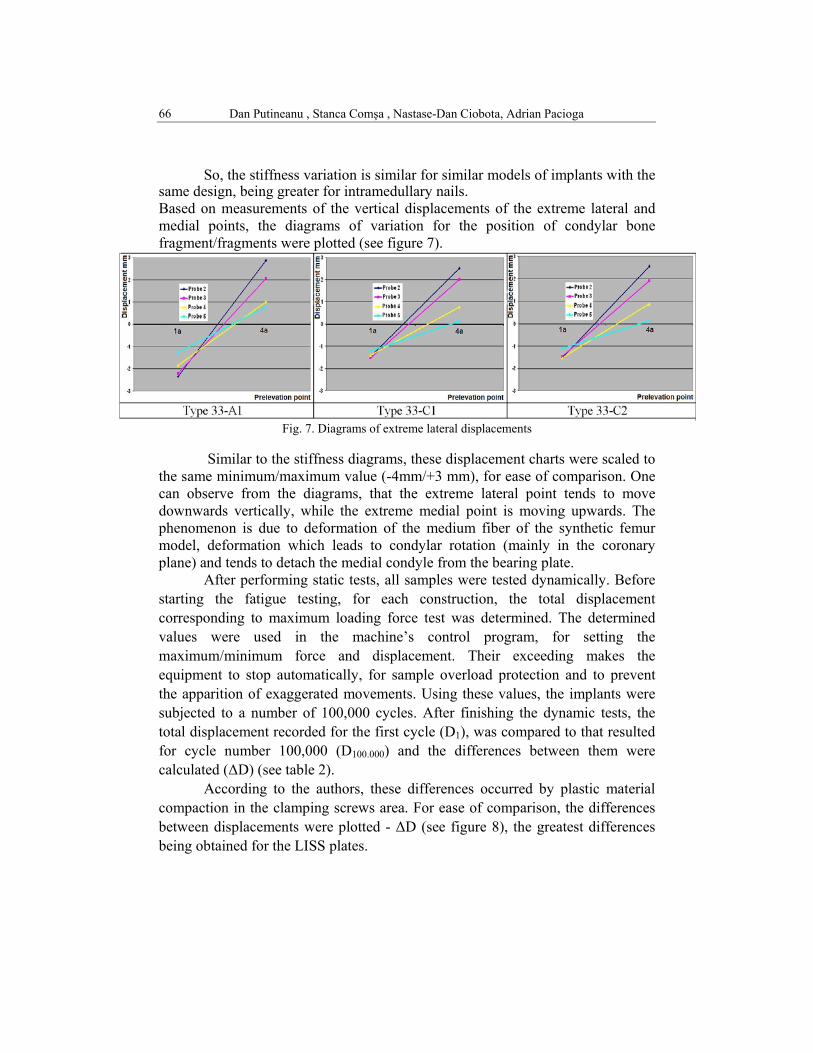
So, the stiffness variation is similar for similar models of implants with the same design, being greater for intramedullary nails. Based on measurements of the vertical displacements of the extreme lateral and medial points, the diagrams of variation for the position of condylar bone fragment/fragments were plotted (see figure 7).
Fig. 7. Diagrams of extreme lateral displacements
Similar to the stiffness diagrams, these displacement charts were scaled to
the same minimum/maximum value (-4mm/+3 mm), for ease of comparison. One can observe from the diagrams, that the extreme lateral point tends to move downwards vertically, while the extreme medial point is moving upwards. The phenomenon is due to deformation of the medium fiber of the synthetic femur model, deformation which leads to condylar rotation (mainly in the coronary plane) and tends to detach the medial condyle from the bearing plate.
After performing static tests, all samples were tested dynamically. Before starting the fatigue testing, for each construction, the total displacement corresponding to maximum loading force test was determined. The determined values were used in the machine’s control program, for setting the maximum/minimum force and displacement. Their exceeding makes the equipment to stop automatically, for sample overload protection and to prevent the apparition of exaggerated movements. Using these values, the implants were subjected to a number of 100,000 cycles. After finishing the dynamic tests, the total displacement recorded for the first cycle (D1), was compared to that resulted for cycle number 100,000 (D100.000) and the differences between them were calculated (ΔD) (see table 2).
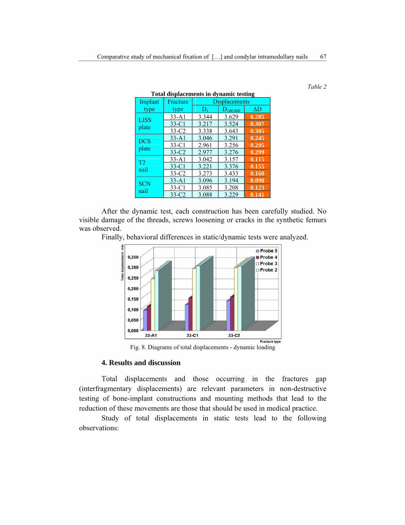
According to the authors, these differences occurred by plastic material compaction in the clamping screws area. For ease of comparison, the differences between displacements were plotted - ΔD (see figure 8), the greatest differences being obtained for the LISS plates.
Comparative study of mechanical fixation of […] and condylar intramedullary nails 67
Table 2 Total displacements in dynamic testing
Implant type
Fracture type
Displacements D1 D100.000 ΔD
LISS plate
33-A1 3.344 3.629 0.28533-C1 3.217 3.524 0.30733-C2 3.338 3.643 0.305
DCS plate
33-A1 3.046 3.291 0.24533-C1 2.961 3.256 0.29533-C2 2.977 3.276 0.299
T2 nail
33-A1 3.042 3.157 0.11533-C1 3.221 3.376 0.15533-C2 3.273 3.433 0.160
SCN nail
33-A1 3.096 3.194 0.09833-C1 3.085 3.208 0.12333-C2 3.088 3.229 0.141
After the dynamic test, each construction has been carefully studied. No
visible damage of the threads, screws loosening or cracks in the synthetic femurs was observed.
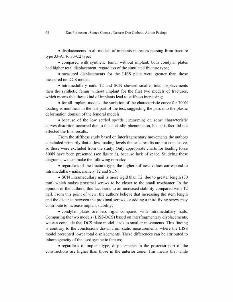
Finally, behavioral differences in static/dynamic tests were analyzed.
Fig. 8. Diagrams of total displacements - dynamic loading
4. Results and discussion
Total displacements and those occurring in the fractures gap (interfragmentary displacements) are relevant parameters in non-destructive testing of bone-implant constructions and mounting methods that lead to the reduction of these movements are those that should be used in medical practice.
Study of total displacements in static tests lead to the following observations:
Dan Putineanu , Stanca Comşa , Nastase-Dan Ciobota, Adrian Pacioga 68
• displacements in all models of implants increases passing from fracture type 33-A1 to 33-C2 type;
• compared with synthetic femur without implant, both condylar plates had higher total displacement, regardless of the simulated fracture type;
• measured displacements for the LISS plate were greater than those measured on DCS model;
• intramedullary nails T2 and SCN showed smaller total displacements then the synthetic femur without implant for the first two models of fractures, which means that these kind of implants lead to stiffness increasing;
• for all implant models, the variation of the characteristic curve for 700N loading is nonlinear in the last part of the test, suggesting the pass into the plastic deformation domain of the femoral models;
• because of the low settled speeds (1mm/min) on some characteristic curves distortion occurred due to the stick-slip phenomenon, but this fact did not affected the final results.
From the stiffness study based on interfragmentary movements the authors concluded primarily that at low loading levels the tests results are not conclusive, so these were excluded from the study. Only appropriate charts for loading force 800N have been presented (see figure 6), because lack of space. Studying these diagrams, we can make the following remarks:
• regardless of the fracture type, the higher stiffness values correspond to intramedullary nails, namely T2 and SCN;
• SCN intramedullary nail is more rigid than T2, due to greater length (30 mm) which makes proximal screws to be closer to the small trochanter. In the opinion of the authors, this fact leads to an increased stability compared with T2 nail. From this point of view, the authors believe that increasing the stem length and the distance between the proximal screws, or adding a third fixing screw may contribute to increase implant stability;
• condylar plates are less rigid compared with intramedullary nails. Comparing the two models (LISS-DCS) based on interfragmentary displacements, we can conclude that DCS plate model leads to smaller movements. This finding is contrary to the conclusions drawn from static measurements, where the LISS model presented lower total displacements. These differences can be attributed to inhomogeneity of the used synthetic femurs;
• regardless of implant type, displacements in the posterior part of the constructions are higher than those in the anterior zone. This means that while
Comparative study of mechanical fixation of […] and condylar intramedullary nails 69
increasing the loading forces, fracture tends to "close" in the posterior direction. The phenomenon can be explained by the medium fiber curvature also in the sagittal plane;
• regardless of the implant model, the displacements are reduced nearby implant and grow while increasing the distance. This makes the stiffness of femoral nails greater not only because of their central position, but also due to lower bending moment applied by reducing force’s arm, leading to reduced movements in both the sagittal and coronal plane;
• no significant differences were found between the stiffness of fractures 33-C1 and 33 -C2 type, and the values obtained for the four implants type do not allow clear conclusions regarding stability differences.
Based on the measurement of vertical displacements extreme lateral and medial points, the diagrams of position variation for the fragment/fragments of condylar bone were plotted, which reveals rotation of the condylar fragments in the coronal plane. It is a little surprising the finding that displacements for fractures 33-C1 and 33-C2 type are slightly lower than those for the 33-A1 type. The explanation for this phenomenon lies in the rotation tendency of the condylar fragments in the coronal plane, which contributes to increase of the total displacement. This rotation tendency was evidenced by measuring the intercondylar gap using a feeler gauge, the maximum thickness variations obtained being 0.3mm in the inferior part of the condyles.
Cyclic tests of constructions, came to support static tests conclusions, in the sense that they showed that the stiffness of femoral stems is greater than that of the condylar plates. Permanent deformations were smaller in T2 and SCN nails, compared with condylar plates. As in the static tests, the stiffness differences between fracture types 33-C1 and 33-C2 were not significant, but the values obtained for cyclic tests have clearly indicated a greater instability in the case of the third fracture type.
This study has some deficiencies as follows: • synthetic femurs models had a high elasticity, which prevented the
similarly in-vivo loading (2100N for a patient weighing 70kg); • due to impossibility to achieve a perfect match between condyles and
support bearing plate (impossible to ensure an identical profile) it is probable that further displacement could be induced;
Dan Putineanu , Stanca Comşa , Nastase-Dan Ciobota, Adrian Pacioga 70
• in this study were not taken into account the existence of variables data introduced by the surrounding soft tissues, which may influence the achieved final results.
5. Conclusions
In the experiments carried out, four models of implants were tested for fixation of extreme distal femoral fractures. For this purpose synthetic femurs were used, three types supracondylar fractures were simulated and constructions obtained with 4 implant types were evaluated in terms of biomechanics. The study demonstrates that the use of condylar plates lead to higher displacements in osteotomy gap than intramedullary nails, in the same loading conditions.
Cyclic tests performed on femoral constructions, once again showed increased stiffness of T2 and SCN nails, because for these constructions permanent deformations were lower than those of condylar plates. Another important conclusion is that instability increases with increasing the number of bone fragments.
Our conclusions are supported by other author’s researches [6], who testing similar models of implants have concluded that intramedullary nails have higher stiffness. Extending the study results from in-vitro to in-vivo situation is limited because different modulus of elasticity of synthetic femurs from natural femurs, but unfortunately the acquisition of bones taken from cadavers was not possible.
R E F E R E N C E S
[1]. De Van C. Mow, Rik Huiskes, Basic orthopaedic biomechanics & mechano-biology, Lippincott Williams & Wilkins, 3rd ed., 2005
[2]. Sear, R., Brian, G. Coulson and S. Namuye, A Mechanical Study of Gap Motion in Cadaveric Femurs Using Short and Long Supracondylar Nails, Journal of Orthopaedic Trauma, Vol. 18, Issue 6, 2004, pp 354-360
[3]. OTA Fracture Classification Committee, Fracture and dislocation compendium, Journal of Orthopaedic Trauma, Vol. 21, No. 10, 2007
[4]. E. A. Avallone, Marks' standard handbook for mechanical engineers, 11th illustrated ed., McGraw-Hill Professional, New York, 2006, pp. 562-567
[5]. P.M.G.P. Moreira, S.D. Pastrama, P.M.S.T. de Castro, Comparative three dimensional fracture analyses of cracked plates, U.P.B. Scientific Bulletin., Series D, Vol. 69, No. 1, 2007, pp. 152-157
[6]. K. Firoozbakhsh, K. Behzadi, Mechanics of retrograde nail versus plate fixation for supracondylar femur fractures, Journal of Orthopaedic Trauma, Vol. 9, No. 9, 1995, pp. 152-157.