Embed Size (px)
Citation preview
Conserved Pyridoxal Protein That Regulates Ile and Val Metabolism
Tomokazu Ito, Jumpei Iimori, Sayuri Takayama, Akihito Moriyama, Ayako Yamauchi, Hisashi Hemmi, Tohru Yoshimura
Department of Applied Molecular Biosciences, Graduate School of Bioagricultural Sciences, Nagoya University, Furou-chou, Chikusa, Nagoya, Aichi, Japan
Escherichia coli YggS is a member of the highly conserved uncharacterized protein family that binds pyridoxal 5=-phosphate(PLP). To assist with the functional assignment of the YggS family, in vivo and in vitro analyses were performed using a yggS-deficient E. coli strain (�yggS) and a purified form of YggS, respectively. In the stationary phase, the �yggS strain exhibited acompletely different intracellular pool of amino acids and produced a significant amount of L-Val in the culture medium. Thelog-phase �yggS strain accumulated 2-ketobutyrate, its aminated compound 2-aminobutyrate, and, to a lesser extent, L-Val. Italso exhibited a 1.3- to 2.6-fold increase in the levels of Ile and Val metabolic enzymes. The fact that similar phenotypes were in-duced in wild-type E. coli by the exogenous addition of 2-ketobutyrate and 2-aminobutyrate indicates that the 2 compoundscontribute to the �yggS phenotypes. We showed that the initial cause of the keto acid imbalance was the reduced availability ofcoenzyme A (CoA); supplementation with pantothenate, which is a CoA precursor, fully reversed phenotypes conferred by theyggS mutation. The plasmid-borne expression of YggS and orthologs from Bacillus subtilis, Saccharomyces cerevisiae, and hu-mans fully rescued the �yggS phenotypes. Expression of a mutant YggS lacking PLP-binding ability, however, did not reverse the�yggS phenotypes. These results demonstrate for the first time that YggS controls Ile and Val metabolism by modulating 2-keto-butyrate and CoA availability. Its function depends on PLP, and it is highly conserved in a wide range species, from bacteria tohumans.
Escherichia coli YggS belongs to a poorly characterized buthighly conserved protein family that binds pyridoxal 5=-phos-
phate (PLP). YggS and its orthologs, which range in molecularmass from 24 to 30 kDa, are highly conserved and are present inalmost all kingdoms of life, including bacteria, plants, yeasts, andmammals. This high degree of conservation indicates that YggSplays an important role in cellular functions, but no definitiveinformation is available thus far.
In the E. coli genome, yggS is located at 66.67 min on the geneticmap and is in a polycistronic operon that is composed of 5 poorlycharacterized genes, yggS-yggT-yggU-yggV-yggW (1). A similar ge-netic localization is also found in the genomes of Salmonella en-terica serovar Typhimurium, Klebsiella pneumoniae, and Pantoeasp. strain At-9b. Among the products of these operons, YggT is anintegral membrane protein that has no clearly defined function,and its orthologs are found in bacteria and plants. We previouslyfound that the introduction of the plasmid-borne yggT into an E.coli TK2420 strain, which lacks 3 major K� uptake systems (Kdp,Trk, and Kup) and requires exogenous K�, enabled us to restorecell growth at high osmotic pressure (1). The YggT homologs arerequired for the biogenesis of the c= heme in the cytochrome b6fcomplex in Chlamydomonas reinhardtii (2), as well as for theproper distribution of nucleoids in chloroplasts and cyanobacte-ria (3). The fourth gene product, YggV, exhibits ITP/XTPase ac-tivity, and a previous study has suggested that it removes misin-corporated bases, such as xanthine or hypoxanthine, from thepool of DNA precursors (4). To date, no functional correlationshave been reported between YggS and other gene products in thecluster, which include YggT, YggU, YggV, and/or YggW.
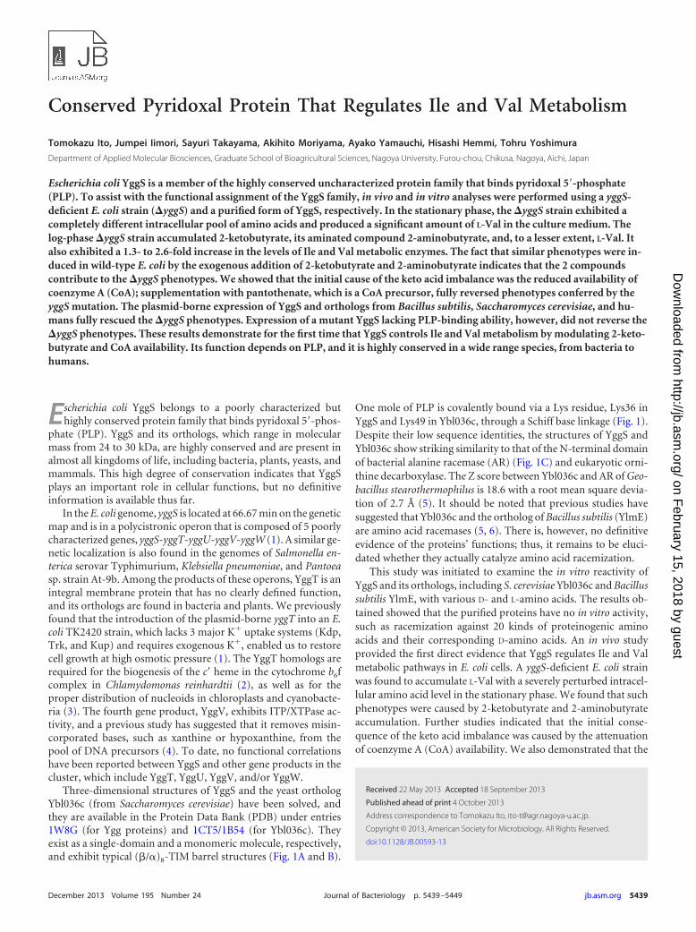
Three-dimensional structures of YggS and the yeast orthologYbl036c (from Saccharomyces cerevisiae) have been solved, andthey are available in the Protein Data Bank (PDB) under entries1W8G (for Ygg proteins) and 1CT5/1B54 (for Ybl036c). Theyexist as a single-domain and a monomeric molecule, respectively,and exhibit typical (�/�)8-TIM barrel structures (Fig. 1A and B).
One mole of PLP is covalently bound via a Lys residue, Lys36 inYggS and Lys49 in Ybl036c, through a Schiff base linkage (Fig. 1).Despite their low sequence identities, the structures of YggS andYbl036c show striking similarity to that of the N-terminal domainof bacterial alanine racemase (AR) (Fig. 1C) and eukaryotic orni-thine decarboxylase. The Z score between Ybl036c and AR of Geo-bacillus stearothermophilus is 18.6 with a root mean square devia-tion of 2.7 Å (5). It should be noted that previous studies havesuggested that Ybl036c and the ortholog of Bacillus subtilis (YlmE)are amino acid racemases (5, 6). There is, however, no definitiveevidence of the proteins’ functions; thus, it remains to be eluci-dated whether they actually catalyze amino acid racemization.
This study was initiated to examine the in vitro reactivity ofYggS and its orthologs, including S. cerevisiae Ybl036c and Bacillussubtilis YlmE, with various D- and L-amino acids. The results ob-tained showed that the purified proteins have no in vitro activity,such as racemization against 20 kinds of proteinogenic aminoacids and their corresponding D-amino acids. An in vivo studyprovided the first direct evidence that YggS regulates Ile and Valmetabolic pathways in E. coli cells. A yggS-deficient E. coli strainwas found to accumulate L-Val with a severely perturbed intracel-lular amino acid level in the stationary phase. We found that suchphenotypes were caused by 2-ketobutyrate and 2-aminobutyrateaccumulation. Further studies indicated that the initial conse-quence of the keto acid imbalance was caused by the attenuationof coenzyme A (CoA) availability. We also demonstrated that the
Received 22 May 2013 Accepted 18 September 2013
Published ahead of print 4 October 2013
Address correspondence to Tomokazu Ito, [email protected].
Copyright © 2013, American Society for Microbiology. All Rights Reserved.
doi:10.1128/JB.00593-13
December 2013 Volume 195 Number 24 Journal of Bacteriology p. 5439 –5449 jb.asm.org 5439
on February 15, 2018 by guest
http://jb.asm.org/
Dow
nloaded from
biological function of YggS depends on PLP and is highly con-served in a wide range of species, from bacteria to humans.
MATERIALS AND METHODSMaterials. Amino acids, leucine dehydrogenase from Bacillus stearother-mophilus, phosphotransacetylase from Leuconostoc mesenteroides, andmalate dehydrogenase from pig heart were from Wako (Osaka, Japan).KOD-plus ver-2 DNA polymerase was from Toyobo (Osaka, Japan). Ci-trate synthase (from porcine heart) was from Sigma (St. Louis, MO).Restriction enzymes were purchased from TaKaRa Shuzo (Kyoto, Japan)or New England BioLabs Inc. (Beverly, MA). Oligonucleotides were or-dered and purchased from Fasmac (Tokyo, Japan) or Operon Biotechnol-ogy (Tokyo, Japan). D-Pantoate was obtained by autoclaving D-pantolac-tone solution (0.5 M D-pantolactone in 0.5 M KOH) for 20 min at 121°C.
Bacterial strains and growth conditions. Bacterial strains and plas-mids used in this study are listed in Table 1. LB or M9-glucose syntheticmedium were used for cell growth. The LB medium contained 1% tryp-tone, 0.5% yeast extract, and 1% NaCl. The M9-glucose medium con-sisted of 0.6% Na2HPO4, 0.3% KH2PO4, 0.1% NH4Cl, 0.05% NaCl, 0.2%glucose, 1 mM MgSO4, 10 �M FeSO4, 10 �M CaCl2, and 0.001% thia-mine-HCl. Agar (1.5%) was added for solid medium. Ampicillin andkanamycin were added to the LB or the M9-glucose medium when re-quired at a final concentration of 100 and 30 �g/ml, respectively. For thegrowth test, cells from overnight culture (in LB medium) were washed,resuspended in phosphate-buffered saline (PBS; 10 mM Na2HPO4, 1.8mM KH2PO4, 140 mM NaCl, 2.7 mM KCl, pH 7.5), and inoculated in theM9 medium at a final optical density at 600 nm (OD600) of 0.02. Growthof the bacteria was measured as the cell OD600 with shaking at 30°C.
Genetic manipulation. The disruption of yggS of E. coli K-12 MG1655was performed using the � Red recombinase system described byDatsenko and Wanner (7). A PCR product was generated by using two60-nucleotide (nt) primers, yggS-H1 and yggS-H2, comprised of 40 nthomologous to the yggS gene and an additional 20 nt complementary tothe template plasmid pKD13, which carries the kanamycin resistancegene. The 100-ng sample of the PCR product was purified from the aga-rose gel and electroporated into wild-type (WT) E. coli MG1655 cellsharboring pKD46, which had been grown in LB solid medium containing30 �g/ml kanamycin at 30°C. Gene knockout was verified through colo-ny-directed PCR. Construction of pUS (pUC19-yggS) and pUSm has
been described previously (1). A control plasmid, pU0, which containspart of the yggS sequence, was obtained by FspI digestion of pUS and thenself-ligation (1). pGEX-yggS, pGEX-YBL036C, pGEX-ylmE, and pGEX-prosc are derived from pGEX vector (GE Healthcare, Piscataway, NJ) andexpress E. coli yggS, S. cerevisiae YBL036C, B. subtilis ylmE, and Homosapiens PROSC, respectively. They were constructed as follows. The E. coliyggS gene was obtained by PCR with a set of primers (yggS-fw and yggS-rv)with genomic DNA of E. coli MG1655 as the template. A set of primers(YBL036C-fw and YBL036C-rv) and the chromosomal DNA from S.cerevisiae BY4742 were used for the amplification of the Ybl036c gene. TheylmE gene was amplified with primers ylmE-fw and ylmE-rv. The genomicDNA from B. subtilis NCIB3610 was used for gene amplification. ThecDNA of PROSC was amplified using the primes hPROSC-fw andhPROSC-rv. Plasmid pOTB7 containing the full-length PROSC cDNA,which was purchased from Open Biosystems Inc. (plasmid MHS1011;Huntsville, AL), was used as the template. Each of the amplified DNAfragments was digested with appropriate restriction enzymes, purifiedfrom the agarose gel, and then cloned into pGEX vector. The proteinswere expressed in E. coli cells as a fusion with N-terminal glutathioneS-transferase (GST). pYggS was constructed by using Champion pET di-rectional TOPO expression kits (Invitrogen, CA) according to the manu-facturer’s protocol. Primer set yggS-pET-fw and yggS-pET-rv and thegenomic DNA of E. coli MG1655 were used for the yggS amplification.Plasmids and primers used in this study are listed in Table 1.
Protein purification. E. coli BL21(DE3) star (Invitrogen) carryingpYggS was cultivated in LB medium containing 100 �g/ml ampicillin at30°C. Isopropyl-�-D-thiogalactoside (IPTG; 1 mM) was added to the me-dium at mid-log phase, and the cells were collected after 12 h of cultiva-tion. All of the following purification steps were performed at 4°C. The cellpellet was suspended in a binding buffer (20 mM Tris-HCl buffer con-taining 500 mM NaCl and 80 mM imidazole, pH 7.9) and disrupted bysonication. After centrifugation at 20,000 � g for 20 min, the clear lysatewas applied to a Ni affinity chromatography equipped with Ni Sepharose6 fast flow (GE Healthcare, WI). The column was washed with a washingbuffer (20 mM Tris-HCl buffer containing 500 mM NaCl and 80 mMimidazole, pH 7.9), and the purified protein was eluted with an elutionbuffer (20 mM Tris-HCl buffer containing 500 mM NaCl and 500 mMimidazole, pH 7.9). The protein was dialyzed with PBS containing 20 �M
FIG 1 Structure of YggS and comparison to bacterial AR. A front view (A) and side view (B) of the YggS structure are represented. The PDB code used for YggSis 1W8G. (C) The superposition of the YggS structure onto that of the monomeric structure of G. stearothermophilus AR (PDB entry 1SFT). (D and E) Stereo viewof the putative active site of YggS (D) and the active site of AR (E).
Ito et al.
5440 jb.asm.org Journal of Bacteriology
on February 15, 2018 by guest
http://jb.asm.org/
Dow
nloaded from
PLP and redialyzed in the same buffer in the absence of PLP to removeunbound PLP. The purified protein was stored at �80°C until use.
Amino acid pool analysis. Cells were grown in M9-glucose mediumto a final OD600 of 0.5 or 2.0. The cell pellet (100 mg) was washed oncewith the ice-cold PBS and was resuspended in 4-fold cell pellet volumes(400 �l) of 5% trichloroacetic acid (TCA) solution. After sonicationand following incubation for 30 min at 4°C, the cell debris was removed bycentrifugation (20,000 � g, 20 min, 4°C). The TCA was removed from thesample by three extractions with a water-saturated diethyl ether. The sam-ples were stored at �20°C until use. The culture medium supernatant (1ml) was deproteinized with 200 �l of 30% TCA, and the TCA was re-moved by 3 extractions with water-saturated diethyl ether. The solutionwas dried under vacuum, and the resultant pellet was then reconstitutedin distilled water. The samples were stored at �20°C until use.
High-performance liquid chromatography (HPLC) analysis. A20-�l aliquot of the sample was applied to an amino acid analysis system(Shimadzu, Kyoto, Japan) equipped with a Shim-pack amino Na column(Shimadzu, Kyoto, Japan) and RF-10A spectrofluorometer after de-rivatization with O-phtalaldehyde and L-acetylcysteine according to themanufacturer’s protocol. Amino acid mixture standard solution (Type H;Wako, Japan) was used for identification and quantification of aminoacids. The enantioselective amino acid analysis was performed as de-scribed previously (8, 9).
Enzymatic determination of L-Val. The reaction mixture (final vol-ume, 250 �l) containing 200 mM glycine-KCl-KOH buffer (pH 10.2), 5
mM NAD�, 1 mU leucine dehydrogenase (1 U is defined as the amount ofenzyme to produce 1 �mol of NADH from L-leucine in 1 min at 30°C),and the deproteinized culture supernatant was incubated at 37°C for 1 h.After incubation, the L-Val concentration was estimated as the NADHformation (increase of the A340) over time.
Keto acid analysis. Intracellular keto acids were extracted from E. colicells as follows. Mid-log-phase cells (OD600, 0.5; 100 mg cells) in M9-glucose medium were suspended in 4 volumes (400 �l) of 0.8 M HClO4
and incubated for 5 min at 4°C. The resultant cell suspension was centri-fuged for 15 min (11,000 � g, 4°C), and the supernatant was 10-folddiluted. The aliquot (200 �l) was mixed to equal amounts in a solutionconsisting of 0.7 mM 1,2-diamino-4,5-methylenedioxybenzene (DMB;Dojindo, Tokyo, Japan), 0.7 M HCl, 1.0 M 2-mercaptoethanol, and 28mM Na2S2O4 and incubated for 4 h at 50°C in a dark place. The resultantsolution was passed through a 0.45-�m filter device and stored at �80°Cuntil use. The separation and quantification of the derivatized keto acidswere performed according to the protocol described by Kato et al. (10).
Determination of total coenzyme A. The levels of total coenzyme Awere determined according to the method previously described by Allredand Guy (11). Briefly, WT and yggS strains were cultivated in M9-glu-cose medium at 30°C. Cultures were grown to log phase (OD600, 0.5),and the cells were collected by centrifugation (8,000 � g, 10 min, 4°C).Cells (80 mg) were resuspended in ice-cold PBS and disrupted by theaddition of HClO4 to a final concentration of 0.4 M. The suspensions wereincubated on ice for 30 min and were vortexed periodically. After centrif-
TABLE 1 E. coli strains, plasmids, and primers used in this study
Strain, plasmid, orprimer Relevant characteristic(s) Reference or source
E. coli strainsMG1655 F� �� rfb-50 rph-1; mutated in ilvG Laboratory collectionMG1655yggS F� �� rfb-50 rph-1 yggS::Kmr; mutated in ilvG This studyXL1-Blue hsdR17 supE44 recA1 endA1 gyrA46 thi relA1 lac-F= [proAB� lacIq lacZM15::Tn10(Tetr)] StratageneBL21 F� dcm ompT hsdS(rB
� mB�) gal[malB�]K-12 (�S) Laboratory collection
BL21(DE3) star F� ompT hsdSB (rB� mB
�) gal dcm rne131 (DE3) Invitrogen
PlasmidspKD13 Kmr gene with FLP recombination target sequence 7pKD46 � Red recombinase expression plasmid 7pU0 Control vector (pUC19 containing partial sequence of yggS) This studypUS yggS expression vector (expresses YggS-His) 1pUSm pUS containing K36A mutation of YggS 1pGEX-4T-1 Empty vector GE HealthcarepGEX-4T-3 Empty vector GE HealthcarepGEX-yggS yggS expression (expresses GST-YggS-His) This studypGEX-ylmE ylmE expression (expresses GST-YlmE) This studypGEX-YBL036C YBL036C expression (expresses GST-ybl036c) This studypGEX-prosc PROSC expression (expresses GST-PROSC) This studypYggS YggS (C-terminal His tag) expression This study
Primersa
yggS-H1 AGCGCCATCGCAGAAGCCATTGATGCCGGGCAGCGTCAATGTGTAGGCTGGAGCTGCTTC This studyyggS-H2 TCGTCCGACATTCCCAGAGAGAGCGTGTCGATATGCGGGTATTCCGGGGATCCGTCGACC This studyyggS-pET-fw CACCATGAACGATATGCGCATAAGCGG This studyyggS-pET-rv TTAATGGTGATGAGGTGATGTTTTTTAGAGTAATCA This studyyggS-fw AAAGAATTCAGAAGGAGATCCTCGGAAAATGAACG This studyyggS-rv AAACTGCAGTTAATGGTGATGATGGTGATGTTTTTTAGAGTAATCACGCGC This studyybl036c-fw GGAATTCAGGAGGATTGCAATATGTCCACTGGTATTACTTATGATG This studyybl036c-rv AACTCGAGCTAGTGATGGTGGTGATGGTGAATGATTCTAGCTTCATTTTTTGGAGGTC This studyhPROSC-fw GGAATCCAGAAGGAGATCCTCGGAAAATGTGGAGCTGGAGAGCTGGCAGCATGTC This studyhPROSC-rv AACTGCAGTCAATGATGGGTGATGATGGTGCTCCTGTGCACCTCCAG This studyylmE-fw AAAGAATTCATGCGTGTTGTTGATAATTTACGAC This studyylmE-rv AATAGTCGACTCATTGCTGTACACCCCCTG This study
a Primer sequences are given in 5= to 3= orientation.
PLP Protein That Regulates Ile-Val Metabolism
December 2013 Volume 195 Number 24 jb.asm.org 5441
on February 15, 2018 by guest
http://jb.asm.org/
Dow
nloaded from
ugation (20,000 � g, 20 min, 4°C), the resultant supernatant was neutral-ized with KOH. The reaction solution (1 ml) contained 500 mM Tris-HCl(pH 7.2), 50 mM KCl, 20 mM malate, 8 mM lithium acetyl phosphate, 2mM NAD�, 3 U citrate synthase, 15 U malate dehydrogenase, 8.4 U phos-photransacetylase, and the cell extract. The reaction was performed at30°C in a cuvette with a 1-cm light path. The rate of NADH formation wasmeasured at 340 nm using a Shimadzu UV-2450 spectrophotometer.
Enzyme assay. Crude cell extracts were prepared using mid-log-phasecells (OD600, 0.5) grown in M9-glucose medium. The cells (80 mg) werecollected by centrifugation and were resuspended in 10 volumes of a dis-ruption buffer (800 �l) containing 100 mM potassium phosphate, 0.5mM dithiothreitol, and 20% glycerol (pH 7.5). The cell suspension wassonicated and centrifuged (20,000 � g, 20 min, 4°C), and the resultantclear lysate was used for the enzyme assay. Protein concentration wasdetermined by the procedure of Bradford with the Bio-Rad protein assaykit (Bio-Rad, Hercules, CA) using bovine serum albumin as the standard.
(i) Threonine dehydratase activity. An aliquot of the cell-free lysate(125 �g of protein) was added to the reaction mixture containing 100mM potassium phosphate buffer (pH 7.5), 40 �M PLP, and 20 mM L-Thrat a final volume of 250 �l. After incubation for 60 min at 30°C, 20 �l ofthe reaction mixture was withdrawn and diluted to 50 �l. A 50-�l aliquotof 0.1% 2,4-dinitrophenylhydrazine (2,4-DNP; Kanto Chemical Co. To-kyo, Japan) dissolved in 2N HCl was added to the mixture and incubatedfor 10 min at 25°C. A 100-�l aliquot of ethanol and a 125-�l aliquot of 2NNaOH were mixed, and chromophore formation was allowed to occur.The absorbance at 515 nm of the sample was determined with a ShimadzuUV-2450 spectrophotometer. The amount of 2-ketobutyrate formed wasestimated by comparison of the absorbance of known concentrations of2-ketobutyrate.
(ii) AHAS activity. Activity of acetohydroxy acid synthase (AHAS)was determined in the reaction mixture containing 100 mM potassiumphosphate buffer, pH 7.5, 10 mM MgCl2, 50 mM pyruvate, 100 �M thi-amine pyrophosphate (TPP), 100 �M flavin adenine dinucleotide (FAD),and the cell-free lysate (300 �g of protein) in a total volume of 500 �l at30°C. The rate of pyruvate consumption was calculated by monitoring thedecrease of the absorbance at 333 nm (ε, 17.5 M cm�1) (12).
(iii) Transaminase B (IlvE) activity. Activity of branched-chainamino acid transaminase (transaminase B; IlvE) was determined accord-ing to the method described by Duggan and Wechsler (13). The standardassay mixture (500 �l) contained 100 mM Tris-HCl, 100 �M PLP, 15 mM�-ketoglutarate, 25 mM L-Val, and the cell extract (0.5 to 1.0 mg/ml), pH8.0, which was incubated for 30 min at 37°C.
(iv) Transaminase C (AvtA) assay. Activity of alanine-valinetransaminase (transaminase C; AvtA) was determined at 37°C accordingto the method described previously, with slight modifications (14). Themethod is based on a different extraction of pyruvate and �-ketoisovalericacid (KIV). The 500-�l reaction mixture contained 100 mM Tris-HCl(pH 8.0), 10 mM KIV, 10 mM L-Ala, 1 mM PLP, and cell extract (1 mgprotein). After 30 min of incubation, 50 �l of 20% metaphosphoric acidwas added to terminate the reaction. The ensuing procedures were per-formed according to the protocol described in reference 14.
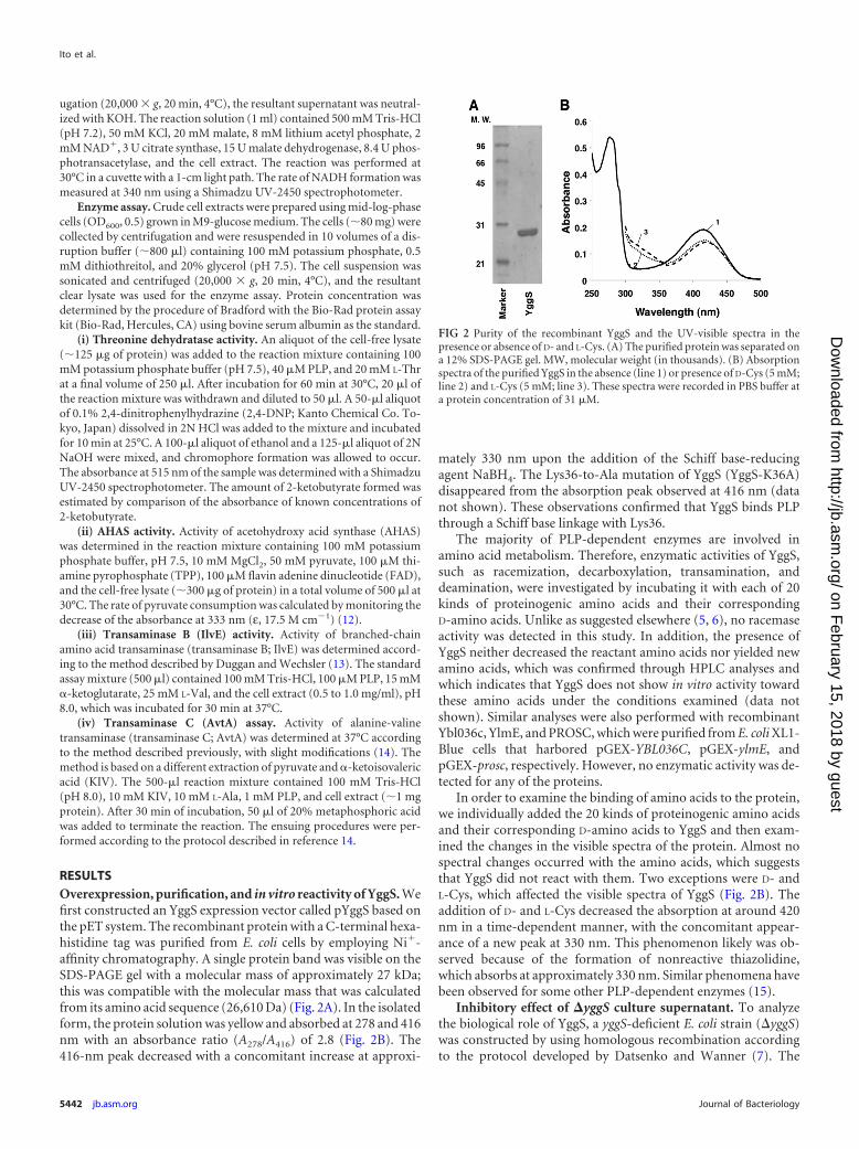
RESULTSOverexpression, purification, and in vitro reactivity of YggS. Wefirst constructed an YggS expression vector called pYggS based onthe pET system. The recombinant protein with a C-terminal hexa-histidine tag was purified from E. coli cells by employing Ni�-affinity chromatography. A single protein band was visible on theSDS-PAGE gel with a molecular mass of approximately 27 kDa;this was compatible with the molecular mass that was calculatedfrom its amino acid sequence (26,610 Da) (Fig. 2A). In the isolatedform, the protein solution was yellow and absorbed at 278 and 416nm with an absorbance ratio (A278/A416) of 2.8 (Fig. 2B). The416-nm peak decreased with a concomitant increase at approxi-
mately 330 nm upon the addition of the Schiff base-reducingagent NaBH4. The Lys36-to-Ala mutation of YggS (YggS-K36A)disappeared from the absorption peak observed at 416 nm (datanot shown). These observations confirmed that YggS binds PLPthrough a Schiff base linkage with Lys36.
The majority of PLP-dependent enzymes are involved inamino acid metabolism. Therefore, enzymatic activities of YggS,such as racemization, decarboxylation, transamination, anddeamination, were investigated by incubating it with each of 20kinds of proteinogenic amino acids and their correspondingD-amino acids. Unlike as suggested elsewhere (5, 6), no racemaseactivity was detected in this study. In addition, the presence ofYggS neither decreased the reactant amino acids nor yielded newamino acids, which was confirmed through HPLC analyses andwhich indicates that YggS does not show in vitro activity towardthese amino acids under the conditions examined (data notshown). Similar analyses were also performed with recombinantYbl036c, YlmE, and PROSC, which were purified from E. coli XL1-Blue cells that harbored pGEX-YBL036C, pGEX-ylmE, andpGEX-prosc, respectively. However, no enzymatic activity was de-tected for any of the proteins.
In order to examine the binding of amino acids to the protein,we individually added the 20 kinds of proteinogenic amino acidsand their corresponding D-amino acids to YggS and then exam-ined the changes in the visible spectra of the protein. Almost nospectral changes occurred with the amino acids, which suggeststhat YggS did not react with them. Two exceptions were D- andL-Cys, which affected the visible spectra of YggS (Fig. 2B). Theaddition of D- and L-Cys decreased the absorption at around 420nm in a time-dependent manner, with the concomitant appear-ance of a new peak at 330 nm. This phenomenon likely was ob-served because of the formation of nonreactive thiazolidine,which absorbs at approximately 330 nm. Similar phenomena havebeen observed for some other PLP-dependent enzymes (15).
Inhibitory effect of �yggS culture supernatant. To analyzethe biological role of YggS, a yggS-deficient E. coli strain (yggS)was constructed by using homologous recombination accordingto the protocol developed by Datsenko and Wanner (7). The
FIG 2 Purity of the recombinant YggS and the UV-visible spectra in thepresence or absence of D- and L-Cys. (A) The purified protein was separated ona 12% SDS-PAGE gel. MW, molecular weight (in thousands). (B) Absorptionspectra of the purified YggS in the absence (line 1) or presence of D-Cys (5 mM;line 2) and L-Cys (5 mM; line 3). These spectra were recorded in PBS buffer ata protein concentration of 31 �M.
Ito et al.
5442 jb.asm.org Journal of Bacteriology
on February 15, 2018 by guest
http://jb.asm.org/
Dow
nloaded from
yggS strain grew well both in rich (LB) and minimal salt (M9-glucose) media. Although no apparent differences were observedin the growth rates between the WT and yggS E. coli strains in theLB medium, the yggS strain exhibited slight growth retardationin the M9-glucose medium (Fig. 3). In the log phase, the initialrate of glucose consumption was almost the same for the 2 strains(data not shown). This suggested that some toxic metabolite(s)was produced by the yggS strain. To examine the toxicity of theyggS culture supernatant, 24-h-old culture supernatant wasadded to freshly prepared M9-glucose medium, and the cellgrowth of WT and yggS strains was examined after 24 h of cul-tivation by measuring their OD600 values. As shown in Fig. 4, cellgrowth was severely impaired by the yggS mutant culture super-natant. In contrast, the culture supernatant of WT (Fig. 4) andyggS/pUS (yggS cells that expressed complementary yggS; datanot shown) strains did not show inhibitory effects, which indi-cated the presence of a toxic compound in the yggS culture su-pernatant. It should be noted that the toxic effect was manifestedregardless of the presence of yggS; growth of both WT and yggSstrains was inhibited by the yggS culture supernatant (Fig. 4).Thus, the toxic molecule was unlikely to be the substrate of YggS.Moreover, this molecule should be a nonprotein compound, be-
cause the autoclaved culture supernatant of the yggS mutantretained its inhibitory effect on E. coli cells (data not shown).
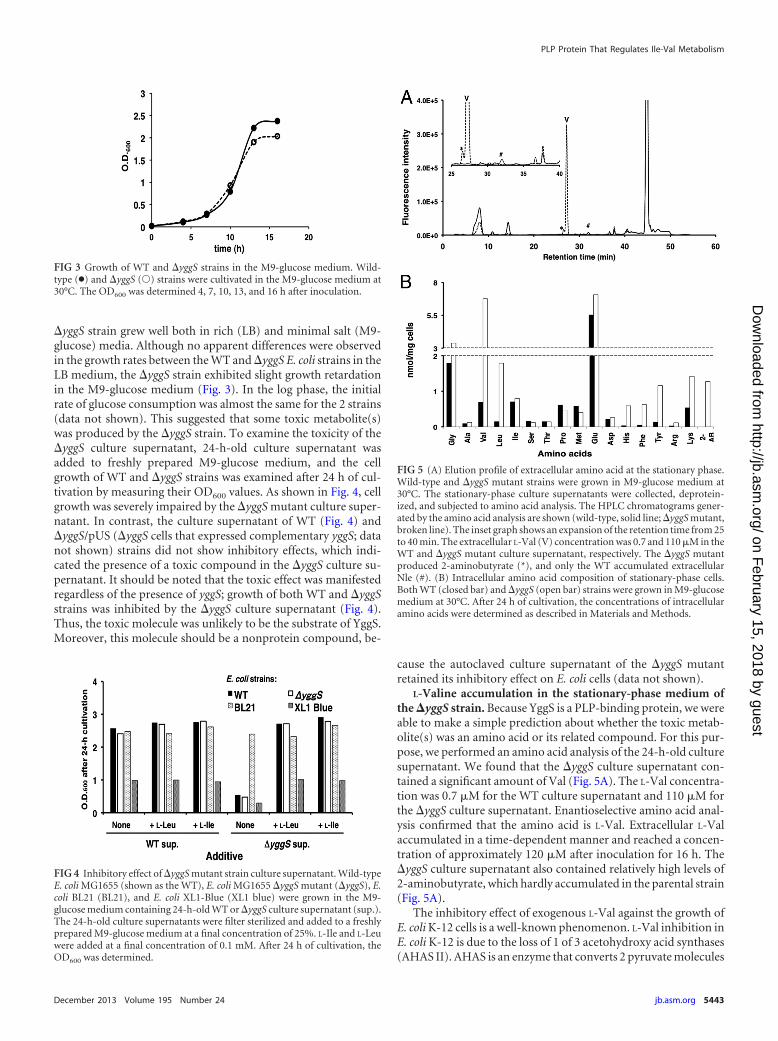
L-Valine accumulation in the stationary-phase medium ofthe �yggS strain. Because YggS is a PLP-binding protein, we wereable to make a simple prediction about whether the toxic metab-olite(s) was an amino acid or its related compound. For this pur-pose, we performed an amino acid analysis of the 24-h-old culturesupernatant. We found that the yggS culture supernatant con-tained a significant amount of Val (Fig. 5A). The L-Val concentra-tion was 0.7 �M for the WT culture supernatant and 110 �M forthe yggS culture supernatant. Enantioselective amino acid anal-ysis confirmed that the amino acid is L-Val. Extracellular L-Valaccumulated in a time-dependent manner and reached a concen-tration of approximately 120 �M after inoculation for 16 h. TheyggS culture supernatant also contained relatively high levels of2-aminobutyrate, which hardly accumulated in the parental strain(Fig. 5A).
The inhibitory effect of exogenous L-Val against the growth ofE. coli K-12 cells is a well-known phenomenon. L-Val inhibition inE. coli K-12 is due to the loss of 1 of 3 acetohydroxy acid synthases(AHAS II). AHAS is an enzyme that converts 2 pyruvate molecules
FIG 3 Growth of WT and yggS strains in the M9-glucose medium. Wild-type (●) and yggS (Œ) strains were cultivated in the M9-glucose medium at30°C. The OD600 was determined 4, 7, 10, 13, and 16 h after inoculation.
FIG 4 Inhibitory effect of yggS mutant strain culture supernatant. Wild-typeE. coli MG1655 (shown as the WT), E. coli MG1655 yggS mutant (yggS), E.coli BL21 (BL21), and E. coli XL1-Blue (XL1 blue) were grown in the M9-glucose medium containing 24-h-old WT or yggS culture supernatant (sup.).The 24-h-old culture supernatants were filter sterilized and added to a freshlyprepared M9-glucose medium at a final concentration of 25%. L-Ile and L-Leuwere added at a final concentration of 0.1 mM. After 24 h of cultivation, theOD600 was determined.
FIG 5 (A) Elution profile of extracellular amino acid at the stationary phase.Wild-type and yggS mutant strains were grown in M9-glucose medium at30°C. The stationary-phase culture supernatants were collected, deprotein-ized, and subjected to amino acid analysis. The HPLC chromatograms gener-ated by the amino acid analysis are shown (wild-type, solid line; yggS mutant,broken line). The inset graph shows an expansion of the retention time from 25to 40 min. The extracellular L-Val (V) concentration was 0.7 and 110 �M in theWT and yggS mutant culture supernatant, respectively. The yggS mutantproduced 2-aminobutyrate (*), and only the WT accumulated extracellularNle (#). (B) Intracellular amino acid composition of stationary-phase cells.Both WT (closed bar) and yggS (open bar) strains were grown in M9-glucosemedium at 30°C. After 24 h of cultivation, the concentrations of intracellularamino acids were determined as described in Materials and Methods.
PLP Protein That Regulates Ile-Val Metabolism
December 2013 Volume 195 Number 24 jb.asm.org 5443
on February 15, 2018 by guest
http://jb.asm.org/
Dow
nloaded from
into an �-acetolactate during L-Val biosynthesis and condensesone pyruvate plus one 2-ketobutyrate to yield an �-aceto-hy-droxybutyrate during L-Ile biosynthesis. The genome of E. coliK-12 contains 3 AHAS genes (AHAS I, II, and III), but the L-Val-insensitive AHAS (AHAS II) gene is inactivated. It is reported thatthe in vitro AHAS activity of E. coli K-12 is inhibited up to 85 to90% by treatment with 1.5 mM L-Val, and that growth of E. coliK-12 is completely blocked with 0.1 mM L-Val (16–18).
To determine whether growth inhibition due to the yggS cul-ture supernatant reflected the accumulation of L-Val, the growthof 2 classes of E. coli strains, (i) E. coli MG1655 and E. coli XL1-Bluecarrying a nonfunctional AHAS II and (ii) E. coli BL21 possessingthe functional enzyme, were compared to the 24-h-old culturesupernatant. As shown in Fig. 4, the growth of both E. coliMG1655 and E. coli XL1-Blue were severely inhibited by the yggSculture supernatant. In contrast, the growth of E. coli BL21 wasunchanged with the yggS culture supernatant. Inclusion of L-Ileand L-Leu, which alleviate L-Val toxicity, reduced the inhibitoryeffect of the yggS supernatant (Fig. 4). These results validated ourhypothesis that the inhibitory effect stemmed, in part, from L-Val.
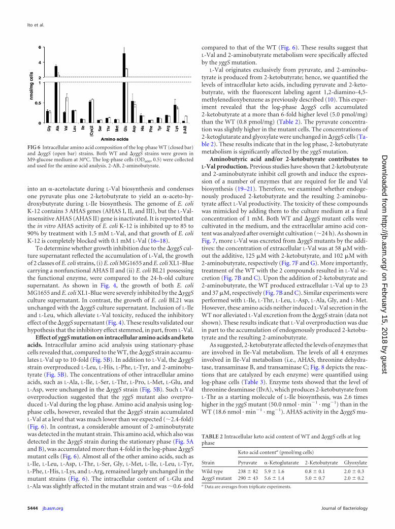
Effect of yggS mutation on intracellular amino acids and ketoacids. Intracellular amino acid analysis using stationary-phasecells revealed that, compared to the WT, the yggS strain accumu-lates L-Val up to 10-fold (Fig. 5B). In addition to L-Val, the yggSstrain overproduced L-Leu, L-His, L-Phe, L-Tyr, and 2-aminobu-tyrate (Fig. 5B). The concentrations of other intracellular aminoacids, such as L-Ala, L-Ile, L-Ser, L-Thr, L-Pro, L-Met, L-Glu, andL-Asp, were unchanged in the yggS strain (Fig. 5B). Such L-Valoverproduction suggested that the yggS mutant also overpro-duced L-Val during the log phase. Amino acid analysis using log-phase cells, however, revealed that the yggS strain accumulatedL-Val at a level that was much lower than we expected (2.4-fold)(Fig. 6). In contrast, a considerable amount of 2-aminobutyratewas detected in the mutant strain. This amino acid, which also wasdetected in the yggS strain during the stationary phase (Fig. 5Aand B), was accumulated more than 4-fold in the log-phase yggSmutant cells (Fig. 6). Almost all of the other amino acids, such asL-Ile, L-Leu, L-Asp, L-Thr, L-Ser, Gly, L-Met, L-Ile, L-Leu, L-Tyr,L-Phe, L-His, L-Lys, and L-Arg, remained largely unchanged in themutant strains (Fig. 6). The intracellular content of L-Glu andL-Ala was slightly affected in the mutant strain and was 0.6-fold
compared to that of the WT (Fig. 6). These results suggest thatL-Val and 2-aminobutyrate metabolism were specifically affectedby the yggS mutation.
L-Val originates exclusively from pyruvate, and 2-aminobu-tyrate is produced from 2-ketobutyrate; hence, we quantified thelevels of intracellular keto acids, including pyruvate and 2-keto-butyrate, with the fluorescent labeling agent 1,2-diamino-4,5-methylenedioxybenzene as previously described (10). This exper-iment revealed that the log-phase yggS cells accumulated2-ketobutyrate at a more than 6-fold higher level (5.0 pmol/mg)than the WT (0.8 pmol/mg) (Table 2). The pyruvate concentra-tion was slightly higher in the mutant cells. The concentrations of2-ketoglutarate and glyoxylate were unchanged in yggS cells (Ta-ble 2). These results indicate that in the log phase, 2-ketobutyratemetabolism is significantly affected by the yggS mutation.
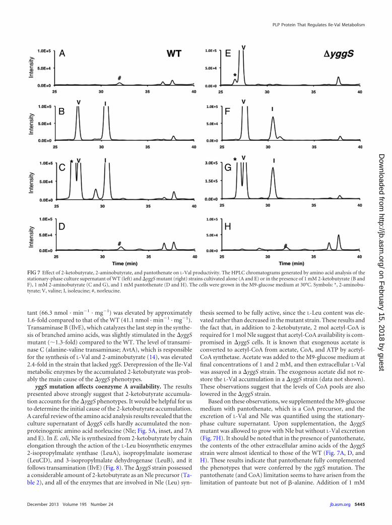
Aminobutyric acid and/or 2-ketobutyrate contributes toL-Val production. Previous studies have shown that 2-ketobutyrateand 2-aminobutyrate inhibit cell growth and induce the expres-sion of a number of enzymes that are required for Ile and Valbiosynthesis (19–21). Therefore, we examined whether endoge-nously produced 2-ketobutyrate and the resulting 2-aminobu-tyrate affect L-Val productivity. The toxicity of these compoundswas mimicked by adding them to the culture medium at a finalconcentration of 1 mM. Both WT and yggS mutant cells werecultivated in the medium, and the extracellular amino acid con-tent was analyzed after overnight cultivation (24 h). As shown inFig. 7, more L-Val was excreted from yggS mutants by the addi-tives: the concentration of extracellular L-Val was at 58 �M with-out the additive, 125 �M with 2-ketobutyrate, and 102 �M with2-aminobutyrate, respectively (Fig. 7F and G). More importantly,treatment of the WT with the 2 compounds resulted in L-Val se-cretion (Fig. 7B and C). Upon the addition of 2-ketobutyrate and2-aminobutyrate, the WT produced extracellular L-Val up to 23and 37 �M, respectively (Fig. 7B and C). Similar experiments wereperformed with L-Ile, L-Thr, L-Leu, L-Asp, L-Ala, Gly, and L-Met.However, these amino acids neither induced L-Val secretion in theWT nor alleviated L-Val excretion from the yggS strain (data notshown). These results indicate that L-Val overproduction was duein part to the accumulation of endogenously produced 2-ketobu-tyrate and the resulting 2-aminobutyrate.
As suggested, 2-ketobutyrate affected the levels of enzymes thatare involved in Ile-Val metabolism. The levels of all 4 enzymesinvolved in Ile-Val metabolism (i.e., AHAS, threonine dehydra-tase, transaminase B, and transaminase C; Fig. 8 depicts the reac-tions that are catalyzed by each enzyme) were quantified usinglog-phase cells (Table 3). Enzyme tests showed that the level ofthreonine deaminase (IlvA), which produces 2-ketobutyrate fromL-Thr as a starting molecule of L-Ile biosynthesis, was 2.6 timeshigher in the yggS mutant (50.0 nmol · min�1 · mg�1) than in theWT (18.6 nmol · min�1 · mg�1). AHAS activity in the yggS mu-
FIG 6 Intracellular amino acid composition of the log-phase WT (closed bar)and yggS (open bar) strains. Both WT and yggS strains were grown inM9-glucose medium at 30°C. The log-phase cells (OD600, 0.5) were collectedand used for the amino acid analysis. 2-AB, 2-aminobutyrate.
TABLE 2 Intracellular keto acid content of WT and yggS cells at logphase
Strain
Keto acid contenta (pmol/mg cells)
Pyruvate �-Ketoglutarate 2-Ketobutyrate Glyoxylate
Wild type 238 � 82 5.9 � 1.6 0.8 � 0.1 2.0 � 0.3yggS mutant 290 � 43 5.6 � 1.4 5.0 � 0.7 2.0 � 0.2a Data are averages from triplicate experiments.
Ito et al.
5444 jb.asm.org Journal of Bacteriology
on February 15, 2018 by guest
http://jb.asm.org/
Dow
nloaded from
tant (66.3 nmol · min�1 · mg�1) was elevated by approximately1.6-fold compared to that of the WT (41.1 nmol · min�1 · mg�1).Transaminase B (IlvE), which catalyzes the last step in the synthe-sis of branched amino acids, was slightly stimulated in the yggSmutant (1.3-fold) compared to the WT. The level of transami-nase C (alanine-valine transaminase; AvtA), which is responsiblefor the synthesis of L-Val and 2-aminobutyrate (14), was elevated2.4-fold in the strain that lacked yggS. Derepression of the Ile-Valmetabolic enzymes by the accumulated 2-ketobutyrate was prob-ably the main cause of the yggS phenotypes.
yggS mutation affects coenzyme A availability. The resultspresented above strongly suggest that 2-ketobutyrate accumula-tion accounts for the yggS phenotypes. It would be helpful for usto determine the initial cause of the 2-ketobutyrate accumulation.A careful review of the amino acid analysis results revealed that theculture supernatant of yggS cells hardly accumulated the non-proteinogenic amino acid norleucine (Nle; Fig. 5A, inset, and 7Aand E). In E. coli, Nle is synthesized from 2-ketobutyrate by chainelongation through the action of the L-Leu biosynthetic enzymes2-isopropylmalate synthase (LeuA), isopropylmalate isomerase(LeuCD), and 3-isopropylmalate dehydrogenase (LeuB), and itfollows transamination (IlvE) (Fig. 8). The yggS strain possesseda considerable amount of 2-ketobutyrate as an Nle precursor (Ta-ble 2), and all of the enzymes that are involved in Nle (Leu) syn-
thesis seemed to be fully active, since the L-Leu content was ele-vated rather than decreased in the mutant strain. These results andthe fact that, in addition to 2-ketobutyrate, 2 mol acetyl-CoA isrequired for 1 mol Nle suggest that acetyl-CoA availability is com-promised in yggS cells. It is known that exogenous acetate isconverted to acetyl-CoA from acetate, CoA, and ATP by acetyl-CoA synthetase. Acetate was added to the M9-glucose medium atfinal concentrations of 1 and 2 mM, and then extracellular L-Valwas assayed in a yggS strain. The exogenous acetate did not re-store the L-Val accumulation in a yggS strain (data not shown).These observations suggest that the levels of CoA pools are alsolowered in the yggS strain.
Based on these observations, we supplemented the M9-glucosemedium with pantothenate, which is a CoA precursor, and theexcretion of L-Val and Nle was quantified using the stationary-phase culture supernatant. Upon supplementation, the yggSmutant was allowed to grow with Nle but without L-Val excretion(Fig. 7H). It should be noted that in the presence of pantothenate,the contents of the other extracellular amino acids of the yggSstrain were almost identical to those of the WT (Fig. 7A, D, andH). These results indicate that pantothenate fully complementedthe phenotypes that were conferred by the yggS mutation. Thepantothenate (and CoA) limitation seems to have arisen from thelimitation of pantoate but not of �-alanine. Addition of 1 mM
FIG 7 Effect of 2-ketobutyrate, 2-aminobutyrate, and pantothenate on L-Val productivity. The HPLC chromatograms generated by amino acid analysis of thestationary-phase culture supernatant of WT (left) and yggS mutant (right) strains cultivated alone (A and E) or in the presence of 1 mM 2-ketobutyrate (B andF), 1 mM 2-aminobutyrate (C and G), and 1 mM pantothenate (D and H). The cells were grown in the M9-glucose medium at 30°C. Symbols: *, 2-aminobu-tyrate; V, valine; I, isoleucine; #, norleucine.
PLP Protein That Regulates Ile-Val Metabolism
December 2013 Volume 195 Number 24 jb.asm.org 5445
on February 15, 2018 by guest
http://jb.asm.org/
Dow
nloaded from
pantoate was effective to decrease the L-Val excretion. �-Alaninesupplementation did not affect L-Val productivity in yggS cells(data not shown).
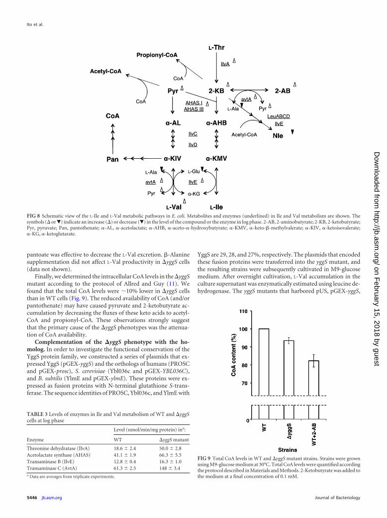
Finally, we determined the intracellular CoA levels in the yggSmutant according to the protocol of Allred and Guy (11). Wefound that the total CoA levels were 10% lower in yggS cellsthan in WT cells (Fig. 9). The reduced availability of CoA (and/orpantothenate) may have caused pyruvate and 2-ketobutyrate ac-cumulation by decreasing the fluxes of these keto acids to acetyl-CoA and propionyl-CoA. These observations strongly suggestthat the primary cause of the yggS phenotypes was the attenua-tion of CoA availability.
Complementation of the �yggS phenotype with the ho-molog. In order to investigate the functional conservation of theYggS protein family, we constructed a series of plasmids that ex-pressed YggS (pGEX-yggS) and the orthologs of humans (PROSCand pGEX-prosc), S. cerevisiae (Ybl036c and pGEX-YBL036C),and B. subtilis (YlmE and pGEX-ylmE). These proteins were ex-pressed as fusion proteins with N-terminal glutathione S-trans-ferase. The sequence identities of PROSC, Ybl036c, and YlmE with
YggS are 29, 28, and 27%, respectively. The plasmids that encodedthese fusion proteins were transferred into the yggS mutant, andthe resulting strains were subsequently cultivated in M9-glucosemedium. After overnight cultivation, L-Val accumulation in theculture supernatant was enzymatically estimated using leucine de-hydrogenase. The yggS mutants that harbored pUS, pGEX-yggS,
FIG 8 Schematic view of the L-Ile and L-Val metabolic pathways in E. coli. Metabolites and enzymes (underlined) in Ile and Val metabolism are shown. Thesymbols ( or�) indicate an increase () or decrease (�) in the level of the compound or the enzyme in log phase. 2-AB, 2-aminobutyrate; 2-KB, 2-ketobutyrate;Pyr, pyruvate; Pan, pantothenate; �-AL, �-acetolactate; �-AHB, �-aceto-�-hydroxybutyrate; �-KMV, �-keto-�-methylvalerate; �-KIV, �-ketoisovalerate;�-KG, �-ketoglutarate.
TABLE 3 Levels of enzymes in Ile and Val metabolism of WT and yggScells at log phase
Enzyme
Level (nmol/min/mg protein) ina:
WT yggS mutant
Threonine dehydratase (IlvA) 18.6 � 2.4 50.0 � 2.8Acetolactate synthase (AHAS) 41.1 � 1.9 66.3 � 5.5Transaminase B (IlvE) 12.8 � 0.4 16.3 � 1.0Transaminase C (AvtA) 61.3 � 2.5 148 � 3.4a Data are averages from triplicate experiments.
FIG 9 Total CoA levels in WT and yggS mutant strains. Strains were grownusing M9-glucose medium at 30°C. Total CoA levels were quantified accordingthe protocol described in Materials and Methods. 2-Ketobutyrate was added tothe medium at a final concentration of 0.1 mM.
Ito et al.
5446 jb.asm.org Journal of Bacteriology
on February 15, 2018 by guest
http://jb.asm.org/
Dow
nloaded from
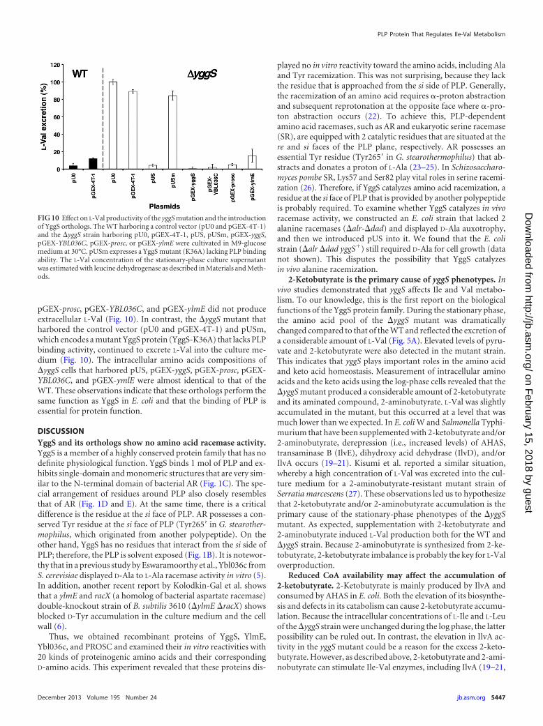
pGEX-prosc, pGEX-YBL036C, and pGEX-ylmE did not produceextracellular L-Val (Fig. 10). In contrast, the yggS mutant thatharbored the control vector (pU0 and pGEX-4T-1) and pUSm,which encodes a mutant YggS protein (YggS-K36A) that lacks PLPbinding activity, continued to excrete L-Val into the culture me-dium (Fig. 10). The intracellular amino acids compositions ofyggS cells that harbored pUS, pGEX-yggS, pGEX-prosc, pGEX-YBL036C, and pGEX-ymlE were almost identical to that of theWT. These observations indicate that these orthologs perform thesame function as YggS in E. coli and that the binding of PLP isessential for protein function.
DISCUSSIONYggS and its orthologs show no amino acid racemase activity.YggS is a member of a highly conserved protein family that has nodefinite physiological function. YggS binds 1 mol of PLP and ex-hibits single-domain and monomeric structures that are very sim-ilar to the N-terminal domain of bacterial AR (Fig. 1C). The spe-cial arrangement of residues around PLP also closely resemblesthat of AR (Fig. 1D and E). At the same time, there is a criticaldifference is the residue at the si face of PLP. AR possesses a con-served Tyr residue at the si face of PLP (Tyr265= in G. stearother-mophilus, which originated from another polypeptide). On theother hand, YggS has no residues that interact from the si side ofPLP; therefore, the PLP is solvent exposed (Fig. 1B). It is notewor-thy that in a previous study by Eswaramoorthy et al., Ybl036c fromS. cerevisiae displayed D-Ala to L-Ala racemase activity in vitro (5).In addition, another recent report by Kolodkin-Gal et al. showsthat a ylmE and racX (a homolog of bacterial aspartate racemase)double-knockout strain of B. subtilis 3610 (ylmE racX) showsblocked D-Tyr accumulation in the culture medium and the cellwall (6).
Thus, we obtained recombinant proteins of YggS, YlmE,Ybl036c, and PROSC and examined their in vitro reactivities with20 kinds of proteinogenic amino acids and their correspondingD-amino acids. This experiment revealed that these proteins dis-
played no in vitro reactivity toward the amino acids, including Alaand Tyr racemization. This was not surprising, because they lackthe residue that is approached from the si side of PLP. Generally,the racemization of an amino acid requires �-proton abstractionand subsequent reprotonation at the opposite face where �-pro-ton abstraction occurs (22). To achieve this, PLP-dependentamino acid racemases, such as AR and eukaryotic serine racemase(SR), are equipped with 2 catalytic residues that are situated at there and si faces of the PLP plane, respectively. AR possesses anessential Tyr residue (Tyr265= in G. stearothermophilus) that ab-stracts and donates a proton of L-Ala (23–25). In Schizosaccharo-myces pombe SR, Lys57 and Ser82 play vital roles in serine racemi-zation (26). Therefore, if YggS catalyzes amino acid racemization, aresidue at the si face of PLP that is provided by another polypeptideis probably required. To examine whether YggS catalyzes in vivoracemase activity, we constructed an E. coli strain that lacked 2alanine racemases (alr-dad) and displayed D-Ala auxotrophy,and then we introduced pUS into it. We found that the E. colistrain (alr dad yggS�) still required D-Ala for cell growth (datanot shown). This disputes the possibility that YggS catalyzesin vivo alanine racemization.
2-Ketobutyrate is the primary cause of yggS phenotypes. Invivo studies demonstrated that yggS affects Ile and Val metabo-lism. To our knowledge, this is the first report on the biologicalfunctions of the YggS protein family. During the stationary phase,the amino acid pool of the yggS mutant was dramaticallychanged compared to that of the WT and reflected the excretion ofa considerable amount of L-Val (Fig. 5A). Elevated levels of pyru-vate and 2-ketobutyrate were also detected in the mutant strain.This indicates that yggS plays important roles in the amino acidand keto acid homeostasis. Measurement of intracellular aminoacids and the keto acids using the log-phase cells revealed that theyggS mutant produced a considerable amount of 2-ketobutyrateand its aminated compound, 2-aminobutyrate. L-Val was slightlyaccumulated in the mutant, but this occurred at a level that wasmuch lower than we expected. In E. coli W and Salmonella Typhi-murium that have been supplemented with 2-ketobutyrate and/or2-aminobutyrate, derepression (i.e., increased levels) of AHAS,transaminase B (IlvE), dihydroxy acid dehydrase (IlvD), and/orIlvA occurs (19–21). Kisumi et al. reported a similar situation,whereby a high concentration of L-Val was excreted into the cul-ture medium for a 2-aminobutyrate-resistant mutant strain ofSerratia marcescens (27). These observations led us to hypothesizethat 2-ketobutyrate and/or 2-aminobutyrate accumulation is theprimary cause of the stationary-phase phenotypes of the yggSmutant. As expected, supplementation with 2-ketobutyrate and2-aminobutyrate induced L-Val production both for the WT andyggS strain. Because 2-aminobutyrate is synthesized from 2-ke-tobutyrate, 2-ketobutyrate imbalance is probably the key for L-Valoverproduction.
Reduced CoA availability may affect the accumulation of2-ketobutyrate. 2-Ketobutyrate is mainly produced by IlvA andconsumed by AHAS in E. coli. Both the elevation of its biosynthe-sis and defects in its catabolism can cause 2-ketobutyrate accumu-lation. Because the intracellular concentrations of L-Ile and L-Leuof the yggS strain were unchanged during the log phase, the latterpossibility can be ruled out. In contrast, the elevation in IlvA ac-tivity in the yggS mutant could be a reason for the excess 2-keto-butyrate. However, as described above, 2-ketobutyrate and 2-ami-nobutyrate can stimulate Ile-Val enzymes, including IlvA (19–21,
FIG 10 Effect on L-Val productivity of the yggS mutation and the introductionof YggS orthologs. The WT harboring a control vector (pU0 and pGEX-4T-1)and the yggS strain harboring pU0, pGEX-4T-1, pUS, pUSm, pGEX-yggS,pGEX-YBL036C, pGEX-prosc, or pGEX-ylmE were cultivated in M9-glucosemedium at 30°C. pUSm expresses a YggS mutant (K36A) lacking PLP bindingability. The L-Val concentration of the stationary-phase culture supernatantwas estimated with leucine dehydrogenase as described in Materials and Meth-ods.
PLP Protein That Regulates Ile-Val Metabolism
December 2013 Volume 195 Number 24 jb.asm.org 5447
on February 15, 2018 by guest
http://jb.asm.org/
Dow
nloaded from
27). In addition, we found that supplementation with L-Ile andL-Leu, which reduce IlvA activity through allosteric inhibition,could not reverse L-Val secretion in the yggS strain. These obser-vations suggest that the derepression of IlvA is not the primarycause but the result of excess 2-ketobutyrate.
In contrast, we provide a plausible explanation for the keto acidaccumulation, which is the attenuation of CoA availability. A firsthint about the cause of 2-ketobutyrate accumulation evolvedfrom the observation that the yggS strain hardly accumulatedNle. We showed that total CoA levels were 10% less in a yggSstrain than those in the WT, and the supplementation with pan-tothenate, which is a CoA precursor, was very effective in revers-ing the stationary-phase phenotypes of the yggS strain (Fig. 7 and9). A decrease in CoA availability is assumed to prevent the effi-cient conversion of pyruvate and 2-ketobutyrate to acetyl-CoAand propionyl-CoA, respectively, thereby leading to pyruvate and2-ketobutyrate accumulation. A similar situation was observed byBlombach et al. and Valle et al. (28, 29). They recently reportedthat the inactivation of pyruvate dehydrogenase (aceE), whichcatalyzes the transformation of pyruvate into acetyl-CoA, led topyruvate accumulation and L-Val excretion in Corynebacteriumglutamicum and E. coli (28, 29). Radmacher et al. showed that a C.glutamicum ilvA panBC strain with plasmid-borne ilvBNCDcould overproduce L-Val under pantothenate-limiting conditions(30).
On the other hand, we cannot rule out the possibility that thepantothenate and/or CoA limitation stems from the excess 2-ke-tobutyrate. Powers et al. and Primerano et al. previously suggestedthat CoA synthesis was compromised by 2-ketobutyrate accumu-lation by demonstrating that 2-ketobutyrate is a competitive sub-strate for ketopantoate hydroxymethyltransferase, which is thefirst enzyme in pantothenate biosynthesis, in E. coli K-12 and S. Ty-phimurium (31, 32). Accordingly, we found that 2-ketobutyrate sig-nificantly lowers the intracellular CoA levels in E. coli (Fig. 9). There-fore, it is necessary to determine the molecular basis of YggS-mediated 2-ketobutyrate accumulation and CoA limitation.
YggS function depends on PLP and is highly conserved invarious species. In an attempt to investigate the functional con-servation of the YggS protein family, we found that all of the yggSstrains that expressed yggS or its orthologs (of B. subtilis, S. cerevi-siae, and humans) grew without L-Val overproduction (Fig. 10).In contrast, YggS-K36A expression did not affect the L-Val pro-ductivity of yggS cells (Fig. 10). This observation led us realizethat the in vivo function of YggS is highly conserved in a widerange of species, from bacteria to humans, and it depends on PLP.As described above, YggS and its orthologs (PROSC, Ybl036c, andYlmE) share 30% sequence identity. However, their highly con-served residues are located near the PLP (data not shown); there-fore, they lack conservation of the protein surface. This findingsuggests that YggS function is not mediated by protein-proteininteractions.
Valle et al. showed that biofilms of E. coli and other Gram-negative bacteria accumulate high levels of Val under stationary-phase-like conditions (29). 2-Ketobutyrate is known to regulatethe phosphoenolpyruvate-sugar phosphotransferase system (PTS) inamino acid starvation and to be served as an alarmone to governthe shift from anaerobic to aerobic growth (33). ylmE of B. subtilisis reported to be involved in biofilm disassembly (6). These piecesof evidence suggest to us that yggS and the orthologs play a regu-
latory role under specific conditions, such as low nutrients, andwithin biofilm by valine or 2-ketobutyrate signaling.
Further studies may shed light on the conserved processes thatare mediated by this unique PLP-dependent protein.
ACKNOWLEDGMENTS
This work was supported in part by Grant-in-Aid for Young Scientists (B)24780098 (to T.I.) and by funding from the Takano Life Science ResearchFoundation and the Towa Foundation for Food Research (to T.I.).
REFERENCES1. Ito T, Uozumi N, Nakamura T, Takayama S, Matsuda N, Aiba H,
Hemmi H, Yoshimura T. 2009. The implication of YggT of Escherichiacoli in osmotic regulation. Biosci. Biotechnol. Biochem. 73:2698 –2704.
2. Kuras R, Saint-Marcoux D, Wollman FA, de Vitry C. 2007. A specificc-type cytochrome maturation system is required for oxygenic photosyn-thesis. Proc. Natl. Acad. Sci. U. S. A. 104:9906 –9910.
3. Kabeya Y, Nakanishi H, Suzuki K, Ichikawa T, Kondou Y, Matsui M,Miyagishima SY. 2010. The YlmG protein has a conserved function re-lated to the distribution of nucleoids in chloroplasts and cyanobacteria.BMC Plant Biol. 10:57. doi:10.1186/1471-2229-10-57.
4. Bradshaw JS, Kuzminov A. 2003. RdgB acts to avoid chromosome frag-mentation in Escherichia coli. Mol. Microbiol. 48:1711–1725.
5. Eswaramoorthy S, Gerchman S, Graziano V, Kycia H, Studier FW,Swaminathan S. 2003. Structure of a yeast hypothetical protein selectedby a structural genomics approach. Acta Crystallogr. D Biol. Crystallogr.59:127–135.
6. Kolodkin-Gal I, Romero D, Cao S, Clardy J, Kolter R, Losick R. 2010.D-amino acids trigger biofilm disassembly. Science 328:627– 629.
7. Datsenko KA, Wanner BL. 2000. One-step inactivation of chromosomalgenes in Escherichia coli K-12 using PCR products. Proc. Natl. Acad. Sci.U. S. A. 97:6640 – 6645.
8. Ito T, Hemmi H, Kataoka K, Mukai Y, Yoshimura T. 2008. A novelzinc-dependent D-serine dehydratase from Saccharomyces cerevisiae.Biochem. J. 409:399 – 406.
9. Ito T, Murase H, Maekawa M, Goto M, Hayashi S, Saito H, Maki M,Hemmi H, Yoshimura T. 2012. Metal ion dependency of serine racemasefrom Dictyostelium discoideum. Amino Acids 43:1567–1576.
10. Kato S, Kito Y, Hemmi H, Yoshimura T. 2011. Simultaneous determi-nation of D-amino acids by the coupling method of D-amino acid oxidasewith high-performance liquid chromatography. J. Chromatogr. B Analyt.Technol. Biomed. Life Sci. 879:3190 –3195.
11. Allred JB, Guy DG. 1969. Determination of coenzyme A and acetyl CoAin tissue extracts. Anal. Biochem. 29:293–299.
12. Hasegawa S, Uematsu K, Natsuma Y, Suda M, Hiraga K, Jojima T, InuiM, Yukawa H. 2012. Improvement of the redox balance increases L-valineproduction by Corynebacterium glutamicum under oxygen deprivationconditions. Appl. Environ. Microbiol. 78:865– 875.
13. Duggan DE, Wechsler JA. 1973. An assay for transaminase B enzymeactivity in Escherichia coli K-12. Anal. Biochem. 51:67–79.
14. McGilvray D, Umbarger HE. 1974. Regulation of transaminase C syn-thesis in Escherichia coli: conditional leucine auxotrophy. J. Bacteriol. 120:715–723.
15. Cook SP, Galve-Roperh I, Martínez del Pozo A, Rodríguez-Crespo I.2002. Direct calcium binding results in activation of brain serine racemase.J. Biol. Chem. 277:27782–27792.
16. De Felice M, Squires C, Levinthal M, Guardiola J, Lamberti A, Iacca-rino M. 1977. Growth inhibition of Escherichia coli K-12 by L-valine: aconsequence of a regulatory pattern. Mol. Gen. Genet. 156:1–7.
17. De Felice M, Squires C, Levinthal M. 1978. A comparative study of theacetohydroxy acid synthase isoenzymes of Escherichia coli K-12. Biochim.Biophys. Acta 541:9 –17.
18. Jackson JH, Herring PA, Patterson EB, Blatt JM. 1993. A mechanism forvaline-resistant growth of Escherichia coli K-12 supported by the valine-sensitive acetohydroxy acid synthase IV activity from ilvJ662. Biochimie75:759 –765.
19. Freundlich M, Clarke LP. 1968. Control of isoleucine, valine and leucinebiosynthesis. V. Dual effect of alpha-aminobutyric acid on repression andend product inhibition in Escherichia coli. Biochim. Biophys. Acta 170:271–281.
20. LaRossa RA, Van Dyk TK, Smulski DR. 1987. Toxic accumulation of
Ito et al.
5448 jb.asm.org Journal of Bacteriology
on February 15, 2018 by guest
http://jb.asm.org/
Dow
nloaded from
alpha-ketobutyrate caused by inhibition of the branched-chain aminoacid biosynthetic enzyme acetolactate synthase in Salmonella typhimu-rium. J. Bacteriol. 169:1372–1378.
21. Shaw KJ, Berg CM. 1980. Substrate channeling: alpha-ketobutyrate in-hibition of acetohydroxy acid synthase in Salmonella typhimurium. J. Bac-teriol. 143:1509 –1512.
22. Soda K, Yoshimura T, Esaki N. 2001. Stereospecificity for the hydrogentransfer of pyridoxal enzyme reactions. Chem. Rec. 1:373–384.
23. Shaw JP, Petsko GA, Ringe D. 1997. Determination of the structure ofalanine racemase from Bacillus stearothermophilus at 1.9-A resolution.Biochemistry 36:1329 –1342.
24. Watanabe A, Yoshimura T, Mikami B, Esaki N. 1999. Tyrosine 265 ofalanine racemase serves as a base abstracting alpha-hydrogen from l-ala-nine: the counterpart residue to lysine 39 specific to D-alanine. J. Biochem.126:781–786.
25. Watanabe A, Yoshimura T, Mikami B, Hayashi H, Kagamiyama H,Esaki N. 2002. Reaction mechanism of alanine racemase from Bacillusstearothermophilus: x-ray crystallographic studies of the enzyme boundwith N-(5=-phosphopyridoxyl)alanine. J. Biol. Chem. 277:19166 –19172.
26. Goto M, Yamauchi T, Kamiya N, Miyahara I, Yoshimura T, Mihara H,Kurihara T, Hirotsu K, Esaki N. 2009. Crystal structure of a homolog ofmammalian serine racemase from Schizosaccharomyces pombe. J. Biol.Chem. 284:25944 –25952.
27. Kisumi M, Komatsubara S, Chibata I. 1971. Valine accumulation byalpha-aminobutyric acid-resistant mutants of Serratia marcescens. J. Bac-teriol. 106:493– 499.
28. Blombach B, Schreiner ME, Holátko J, Bartek T, Oldiges M, EikmannsBJ. 2007. L-valine production with pyruvate dehydrogenase complex-deficient Corynebacterium glutamicum. Appl. Environ. Microbiol. 73:2079 –2084.
29. Valle J, Da Re S, Schmid S, Skurnik D, D’Ari R, Ghigo JM. 2008. Theamino acid valine is secreted in continuous-flow bacterial biofilms. J. Bac-teriol. 190:264 –274.
30. Radmacher E, Vaitsikova A, Burger U, Krumbach K, Sahm H, EggelingL. 2002. Linking central metabolism with increased pathway flux: L-valineaccumulation by Corynebacterium glutamicum. Appl. Environ. Microbiol.68:2246 –2250.
31. Powers SG, Snell EE. 1976. Ketopantoate hydroxymethyltransferase. II.Physical, catalytic, and regulatory properties. J. Biol. Chem. 251:3786 –3793.
32. Primerano DA, Burns RO. 1982. Metabolic basis for the isoleucine,pantothenate or methionine requirement of ilvG strains of Salmonellatyphimurium. J. Bacteriol. 150:1202–1211.
33. Daniel J, Dondon L, Danchin A. 1983. 2-Ketobutyrate: a putative alar-mone of Escherichia coli. Mol. Gen. Genet. 190:452– 458.
PLP Protein That Regulates Ile-Val Metabolism
December 2013 Volume 195 Number 24 jb.asm.org 5449
on February 15, 2018 by guest
http://jb.asm.org/
Dow
nloaded from


![BMC Bioinformatics BioMed Central · – conserved, essential ones [26], whereas Borrelia has numerous plasmids that mostly encode poorly conserved genes [27]. Probably more telling](https://img.pdfslide.tips/doc/110x75/5f6c9bf3f6891336585163a7/bmc-bioinformatics-biomed-central-a-conserved-essential-ones-26-whereas-borrelia.jpg)