Embed Size (px)
Citation preview
of January 10, 2019.This information is current as
βRegulatory T Cells, and TGF- by IL-10,Trichinella spiralisStage
Coordinated Control of Immunity to Muscle
Susan K. Bliss, Diana Meskill and Judith A. AppletonDaniel P. Beiting, Lucille F. Gagliardo, Matthias Hesse,
http://www.jimmunol.org/content/178/2/1039doi: 10.4049/jimmunol.178.2.1039
2007; 178:1039-1047; ;J Immunol
Referenceshttp://www.jimmunol.org/content/178/2/1039.full#ref-list-1
, 37 of which you can access for free at: cites 64 articlesThis article
average*
4 weeks from acceptance to publicationFast Publication! •
Every submission reviewed by practicing scientistsNo Triage! •
from submission to initial decisionRapid Reviews! 30 days* •
Submit online. ?The JIWhy
Subscriptionhttp://jimmunol.org/subscription
is online at: The Journal of ImmunologyInformation about subscribing to
Permissionshttp://www.aai.org/About/Publications/JI/copyright.htmlSubmit copyright permission requests at:
Email Alertshttp://jimmunol.org/alertsReceive free email-alerts when new articles cite this article. Sign up at:
Print ISSN: 0022-1767 Online ISSN: 1550-6606. Immunologists All rights reserved.Copyright © 2007 by The American Association of1451 Rockville Pike, Suite 650, Rockville, MD 20852The American Association of Immunologists, Inc.,
is published twice each month byThe Journal of Immunology
by guest on January 10, 2019http://w
ww
.jimm
unol.org/D
ownloaded from
by guest on January 10, 2019
http://ww
w.jim
munol.org/
Dow
nloaded from
Coordinated Control of Immunity to Muscle StageTrichinella spiralis by IL-10, Regulatory T Cells, and TGF-�1
Daniel P. Beiting,* Lucille F. Gagliardo,* Matthias Hesse,† Susan K. Bliss,*Diana Meskill,* and Judith A. Appleton2†
We previously demonstrated that IL-10 is critical in the control of acute inflammation during development of Trichinella spiralisin the muscle. In this study, we use gene-targeted knockout mice, adoptive transfer of specific T cell populations, and in vivo Abtreatments to determine the mechanisms by which inflammation is controlled and effector T cell responses are moderated duringmuscle infection. We report that CD4�CD25� effector T cells, rather than CD4�CD25� regulatory T cells, suppress inflammationby an IL-10-dependent mechanism that limits IFN-� production and local inducible NO synthase induction. Conversely, we showthat depletion of regulatory T cells during infection results in exaggerated Th2 responses. Finally, we provide evidence that, in theabsence of IL-10, TGF-� participates in control of local inflammation in infected muscle and promotes parasite survival. TheJournal of Immunology, 2007, 178: 1039–1047.
T richinella spiralis is a natural pathogen of rodents thatestablishes chronic infection in skeletal muscle and is wellsuited for the study of helminth-induced immune suppres-
sion. During a relatively brief intestinal phase, adult female wormsrelease newborn larvae (NBL)3 that rapidly enter mesentericvenules (1), disseminate throughout the host, and eventually enterskeletal muscle. The diaphragm, tongue, and masseter muscles areall preferred sites of infection in mice (2). Each NBL invades asingle, terminally differentiated muscle cell (myotube) and, over aperiod of 20 days, both the parasite and the host cell undergo a processof coordinated growth and development, marked by dramatic host cellremodeling (3–6). If larvae survive this developmental period, theyestablish a chronic infection that is essential for transmission. To in-vestigate the mechanisms that control T cell responses specifically tomuscle stage T. spiralis, we synchronously infected mice with NBLdelivered i.v. Using this route of infection, we can evaluate the im-mune response to muscle larvae without the confounding influencesof intestinal adult worm survival and fecundity.
Chronic helminth infections are often associated with polarizedTh2 responses and the production of the anti-inflammatory cyto-kines IL-10 and TGF-� (7–9). It is unclear which cell types serveas critical sources of IL-10 to regulate inflammation and limit Tcell responses in vivo. Moreover, there is a paucity of studies thathave addressed the importance of IL-10/TGF-� cooperation in
vivo during infection. Understanding the specific cell types re-quired for IL-10-mediated suppression during helminth infectionand the additional cytokine signals important for this process willhelp to elucidate the mechanisms of long-term T cell modulation.
Naturally occurring regulatory T cells (Treg), defined by thesurface expression of CD4 and the IL-2R �-chain (CD25) andintracellular expression of the transcription factor forkhead boxP3 (Foxp3), regulate Th1-driven intestinal inflammation throughmechanisms that require IL-10 and TGF-� (10, 11). In recentyears, these suppressive cells have been implicated as potentialregulators of helminth-induced responses, but the nature of Treg-mediated suppression and the role of IL-10 in this process remaincontroversial. For example, in mice immunized with eggs from thehuman helminth parasite Schistosoma mansoni, the Treg cell pop-ulation expands in parallel with effector T cells (Teff) and limitsboth Th1 and Th2 responses in an IL-10-independent manner (12,13). In contrast, earlier studies conducted in S. mansoni-infectedmice showed that both CD4�CD25� Treg and CD4�CD25� Teffisolated from liver granulomas produced IL-10 in response to eggAgs (14, 15) and suppressed egg-specific IFN-� production afteradoptive transfer into IL-10�/� mice (15). Conversely, whenTreg were depleted in mice infected with the filarial parasiteLitomosoides sigmodontis, production of the Th2 cytokines IL-4and IL-5, as well as IL-10, were increased (16). These varyingresults underscore the importance of examining Treg-mediatedsuppression during other helminth infections to more completelyunderstand the mechanisms by which regulation of effector func-tion is achieved.
In addition to limiting inflammation, tight regulation of Teffresponses can promote parasite persistence. It is clear that TH2responses are protective against intestine-dwelling helminth para-sites (17). IL-4 and IL-13 promote mastocytosis (18), smooth mus-cle hypercontractility (19), goblet cell hyperplasia, and mucus se-cretion that are critical to the timely expulsion of nematodes fromthe gut (20–22). In the absence of IL-10, the intestinal nematodeTrichuris muris induces more pronounced Th1 responses, therebylimiting Th2-driven expulsion, prolonging infection and causingincreased mortality (23). Helminth parasites often have tissue mi-gratory phases and occupy more than one anatomical niche duringthe life cycle, yet little is known about what constitutes a protectiveresponse against parenteral stages of helminths where the pathogen
*James A. Baker Institute for Animal Health and †Department of Microbiology andImmunology, College of Veterinary Medicine, Cornell University, Ithaca, NY 14853
Received for publication August 14, 2006. Accepted for publication November9, 2006.
The costs of publication of this article were defrayed in part by the payment of pagecharges. This article must therefore be hereby marked advertisement in accordancewith 18 U.S.C. Section 1734 solely to indicate this fact.1 This research was supported by a grant to J.A.A. from the National Institutes ofHealth (A114490). D.P.B. was supported by Training Grant T32-AI07643.2 Address correspondence and reprint requests to Dr. Judith A. Appleton, James A.Baker Institute for Animal Health, College of Veterinary Medicine, Cornell Univer-sity, Ithaca, NY 14853. E-mail address: [email protected] Abbreviations used in this paper: NBL, newborn larvae; Treg, regulatory T cell;Foxp3, forkhead box P3; Teff, effector T cell; KO, knockout; CLN, cervical lymphnode; dpi, days postinjection; WT, wild type; iNOS, inducible NO synthase; 1°/2°,primary/secondary.
Copyright © 2007 by The American Association of Immunologists, Inc. 0022-1767/07/$2.00
The Journal of Immunology
www.jimmunol.org
by guest on January 10, 2019http://w
ww
.jimm
unol.org/D
ownloaded from
must often be destroyed rather than simply displaced from its hab-itat. Although the S. mansoni granuloma has been invaluable inunderstanding how IL-10 limits tissue pathology (24), egg depositionin the liver is not essential to the completion of the S. mansoni lifecycle, making it difficult to link the control of immunity with survivaland transmission of the parasite.
In this study, we use gene-targeted knockout (KO) mice, adop-tive transfer of specific T cell populations, and in vivo Ab treat-ments to determine the mechanisms by which inflammation is con-trolled and Teff responses are regulated during muscle infectionwith T. spiralis. The results reveal a cooperative interplay amongIL-10, TGF-�, Teff, and Treg that ensures parasite survival whileprotecting the host from inflammatory disease.
Materials and MethodsRats and mice
Adult Albino Oxford strain rats were produced and maintained in theJames A. Baker Institute Vivarium. C57BL/10SgSnAi�(ko)IL10 (IL-10�/�)and C57BL/10SgSnAi�(ko)IL10�(ko)RAG2 (RAG2�/� � IL-10�/�) were bredat the Cornell Transgenic Mouse Core Facility and maintained at the JamesA. Baker Institute. Six-week-old C.129S2-STAT6tm1Gru/J (STAT6�/�)mice were obtained from The Jackson Laboratory. Age- and gender-matched C57BL/10SgSnAiTac and BALB/cAnNTac mice were obtained fromTaconic Farms. RAG2�/� � IL-10�/� were maintained in a Bioclean iso-lation unit (Lab Products). Mice and rats were fed autoclaved, pelletedration (5K67; The Jackson Laboratory), and acidified water (pH 3). Animalcare was in accordance with the guidelines of the American Association forAccreditation of Laboratory Animal Care and experiments were performedwith the approval of the Institutional Animal Care and Use Committee ofCornell University.
Parasite
T. spiralis (pig strain) infectious larvae (L1) were recovered from musclesof irradiated Albino Oxford rats by digestion with 1% pepsin in acidifiedwater as described by Crum et al. (25). Newborn larvae were recoveredfrom adult worm cultures prepared as described previously (26). Mice wereadministered a single injection into the lateral tail vein of 25,000 NBLsuspended in 0.25 ml of serum-free DMEM (Mediatech). Infection by thisroute bypasses the intestinal phase of infection, eliminating the intestinalimmune response as a confounding variable. In addition, injection of NBLresults in synchronous development of nurse cells and the host response tomuscle infection. Typically, we find that approximately one-half of theNBL injected establish chronic infection (data not shown). In other exper-iments, mice were injected with 5,000 NBL, rested for 3 mo, and thenchallenged i.v. with 20,000 NBL. Mice were euthanized by CO2 inhalationat the times indicated in each experiment. Muscle larvae burdens weredetermined as described previously (26). To evaluate infectivity of para-sites that developed in experimental mice, muscle larvae recovered fromindividual mice were pooled and C57BL/10 mice were infected with 250L1. At �30 days after oral infection, muscle burdens were determined toconfirm that larvae from different treatment groups were equally infective.Somatic Ags from mature muscle larvae were prepared from whole larvalhomogenate as described previously (27).
Antibodies
Rat mAb to the IL-2R �-chain (CD25; IgG1, clone PC61 (28)); mousemAb to TGF-� (IgG1, clone 1D11 (29); American Type Culture Collec-tion); and mouse mAb to equine influenza virus (IgG1, clone 5 (30)) wereaffinity purified from culture supernatant using protein G and fast perfor-mance liquid chromatography (Akta FPLC; Amersham Biosciences). Thecolumn was equilibrated with 0.02 M sodium phosphate (pH 7.0), andbound protein was eluted with 0.1 M glycine-HCl (pH 2.7). Fractions wereneutralized with 1 M Tris-HCl (pH 9.0). Purified Ab was dialyzed againstPBS. Total rat Ig was precipitated from normal sera with 40% ammoniumsulfate (Sigma-Aldrich) and dialyzed as described above. After endotoxinremoval over polymyxin B columns (Detoxi-Gel; Pierce), all Ab prepara-tions contained �1.0 unit endotoxin/mg as determined using the Limulusamebocyte lysate pyrochrome assay (Associates of Cape Cod).
For flow cytometry experiments, FITC-conjugated anti-CD4 (cloneGK1.5, 1 �g/ml; eBioscience), allophycocyanin-conjugated anti-Foxp3(clone FJK-16s, 5 �g/ml), biotinylated anti-CD25 (clone 7D4; recognizesan epitope distinct from that bound by clone PC61, 1 �g/ml; BD Pharm-ingen), and streptavidin-PE (1 �g/ml; eBioscience) were used. For immu-
nohistochemistry, rabbit polyclonal Ab raised against the C terminus ofmouse inducible NO synthase (iNOS, 0.34 �g/ml; Lab Vision) was de-tected with biotinylated goat anti-rabbit Ig (1 �g/ml; Vector Laboratories).Negative control sections were treated with either biotinylated mouse anti-llama IgG1 (31) or normal rabbit serum.
Histology and immunohistochemistry
Histochemical staining was conducted as described previously (26). Forimmunohistochemistry, formalin-fixed, paraffin-embedded sections weremounted on poly-L-lysine-coated glass slides, deparaffinized for 10 min inxylene, and rehydrated through graded alcohol baths. Ag retrieval wasconducted by steaming tissue sections in a rice steamer (Black & Decker)for 40 min in 10 mM citric acid (pH 6.0). Sections were allowed to cool for20 min at room temperature. Staining was conducted as described previ-ously (26). Slides were dehydrated through graded alcohol baths, cleared inxylene, and mounted in Permount (Fisher Scientific).
Recovery and culture of lymphocytes from cervical lymphnodes (CLN)
CLN drain the tongue and masseter muscles in the mouse and were used tomonitor the immune response to muscle infection. CLN were excised frominfected mice and kept on ice in DMEM supplemented with 10% FCS(Atlanta Biologicals), 0.1 M nonessential amino acids (Invitrogen LifeTechnologies), 30 mM HEPES (Invitrogen Life Technologies), and 50 �M2-ME (Sigma-Aldrich). CLN from individual mice were manually dis-persed, under aseptic conditions, in petri dishes using a 12-ml syringepestle. Cells were passed through sterile 100-�m screens, washed withmedium, and cell number was determined using a Coulter Counter (modelZ2; Beckman Coulter). Cells were plated in triplicate at 1 � 106 cells/wellin 200 �l on 96-well tissue culture plates (Costar, Corning) and stimulatedwith 10 �g/well somatic larval Ag to elicit cytokine secretion from para-site-specific T cells. Additional triplicate wells were cultured either in theabsence of Ag (medium only) or in the presence of plate-bound anti-CD3(no azide, low endotoxin; clone 145-2C11, BD Pharmingen) as a pan-T cellstimulus. Cells were incubated in 8% CO2 and supernatants were collectedafter 72 h and stored at �20°C.
Cytokine-specific ELISA
ELISA to measure concentrations of IL-4, IL-5, IL-10 and IFN-� wasconducted as described previously (32). IL-13 ELISA used capture (clone38321, 2 �g/ml) and detecting Abs (goat polyclonal, 0.1 �g/ml) from R&DSystems. The sensitivities of the assays were 100 pg/ml (IL-5, 10, 13 andIFN-�) or 20 pg/ml (IL-4).
Flow cytometry
Cells were recovered from individual diaphragms by digestion in collagenaseI and stained for flow cytometric analysis as described previously (26).
Adoptive transfer experiments
Single-cell suspensions were prepared from CLN recovered from wild-type(WT; n � 8) and IL-10�/� (n � 8) donor mice at 20 days postinjection(dpi) as described above. CD4�CD25� and CD4�CD25� cells were pu-rified from CLN using the Mouse Regulatory T Cell Isolation kit (MiltenyiBiotec) and an AutoMACS magnetic cell separator. CLN cells were sub-jected to two rounds of negative selection for CD4� T cells and were 97%pure in flow cytometric analysis (data not shown). CD25� Treg andCD25� Teff populations were then enriched by positive and negativeselection. Treg carried 6% CD4�CD25� cells and Teff cells carried3% CD4�CD25� cells. RAG2�/�/IL10�/� mice received 1 � 104 CD4�
CD25� cells and 1 � 105 CD4�CD25� cells i.p. Mice were i.v. infected with25,000 NBL the following day. At 13 dpi, CLN cells and tongue were col-lected for cytokine assays and histochemistry, respectively.
In vivo Ab treatments
For depletion of CD25� cells, mice were injected once i.p. with 750 �g ofanti-CD25 or rat Ig (control). The following day, mice were injected with25,000 NBL. At 20 dpi, cells were recovered from diaphragm and stainedfor flow cytometric analysis to confirm extent of Treg depletion. In separateexperiments, mice were administered 1 mg of neutralizing Ab to TGF-� ormouse anti-influenza (control) each week beginning on day �1. Micewere infected on day 0 and sera collected at 20 dpi were assayed foractive TGF-�.
1040 IL-10, TGF-�, Treg LIMIT CELLULAR RESPONSES TO Trichinella
by guest on January 10, 2019http://w
ww
.jimm
unol.org/D
ownloaded from
Luminescence-based TGF-� bioassay
Active TGF-� was assayed in sera as described previously (16) using minklung epithelial cells (clone 32, provided by Dr. D. Rifkin, New YorkUniversity Medical Center, New York, NY) cultured in X-VIVO-15serum-free medium (Cambrex). Luminescence was measured using theluminescent substrate system (Promega) and a Veritas Microplate Lumi-nometer equipped with reagent injectors (Turner). The concentration ofactive or total TGF-� in sera was determined using a standard curve pre-pared with recombinant human TGF-�1 (Roche Diagnostic Systems).
Statistical analysis
Each experiment was conducted two or three times. Means and SDs werecalculated from data collected from individual mice. Significance was de-termined using a Student’s t test or one-way ANOVA with Tukey’s posthoc analysis. Statistical analysis was performed with GraphPad Prism 4software.
ResultsInfluence of IL-10 on the development of the T cell response tomuscle infection
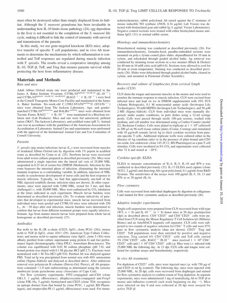
We previously demonstrated that IL-10 limits inflammation duringparasite development in the muscle, but does not contribute toimmune modulation during chronic infection (26). To better definethe influence of IL-10 on the cellular response to muscle infection,we examined the cytokine secretion profile of CLN cells recoveredfrom WT and IL-10�/� mice during parasite development (8 dpi;acute muscle infection) and at the completion of parasite develop-ment (21 dpi; transition to chronic infection). After stimulationwith somatic larval Ags, WT CLN cells produced IL-10 and IFN-�(Fig. 1A) at 8 dpi. At 21 dpi, IL-10 production was increased, IL-5and IL-13 production was detectable, and IFN-� production wasunchanged (Fig. 1B). In the absence of IL-10, cells producednearly eight times more IFN-� at 8 dpi (Fig. 1C) and 21 dpi (Fig.1D), compared with WT counterparts. In contrast, Th2 cytokineproduction was not significantly influenced by IL-10 deficiency.Parallel cultures stimulated with anti-CD3 yielded similar cytokineprofiles, although the magnitude of the responses was greater thanthat obtained with specific Ag stimulation (data not shown). Wedid not detect significant levels of IL-4, IL-5, IL-10, IL-13, orIFN-� in uninfected WT or IL-10�/� mice after stimulation withsomatic Ag (data not shown).
IFN-� is a potent activator of macrophage activity, stimulatingproduction of iNOS and enhanced generation of reactive oxygenintermediates. We have shown previously that the cellular infiltraterecruited to infected muscle is rich in macrophages. Sections ofinfected tongue were stained with Ab to iNOS to determinewhether the increase in IFN-� observed in IL-10�/� mice wouldtranslate to enhanced macrophage activation in vivo. Nurse cells inWT mice rarely had iNOS� cells at 21 dpi (Fig. 1E). In contrast,iNOS-producing cells were abundant near infected myotubes ofIL-10�/� mice (Fig. 1F). These data suggest that IL-10 limits in-flammation during parasite development by suppressing IFN-�levels and limiting local iNOS production, but does not influencethe development of a Th2 response later in infection.
Influence of Th2 cytokines on the inflammatory response tomuscle infection
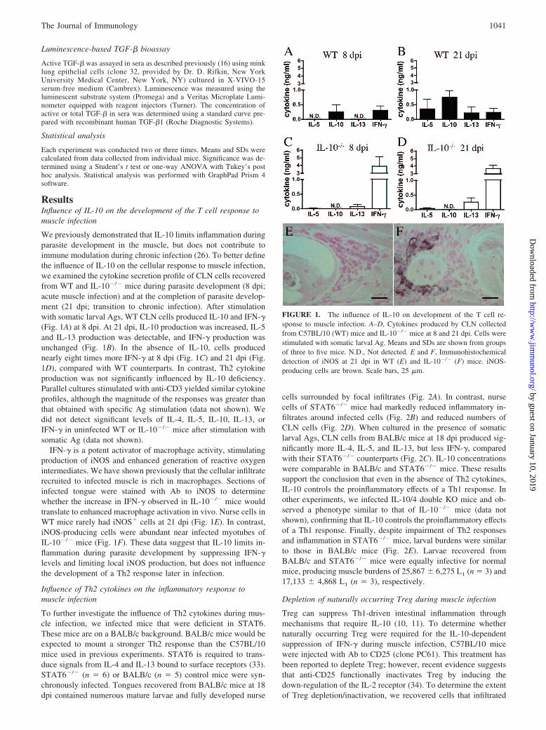
To further investigate the influence of Th2 cytokines during mus-cle infection, we infected mice that were deficient in STAT6.These mice are on a BALB/c background. BALB/c mice would beexpected to mount a stronger Th2 response than the C57BL/10mice used in previous experiments. STAT6 is required to trans-duce signals from IL-4 and IL-13 bound to surface receptors (33).STAT6�/� (n � 6) or BALB/c (n � 5) control mice were syn-chronously infected. Tongues recovered from BALB/c mice at 18dpi contained numerous mature larvae and fully developed nurse
cells surrounded by focal infiltrates (Fig. 2A). In contrast, nursecells of STAT6�/� mice had markedly reduced inflammatory in-filtrates around infected cells (Fig. 2B) and reduced numbers ofCLN cells (Fig. 2D). When cultured in the presence of somaticlarval Ags, CLN cells from BALB/c mice at 18 dpi produced sig-nificantly more IL-4, IL-5, and IL-13, but less IFN-�, comparedwith their STAT6�/� counterparts (Fig. 2C). IL-10 concentrationswere comparable in BALB/c and STAT6�/� mice. These resultssupport the conclusion that even in the absence of Th2 cytokines,IL-10 controls the proinflammatory effects of a Th1 response. Inother experiments, we infected IL-10/4 double KO mice and ob-served a phenotype similar to that of IL-10�/� mice (data notshown), confirming that IL-10 controls the proinflammatory effectsof a Th1 response. Finally, despite impairment of Th2 responsesand inflammation in STAT6�/� mice, larval burdens were similarto those in BALB/c mice (Fig. 2E). Larvae recovered fromBALB/c and STAT6�/� mice were equally infective for normalmice, producing muscle burdens of 25,867 � 6,275 L1 (n � 3) and17,133 � 4,868 L1 (n � 3), respectively.
Depletion of naturally occurring Treg during muscle infection
Treg can suppress Th1-driven intestinal inflammation throughmechanisms that require IL-10 (10, 11). To determine whethernaturally occurring Treg were required for the IL-10-dependentsuppression of IFN-� during muscle infection, C57BL/10 micewere injected with Ab to CD25 (clone PC61). This treatment hasbeen reported to deplete Treg; however, recent evidence suggeststhat anti-CD25 functionally inactivates Treg by inducing thedown-regulation of the IL-2 receptor (34). To determine the extentof Treg depletion/inactivation, we recovered cells that infiltrated
FIGURE 1. The influence of IL-10 on development of the T cell re-sponse to muscle infection. A–D, Cytokines produced by CLN collectedfrom C57BL/10 (WT) mice and IL-10�/� mice at 8 and 21 dpi. Cells werestimulated with somatic larval Ag. Means and SDs are shown from groupsof three to five mice. N.D., Not detected. E and F, Immunohistochemicaldetection of iNOS at 21 dpi in WT (E) and IL-10�/� (F) mice. iNOS-producing cells are brown. Scale bars, 25 �m.
1041The Journal of Immunology
by guest on January 10, 2019http://w
ww
.jimm
unol.org/D
ownloaded from
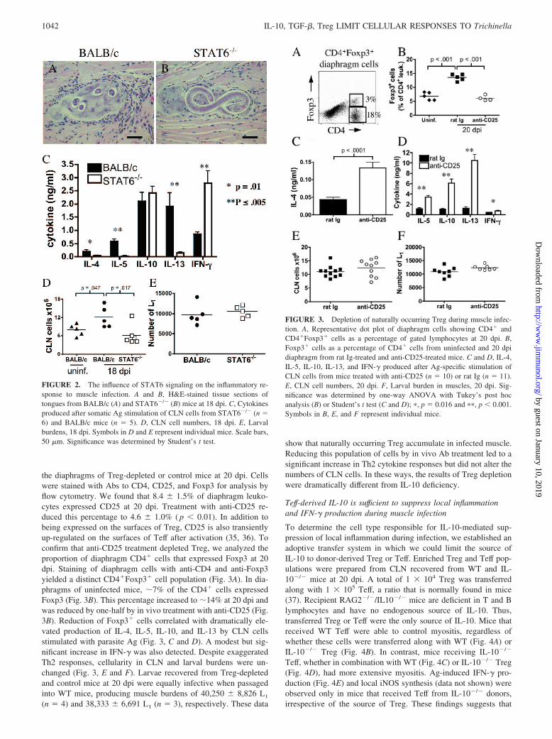
the diaphragms of Treg-depleted or control mice at 20 dpi. Cellswere stained with Abs to CD4, CD25, and Foxp3 for analysis byflow cytometry. We found that 8.4 � 1.5% of diaphragm leuko-cytes expressed CD25 at 20 dpi. Treatment with anti-CD25 re-duced this percentage to 4.6 � 1.0% ( p � 0.01). In addition tobeing expressed on the surfaces of Treg, CD25 is also transientlyup-regulated on the surfaces of Teff after activation (35, 36). Toconfirm that anti-CD25 treatment depleted Treg, we analyzed theproportion of diaphragm CD4� cells that expressed Foxp3 at 20dpi. Staining of diaphragm cells with anti-CD4 and anti-Foxp3yielded a distinct CD4�Foxp3� cell population (Fig. 3A). In dia-phragms of uninfected mice, �7% of the CD4� cells expressedFoxp3 (Fig. 3B). This percentage increased to �14% at 20 dpi andwas reduced by one-half by in vivo treatment with anti-CD25 (Fig.3B). Reduction of Foxp3� cells correlated with dramatically ele-vated production of IL-4, IL-5, IL-10, and IL-13 by CLN cellsstimulated with parasite Ag (Fig. 3, C and D). A modest but sig-nificant increase in IFN-� was also detected. Despite exaggeratedTh2 responses, cellularity in CLN and larval burdens were un-changed (Fig. 3, E and F). Larvae recovered from Treg-depletedand control mice at 20 dpi were equally infective when passagedinto WT mice, producing muscle burdens of 40,250 � 8,826 L1
(n � 4) and 38,333 � 6,691 L1 (n � 3), respectively. These data
show that naturally occurring Treg accumulate in infected muscle.Reducing this population of cells by in vivo Ab treatment led to asignificant increase in Th2 cytokine responses but did not alter thenumbers of CLN cells. In these ways, the results of Treg depletionwere dramatically different from IL-10 deficiency.
Teff-derived IL-10 is sufficient to suppress local inflammationand IFN-� production during muscle infection
To determine the cell type responsible for IL-10-mediated sup-pression of local inflammation during infection, we established anadoptive transfer system in which we could limit the source ofIL-10 to donor-derived Treg or Teff. Enriched Treg and Teff pop-ulations were prepared from CLN recovered from WT and IL-10�/� mice at 20 dpi. A total of 1 � 104 Treg was transferredalong with 1 � 105 Teff, a ratio that is normally found in mice(37). Recipient RAG2�/�/IL10�/� mice are deficient in T and Blymphocytes and have no endogenous source of IL-10. Thus,transferred Treg or Teff were the only source of IL-10. Mice thatreceived WT Teff were able to control myositis, regardless ofwhether these cells were transferred along with WT (Fig. 4A) orIL-10�/� Treg (Fig. 4B). In contrast, mice receiving IL-10�/�
Teff, whether in combination with WT (Fig. 4C) or IL-10�/� Treg(Fig. 4D), had more extensive myositis. Ag-induced IFN-� pro-duction (Fig. 4E) and local iNOS synthesis (data not shown) wereobserved only in mice that received Teff from IL-10�/� donors,irrespective of the source of Treg. These findings suggests that
FIGURE 2. The influence of STAT6 signaling on the inflammatory re-sponse to muscle infection. A and B, H&E-stained tissue sections oftongues from BALB/c (A) and STAT6�/� (B) mice at 18 dpi. C, Cytokinesproduced after somatic Ag stimulation of CLN cells from STAT6�/� (n �6) and BALB/c mice (n � 5). D, CLN cell numbers, 18 dpi. E, Larvalburdens, 18 dpi. Symbols in D and E represent individual mice. Scale bars,50 �m. Significance was determined by Student’s t test.
FIGURE 3. Depletion of naturally occurring Treg during muscle infec-tion. A, Representative dot plot of diaphragm cells showing CD4� andCD4�Foxp3� cells as a percentage of gated lymphocytes at 20 dpi. B,Foxp3� cells as a percentage of CD4� cells from uninfected and 20 dpidiaphragm from rat Ig-treated and anti-CD25-treated mice. C and D, IL-4,IL-5, IL-10, IL-13, and IFN-� produced after Ag-specific stimulation ofCLN cells from mice treated with anti-CD25 (n � 10) or rat Ig (n � 11).E, CLN cell numbers, 20 dpi. F, Larval burden in muscles, 20 dpi. Sig-nificance was determined by one-way ANOVA with Tukey’s post hocanalysis (B) or Student’s t test (C and D); �, p � 0.016 and ��, p � 0.001.Symbols in B, E, and F represent individual mice.
1042 IL-10, TGF-�, Treg LIMIT CELLULAR RESPONSES TO Trichinella
by guest on January 10, 2019http://w
ww
.jimm
unol.org/D
ownloaded from

IL-10 produced by CD4�CD25� cells is sufficient to suppress my-ositis and IFN-�.
Effect of TGF-� neutralization on the cytokine and inflammatoryresponse to muscle infection
Both surface-bound and secreted TGF-� have been shown to beimportant mechanisms of Treg-mediated control of Teff (38–40).To evaluate a role for TGF-� in control of myositis, we neutralizedthis cytokine in vivo by weekly administration of a mAb (clone1D11) that neutralizes activity of TGF-�1 and �2 isoforms (29).We determined the extent of neutralization using a bioassay thatmeasures the active form of TGF-� (41). Muscle infection with T.spiralis induced a significant elevation of active TGF-� in sera ofIL-10�/� but not WT mice (Fig. 5A). Treatment with TGF-�-neutralizing Ab reduced circulating levels of the active cytokine topreinfection levels (Fig. 5A). CLN from WT mice treated withanti-TGF-� produced significantly more IL-13 and less IFN-�compared with control Ig-treated WT mice (Fig. 5B). In contrast,neutralization of TGF-� in IL-10�/� mice caused a reduction inIL-13 production (Fig. 5C).
As expected, WT mice treated with control Ig had limited my-ositis at 20 dpi, whereas IL-10�/� mice treated with control Ig hadlarger infiltrates surrounding nurse cells (Fig. 5, D and F). Cellularinfiltration was moderate in WT mice treated with anti-TGF-�
(Fig. 5E). Mice deficient in IL-10 and treated with anti-TGF-� hadthe most extensive cellular infiltration of muscle (Fig. 5G) and themost cellular CLN (Fig. 5H). Although IL-10 or TGF-� alone didnot significantly protect muscle larvae from destruction, the combined
FIGURE 4. Contributions of CD4�CD25� and CD4�CD25� cells toIL-10-mediated suppression of inflammation in infected muscle. A–D,H&E-stained sections of diaphragm from RAG2�/�/IL-10�/� mice at 13dpi that received the following: A, CD4�CD25� and CD4�CD25� cellsfrom WT donor mice; B, WT CD4�CD25� cells and IL-10�/�
CD4�CD25� cells; C, IL-10�/� CD4�CD25� cells and WT CD4�CD25�
cells; or D, CD4�CD25� and CD4�CD25� cells from IL-10�/� donors. E,IFN-� produced by CLN cells from recipient mice after stimulation withsomatic Ags. Symbols represent individual mice. Significance was de-termined by one-way ANOVA with Tukey’s post hoc analysis. Scalebars, 50 �m.
FIGURE 5. Influence of TGF-� on muscle infection in WT and IL-10�/� mice. A, Active TGF-� in sera of uninfected (n � 5) and 20 dpi WTand IL10�/� mice. B and C, Cytokines produced after somatic Ag stimu-lation of CLN cells from WT mice treated with anti-TGF-� (n � 7) orcontrol Ig (n � 8) (B), or IL-10�/� mice treated with anti-TGF-� (n � 8)or control Ig (n � 6) (C). N.D., Not detected. �, p � 0.05 and ��, p � 0.001as determined by Student’s t test. D–G, H&E-stained tongue sections col-lected at 20 dpi from WT and IL-10�/� (KO) mice treated with control Igor anti-TGF-�. H, CLN cell numbers, 20 dpi. I, Larval burdens in muscles,20 dpi. Scale bars, 50 �m. Symbols represent individual mice in H and I.Significance was determined by one-way ANOVA and Tukey’s post hoc test.
1043The Journal of Immunology
by guest on January 10, 2019http://w
ww
.jimm
unol.org/D
ownloaded from

deficiency in both cytokines resulted in a significant reduction inmuscle larvae burden compared with WT mice treated with controlIg (Fig. 5I). Viable larvae recovered from all four groups of micewere equally infective when passaged into normal mice (n � 5 foreach group) producing muscle burdens of 15,200 � 4,361 L1 (WT,control Ig); 23,600 � 4,347 L1 (WT, anti-TGF-�); 25,880 �11,140 L1 (IL-10�/�, control Ig); and 16,240 � 6,121 L1 (IL-10�/�, anti-TGF-�).
Effects of Th2-polarized or mixed Th1/Th2 responses on survivalof muscle stage parasites
We reasoned that T cell responses capable of destroying larvaewould serve as logical targets of suppression during infection. Tobetter understand the type of immune response that is required fordestruction of muscle larvae, we developed an in vivo challengemodel in which WT and IL-10�/� mice were first infected with alow dose of parasites (5,000 NBL), rested for 3 mo to allow thelarvae to establish a chronic infection, and then challenged with20,000 NBL to elicit a memory response (primary/secondary (1°/2°)). For controls, additional mice received either the priming doseonly (1°/�), or the challenge dose only (�/2°). WT mice receivingboth priming and challenge infections mounted a polarized Th2response characterized by IL-5, IL-10, and IL-13 production, withlittle IFN-� (Fig. 6A). In contrast, cells from IL-10�/� mice pro-duced significantly more IFN-�, but similar quantities of IL-5 andIL-13, compared with WT mice (Fig. 6A). If larvae survived theTh2-polarized response to challenge infection in WT mice, wewould expect 1°/2° mice to harbor the combined burdens of the 1°infection (2,221 � 575 L1) and the 2° infection (9,066 � 2,505 L1)to give an expected burden of �11,287 L1, yet 1°/2° WT mice hada mean muscle burden of only 8,874 L1 � 3,679 at 20 days post-challenge (Fig. 6B). If larvae survived the mixed response to chal-lenge in IL-10�/� mice, we would expect 1°/2° mice yield wormburdens of �9,432 L1, rather than the observed mean burden of4,915 � 2,878 (48% killing; Fig. 6B).
Although the worm burdens in the challenge groups were notreduced sufficiently to achieve statistical significance, killing oflarvae was evident in histological sections of the diaphragm. WTmice receiving only the secondary infection had no cellular infil-trate surrounding developing larvae at 8 dpi (Fig. 6C). In contrast,WT 1°/2° mice had extensive infiltrates in muscle (Fig. 6E), char-acterized by a cellular infiltrate rich in eosinophils (Fig. 6E, inset).Compared with WT mice, IL-10�/� mice receiving only a sec-ondary infection had more intense inflammatory reactions sur-rounding developing larvae (Fig. 6D). The most severe myositiswas seen in IL-10�/� 1°/2° mice (Fig. 6F), in which an infiltratelargely comprised of mononuclear cells and nearly devoid of eo-sinophils (Fig. 6F, inset) incarcerated some larvae. Entry of largenumbers of inflammatory cells into the nurse cell occurs duringnurse cell destruction and parasite death. Large numbers of iNOS-producing cells were detected in the diaphragms of primed andchallenged IL-10�/� (Fig. 6H) but not WT mice (Fig. 6G). Sig-nificant increases in CLN size occurred along with increased my-ositis, with 1°/2° IL-10�/� mice having the most cellular CLN(Fig. 6I). Thus, the exaggerated local Th1 response was associatedwith larval killing.
DiscussionOur previous work demonstrated a role for IL-10 in limiting in-flammation during T. spiralis development in the muscle, yet thecell types responsible for this modulation, as well as the mecha-nism by which IL-10 effected suppression, were not identified. Thedata presented here show that IL-10 limits acute inflammation,suppresses IFN-� levels, and prevents iNOS production by inflam-matory cells recruited to the site of infection. In our system, IL-10deficiency does not influence the production of IL-4, IL-5, or IL-13during muscle infection, nor does it impair the generation of par-asite-specific IgG1 (26), suggesting that IL-10-producing cells spe-cifically suppress Th1 responses during muscle infection. Our re-sults contrast with findings reported from S. mansoni-infected
FIGURE 6. Response to NBL challenge. A, Cytokines produced aftersomatic Ag stimulation of CLN cells recovered from WT and IL-10�/�
mice 8 days postchallenge. B, Larval burdens in muscles, 21 dpi. C–F,H&E-stained sections of tongue from C57BL/10 (WT) and IL-10�/� micereceiving prime and challenge (1°/2°) or only the secondary infection (�/2°). G and H, Immunohistochemical detection of iNOS in sections of di-aphragm from WT (G) and IL-10�/� (H) mice. I, CLN cell numbers, 8 dpi.Arrows, Immature larvae. Scale bars, 50 �m. Symbols represent individualmice in B and I. �, p � 0.023 as determined by Student’s t test.
1044 IL-10, TGF-�, Treg LIMIT CELLULAR RESPONSES TO Trichinella
by guest on January 10, 2019http://w
ww
.jimm
unol.org/D
ownloaded from

mice, where IL-10 limits not only IFN-� production, but also pro-duction of the Th2 cytokines IL-4 and IL-5 (42, 43). This differ-ence in the targets of regulation may relate to the different habitatsoccupied by the two organisms. Although S. mansoni eggs areextracellular in liver and gut, larval T. spiralis are intracellular inmuscle. The suppression of Th1 responses by IL-10 during T.spiralis infection may reflect a biological adaptation to limitintracellular killing mechanisms during larval developmentwithin muscle cells.
Our data are compatible with a role for IFN-� in promotinginflammation observed in IL-10�/� mice but do not eliminate pos-sible influences of other proinflammatory mediators (e.g., TNF-�).In a murine model of Th1-driven intestinal inflammation, neutral-ization of IFN-� in IL-10�/� animals was effective in preventingcolitis if given early, but did not ameliorate established disease ifadministered later (44, 45), suggesting that IFN-� drives inflam-mation, but may not sustain it. IL-10 also preferentially limits Th1responses induced by filarial nematodes (46). Th1 stimuli havebeen investigated in filarial nematodes, and it has been reportedthat surface Ags of the endosymbiotic bacterium Wolbachia sp.stimulate Th1 responses by engaging TLR2 and TLR4 (47).Similar endosymbionts have not been identified in the genusTrichinella and the stimulus for Th1 cells has not been investi-gated. We observed that the Th1 response is prominent only duringacute infection, a time that is coincident with significant damage tohost muscle cells caused by parasite migration and growth. It hasbeen reported that tissue damage releases extracellular matrix pro-teins that can trigger activation of dendritic cells and macrophages.For example, heparan sulfate, a major component of extracellularmatrix and cell membranes, stimulates TLR4-dependent matura-tion of dendritic cells resulting in production of proinflammatorycytokines TNF-�, IL-6, and IL-1� (48, 49). Skeletal muscle and T.spiralis nurse cells are rich in heparan sulfate (50). Leukocytesrecruited to sites of infection may be exposed to cytoplasmic con-tents of muscle cells that stimulate production of proinflammatorycytokines. IL-10 may be produced to control such pathologicalresponses to tissue damage.
Treg have been shown to require IL-10 to mediate protectionfrom Th1-driven intestinal inflammation (10, 11); therefore, weinvestigated whether this cell population was important in controlof Th1 responses and myositis during T. spiralis infection. Infec-tion induced an enrichment of CD4�Foxp3� Treg in the dia-phragm. We found that reducing the proportion of Treg in theinfected diaphragm by anti-CD25 treatment was associated withincreased parasite-specific Th2 cytokine production but did notinfluence myositis. Because anti-CD25 treatment did not depleteall Foxp3� cells, it is possible that the residual Treg may havelimited the inflammatory response in depleted mice; however, thecontrasting responses in IL-10�/� and Treg-depleted mice suggestthat the two modulators work independently.
We took a mechanistic approach to identifying cells that medi-ate IL-10-dependent suppression of inflammation. Focusing onCD4 T cell populations, we conducted adoptive transfer experi-ments with CD4�CD25� and CD4�CD25� cells derived frominfected WT and IL-10�/� donors. Because recipient mice wereRAG2�/�/IL-10�/�, the source of IL-10 was restricted to donorTreg and/or Teff. We found that IL-10 production by CD4�
CD25� cells was sufficient to control myositis and IFN-� produc-tion. IL-10 production has been described in suppressive CD4 Tcell populations that may cosecrete Th2 and/or Th1 cytokines (re-viewed in Ref. 51). These cells typically do not express CD25, andit is possible they may contribute to the suppressive effects weobserved with CD4�CD25� cells. Regardless, our results clearlyillustrate that the CD4� Th cell population must simultaneously
balance effector function with control of inflammation during mus-cle infection, and that IL-10 production contributes to this control.
TGF-� and IL-10 have been shown to have an additive effect inthe suppression of T cell activity in vitro (52) and to reduce the invitro larvicidal activity of IFN-�-activated macrophages againstschistosomula (53), yet few studies have examined this coopera-tive relationship in vivo during infection. Neutralization of TGF-�in IL-10�/� mice infected with T. spiralis resulted in myositis thatwas more severe than that seen in mice deficient in IL-10 orTGF-� alone. More importantly, this treatment resulted in parasitekilling. This constitutes the first demonstration of cooperation be-tween TGF-� and IL-10 that promotes parasite survival while pro-tecting host tissue. TGF-� is a pleiotropic cytokine and our dataraise interesting questions about the nature of its effect on T. spi-ralis infection. TGF-� signaling regulates the development of free-living nematodes, and there is great interest in understandingwhether parasitic nematodes might depend upon host cytokines forgrowth and development (54). We did not observe a significanteffect on parasite survival when TGF-� was neutralized in WTmice; therefore, our data argue against a requirement for hostTGF-� in development of T. spiralis in the muscle. AlthoughIL-10 and TGF-� both limited myositis, the two had very differenteffects on the cytokine response during infection. TGF-� appearedto attenuate the predominant cytokine response whereas IL-10 spe-cifically targeted the Th1 response.
To better understand the types of T cell responses associatedwith parasite killing, we primed and challenged WT and IL-10�/�
mice with NBL. We found that in WT mice, the memory responseto infection was Th2 polarized and was associated with only mod-est reduction in muscle larvae. These results were similar to thoseobtained in Treg-depleted mice, where an enhanced Th2 responseoccurred in the absence of parasite killing. In contrast, in chal-lenged, IL-10-deficient mice, Th2 responses were maintainedwhile IFN-� was markedly increased, resulting in a mixed memoryresponse that was associated with evidence of parasite killing.Similarly, enhanced Th1 responses in IL-10/TGF-�-deficient micedestroyed larvae during primary infection. In aggregate, our resultsshow that poorly regulated Th1 responses can destroy larvae,whereas Th2 responses are largely not destructive. Our resultsdiffer from those obtained with Brugia pahangi (55) and Lito-mosoides sigmodontis (16), in which elevated Th2 responses afterTreg depletion resulted in parasite clearance. In the case of L.sigmodontis, Treg depletion resulted in elimination of �70% ofthe parasite burden (16).
Tissue distribution and intracellular vs extracellular location ofworms may influence the requirements for parasite destruction(56). Extracellular filarial nematodes are highly susceptible to eosi-nophil-mediated killing (57, 58), while there is limited and con-tradictory evidence that intracellular T. spiralis muscle larvae arevulnerable to attack by eosinophils (59–61). Several expressedsequence tag clusters encoding antioxidants were found in matureT. spiralis muscle larvae, but not in NBL (62), suggesting thatimmature larvae may be vulnerable to oxidative or nitrosativedamage. Indeed, it has been reported that NBL are highly suscep-tible to oxidative killing (63). Evidence from in vitro studies hasshown that TGF-� can impair NO generation in macrophages byreducing the stability and translation of iNOS mRNA and increas-ing degradation of iNOS protein (64). We hypothesize that IL-10and TGF-� cooperate to limit oxidative and/or nitrosative damageto the developing muscle larva or the host muscle cell. The role ofiNOS in parasite killing can be tested using specific inhibitors. Itis important to note that parasite death in the muscle was not as-sociated with significant host morbidity, allowing us to test our
1045The Journal of Immunology
by guest on January 10, 2019http://w
ww
.jimm
unol.org/D
ownloaded from
hypothesis and examine potential killing mechanisms in otherwisehealthy animals.
Collectively, our data support a model of T cell suppressionduring muscle infection whereby Teff-derived IL-10 limits acutemyositis, IFN-� levels, and local iNOS production. In the presenceof IL-10, Th2 cytokines are produced and that production is reg-ulated by CD4�Foxp3� cells. TGF-�, produced by unknown celltypes, helps balance the T cell response and limits inflammation. Incombination, these modulatory influences protect parasites in themuscle and limit tissue damage.
AcknowledgmentsWe thank Dr. Daniel Rifkin for providing mink-lung epithelial cells andDr. Edward Pearce for helpful suggestions.
DisclosuresThe authors have no financial conflict of interest.
References1. Bell, R. G., and C. H. Wang. 1987. The Trichinella spiralis newborn larvae:
production, migration and immunity in vivo. Wiad. Parazytol. 33: 453–478.2. Stewart, G. L., and L. M. Charniga. 1980. Distribution of Trichinella spiralis in
muscles of the mouse. J. Parasitol. 66: 688–689.3. Ritterson, A. L. 1966. Nature of the cyst of Trichinella spiralis. J. Parasitol. 52:
157–161.4. Humes, A. G., and R. P. Akers. 1952. Vascular changes in the cheek pouch of the
golden hamster during infection with Trichinella spiralis larvae. Anat. Rec. 114:103–113.
5. Jasmer, D. P. 1993. Trichinella spiralis infected skeletal muscle cells arrest inG2-M and cease muscle gene expression. J. Cell Biol. 121: 785–793.
6. Despommier, D. 1975. Adaptive changes in muscle fibers infected withTrichinella spiralis. Am. J. Pathol. 78: 477–496.
7. King, C. L., S. Mahanty, V. Kumaraswami, J. S. Abrams, J. Regunathan, K.Jayaraman, E. A. Ottesen, and T. B. Nutman. 1993. Cytokine control of parasite-specific anergy in human lymphatic filariasis: preferential induction of a regulatory Thelper type 2 lymphocyte subset. J. Clin. Invest. 92: 1667–1673.
8. Babu, S., C. P. Blauvelt, V. Kumaraswami, and T. B. Nutman. 2006. Regulatorynetworks induced by live parasites impair both Th1 and Th2 pathways in patentlymphatic filariasis: implications for parasite persistence. J. Immunol. 176:3248–3256.
9. Doetze, A., J. Satoguina, G. Burchard, T. Rau, C. Loliger, B. Fleischer, andA. Hoerauf. 2000. Antigen-specific cellular hyporesponsiveness in a chronic hu-man helminth infection is mediated by T 3/Tr 1-type cytokines IL-10 and trans-forming growth factor-� but not by a Th1 to Th2 shift. Int. Immunol. 12: 623–630.
10. Asseman, C., S. Mauze, M. W. Leach, R. L. Coffman, and F. Powrie. 1999. Anessential role for interleukin 10 in the function of regulatory T cells that inhibitintestinal inflammation. J. Exp. Med. 190: 995–1004.
11. Maloy, K. J., L. Salaun, R. Cahill, G. Dougan, N. J. Saunders, and F. Powrie.2003. CD4�CD25� TR cells suppress innate immune pathology through cyto-kine-dependent mechanisms. J. Exp. Med. 197: 111–119.
12. Taylor, J. J., M. Mohrs, and E. J. Pearce. 2006. Regulatory T cell responsesdevelop in parallel to Th responses and control the magnitude and phenotype ofthe Th effector population. J. Immunol. 176: 5839–5847.
13. Baumgart, M., F. Tompkins, J. Leng, and M. Hesse. 2006. Naturally occurringCD4�Foxp3� regulatory T cells are an essential, IL-10-independent part of theimmunoregulatory network in Schistosoma mansoni egg-induced inflammation.J. Immunol. 176: 5374–5387.
14. Hesse, M., C. A. Piccirillo, Y. Belkaid, J. Prufer, M. Mentink-Kane, M. Leusink,A. W. Cheever, E. M. Shevach, and T. A. Wynn. 2004. The pathogenesis ofschistosomiasis is controlled by cooperating IL-10-producing innate effector andregulatory T cells. J. Immunol. 172: 3157–3166.
15. McKee, A. S., and E. J. Pearce. 2004. CD25�CD4� cells contribute to Th2polarization during helminth infection by suppressing Th1 response development.J. Immunol. 173: 1224–1231.
16. Taylor, M. D., L. LeGoff, A. Harris, E. Malone, J. E. Allen, and R. M. Maizels.2005. Removal of regulatory T cell activity reverses hyporesponsiveness andleads to filarial parasite clearance in vivo. J. Immunol. 174: 4924–4933.
17. Finkelman, F. D., T. Shea-Donohue, S. C. Morris, L. Gildea, R. Strait,K. B. Madden, L. Schopf, and J. F. Urban, Jr. 2004. Interleukin-4- and interleu-kin-13-mediated host protection against intestinal nematode parasites. Immunol.Rev. 201: 139–155.
18. Fallon, P. G., H. E. Jolin, P. Smith, C. L. Emson, M. J. Townsend, R. Fallon, andA. N. McKenzie. 2002. IL-4 induces characteristic Th2 responses even in thecombined absence of IL-5, IL-9, and IL-13. Immunity 17: 7–17.
19. Akiho, H., Y. Deng, P. Blennerhassett, H. Kanbayashi, and S. M. Collins. 2005.Mechanisms underlying the maintenance of muscle hypercontractility in a modelof postinfective gut dysfunction. Gastroenterology 129: 131–141.
20. Urban, J. F., Jr., N. Noben-Trauth, D. D. Donaldson, K. B. Madden, S. C. Morris,M. Collins, and F. D. Finkelman. 1998. IL-13, IL-4R�, and Stat6 are required forthe expulsion of the gastrointestinal nematode parasite Nippostrongylus brasil-iensis. Immunity 8: 255–264.
21. Urban, J. F., Jr., I. M. Katona, W. E. Paul, and F. D. Finkelman. 1991. Interleukin4 is important in protective immunity to a gastrointestinal nematode infection inmice. Proc. Natl. Acad. Sci. USA 88: 5513–5517.
22. Else, K. J., F. D. Finkelman, C. R. Maliszewski, and R. K. Grencis. 1994. Cy-tokine-mediated regulation of chronic intestinal helminth infection. J. Exp. Med.179: 347–351.
23. Schopf, L. R., K. F. Hoffmann, A. W. Cheever, J. F. Urban, Jr., and T. A. Wynn.2002. IL-10 is critical for host resistance and survival during gastrointestinalhelminth infection. J. Immunol. 168: 2383–2392.
24. Hoffmann, K. F., A. W. Cheever, and T. A. Wynn. 2000. IL-10 and the dangersof immune polarization: excessive type 1 and type 2 cytokine responses inducedistinct forms of lethal immunopathology in murine schistosomiasis. J. Immunol.164: 6406–6416.
25. Crum, E. D., D. D. Despommier, and D. D. McGregor. 1977. Immunity toTrichinella spiralis I: transfer of resistance by two classes of lymphocytes. Im-munology 33: 787–795.
26. Beiting, D. P., S. K. Bliss, D. H. Schlafer, V. L. Roberts, and J. A. Appleton.2004. Interleukin-10 limits local and body cavity inflammation during infectionwith muscle-stage Trichinella spiralis. Infect. Immun. 72: 3129–3137.
27. Appleton, J. A., and L. Usack. 1993. Identification of potential antigenic targetsfor rapid expulsion of Trichinella spiralis. Mol. Biochem. Parasitol. 58: 53–62.
28. Lowenthal, J. W., P. Corthesy, C. Tougne, R. Lees, H. R. MacDonald, andM. Nabholz. 1985. High and low affinity IL 2 receptors: analysis by IL 2 disso-ciation rate and reactivity with monoclonal anti-receptor antibody PC61. J. Im-munol. 135: 3988–3994.
29. Dasch, J. R., D. R. Pace, W. Waegell, D. Inenaga, and L. Ellingsworth. 1989.Monoclonal antibodies recognizing transforming growth factor-�: bioactivityneutralization and transforming growth factor �2 affinity purification. J. Immunol.142: 1536–1541.
30. Appleton, J. A., D. F. Antczak, and A. D. Lopes. 1987. Characterization of theequine influenza virus H3 with monoclonal antibodies. Arch. Virol. 94: 339–346.
31. Daley, L. P., L. F. Gagliardo, M. S. Duffy, M. C. Smith, and J. A. Appleton. 2005.Application of monoclonal antibodies in functional and comparative investiga-tions of heavy-chain immunoglobulins in new world camelids. Clin. Diagn. Lab.Immunol. 12: 380–386.
32. Bliss, S. K., A. Alcaraz, and J. A. Appleton. 2003. IL-10 prevents liver necrosisduring murine infection with Trichinella spiralis. J. Immunol. 171: 3142–3147.
33. Kaplan, M. H., U. Schindler, S. T. Smiley, and M. J. Grusby. 1996. Stat6 isrequired for mediating responses to IL-4 and for development of Th2 cells. Im-munity 4: 313–319.
34. Kohm, A. P., J. S. McMahon, J. R. Podojil, W. S. Begolka, M. Degutes,D. J. Kasprowicz, S. F. Ziegler, and S. D. Miller. 2006. Cutting edge: anti-CD25monoclonal antibody injection results in the functional inactivation, not deple-tion, of CD4�CD25� T regulatory cells. J. Immunol. 176: 3301–3305.
35. Konaka, Y., M. A. Norcross, V. C. Maino, and R. T. Smith. 1981. Anti-Thy-1-mediated T cell activation: role of soluble factors and expression of interleukin 2receptors on T cells. Eur. J. Immunol. 11: 445–450.
36. Kroczek, R. A., C. D. Black, J. Barbet, L. J. Edison, and E. M. Shevach. 1987.Induction of IL-2 receptor expression in vivo: response to allogeneic cells. Trans-plantation 44: 547–553.
37. Sakaguchi, S., N. Sakaguchi, M. Asano, M. Itoh, and M. Toda. 1995. Immuno-logic self-tolerance maintained by activated T cells expressing IL-2 receptor�-chains (CD25): breakdown of a single mechanism of self-tolerance causesvarious autoimmune diseases. J. Immunol. 155: 1151–1164.
38. Kitani, A., I. Fuss, K. Nakamura, F. Kumaki, T. Usui, and W. Strober. 2003.Transforming growth factor (TGF)-�1-producing regulatory T cells induceSmad-mediated interleukin 10 secretion that facilitates coordinated immunoregu-latory activity and amelioration of TGF-�1-mediated fibrosis. J. Exp. Med. 198:1179–1188.
39. Nakamura, K., A. Kitani, I. Fuss, A. Pedersen, N. Harada, H. Nawata, andW. Strober. 2004. TGF-� 1 plays an important role in the mechanism ofCD4�CD25� regulatory T cell activity in both humans and mice. J. Immunol.172: 834–842.
40. Nakamura, K., A. Kitani, and W. Strober. 2001. Cell contact-dependent immu-nosuppression by CD4�CD25� regulatory T cells is mediated by cell surface-bound transforming growth factor �. J. Exp. Med. 194: 629–644.
41. Abe, M., J. G. Harpel, C. N. Metz, I. Nunes, D. J. Loskutoff, and D. B. Rifkin.1994. An assay for transforming growth factor-� using cells transfected with aplasminogen activator inhibitor-1 promoter-luciferase construct. Anal. Biochem.216: 276–284.
42. Sadler, C. H., L. I. Rutitzky, M. J. Stadecker, and R. A. Wilson. 2003. IL-10 iscrucial for the transition from acute to chronic disease state during infection ofmice with Schistosoma mansoni. Eur. J. Immunol. 33: 880–888.
43. Wynn, T. A., R. Morawetz, T. Scharton-Kersten, S. Hieny, H. C. Morse, III,R. Kuhn, W. Muller, A. W. Cheever, and A. Sher. 1997. Analysis of granulomaformation in double cytokine-deficient mice reveals a central role for IL-10 inpolarizing both T helper cell 1- and T helper cell 2-type cytokine responses invivo. J. Immunol. 159: 5014–5023.
44. Kullberg, M. C., J. M. Ward, P. L. Gorelick, P. Caspar, S. Hieny, A. Cheever,D. Jankovic, and A. Sher. 1998. Helicobacter hepaticus triggers colitis in spe-cific-pathogen-free interleukin-10 (IL-10)-deficient mice through an IL-12- and �interferon-dependent mechanism. Infect. Immun. 66: 5157–5166.
45. Kullberg, M. C., A. G. Rothfuchs, D. Jankovic, P. Caspar, T. A. Wynn, P. L.Gorelick, A. W. Cheever, and A. Sher. 2001. Helicobacter hepaticus-induced colitisin interleukin-10-deficient mice: cytokine requirements for the induction and main-tenance of intestinal inflammation. Infect. Immun. 69: 4232–4241.
1046 IL-10, TGF-�, Treg LIMIT CELLULAR RESPONSES TO Trichinella
by guest on January 10, 2019http://w
ww
.jimm
unol.org/D
ownloaded from
46. Osborne, J., and E. Devaney. 1999. Interleukin-10 and antigen-presenting cellsactively suppress Th1 cells in BALB/c mice infected with the filarial parasiteBrugia pahangi. Infect. Immun. 67: 1599–1605.
47. Brattig, N. W., C. Bazzocchi, C. J. Kirschning, N. Reiling, D. W. Buttner,F. Ceciliani, F. Geisinger, H. Hochrein, M. Ernst, H. Wagner, et al. 2004. Themajor surface protein of Wolbachia endosymbionts in filarial nematodes elicitsimmune responses through TLR2 and TLR4. J. Immunol. 173: 437–445.
48. Johnson, G. B., G. J. Brunn, Y. Kodaira, and J. L. Platt. 2002. Receptor-mediatedmonitoring of tissue well-being via detection of soluble heparan sulfate by Toll-like receptor 4. J. Immunol. 168: 5233–5239.
49. Kodaira, Y., S. K. Nair, L. E. Wrenshall, E. Gilboa, and J. L. Platt. 2000. Phe-notypic and functional maturation of dendritic cells mediated by heparan sulfate.J. Immunol. 165: 1599–1604.
50. Beiting, D. P., P. W. Park, and J. A. Appleton. 2006. Synthesis of syndecan-1 byskeletal muscle cells is an early response to infection with Trichinella spiralis butis not essential for nurse cell development. Infect. Immun. 74: 1941–1943.
51. Hawrylowicz, C. M., and A. O’Garra. 2005. Potential role of interleukin-10-secreting regulatory T cells in allergy and asthma. Nat. Rev. Immunol. 5:271–283.
52. Zeller, J. C., A. Panoskaltsis-Mortari, W. J. Murphy, F. W. Ruscetti, S. Narula,M. G. Roncarolo, and B. R. Blazar. 1999. Induction of CD4� T cell alloantigen-specific hyporesponsiveness by IL-10 and TGF-�. J. Immunol. 163: 3684–3691.
53. Oswald, I. P., R. T. Gazzinelli, A. Sher, and S. L. James. 1992. IL-10 synergizeswith IL-4 and transforming growth factor-� to inhibit macrophage cytotoxic ac-tivity. J. Immunol. 148: 3578–3582.
54. Viney, M. E., F. J. Thompson, and M. Crook. 2005. TGF-� and the evolution ofnematode parasitism. Int. J. Parasitol. 35: 1473–1475.
55. Gillan, V., and E. Devaney. 2005. Regulatory T cells modulate Th2 responsesinduced by Brugia pahangi third-stage larvae. Infect. Immun. 73: 4034–4042.
56. Mulcahy, G., S. O’Neill, J. Fanning, E. McCarthy, and M. Sekiya. 2005. Tissuemigration by parasitic helminths—an immunoevasive strategy? Trends Parasitol.21: 273–277.
57. Martin, C., L. Le Goff, M. N. Ungeheuer, P. N. Vuong, and O. Bain. 2000.Drastic reduction of a filarial infection in eosinophilic interleukin-5 transgenicmice. Infect. Immun. 68: 3651–3656.
58. Volkmann, L., O. Bain, M. Saeftel, S. Specht, K. Fischer, F. Brombacher, K. I.Matthaei, and A. Hoerauf. 2003. Murine filariasis: interleukin 4 and interleukin5 lead to containment of different worm developmental stages. Med. Microbiol.Immunol. 192: 23–31.
59. Gurish, M. F., A. Humbles, H. Tao, S. Finkelstein, J. A. Boyce, C. Gerard,D. S. Friend, and K. F. Austen. 2002. CCR3 is required for tissue eosinophiliaand larval cytotoxicity after infection with Trichinella spiralis. J. Immunol. 168:5730–5736.
60. Herndon, F. J., and S. G. Kayes. 1992. Depletion of eosinophils by anti-IL-5monoclonal antibody treatment of mice infected with Trichinella spiralis does notalter parasite burden or immunologic resistance to reinfection. J. Immunol. 149:3642–3647.
61. Hokibara, S., M. Takamoto, A. Tominaga, K. Takatsu, and K. Sugane. 1997.Marked eosinophilia in interleukin-5 transgenic mice fails to prevent Trichinellaspiralis infection. J. Parasitol. 83: 1186–1189.
62. Mitreva, M., D. P. Jasmer, J. Appleton, J. Martin, M. Dante, T. Wylie, S. W.Clifton, R. H. Waterston, and J. P. McCarter. 2004. Gene discovery in the ad-enophorean nematode Trichinella spiralis: an analysis of transcription from threelife cycle stages. Mol. Biochem. Parasitol. 137: 277–291.
63. Kazura, J. W., and S. R. Meshnick. 1984. Scavenger enzymes and resistanceto oxygen mediated damage in Trichinella spiralis. Mol. Biochem. Parasitol.10: 1–10.
64. Vodovotz, Y., C. Bogdan, J. Paik, Q. W. Xie, and C. Nathan. 1993. Mechanismsof suppression of macrophage nitric oxide release by transforming growth factor�. J. Exp. Med. 178: 605–613.
1047The Journal of Immunology
by guest on January 10, 2019http://w
ww
.jimm
unol.org/D
ownloaded from