Embed Size (px)
Citation preview

Section 1
Core Issues
COPYRIG
HTED M
ATERIAL

2
Perioperative Homeostasis
Paul Wicker 1
LEARNING OUTCOMES ❏ Understand homeostasis . ❏ Discuss fl uid and electrolyte balance in the perioperative
patient. ❏ Describe the structure, function and regulation of the cardiovas-
cular and respiratory system . ❏ Describe blood pressure regulation . ❏ Discuss the implications of the metabolic response and the
stages of wound healing .
INTRODUCTION Teamwork is the focus of good perioperative practice. Nowhere is teamwork more obvious than within the human body itself, where the close and effi cient functioning of all the individual parts is essential for survival.
The purpose of this introductory chapter is to set the context of perioperative care and to identify links between the patient ’ s anatomy and physiology, and perioperative care. Everything that happens to the perioperative patient during surgery and anaesthesia has an effect on his or her anatomy and physiology. This makes it important to understand the internal maintenance and control of the body ’ s systems, and the external control through medical interventions.
The human body is a complex system of parts which can protect itself against major changes to its own internal environ-ment. It does this by preserving a fi ne balance between all its major organs and systems, by maintaining fl uids and electro-lytes, blood pressure and oxygenation between particular limits to ensure effi cient functioning. Every part of the system is

3
Perioperative Homeostasis 1
related: for example, the lungs absorb oxygen which is trans-ported by the blood to muscles such as the heart, allowing it to beat and maintain blood pressure. The blood pressure in turn pushes the oxygenated blood around the body to the tissue cells. The blood then progresses back to the lungs where it supplies the lung tissue itself, as well as going on to provide a further source of oxygenated blood to all the cells of the body.
The term homeostasis is often used to refer to the mainte-nance of a constant environment. In perioperative care; however, it is better to see homeostasis as a dynamic process that results in a peak state for the body under existing circumstances (Clancy et al . 2002 ). Hence, for example, blood pressure may or may not be maintained at preoperative levels during the peri-operative experience, and during surgery a low blood pressure may be helpful to reduce bleeding, ensuring a bloodless fi eld. However, the main principles of control still hold true – main-taining an ideal environment for body processes to take place under current circumstances. The aim of medical interventions, as external homeostatic controllers, is to support the body ’ s natural ability to maintain this dynamic homeostatic environment.
The topics associated with homeostasis are huge and this chapter will highlight selected areas of interest and relate them to clinical issues raised later in the book. This chapter therefore, describes some of the ways in which the human body maintains equilibrium, how anaesthesia and surgery affect this balance and how medical interventions support the return to normal homeostasis. Finally, this chapter will look at the human body ’ s response to stress and the process of wound healing.
PRINCIPLES OF HOMEOSTASIS To maintain homeostasis naturally, the body needs to:
� detect and analyse changes; � take measures to address the changes; � evaluate the effect of measures taken.
Control mechanisms carry out these processes, acting as recep-tors (detecting the changes), analysers (interpreting the changes)

1 Caring for the Perioperative Patient
4
and effectors (acting on the changes to minimise, maximise or regulate them). These mechanisms detect changes in normal values and try to bring them back to within the normal homeo-static range. An example of this system is the acid – base balance, where the buffer systems (described later in this chapter) act in unison to maintain the pH of body fl uids within a normal range of around 7.35 – 7.45. Under normal circumstances, the body ’ s buffer systems are able to maintain this balance; however, during illness, disease or because of trauma such as surgery or anaesthesia, the body may need support from external controls (medical interventions) to regain equilibrium.
Most of the body ’ s own control mechanisms work through negative feedback – a change occurs to the body ’ s environment and then mechanisms to cancel the changes are activated. Blood glucose regulation, for example, involves either the release of insulin to lower blood glucose or the release of glucagon to raise it. In either case, blood sugar levels are brought back to within the normal range.
Occasionally the control mechanisms work through positive feedback and a control mechanism to promote changes is acti-vated – for example, blood clotting, where the blood undergoes changes that allow it to clot and therefore reduce blood loss. Once the need has passed, the blood then returns to its normal state.
Internally, the organs act as both independent and interactive homeostatic controllers. This topic goes beyond the scope of this book. However, in the perioperative patient, several systems are specifi cally important because they are critical to the patient ’ s immediate survival and are also the target of many periopera-tive interventions. Fluid and electrolyte balances are important perioperative considerations because of the potential for blood loss and possible hypovolaemia. Medical interventions that support the effects of blood and fl uid loss, and help maintain electrolyte balance, therefore, merit consideration. The regula-tion of the respiratory and cardiovascular systems is also crucial in both the long and the short term. The aim of many anaesthetic functions is to control these systems to provide the optimum physiological environment during surgery.

5
Perioperative Homeostasis 1
WATER AND ELECTROLYTE HOMEOSTASIS Table 1.1 describes the fl uid compartments of the body. Total body water is distributed among all the fl uid compartments of the body. Of the 40 litres present in a 68 kg (150 lb) male, 65% is intracellular and 35% is extracellular. Extracellular fl uid is com-posed of 25% tissue fl uid, 8% blood plasma and lymph, and 2% transcellular fl uid such as cerebrospinal fl uid (CSF) and syno-vial fl uid.
Osmosis is the main process that distributes fl uid throughout these compartments (Watson & Fawcett 2003 ). The term ‘ osmo-larity ’ refers to the concentration of solutes (such as potassium) in the solution (such as water). Osmosis is a special form of diffusion which involves the passage of water across a selec-tively permeable cell membrane that is freely permeable to water but not freely permeable to solutes. This process aims to equalise the concentrations of the solution on each side of the membrane. In osmosis, water will fl ow across a membrane toward the solution that has the higher concentration of solutes, because that is where the concentration of water is lowest (Figure 1.1 ). A solution with a high osmolarity has high osmotic pressure.
Table 1.1 Terminology associated with fl uid compartments.
Fluid compartment Description
Extracellular (extracellular fl uid = ECF) Outside cells
Intracellular (intracellular fl uid = ICF) Inside cells
Interstitial Between cells
Intravascular Inside blood vessels (this is also extracellular)
Extravascular Outside blood vessels (this may be intracellular, interstitial or transcellular)
Transcellular Within hollow spaces, such as cerebrospinal fl uid, the joints, the gastrointestinal tract, the urinary tract and the ducts of glands. Also called the ‘ third space ’

1 Caring for the Perioperative Patient
6
An important effect of the movement of ions and water across cell membranes is the development of an electrical charge across the membrane. The resting potential is the charge across the membrane of an undisturbed cell. When the cell is stimulated, the electrical charge may be increased, causing an ‘ action poten-tial ’ . In nerves, for example, this represents the movement of information away from the cell body and down the nerve. In muscles, the action potential causes the contraction of muscle fi bres.
Other methods which the cell uses to transport water and electrolytes include:
� fi ltration, which is a passive process where hydrostatic pres-sure (blood pressure) forces fl uid and solutes across a mem-brane barrier;
� carrier - mediated transport, which involves specialised cell proteins binding to ions or organic substances, facilitating their entry to or exit from the cell;
� vesicular transport, which involves the movement of materi-als within small membranous sacs or vesicles.
Fig. 1.1 Osmosis.
Fluid compartments
Solvent
Osmosis
Semi-permeable membrane
Solute

7
Perioperative Homeostasis 1
Fluid b alance
A person is in a state of fl uid balance when water gain equals water loss; for example, a water intake of 2.5 litres a day, taken in by food and drink, is balanced by water loss via routes such as faeces, expired air, sweat and urine.
Fluid intake is mainly controlled by the thirst centre in the hypothalamus which generates the sensation of thirst. Water output is regulated by varying urine volume. This system is mainly controlled by the antidiuretic hormone (ADH), but also to a lesser extent by aldosterone and atrial natriuretic factor (ANF). Blood osmolarity is maintained because both sodium and water are either retained or excreted.
Changes in the osmotic pressure exerted by plasma infl uence the release of ADH. High osmotic pressure (i.e. dehydration) leads to release of ADH; low osmotic pressure (i.e. hydration) reduces excretion of ADH. This system is so effi cient that under normal conditions water balance is maintained to within 2% of the normal homeostatic range (Clancy et al . 2002 ).
ANF is a peptide released by walls of the cardiac atrium in response to high sodium chloride (NaCl) concentration, high extracellular fl uid volume or high blood volume. ANF inhibits NaCl reabsorption in the distal convoluted tubule and cortical collecting duct of the kidneys. It also dilates the afferent glo-merular arteriole and constricts the efferent glomerular arteri-ole. This increases the glomerular fi ltration rate, which increases NaCl excretion, raises urinary fi ltration rate and therefore increases the rate of urine production.
Volume depletion (hypovolaemia) is a loss of total body water volume when osmolarity remains normal. Vomiting, diarrhoea, burns, haemorrhage or renal failure can cause this. Addison ’ s disease results in dehydration leading to loss of total body water volume with an associated rise in osmolarity. This condition can also be caused by lack of drinking water, diabetes, profuse sweating or diuretics. Infants are more vulnerable to this condition.
Electrolyte b alance
The balance of major electrolytes such as sodium (Na + ), potas-sium (K + ), calcium (Ca 2 + ), hydrogen (H + ) and bicarbonate ( HCO3
−

1 Caring for the Perioperative Patient
8
) is essential to ensure homeostasis and the proper functioning of the body ’ s processes (Saladin 2009 ).
Maintaining electrolyte and water balance are three major hormones: ADH, which promotes water retention indepen-dently of Na + and K + concentration; aldosterone, which pro-motes retention of water and Na + , and secretion of K + ; and ANF which increases NaCl secretion.
Electrolyte m etabolism
Sodium and p otassium Sodium and potassium levels are critical to homeostasis because of their many roles. These two electrolytes contribute to main-taining membrane potentials, a major role of the sodium – potas-sium pump. Sodium is also responsible for 90 – 95% of osmolarity of extracellular fl uid (ECF) and potassium is the primary cation in intracellular fl uid (ICF).
The normal sodium intake of around 3 – 7 g/day exceeds the 0.5 g/day needed for survival. Normal blood level ranges are:
� Na + , 130 – 145 mmol/litre; � K + , 3.5 − 5.5 mmol/litre.
The homeostasis of water, sodium and potassium levels occurs by various linked systems:
� aldosterone/ANF; � ADH; � oestrogen/progesterone; � salt craving.
Intercalated cells in the collecting duct of the kidneys also control potassium levels (Saladin 2009 ).
Calcium ECF contains a low concentration of calcium; however calcium exerts great infl uence on the body systems. Low calcium con-centration increases the excitability of cells and in muscle cells this may lead to tetany. High concentrations of calcium ions

9
Perioperative Homeostasis 1
make the cells less excitable and may lead to symptoms such as muscle weakness and bowel stasis. Other roles of calcium include skeletal mineralisation, muscle contraction, exocytosis (release of substances such as hormones from the vesicles of certain tissue cells) and blood clotting. Blood levels of 2.25 – 2.9 mmol/litre are normal.
Calcitonin is a hormone that takes part in calcium and phos-phorus metabolism and affects bone deposition and resorption. Low intracellular Ca 2 + levels infl uences calcitonin. The thyroid gland produces most calcitonin.
Phosphate Phosphate is concentrated in ICF and variations in levels are well tolerated. Roles include being an ingredient of nucleic acids, phospholipids and some enzymes and coenzymes such as adenosine triphosphate. Phosphate also activates enzymes in metabolic pathways and buffers pH. Maintenance and control of phosphate homeostasis is by tubular reabsorption in the kidneys.
Chloride Sodium and chloride homeostasis are linked. Chloride pre-serves osmolarity of ECF and plays a part in stomach acid production. The so - called ‘ chloride shift ’ (infl ux of chloride ions into cells) helps to maintain pH and electrical neutrality within cells. Chloride has a strong attraction to Na + , K + and Ca 2 + , and is retained or secreted with Na + by the kidneys.
Acid – b ase b alance
The acid − base balance is a critical part of homeostasis – the normal pH range of ECF (including blood) is 7.35 – 7.45. Several processes within the body affect acid – base balance mainte-nance. For example, normal metabolism produces substances such as lactic acids, phosphoric acids, fatty acids, ketones and carbonic acids, which all affect pH. Even absorption of acidic foods may alter blood pH (Saladin 2009 ). Maintenance of acid − base balance is through buffer systems in the blood, res-piration and renal systems.

1 Caring for the Perioperative Patient
10
Blood - b ased b uffers The blood itself contains three buffering systems that help to stabilise pH – the bicarbonate buffer, the phosphate buffer and the protein buffer. The bicarbonate buffering system works through the association and disassociation of carbon dioxide, water, hydrogen, carbonic acid and bicarbonate, according to the following formula:
CO H O H CO HCO H2 2 2 3 3+ ↔ ↔ + +
One molecule of carbon dioxide (CO 2 ) combines with one mol-ecule of water (H 2 O) to become one molecule of carbonic acid (H 2 CO 3 ). The release of hydrogen ions from the carbonic acid increases the acidity of blood.
The carbonic acid molecule is not especially stable and will break down in one of two ways. The carbonic acid molecule may break down back to carbon dioxide and water; the equation moves to the left, resulting in alkalosis. Alternatively it could break down into one molecule of bicar-bonate and one hydrogen ion and the equation moves to the right, resulting in acidosis. The chemical reactions have the effect of equalising the levels of bicarbonate/hydrogen and carbon dioxide/water – so regulating the balance between alkalinity and acidity.
The phosphate buffer system works as follows:
H PO HPO H2 4 42↔ +− +
Again, an increase in hydrogen ions increases acidity. The protein buffer system works because the acidic side
groups of protein molecules release hydrogen ions (increasing acidity) and amino side groups bind hydrogen ions (increasing alkalinity).
Respiratory b uffer The second main buffering system is a bicarbonate buffer within the respiratory system. This system is nearly three times more powerful as a buffer system than blood. The process is the same as the bicarbonate system in blood.

11
Perioperative Homeostasis 1
Renal b uffer The third and most powerful buffer system to adjust pH is renal control. The renal tubules secrete H + into urine and this secre-tion involves chemical processes using ammonium chloride and phosphate. The pH of urine affects the pH of blood by the dif-fusion of hydrogen ions through the membranes of the renal tubules into the kidney ’ s capillaries (Saladin 2009 ).
Acidosis and a lkalosis Increasing acid or a signifi cant loss of bicarbonate results in acidosis. H + ions diffuse into cells where they are buffered by the protein buffer system, and simultaneously K + ions are driven into the ECF. The result is membrane hyperpolarisation, which affects, for example, muscle and nerve cells function. Alkalosis is essentially the opposite of this: H + ions diffuse out of the cells, K + ions diffuse in and the membrane becomes hypopolarised.
Acidosis has several possible causes, for example carbon dioxide retention (leading to increased carbonic acid); ketone or organic acid production (such as ketoacidosis or lactic acidosis); the use of acidic drugs; or renal failure resulting in the inade-quate excretion of H + ions. The patient may suffer symptoms of headache, blurred vision, fatigue and weakness.
Metabolic alkalosis may be the result of various conditions including, for example, inadequate generation of metabolic acids, overuse of antacids or severe vomiting. Symptoms may include weakness, muscle cramps and dizziness.
Perioperative i mplications of fl uid and e lectrolyte b alance
Various factors present during anaesthesia and surgery can lead to water imbalances which must either be controlled by the body systems or supported through medical interventions (Heitz & Horne 2004 ).
Preoperative fasting, while necessary to reduce the risk of aspiration of stomach contents during induction or recovery from anaesthesia, may also result in dehydration in elderly, young or sick patients.
Surgery may have a signifi cant effect on water balance because of rapid changes in water level and distribution. High

1 Caring for the Perioperative Patient
12
blood loss, for example, and resulting hypovolaemia can be quickly fatal. Hypovolaemia can also develop because of acute renal failure following disruption of perfusion of the kidneys, respiratory losses during ventilation or insensible losses through sweating because of postoperative pyrexia or disturbances in temperature. Other reasons include blood loss from wound drains, loss of peritoneal fl uid and loss by vomiting, diarrhoea and evaporation by exposed moist tissues.
Surgery also has an effect on other systems of the body, espe-cially the endocrine system, which can lead to changes in the homeostasis of water and electrolytes. For example, ADH is released because of hypotension, blood loss or dehydration, leading to water retention. Adrenaline (epinephrine) is released because of surgical stress and can lead to sodium and water retention by the kidneys (Clancy et al . 2002 ).
Management of the water balance is necessary because of these perioperative challenges. Fluid balance charts are the most common means of recording and estimating the fl uid needs of patients. Consumed or infused fl uids are matched against fl uid losses such as urine output, exudates from wounds or blood loss. Weighing swabs during surgery gives an estimate of the volume of blood loss. The continuing assessment of patients to guard against dehydration or overhydration is therefore essen-tial during all phases of their care (Hatfi eld & Tronson 2008 ).
Conditions such as renal depression, blood loss, vomiting or diarrhoea are likely to affect the perioperative electrolyte balance. Vomiting in particular leads to the loss of Na + , Cl − and K + ions, and gastric acids, leading to metabolic alkalosis. Anaesthetic drugs, such as morphine can stimulate vomiting, and so antiemetics (such as metoclopramide, cyclizine or ondansetron) are often administered concurrently. The prob-lems of unproven effi cacy and sometimes serious side effects of antiemetics have led to alternatives such as acupressure and acupuncture being evaluated in some areas (Abraham 2008 ).
Diarrhoea is common following abdominal surgery for various reasons, such as the use of enemas, use of antiemetic drugs, and following trauma or excision of the large or small bowel. When there is diarrhoea the bowel secretes several litres of fl uid a day, which is high in bicarbonate, and may therefore

13
Perioperative Homeostasis 1
lead to metabolic acidosis and overall electrolyte loss (Waugh & Grant 2004 ).
Intravenous r eplacement The perioperative infusion of fl uid and blood products supports homeostasis. Using fl uid replacement therapies it is possible to infl uence the water and electrolyte content of the fl uid compart-ments to achieve the desired result. It is important to realise that surgery and anaesthesia may have altered the body ’ s needs either permanently, because of anatomical and physiological changes, or temporarily during the recovery phase and period of return to normality. The continuing assessment of the patient ’ s needs is part of the science of anaesthesia and is beyond the scope of this book. However, following diagnosis and establishment of the patient ’ s requirements, fl uid and elec-trolyte replacement therapy will involve the practitioner in the use of colloids, crystalloids and blood products.
Colloids are plasma expanders – they selectively increase fl uid in plasma while having a small effect on the intracellular compartments. Colloids include the protein - based gelatin (Gelofusine) and the carbohydrate - based dextran. They work by increasing or restoring the colloid pressure of plasma, result-ing in increased fl uid movement into the intravascular space. Infusion provides a short - term increase in plasma volume and their effects reduce as they are excreted.
Crystalloid infusions provide electrolytes and water, and support both intracellular and extracellular compartments. Crystalloids can be hypertonic, hypotonic or isotonic. Hypertonic solutions draw water out of cells, causing them to shrink. Mannitol is an example of such an infusate that shrinks brain cells and reduces pressure inside the cranium. Hypotonic solu-tions draw water into cells causing them to swell. There is a potential danger of lysis, where the cell membranes burst, and therefore it is rare to use hypotonic solutions. Cells suspended in an isotonic solution would neither increase nor decrease in size because the osmotic pressure of the fl uid inside the cells is equal to that outside the cells. Isotonic solutions are widely used because they support both the intracellular and extracellular compartments.

1 Caring for the Perioperative Patient
14
Isotonic solutions, such as saline 0.9%, dextrose 5% and Hartmann ’ s solution, have an osmotic pressure similar to that of plasma and therefore move between the compartments in a similar way to normal body fl uids. In hypovolaemia, isotonic crystalloids, the most common of which is sodium chloride 0.9% (normal saline), replace fl uid in the intravascular compartment and will slowly diffuse into the intracellular space.
Dextrose 5% is a solution of glucose in water. At a concentra-tion of 5% it has an equal osmolarity to body fl uids and so on infusion it remains largely in the intravascular compartment since it cannot diffuse rapidly into the cells. However, the body ’ s normal metabolic processes use up the glucose, leaving behind water, which is hypotonic. Intracellular volume increases as this water is drawn into the cells. Dextrose 5% is therefore effective at increasing both extracellular and intracellular fl uid. The 25 g of glucose in 0.5% solution also provides a little energy – roughly equivalent to two chocolate biscuits per 500 ml!
Hartmann ’ s solution provides a more complex mix of electro-lytes, which closely resembles that of extracellular fl uid, except for the presence of lactate rather than the bicarbonate normally found in blood. Other less common infusates include potassium chloride infusion, combinations such as dextrose/saline infu-sions and hyper/hypotonic variations of these solutions, such as 2.5% sodium chloride (Hatfi eld & Tronson 2008 ).
THE CARDIOVASCULAR SYSTEM For the body to stay alive, each of its cells must receive a con-tinuous supply of nutrition and oxygen. Simultaneously, the body removes carbon dioxide and other materials produced by the cells. The body ’ s circulatory system – the heart and blood vessels – continuously support this process of nutrition delivery and waste removal. The circulatory system pumps blood from the heart to the lungs to receive oxygen. Blood then returns to the heart to be pumped throughout the body and then returns to the heart to begin again. The lymphatic system, which is an important constituent of the circulatory system, collects inter-stitial fl uid and returns it to the blood. The cardiovascular system and the respiratory system are linked and could be seen as interlocking and interdependent systems. They are jointly

15
Perioperative Homeostasis 1
responsible for carrying oxygen from the air to the bloodstream and tissues, and expelling the waste product of carbon dioxide.
Arteries and v eins
Both arteries and veins consist of three major layers called ‘ tunica ’ (Figure 1.2 ). Endothelium lines the inner layer of the vessel, the tunica intima. The tunica media is the middle layer, which is much thicker in arteries and contains an extra thick layer of smooth muscle that can constrict to reduce the diameter of the vessel. Blood vessels have an outer layer called the tunica adventitia. Arteries use smooth muscle contractions to alter their internal diameter, which increases or decreases the resis-tance to the fl ow of blood provided by pressure from the heart. Smooth muscle in veins can only contract weakly and so there are one - way valves that aid the fl ow of blood. Squeezing veins, by contracting muscles (such as calf muscles in the leg), pro-duces blood fl ow through the one - way valves. Venous pressure
Fig. 1.2 Anatomy of an artery and a vein.
External elastic membrane
Internal elastic membrane
ARTERY
Tunica intima
Endothelium
Circular muscles
Fat cells
Vessels of adventitia
Tunica adventitia
Circular muscles
Tunica intima
VEIN

1 Caring for the Perioperative Patient
16
is low in comparison with arterial pressure because the blood has lost the pressure exerted by the heart after moving through the smaller blood vessels and capillaries.
Arteries carry blood loaded with oxygen and nutrients away from the heart to all parts of the body. The only exception to this rule is the pulmonary artery which in fact carries deoxygen-ated blood from the heart to the lungs. Eventually arteries divide into smaller arterioles and then into even smaller capil-laries, the smallest of all blood vessels. The network of tiny capillaries is where the exchange of oxygen and carbon dioxide between blood and body cells takes place.
The return of blood via the heart to the lungs for reoxygen-ation is of equal importance. Capillaries join to form venules and then veins, which fl ow into larger main veins, until fi nally they deliver deoxygenated blood back to the heart.
The h eart
The heart consists mainly of cardiac muscle which works like a pump and contracts automatically (without conscious thought) to send blood to the lungs and the rest of the body. The heart consists of four chambers: each half of the heart consists of an upper chamber (called the atrium) and a larger lower chamber (called the ventricle). The major blood vessels entering and leaving the heart are shown in Figure 1.3 .
The aorta is the largest artery in the body. It extends upward from the left ventricle of the heart, arches over the heart to the left, and descends just in front of the spinal column. The fi rst portion of the aorta is the ascending aorta, which curves into the arch of the aorta. Three major arteries originate from the aortic arch: the brachiocephalic artery (which then branches into the right common carotid artery and the right subclavian artery), the left common carotid artery and the left subclavian artery.
The c ardiac c ycle Each heartbeat consists of a ‘ cardiac cycle ’ . As the heart relaxes, both atrium chambers fi ll with blood: deoxygenated blood comes into the right side from the superior and inferior vena cava, and oxygenated blood returns to the left side from the lungs via the pulmonary veins. The heart valves open and the

17
Perioperative Homeostasis 1
Fig. 1.3 The heart.
Superiorvena cava
Aorta
Pulmonaryartery
Pulmonaryvein
Inferiorvena cava
Rightatrium
Tricuspidvalve
Leftatrium
Mitralvalve
Rightventricle
Leftventricle
Aorticvalve
Pulmonaryvalve
atria contract (systole) and force the blood into the ventricles. The ventricles then contract to pump the deoxygenated blood through the pulmonary valve into the lungs and the oxygenated blood through the aortic valve into the body ’ s main circulatory system. The atria relax once more (diastole) and fi ll with blood to restart the cycle. As the valves slap shut to prevent the blood ’ s

1 Caring for the Perioperative Patient
18
backfl ow, they make a noise described as the ‘ lub − dub ’ sound of a heartbeat (Tortora 2008 ).
Conduction of impulses within specialised muscle tissue in the heart itself largely controls this process. The sinuatrial node, found in the right atrium, starts an impulse which spreads throughout the atrium, causing atrial contraction. It then arrives at the atrioventricular (AV) node which ‘ forwards ’ it to the bundles of His and the Purkinje fi bres, spreading the signal throughout the ventricles, which cause these parts to contract. The intrinsic properties of these nodes normally control the rate of heartbeat; however, the autonomic nervous system (produc-ing emotions such as anxiety or fear) and hormones such as thyroxine and adrenaline (epinephrine) also infl uence the rate of the heartbeat. The electrical activity of the heart produces the signals that can be picked up by electrocardiographs.
This outline of the cardiovascular system serves only to show the complexity of the system. Reference to texts on anatomy and physiology are essential for a full understanding of this system (e.g. Tortora 2008 ).
The cardiovascular system delivers oxygen and nutrients to the cells of the body, and removes waste products. This system is inextricably linked to the survival of the patient and is thus vitally important in perioperative care (Clancy et al . 2002 ). Two important areas for the perioperative patient will now be con-sidered – interpreting cardiac rhythms and the homeostasis of blood pressure.
The e lectrocardiogram The electrocardiogram (ECG) measures the electrical changes of the heart as it goes through the cardiac cycle. It is therefore useful for diagnosis of abnormal cardiac rhythms and other cardiac pathology. Therefore, ECG machines provide early warning of cardiac problems during anaesthesia and surgery.
Normal s inus r hythm ( NSR ) The sinus node produces an electrical impulse that launches a normal heart rhythm. This signal radiates through the right and left atrial muscles, producing electrical changes represented by the P - wave on the ECG. The electrical impulse stimulates the

19
Perioperative Homeostasis 1
atria causing them to contract. This contraction of cardiac muscle is known as systole. The electrical impulse then contin-ues to travel into and through specialised cardiac tissue known as the AV node, which conducts electricity at a slower pace, and forwards the impulse into the ventricles causing ventricular systole. The slower conduction rate will create a pause (PR interval) before stimulation of the ventricles. The pause between atrial and ventricular systole allows blood to empty into the ventricles from the atria before ventricular contraction, which propels blood out towards the aorta and the pulmonary artery. The QRS complex of waves on the ECG represents ventricular contraction. The T - wave follows, representing the electrical changes in the ventricles as they are relaxing. The QRS complex hides the electrical changes produced by atrial relaxation. The cardiac cycle then repeats itself after a short pause (Jevon & Ewens 2002 ).
Therefore, a cardiac cycle is represented on an ECG by P - waves which are followed after a brief pause by a QRS complex, then a T - wave (Figure 1.4 ).
Normal sinus rhythm (Figure 1.5 ) suggests that the rhythm produced by the sinus node is travelling through the tissues of the heart in a normal fashion and rate. The normal range of heart rate varies by individual and is infl uenced by factors such as age, health, body mass index, fi tness and emotional state. An adult ’ s heart rate is around 60 – 80 beats per minute at rest. A newborn infant may have a heart rate up to 150 beats per minute, while a child of 5 years old may have a heart rate of 100 beats per minute.
Fig. 1.4 ECG of a single cardiac cycle.
P
Q S
RT

1 Caring for the Perioperative Patient
20
Tachycardia Sinus tachycardia is a fast heart rate which occurs with a normal heart rhythm (Figure 1.6 ). This means that although the impulses producing the heartbeats are normal, they are occurring at a faster pace. This occurs because of conditions such as shock and drug actions, as well as exercise, excitement, anxiety or as a reaction to stress.
Supraventricular t achycardia ( SVT ) This is an abnormal heart rhythm because the sinus node does not produce the impulse stimulating the heart, which instead comes from tissues around the AV node. Rapid generation of these abnormal electrical impulses leads to a heartbeat that may reach up to 280 beats per minute (Figure 1.7 ).
Ventricular tachycardia is similar; however, it results from abnormal tissues in the ventricles producing a rapid and irregu-lar heart rhythm. Poor cardiac output usually accompanies this rapid heart rhythm and can therefore be signifi cant during surgery.
Fig. 1.5 Normal sinus rhythm.
Fig. 1.6 Tachycardia.

21
Perioperative Homeostasis 1
Atrial fl utter This abnormal rapid heart rhythm arises because the impulse which arises from the abnormal tissue in the atria bypasses the AV node. Without the dampening effect of the AV node, the impulse is repeated rapidly, resulting in the faster abnormal rhythm (Figure 1.8 ).
Sinus b radycardia Sinus bradycardia presents as a slow heart rate with a normal sinus rhythm (Figure 1.9 ). It is usually benign, although in some circumstances it may need treatment, for example when caused by stimulation of the vagus nerve during surgery. It is also a result of using medications such as beta - blockers (see Chapter 3 ).
Atrioventricular b lock ( AVB ) This abnormal rhythm occurs because of a block in conduction between the sinus node and the AV node. There are various types of AV block depending upon the mechanism of block. For
Fig. 1.7 Supraventricular tachycardia.
Fig. 1.8 Atrial fl utter.

1 Caring for the Perioperative Patient
22
example, second - degree heart block occurs when some signals from the atria do not reach the ventricles, resulting in ‘ dropped beats ’ (Figure 1.10 ). Third - degree or complete AV block results in a total lack of atrial impulses passing through the AV node and the ventricles therefore create their own rhythm. This rhythm is usually extremely slow and so the heartbeat must be raised to within the normal range with a pacemaker.
Premature a trial c ontraction ( PAC ) The sinoatrial node fi res early, causing the atria to contract early in the cycle, resulting in an irregular rhythm (Figure 1.11 ).
Premature v entricular c ontraction ( PVC ) The AV node fi res early, causing the ventricles to contract early in the cycle, resulting in an irregular rhythm (Figure 1.12 ).
Atrial fi brillation This is a result of many sites within the atria fi ring electrical impulses in an irregular fashion, causing irregular heart rhythm
Fig. 1.9 Sinus bradycardia.
Fig. 1.10 Atrioventricular block.

23
Perioperative Homeostasis 1
Fig. 1.11 Premature atrial contractions.
Fig. 1.12 Premature ventricular contractions.
Fig. 1.13 Atrial fi brillation.
(Figure 1.13 ). This abnormal heart rhythm is unusual in children.
Asystole Asystole represents the lack of electrical activity of the heart and therefore the ending of heartbeats. Note the absence of a com-pletely straight line which signals continuing residual electrical activity (Figure 1.14 ).

1 Caring for the Perioperative Patient
24
Blood p ressure h omeostasis
There are two basic mechanisms for regulating blood pressure: short - term mechanisms, which regulate blood vessel diameter, heart rate and contractility; and long - term mechanisms, which regulate blood volume (Clancey et al . 2002 ).
Nervous c ontrol of b lood p ressure The sympathetic and parasympathetic nervous systems mainly provide the nervous control of the blood pressure. The vagus nerve provides the parasympathetic nerve supply to the heart. Stimulation of the vagus nerve (e.g. during surgery on the vagus such as a vagotomy) leads to a paradoxical decrease in sympathetic nervous system activity. Blood pressure falls because of vasodilation, a lower heart rate (bradycardia) and lower cardiac output.
Sympathetic nerve fi bres stimulate vasomotor fi bres within the smooth muscle of arteries, resulting in vasoconstriction; this causes blood pressure to rise. Lack of sympathetic stimulation results in relaxation of the arteries, an increased arterial diam-eter, and therefore reduces blood pressure.
In the heart, sympathetic activity stimulates the sympathetic cardiac nerves, which results in increased heart rate and con-tractility, higher cardiac output and increased blood pressure. Simultaneously the vagus (parasympathetic) nerve displays decreased activity.
Fig. 1.14 Asystole.

25
Perioperative Homeostasis 1
Hormonal c ontrol of b lood p ressure Increased sympathetic impulses to the adrenal glands lead to the release of adrenaline (epinephrine) and noradrenaline (nor-epinephrine) into the bloodstream. These hormones act on chemoreceptors to increase heart rate, contractility and vaso-constriction. The effect is slower acting and more prolonged than nervous system control.
Short - t erm r egulation of r ising b lood p ressure Baroreceptors, specialised areas of tissue which are sensitive to pressure, provide short - term control of rising blood pressure. Rising blood pressure leads to stretching of arterial walls and stimulation of baroreceptors in the carotid sinus, aortic arch and other large arteries of the neck and thorax. The baroreceptors send an increased frequency of impulses to the brain, which leads to increased parasympathetic activity and decreased sym-pathetic activity. This results in a decreased heart rate and an increase in arterial diameter, which aim to reverse the increas-ing blood pressure (Jevon & Ewens 2002 ).
Short - t erm r egulation of f alling b lood p ressure Falling blood pressure inhibits the baroreceptors, leading to a decrease in impulses sent to the brain. This causes a paradoxical increase in sympathetic activity leading to three effects:
� increased heart rate and increased contractility; � increased vasoconstriction; � release of adrenaline (epinephrine) and noradrenaline (nor-
epinephrine) from the adrenal glands, which increases heart rate, contractility and vasoconstriction.
The combined effect helps to increase blood pressure.
Long - t erm r egulation of b lood p ressure Long - term control of blood pressure is primarily accomplished by altering blood volume. The loss of blood through haemor-rhage, accident or donating a pint of blood will lower blood pressure and trigger processes to restore blood volume and therefore blood pressure back to normal. Long - term regulatory

1 Caring for the Perioperative Patient
26
processes conserve body fl uids by renal mechanisms and stimu-late intake of water to normalise blood volume and blood pres-sures. (See section on water/electrolyte balance pages 5 – 14 .)
Haemorrhage ( l oss of b lood) When there is loss of blood, blood pressure and blood volume decrease. Juxtaglomerular cells (a small endocrine organ associ-ated with individual nephrons within the kidneys) monitor changes in the blood pressure. If blood pressure falls too low, these specialised cells release the enzyme renin into the blood-stream and launch the renin/angiotensin mechanism. This process consists of a series of steps aimed at increasing blood volume and blood pressure.
The fi rst step of this process is angiotensin I formation. As renin travels through the bloodstream, it binds to an inactive plasma protein, angiotensinogen, activating it to become angio-tensin I.
The second step is the conversion of angiotensin I to angio-tensin II as it passes through the lung capillaries. Angiotensin II is a vasoconstrictor and therefore raises blood pressure in the body ’ s arterioles, however, its main effect is on the adrenal gland. Here, in the third step, it stimulates the cells of the adrenal cortex to release the hormone aldosterone.
Aldosterone stimulates increased sodium reabsorption from the tubule cells. The increased sodium levels in the tubules make sodium move into the bloodstream, closely followed by water.
Increase in o smolarity Dehydration resulting from sweating, diarrhoea or excessive urine fl ow will cause an increase in osmolarity of the blood and result in a decrease in blood volume and blood pressure. As osmolarity increases there is both a short - and long - term effect. In the long term, the hypothalamus sends a signal to the poste-rior pituitary to release ADH, which increases water reabsorp-tion in the distal convoluted tubules and collecting tubules of the kidney. Water moves back into the capillaries, decreasing the osmolarity of the blood, increasing the blood volume, and therefore increasing the blood pressure.

27
Perioperative Homeostasis 1
A short - term effect of increased osmolarity is the activation of the thirst centre in the hypothalamus. The thirst centre stimu-lates the individual to drink more water and thus rehydrates the blood and the extracellular fl uid, restoring blood volume and therefore blood pressure.
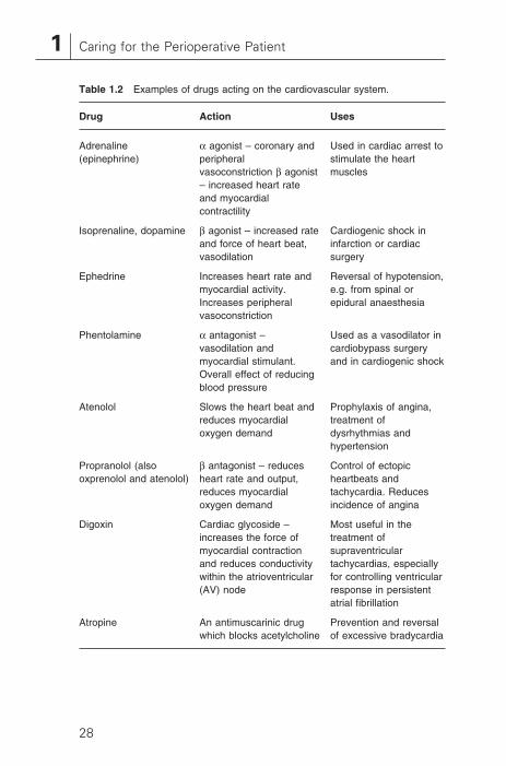
Pharmacology and b lood p ressure h omeostasis There are many chemicals that infl uence blood fl ow and blood vessel diameter and therefore have a direct action on blood pressure. Table 1.2 gives some examples of drugs that act on the cardiovascular system. See Chapter 3 on pharmacology for further discussion of such drugs.
Shock
Shock is a condition that arises from a failure of the circulatory system to deliver oxygen and nutrients to the tissues of the body, and to remove waste products. Before going into shock in detail it is important to understand the relationship between cardiac output, peripheral resistance and blood pressure. Control of blood circulation is through the interaction of blood volume (provided via cardiac output), blood vessel diameter (vasocon-striction – especially peripherally) and the pressure gradient that ‘ pushes ’ blood through the tissues (blood pressure).
Cardiac output is the volume of blood per minute ejected from the heart. The volume of blood ejected every beat is the stroke volume.
Cardiac output stroke volume heart rate ml beats pe
= ×= ×140 60 rr minute
ml per minute= 8400
This is the cardiac output for an average heart beating 60 times per minute with a stroke volume per ventricle of 70 ml (so 140 ml total).
Various factors affect stroke volume. For example, low venous return reduces the volume of blood refi lling the heart between beats (end - diastolic volume) and therefore reduces stroke volume. Cardiac contractility affects the percentage of blood ejected from the heart on every beat – a strong contraction will empty the heart more effi ciently than a weak contraction. A
1 Caring for the Perioperative Patient
28
Table 1.2 Examples of drugs acting on the cardiovascular system.
Drug Action Uses
Adrenaline (epinephrine)
α agonist – coronary and peripheral vasoconstriction β agonist – increased heart rate and myocardial contractility
Used in cardiac arrest to stimulate the heart muscles
Isoprenaline, dopamine β agonist – increased rate and force of heart beat, vasodilation
Cardiogenic shock in infarction or cardiac surgery
Ephedrine Increases heart rate and myocardial activity. Increases peripheral vasoconstriction
Reversal of hypotension, e.g. from spinal or epidural anaesthesia
Phentolamine α antagonist – vasodilation and myocardial stimulant. Overall effect of reducing blood pressure
Used as a vasodilator in cardiobypass surgery and in cardiogenic shock
Atenolol Slows the heart beat and reduces myocardial oxygen demand
Prophylaxis of angina, treatment of dysrhythmias and hypertension
Propranolol (also oxprenolol and atenolol)
β antagonist – reduces heart rate and output, reduces myocardial oxygen demand
Control of ectopic heartbeats and tachycardia. Reduces incidence of angina
Digoxin Cardiac glycoside – increases the force of myocardial contraction and reduces conductivity within the atrioventricular (AV) node
Most useful in the treatment of supraventricular tachycardias, especially for controlling ventricular response in persistent atrial fi brillation
Atropine An antimuscarinic drug which blocks acetylcholine
Prevention and reversal of excessive bradycardia

29
Perioperative Homeostasis 1
huge variety of factors such as autonomic nervous stimulation, hormones and drugs affect the heart rate.
Peripheral resistance refers to the tissue ’ s resistance to blood fl ow. The diameter of blood vessels directly infl uences the resis-tance to blood fl ow – narrow vessels conduct blood at a slower rate than wide vessels. Control of blood vessel diameter occurs at a local tissue level through the release of lactic acid and other metabolites of normal cellular function. These metabolites cause local vasodilatation and therefore reduce peripheral vasocon-striction. Control of peripheral resistance occurs centrally through neural and hormonal activity, in particular the sympa-thetic nervous system.
Blood pressure is also subject to many controls. The higher the pressure gradient the faster blood will fl ow. The difference between the heart contractions (systole) and the relaxation phase (diastole) produces a pressure gradient. In humans, sys-tolic pressure is normally around 120 mmHg and diastolic pres-sure is around 80 mmHg. The difference between these two measurements is the pulse pressure and it is this pressure that represents the pressure gradient. Pulse pressure is infl uenced by a combination of the contractility of the heart, the circulating volume and the peripheral resistance.
Shock, therefore, can be defi ned as acute circulatory failure leading to inadequate tissue perfusion, resulting in generalised cellular hypoxia and end - organ injury. It is caused by a disrup-tion to the cardiovascular system and inadequate compensation to maintain tissue perfusion (Jevon & Ewens 2002 ).
Shock can be classifi ed according to its three known causes:
� a fault of the heart, which is cardiogenic shock; � a fault of the vascular system, which is distributive shock; � a fault of fl uid regulation, which is hypovolaemic shock.
Keeping in mind the previous discussion on blood pressure regulation, it can be seen that hypotension and shock are there-fore caused by a problem with heart rate, stroke volume or peripheral resistance.
A clinical approach to shock (Table 1.3 ) identifi es the main clinical problems associated with shock that can be treated by medical interventions such as drugs or surgery:

1 Caring for the Perioperative Patient
30
Table 1.3 A clinical approach to shock.
Overall effect Physiological problem
Clinical focus
Failure of cardiac output
Heart rate Inappropriate heart rate
• Heart block/pacemaker • Hypotension • Bradycardia
Stroke volume
Inadequate fi lling time
• Tachycardia • Arrhythmias
Failure to receive blood
• Haemorrhage • Hypovolaemia • Dehydration • Inadequate fl uid intake • Excessive fl uid loss • Interstitial fl uid loss (e.g.
bowel surgery, pancreatitis)
• Infl ow obstruction • Mitral stenosis • Tamponade
Failure to eject blood
• Muscle dysfunction • Myocardial ischaemia or
fi brosis • Valvular/septal damage • Ventricular damage • Aortic regurgitation • Septal defects • Outfl ow obstruction • Pulmonary obstruction • Aortic/pulmonary stenosis
Failure of peripheral resistance
Inappropriate vasodilation
• Anaphylaxis • Shock • Sepsis
� low blood pressure, because of inadequate cardiac output or low peripheral resistance;
� low cardiac output, caused by a problem with heart rate or stroke volume;
� heart rate abnormalities – too fast (tachycardia) or too slow (bradycardia);

31
Perioperative Homeostasis 1
� stroke volume abnormalities, caused by failure to receive blood, failure to eject blood or inadequate volume;
� low peripheral vascular resistance, because of inappropriate vasodilation.
Hypovolaemic s hock During shock, the body protects itself from hypovolaemia by a series of refl ex mechanisms involving the cardiovascular and neurohormonal systems as described previously. The result is a decrease in cardiac output and increased peripheral resis-tance. The selective shunting of blood occurs to essential organs such as the brain, heart and kidneys, which are further pro-tected by autoregulatory refl exes. This state is ‘ compensated shock ’ and may occur with up to 20% of blood loss (approxi-mately 1 litre) (University of Pennsylvania 2006 ).
The clinical signs of compensated shock may be subtle: blood pressure may be normal; there is tachycardia, cold and clammy peripheries, decreased capillary refi ll; and a widened gap between core and peripheral temperature (Astiz et al. 1993 ).
As circulating volume decreases (1 – 2 litres blood loss) blood pressure begins to fall, resulting in increased peripheral vaso-constriction and tachycardia. Blood pressure may become unre-cordable and there are signs of end - organ failure (oliguria and confusion) following the loss of more than 40% (2 litres) of circulating volume. The drop in urinary output is a reliable way of identifying progressive loss of circulating volume.
Treatment of h ypovolaemic s hock Treatment of shock addresses the cause by replacing lost fl uids and supporting the body ’ s essential systems against the effects of hypovolaemia. Techniques and protocols are constantly being reviewed as new research evidence becomes available.
Replacement of lost fl uids by colloids and crystalloids is nor-mally a priority for treating shock. Progressively worsening shock necessitates monitoring of arterial and central venous pressure (CVP) to assess the effects of fl uid replacement. The end point of fl uid replacement will be: blood pressure within normal limits; urinary output of greater than 1 ml/kg; CVP of over 12 mmHg; and lactate readings of less than 2 mmol/litre

1 Caring for the Perioperative Patient
32
(University of Pennsylvania 2006 ). If the patient remains hypo-tensive after the volume replacement, then the problem lies with the cardiovascular system which must be supported through medical interventions. Noradrenaline (norepineph-rine) and low dosage vasopressin may increase stroke volume, which can be measured with a pulmonary artery catheter or oesophageal Doppler. Dobutamine may be useful at this point to increase the effi ciency of the heart pump.
The stroke volume, CVP, pulmonary capillary wedge pres-sure (PCWP) and venous oxygen saturation may guide fl uid resuscitation. Since over - transfusion of patients rarely occurs during shock, non - invasive monitors such as the oesophageal Doppler may provide more rapid and less dangerous measure-ment of stroke volume. Close monitoring is essential since the patient ’ s fl uid status may change as the body recovers from shock or as the effects of medical interventions progress (Pinsky & Payen 2004 )).
THE RESPIRATORY SYSTEM The respiratory system transports gases between the blood-stream and the outside air. Blood delivers oxygen to the tissues of the body, while carbon dioxide from tissue activity is returned to the lungs. The carbon dioxide waste leaves the body during exhalation.
Breathing is the process of moving air into and out of the lungs. An adult normally breathes from 14 to 20 times per minute, rising to 80 breaths per minute on effort and dropping to 8 or 10 breaths per minute at rest. A child ’ s rate of breathing at rest is faster than an adult ’ s at rest, and a newborn baby has a rate of about 40 breaths per minute. In adults the tidal volume (amount of air taken in a normal breath) is about 0.5 litres. The vital capacity (the maximum amount) is about 4.8 litres in an adult male.
The process of breathing comprises two phases, inspiration and expiration. The lungs themselves have no muscle tissue so the ribcage and the diaphragm control their movements.
The diaphragm is a large, dome - shaped muscle that lies just under the lungs, which fl attens when stimulated. This expands the volume of the thoracic cavity. The rib muscles also contract

33
Perioperative Homeostasis 1
on stimulation, pulling the ribcage up and out, also expanding the thoracic cavity. The increased volume of the thoracic cavity creates a partial vacuum which sucks air into the lungs. The diaphragm and rib muscles relax when the nervous stimulation ends, the thoracic cavity shrinks and exhalation occurs.
Conscious control and overriding of the respiratory centre alter the rhythm, for example when singing or whistling, or when holding the breath.
Structure
The respiratory system extends from the nose to the lungs and is divided into the upper and lower respiratory tracts. The upper respiratory tract consists of the nose and the pharynx, or throat. The lower respiratory tract includes the larynx, or voice box; the trachea, or windpipe, which splits into two main branches called bronchi; tiny branches of the bronchi called bronchioles; and fi nally the lungs. The nose, pharynx, larynx, trachea, bronchi and bronchioles conduct air to and from the lungs. The lungs interact with the cardiovascular system to deliver oxygen and remove carbon dioxide.
Nose and n asal c avity Capillaries in the nose and nasal cavity warm and humidify the air. Hairs and mucus inside the nasal cavity help to trap dust and other particles to protect the lungs. Stimulation of chemo-receptors inside the nose activates the olfactory nerve which eventually leads to the sensation of smell.
Pharynx The pharynx is a short, funnel - shaped tube about 13 cm long that links the nose and the larynx. The pharynx transports air to the larynx and is lined with a protective mucous membrane and ciliated cells to remove impurities. The pharynx also houses the tonsils, which are lymphatic tissues that contain white blood cells. The tonsils help to protect against upper respiratory tract infections. High in the rear wall of the pharynx are the adenoids. Located at the back of the pharynx on either side of the tongue are the palatine tonsils. The lingual tonsils are found at the base of the tongue. The tonsils can become swollen with infection

1 Caring for the Perioperative Patient
34
(tonsillitis), causing various symptoms associated with airway blockage and sepsis.
Viral infections such as the common cold, infl uenza, German measles (rubella), herpes and infectious mononucleosis cause pharyngitis, giving symptoms of a sore throat. Infection can also be caused by diphtherial, chlamydial, streptococcal and staphy-lococcal bacteria.
Larynx Air moves from the pharynx to the larynx and on to the trachea. The larynx is about 5 cm long and consists of several layers of cartilage. The Adam ’ s apple is a prominent bulge visible on the neck formed by a projection in the cartilage.
The larynx also produces sound, prevents food and fl uid from entering the trachea, and helps fi lter air. The presence of food or fl uid in the larynx produces a cough refl ex. If the cough refl ex does not work, a person can choke.
The larynx houses two pairs of vocal cords made of elastic connective tissue covered by folds of mucous membrane. One pair, the false vocal cords, narrows the glottis (the pharyngeal opening of the larynx) during swallowing. Below this and extending as far as the thyroid cartilage are the true vocal cords. Sound is created when this pair of cords vibrates as air passes through them.
Laryngitis, which often accompanies colds, is the larynx ’ s most common affl iction and can lead to voice loss. Other diseases include croup, diphtheria and cancer. Cancer is often treated by radiotherapy and surgery – partial or total laryngectomy.
Trachea The trachea extends from the larynx to the right and left primary bronchi in the lungs (Tortora 2008 ). The trachea is lined by cili-ated mucous membrane; the mucus traps tiny particles and the cilia move the mucus up and out of the respiratory tract. Rings of cartilage reinforce the trachea and prevent it from collapsing. If the airway blocks above the larynx, a tracheostomy may be performed to bypass the blockage and ease breathing.
35
Perioperative Homeostasis 1
Alternatively, the patient may need intubation using an endo-tracheal tube.
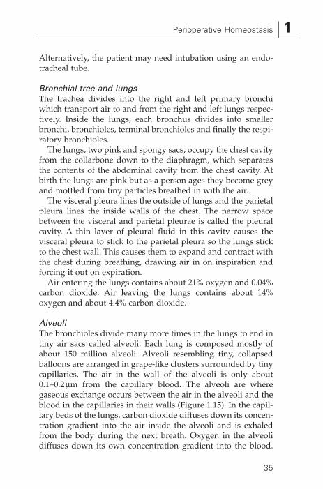
Bronchial t ree and l ungs The trachea divides into the right and left primary bronchi which transport air to and from the right and left lungs respec-tively. Inside the lungs, each bronchus divides into smaller bronchi, bronchioles, terminal bronchioles and fi nally the respi-ratory bronchioles.
The lungs, two pink and spongy sacs, occupy the chest cavity from the collarbone down to the diaphragm, which separates the contents of the abdominal cavity from the chest cavity. At birth the lungs are pink but as a person ages they become grey and mottled from tiny particles breathed in with the air.
The visceral pleura lines the outside of lungs and the parietal pleura lines the inside walls of the chest. The narrow space between the visceral and parietal pleurae is called the pleural cavity. A thin layer of pleural fl uid in this cavity causes the visceral pleura to stick to the parietal pleura so the lungs stick to the chest wall. This causes them to expand and contract with the chest during breathing, drawing air in on inspiration and forcing it out on expiration.
Air entering the lungs contains about 21% oxygen and 0.04% carbon dioxide. Air leaving the lungs contains about 14% oxygen and about 4.4% carbon dioxide.
Alveoli The bronchioles divide many more times in the lungs to end in tiny air sacs called alveoli. Each lung is composed mostly of about 150 million alveoli. Alveoli resembling tiny, collapsed balloons are arranged in grape - like clusters surrounded by tiny capillaries. The air in the wall of the alveoli is only about 0.1 − 0.2 μ m from the capillary blood. The alveoli are where gaseous exchange occurs between the air in the alveoli and the blood in the capillaries in their walls (Figure 1.15 ). In the capil-lary beds of the lungs, carbon dioxide diffuses down its concen-tration gradient into the air inside the alveoli and is exhaled from the body during the next breath. Oxygen in the alveoli diffuses down its own concentration gradient into the blood.

1 Caring for the Perioperative Patient
36
The oxygenated blood leaves the lungs, returns to the heart by the pulmonary arteries and then continues out of the heart into the body to restart the process (Figure 1.16 ).
The r ole of s urfactant Some of the cells forming the alveoli secrete a chemical called surfactant , which decreases surface tension of the fl uid in the alveoli. This reduces the attraction between water molecules and prevents the walls of the alveolus from collapsing.
Infant respiratory distress syndrome (IRDS) is caused by a defi ciency in surfactant. This syndrome is often seen in prema-
Fig. 1.15 Terminal bronchiole and alveoli.
Alveolarcluster
Bronchiole
Venule
Alveolus
Arteriole

37
Perioperative Homeostasis 1
ture babies who may be unable to produce adequate amounts of surfactant. The alveoli collapse on exhalation making reinfl a-tion diffi cult. Giving surfactant and hormones can stimulate the surfactant - producing cells.
Regulation of b reathing
Aerobic respiration is the process within cells in which nutrients and oxygen build the energy molecule adenosine triphosphate (ATP) through a process known as the Krebs ’ cycle. The overall effect of aerobic respiration is that body cells use oxygen to metabolise glucose, forming carbon dioxide as a waste product.
The oxygen and carbon dioxide concentrations in various parts of the body are measured as partial pressures – the pres-sure exerted by any one gas absorbed within a fl uid. For example, pO 2 is the partial pressure of oxygen in the blood – the pressure exerted by oxygen which contributes to the entire pres-sure of all the absorbed gases.
The rate and pattern of breathing is controlled by a cluster of nerve cells in the brain stem called the respiratory centre – a circuit of neurons in the base of the brain (the medulla oblon-gata and pons). Motor neurons from the respiratory control centre innervate the diaphragm and chest muscles. When stim-ulated, they contract and change the volume of the thoracic cavity. The respiratory control centre also receives input from many other neurons.
Fig. 1.16 Exchange of oxygen and carbon dioxide between the blood and lungs.
Inspired air Oxygenated blood Tissues
Expired air Deoxygenated blood Tissues
1 Caring for the Perioperative Patient
38
Nerves from the higher brain centres controlling emotion stimulate or depress the respiratory control centre and cause changes in the rate and depth of breathing when the person is excited or relaxed. Sensory neurons such as proprioceptors from the joints and chemoreceptors from the arteries also inter-act with the respiratory control centre. On exercising, action potentials from proprioceptors in joints stimulate the respira-tory control centre and increase the rate of breathing.
However, the levels of carbon dioxide in the blood and cere-brospinal fl uid (CSF) are the major factors regulating both the rate and depth of breathing (Figure 1.17 ). If carbon dioxide levels in the blood increase, the carbon dioxide will diffuse into the CSF. Central chemoreceptors in the medulla oblongata of the brain stimulate the respiratory control centre, increasing the rate and depth of breathing and so reducing pCO 2 .
Hypercapnia is caused by high carbon dioxide concentration in blood. The carbonic acid equation moves to the right and the hydrogen ion concentration increases (see page 10 ). The increase in hydrogen ion concentration increases the acidity of blood and CSF, and can cause respiratory acidosis. The respiratory control centre will respond by increasing the speed and depth of breath-ing, causing carbon dioxide to diffuse from blood to the lungs more rapidly.
Hypocapnia is caused by low partial pressure of carbon dioxide in blood. In this case, the carbonic acid equation moves to the left, and hydrogen ion concentration will decrease as a new equilibrium is reached (see page 10 ). The decrease in blood hydrogen ion concentration causes respiratory alkalosis. The respiratory control centre reacts by decreasing the respiratory rate, causing carbon dioxide from cellular respiration to build up in the bloodstream. The carbonic acid equation moves to the right again, reversing the respiratory alkalosis. The respiratory control centre constantly adjusts the depth and rate of breathing to maintain the proper balance. Although some degree of con-scious control can be exerted over the amount of air inhaled, the most important factors controlling breathing are the carbon dioxide and hydrogen ion concentrations of the blood and CSF (Jardins 2007 ).

39
Perioperative Homeostasis 1
Fig
. 1.
17
Reg
ulat
ion
of b
reat
hing
and
res
pira
tion
by c
arbo
n di
oxid
e co
ncen
trat
ion
in t
he b
lood
: (a
) de
crea
sed;
(b)
incr
ease
d.
Dec
reas
ed c
arbo
n di
oxid
eco
ncen
trat
ion
in b
lood
Per
iphe
ral
chem
orec
epto
rsge
nera
te fe
wer
ne
rve
impu
lses
Dec
reas
ed c
arbo
n di
oxid
eco
ncen
trat
ion
in C
SF
Dec
reas
ed h
ydro
gen
ion
conc
entr
atio
n in
CS
F
Cen
tral
che
mor
ecep
tors
gene
rate
few
er n
erve
impu
lses
Res
pira
tory
con
trol
cent
re g
ener
ates
few
er n
erve
impu
lses
Dia
phra
gm a
nd o
ther
res
pira
tory
mus
cles
con
trac
t slo
wer
Incr
ease
d ca
rbon
dio
xide
conc
entr
atio
n in
blo
od
Incr
ease
d ca
rbon
dio
xide
conc
entr
atio
n in
blo
odP
erip
hera
lch
emor
ecep
tors
gene
rate
mor
e ne
rve
impu
lses
Incr
ease
d ca
rbon
dio
xide
conc
entr
atio
n in
CS
F
Incr
ease
d hy
drog
en io
nco
ncen
trat
ion
in C
SF
Cen
tral
che
mor
ecep
tors
gene
rate
mor
e ne
rve
impu
lses
Res
pira
tory
con
trol
cent
re g
ener
ates
mor
e ne
rve
impu
lses
Dia
phra
gm a
nd o
ther
res
pira
tory
mus
cles
con
trac
t fas
ter
and
stro
nger
Dec
reas
ed c
arbo
n di
oxid
eco
ncen
trat
ion
in b
lood
(b)
(a)

1 Caring for the Perioperative Patient
40
Diseases and d isorders
There are many diseases and disorders of the respiratory system which can affect any part of the respiratory tract.
Infection by a huge variety of cold viruses is one of the most common ailments affecting the membranes of the nasal pas-sages and pharynx. The immune system fi ghts back by increas-ing blood fl ow to the area, bringing numerous white blood cells to the scene. This infl ammatory reaction causes the membranes to swell and increase the secretion of mucus, resulting in the stuffy and runny nose associated with colds. The infection can spread to the lower respiratory tract, to the middle ear or to the sinuses where it causes sinusitis.
The respiratory system is prone to allergic reactions, such as hay fever and asthma, which are caused when the immune system is stimulated by pollen, dust or other irritants. A runny nose, watery eyes and sneezing characterise hay fever. In asthma, temporary constriction and infl ammation of the bronchi and bronchioles causes diffi culty in breathing. An asthma attack is typically mild in otherwise healthy patients, but can be severe enough to be life - threatening when anaesthesia and surgery, or other concurrent conditions compromise the patient.
Laryngitis is an infl ammation of the larynx resulting from causes such as viral infections, trauma caused by endotracheal tubes, irritants such as cigarette smoke or overuse of the voice. Laryngitis may result in hoarseness or whispering until the swelling subsides. Bronchitis, caused by viral or bacterial infec-tion or by irritating chemicals, is an infl ammation of the mem-branes that line the bronchi or bronchioles. Infections with bacteria or viruses can lead to pneumonia, a potentially serious condition of the lungs where fl uid and infl ammation prevent the fl ow of oxygen and carbon dioxide between the capillaries and the air in the alveoli.
Tuberculosis bacteria attack the lungs and sometimes other body tissues as well. Untreated infections in the lungs destroy lung tissue. In the past, antibiotics have controlled tuberculosis, but recently, new antibiotic - resistant strains of the tuberculosis bacterium have evolved. These new strains now pose a signifi -cant public health problem. Immunocompromised patients are particularly prone to tuberculosis.

41
Perioperative Homeostasis 1
In emphysema the alveolar bundles coalesce, resulting in an overall smaller surface area for the exchange of gases. Weakened bronchioles collapse on exhalation, trapping air in the alveoli. This eventually hinders the exchange of oxygen and carbon dioxide with the circulatory system, leading to hypoxia and diffi culty in breathing. Emphysema is a non - contiguous disease that can result from various causes including a genetic tendency to the condition, smog, cigarette smoke or infection.
Exposure to cancer - causing agents, such as tobacco smoke, asbestos or radiation, can lead to lung cancer in individuals with a genetic inclination to the disease. Cancerous tumours can start in the bronchi, bronchioles or in the alveolar lung tissue. Treatments are more effective on early detection of lung cancer, before it has spread to other parts of the body, and provide a good prognosis for full recovery. The prognosis is poor if the cancer has had the opportunity to spread.
Respiratory distress syndrome (RDS) is the name for a cluster of symptoms that suggest severe failure of the lungs. In infants, RDS is termed infant respiratory distress syndrome (IRDS). As mentioned above, IRDS is commonly found in premature infants when the alveoli fail to expand fully during inhalation. Expansion of the alveoli requires surfactant, but in many pre-mature infants the alveoli cannot produce this substance. Treatment of IRDS is by artifi cial ventilation and giving surfac-tant until the alveoli begin producing surfactant on their own. Severe damage to the lungs caused by, for example, trauma, poisonous gases or as a response to infl ammation in the lungs, causes acute (adult) respiratory distress syndrome (ARDS). ARDS is a life - threatening condition with a survival rate of about 50%.
TRAUMA AND WOUND HEALING Anaesthesia and surgery cause stress to the body, so it comes as no surprise that the perioperative patient presents many of the responses found with naturally occurring stressors. This section describes the body ’ s metabolic response to anaesthesia and surgery and then, the process of wound healing.
Stressors such as surgery and anaesthesia, as well as others such as injury, burns, vascular occlusion, dehydration, starva-

1 Caring for the Perioperative Patient
42
tion, sepsis, acute medical illness or psychological stress, may launch the metabolic response to trauma. The body responds locally by an infl ammatory response designed to protect tissues from further damage and to encourage repair. The purpose of the somatic response is to conserve fl uid and provide energy for tissue repair.
Two phases characterise the somatic response. Initially the body produces an acute catabolic reaction where the patient is in shock, characterised by:
� depression of enzymatic activity; � decreased oxygen consumption; � low cardiac output; � low core temperature; � lactic acidosis.
An anabolic phase follows where fat and protein stores are regained and weight increases, characterised by:
� increased cardiac output; � increased oxygen consumption; � increased glucose production; � lactic acid may be normal.
The state of normal homeostasis returns as the triggers resolve and the body returns to its normal metabolic balance (Tortora 2008 )).
The form of the metabolic response depends chiefl y on the degree of trauma suffered. Other contributing factors include the infl uence of drugs, sepsis, underlying systemic disease, underlying nutritional state and effi cacy of the medical interventions. Although the metabolic response is protective, it can become harmful if excessive or prolonged. The aim of medical interventions is to revive body systems, control pain and temperature, and provide acceptable fl uid and nutrition.
Factors s tarting the m etabolic r esponse
The factors that start the metabolic response include hypovo-laemia, factors exuded by the wound, hormonal responses, sepsis and the infl ammatory response.

43
Perioperative Homeostasis 1
Hypovolaemia is common in major surgery but the body ’ s own response and the early use of fl uid replacement therapy may signifi cantly reduce the metabolic response. Pain and anxiety can also initiate a hormonal response, but this can be adjusted by analgesia so that a metabolic response is not stimulated.
Tissue injury activates two specifi c responses, infl ammatory (humoral) and cellular. Products of these responses play a role in organ dysfunction. For example, the infl ammatory mediators of injury have been implicated in membrane dysfunction, leading to various conditions affecting every organ of the body.
The infl ammatory response is a complex collection of reac-tions involving macrophages, polymorphonuclear leucocytes and phagocytic cells, such as neutrophils and eosinophils. Normal phagocytosis (engulfi ng of foreign bodies by phago-cytes) is one of the primary activations of the metabolic response. This results in responses such as neutrophil aggregation, and secretion of histamine and serotonin, which may increase vas-cular permeability and vasodilation. A combination of these reactions results in the infl ammatory response.
The action and release of adrenaline (epinephrine), noradren-aline (norepinephrine), cortisol and glucagon are increased, while other hormones are decreased during trauma. The hypothalamus has a major role in coordinating the stress response through endocrine actions by the pituitary and the sympathetic and parasympathetic nervous systems. The pituitary gland responds to trauma by increasing adrenocorti-cotrophic hormone (ACTH), prolactin and growth hormone levels.
Pain receptors, osmoreceptors, baroreceptors and chemore-ceptors stimulate the hypothalamus to induce sympathetic nerve activity. Stimulation of the pain receptors results in the secretion of endogenous opiates, which adapt the response to pain.
Hypotension, hypovolaemia and hyponatraemia stimulate the anterior pituitary to secrete ACTH. ACTH stimulates the secretion of antidiuretic hormone (ADH) from the anterior hypothalamus, aldosterone from the adrenal cortex and renin from the juxtaglomerular apparatus of the kidney. This has the

1 Caring for the Perioperative Patient
44
overall effect of increasing water reabsorbtion and thereby increasing blood volume.
Reactions to changes in glucose concentration include the release of insulin from the cells of the pancreas, while high amino acid levels stimulate the release of glucagon from the pancreatic cells.
Result of the m etabolic r esponse
The stress of major surgery can lead to the initiation of the metabolic response. Many of the interventions carried out on the perioperative patient are aimed at moderating the metabolic response, which if untreated may rapidly prove fatal.
As can be seen from the above, the metabolic response results in systemic infl ammatory responses which increase the activity of the cardiovascular system, refl ected as tachycardia, widened pulse pressure and a greater cardiac output. As metabolic rate increases, there is an increase in oxygen consumption, increased protein catabolism and hyperglycaemia.
The resting energy expenditure can rise to more than 20% above normal, if the patient is well enough to respond. In an inadequate response, oxygen consumption may fall and endo-toxins and anoxia may injure cells and limit their ability to utilise oxygen. Cellular injury, impaired hepatic gluconeogen-esis and lack of oxygen may result in rapid deterioration of processes requiring energy. As a result of the ineffi cient energy - making process, lactate is produced, causing a severe metabolic acidosis.
The oliguria, which often follows major surgery, is a conse-quence of the release of ADH and aldosterone. As well as pro-moting the reabsorption of water, aldosterone also releases large quantities of intracellular potassium into the extracellular fl uid, possibly causing a signifi cant rise in serum potassium, especially if renal function is impaired. Retention of sodium and bicarbonate may contribute to metabolic alkalosis with impair-ment of the delivery of oxygen to the tissues.
Critically ill postoperative patients may develop a glucose intolerance which resembles that found in pregnancy and in diabetic patients. This is as a result of both increased mobilisa-tion and decreased uptake of glucose by the tissues. The turn-

45
Perioperative Homeostasis 1
over of glucose is increased and the serum glucose is higher than normal.
The glucose level following surgery should therefore be care-fully monitored since hyperglycaemia may exacerbate ventila-tory insuffi ciency and may provoke an osmotic diuresis. The optimum blood glucose level is between 4 and 10 mmol/litre. Control of blood glucose is best achieved by titration with intravenous insulin, based on a sliding scale. However, because of the degree of insulin resistance associated with trauma, the quantities required may be considerably higher than normal.
The principal source of energy following trauma is adipose tissue and as much as 200 – 500 g of fat may be broken down daily after major surgery. High levels of ketones are produced as free fatty acids are broken down to provide energy. Because of the possible problems with glucose metabolism, nutritional support of traumatised patients requires a mixture of fat and carbohydrate.
The intake of protein by a healthy adult is between 80 and 120 g of protein, or around 1 – 2 g protein/kg/day. Lack of protein intake leads to the breakdown of skeletal muscle in order to produce essential amino acids. After major surgery, or with sepsis, as much as 20 g/day of urea nitrogen from protein breakdown may be excreted in the urine. A severely ill patient may lose over 600 g of muscle mass per day, leading to marked muscle wasting. Depletion of amino acids also results in atrophy of the intestinal mucosa and failure of the mucosal antibacterial barrier. This may lead to systemic infection and multisystem failure after severe trauma. This is normally fatal in patients who lose more than 40% of body protein because of failing immunocompetence. The protein intake can be improved dra-matically by parenteral or enteral feeding, as long as adequate liver function is present.
Survival after trauma such as surgery depends on a balance between the extent of cellular damage, the effi cacy of the meta-bolic response and the effectiveness of supporting treatment. Hypovolaemia is a major initiating trigger for the metabolic sequence; therefore, adequate fl uid resuscitation to shut off the hypovolaemic stimulus is important during and following

1 Caring for the Perioperative Patient
46
surgery. Fear and pain, tissue injury, hypoxia and toxins from infection add to the initiating factor of hypovolaemia and so it is important that these are also moderated by perioperative interventions in order to reduce the metabolic response.
Patient support during perioperative care should be aimed at reducing the factors triggering the metabolic response. During surgery, important aspects of patient care include adequate fl uid volume, maintenance of oxygen delivery to the tissues, careful handling of tissues and removal of pus or devitalised tissue, combined with control of infection, respiratory and nutritional support to aid defence mechanisms.
Wound h ealing
Wound healing is the process of tissue repair. It involves the interaction of epithelial, endothelial and infl ammatory cells, platelets and fi broblasts which are activated and act together in order to repair the damaged tissue. The overall process can be divided into the three overlapping phases of infl ammation, pro-liferation and maturation (Figure 1.18 ) (Dealey 2005 ).
Infl ammatory p hase ( d ay 0 – 5) Tissue healing follows essentially the same process regardless of the tissue involved or the type of injury sustained, although
Fig. 1.18 Phases of wound repair: infl ammation, proliferation and maturation.
Inflammation
0.5 1 3 10Days
30 100
Proliferation
Maturation
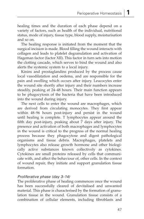
47
Perioperative Homeostasis 1
healing times and the duration of each phase depend on a variety of factors, such as health of the individual, nutritional status, mode of injury, tissue type, blood supply, moisturisation and so on.
The healing response is initiated from the moment that the surgical incision is made. Blood fi lling the wound interacts with collagen and leads to platelet degranulation and activation of Hageman factor (factor XII). This factor in turn sets into motion the clotting cascade, which serves to bind the wound and also alerts the systemic system to a local injury.
Kinins and prostaglandins produced by the process cause local vasodilatation and oedema, and are responsible for the pain and swelling which occurs after injury. Leucocytes enter the wound site shortly after injury and their numbers increase steadily, peaking at 24 – 48 hours. Their main function appears to be phagocytosis of the bacteria that have been introduced into the wound during injury.
The next cells to enter the wound are macrophages, which are derived from circulating monocytes. They fi rst appear within 48 – 96 hours post - injury and persist in the wound until healing is complete. T lymphocytes appear around the fi fth day post - injury, peaking about 7 days after injury. The presence and activation of both macrophages and lymphocytes in the wound is critical to the progress of the normal healing process because they phagocytose and digest pathological organisms and tissue debris. Macrophages, platelets and lymphocytes also release growth hormone and other biologi-cally active substances known collectively as cytokines. Cytokines are small proteins released by cells that communi-cate with, and affect the behaviour of, other cells. In the context of wound repair, they initiate and support granulation tissue formation.
Proliferative p hase ( d ay 3 – 14) The proliferative phase of healing commences once the wound has been successfully cleared of devitalised and unwanted material. This phase is characterised by the formation of granu-lation tissue in the wound. Granulation tissue consists of a combination of cellular elements, including fi broblasts and

1 Caring for the Perioperative Patient
48
infl ammatory cells, along with new capillaries embedded in a loose extracellular matrix of collagen, fi bronectin and hyal-uronic acid. In a process of fi broblast proliferation and syn-thetic activity known as fi broplasia, fi broblasts are induced to proliferate and are attracted into the wound by cytokines. Fibroblasts are responsible for tissue reconstruction through the production of structural proteins, in particular the glyco-protein collagen, which forms the main constituent of the wound matrix and which imparts tensile strength to the scar. The collagen forms into an organised matrix along the wound ’ s stress lines.
Wound revascularisation also occurs at this time. In response to a variety of local mediators, capillary buds sprout from venules, form capillary loops, connect to the bloodstream and then join together to form a capillary plexus.
Re - epithelialisation of the wound surface begins within a couple of hours of the injury. Epithelial cells from the periphery of the wound begin to migrate under the scab and over the underlying viable connective tissue, fi lling the wound. Once the defect is bridged the migrating epithelial cells proliferate through mitosis and the surface layer eventually becomes kera-tinised. Re - epithelialisation is normally complete in less than 48 hours in approximated surgical wounds, but will take longer in larger wounds where there is tissue loss. Epithelialisation can also take place in the absence of fi broplasia and granulation tissue formation in wounds (such as skin graft donor sites) where only the epithelium is damaged. Again, the mediators for re - epithelialisation include a variety of cytokines and other local cellular products.
Maturation p hase ( d ay 7 to 1 y ear) Almost as soon as the extracellular matrix is laid down it becomes cross - linked and aggregated into fi brillar bundles, which gradually provide the healing tissue with increasing stiff-ness and tensile strength through collagen fi brogenesis. The wound develops 20% of its fi nal strength after 3 weeks, with the maximum breaking strength of the scar reaching 70% of that of the intact skin.

49
Perioperative Homeostasis 1
This gradual gain in tensile strength is due not only to con-tinuing collagen deposition, but also to collagen remodelling. Collagen synthesis and catabolism leads to the formation of larger collagen bundles and increased crosslinking. As this occurs, a thin pale scar is formed from collagen which is rela-tively avascular and acellular.
Wounds healed by primary intention are those which require minimal granulisation tissue and minimal loss of wound volume. Large and complex wounds, associated with signifi -cant tissue loss, heal by secondary intention. Granulation tissue gradually fi lls the defect and epithelialisation proceeds slowly from the wound edges. The wound area starts to contract rapidly through the inward movement of the uninjured skin edges caused by an interaction between fi broblast locomotion and collagen reorganisation. The result of the healing process is a scar that helps to restore tissue continuity, strength and function.
REFERENCES Abraham , J. ( 2008 ) Acupressure and acupuncture in preventing and
managing postoperative nausea and vomiting in adults . Journal of Perioperative Practice 18 ( 12 ), 543 – 551 .
Astiz , M.E. , Rackow , E.C. & Weil , M.H. ( 1993 ) Pathophysiology and treatment of circulatory shock . Critical Care Clinics 9 ( 2 ), 183 – 203 .
Clancy , J. , McVicar , A.J. & Baird , N. ( 2002 ) Perioperative Practice: Fundamentals of Homeostasis . Routledge , London .
Dealey , C. ( 2005 ) The Care of Wounds: A Guide for Nurses . Blackwell , Oxford .
Hatfi eld , A. & Tronson , M. ( 2008 ) The Complete Recovery Book , 4th edn . Oxford University Press , Oxford .
Heitz , U. & Horne , M. ( 2004 ) Pocket Guide to Fluid, Electrolyte, and Acid - Base Balance . Mosby , St Louis .
Jardins , T. ( 2007 ) Cardiopulmonary Anatomy & Physiology , 4th edn . Delmar , New York .
Jevon , P. & Ewens , B. (eds) ( 2002 ) Monitoring the Critically Ill Patient . Blackwell Science , Oxford .
Pinsky , M. & Payen , D. ( 2004 ) Functional Hemodynamic Monitoring . Springer - Verlag , New York .
Saladin , K.S. ( 2009 ) Anatomy and Physiology: The Unity of Form and Function , 3rd edn . William C Brown , New York .

1 Caring for the Perioperative Patient
50
Tortora , G.J. ( 2008 ) Principles of Anatomy and Physiology , 12th edn . Wiley , Oxford .
University of Pennsylvania ( 2006 ) Critical Care Medicine Tutorials: Cardiovascular System . www.ccmtutorials.com/cvs/index.htm (accessed 25 January 2009).
Watson , R. & Fawcett , T.N. ( 2003 ) Pathophysiology, Homeostasis and Nursing . Routledge , London .
Waugh , A. & Grant , A. ( 2004 ) Ross and Wilson Anatomy and Physiology in Health and Illness , 9th edn . Churchill Livingstone , New York .


![Budget2015LoulayrapportduCE [Mode de compatibilité]etab.ac-poitiers.fr/coll-loulay/IMG/pdf/budget2015loulayrapportduce_mode_de... · Présentation de l’EPLE ... de mainte nir le](https://img.pdfslide.tips/doc/110x75/5e41ca04d5d9e5454f1ed622/budget2015loulayrapportduce-mode-de-compatibilitetabac-prsentation-de-laeple.jpg)