Embed Size (px)
Citation preview
Kupczyńska et al. Acta Vet Scand (2015) 57:82 DOI 10.1186/s13028-015-0173-4
RESEARCH
Coronary arteries of the European bison (Bison bonasus)Marta Kupczyńska1, Karolina Barszcz1, Katarzyna Olbrych1, Michał Polguj2*, Grzegorz Wysiadecki3, Mirosław Topol3 and Joanna Klećkowska‑Nawrot4
Abstract
Background: The European bison (Bison bonasus) is an endangered species. More information on its anatomy is needed as only few studies have been published. This study is the first report on the morphology of the coronary ves‑sels. Given the anatomical similarity between the European bison and other ruminants, the results of this study can be applied to other species, including endangered ones.
Results: The study was conducted on 70 hearts of European bisons of both sexes, aged 5–20 years, with an aver‑age body weight of 449 kg. A distinct view of subepicardial arterial vessels was obtained by filling them with dyed synthetic latex (LBS 3060) and Plastogen G. There was a division of the common trunk of the left coronary artery into the interventricular paraconal branch and the left circumflex branch in 63 individuals (90 %). In five individuals (7.1 %), the presence of a third vessel, which was a branch of the interventricular septum, was observed. There was a lack of a common trunk in two individuals (2.9 %). Ramifications of the interventricular paraconal branch to the wall of the left ventricle were significantly larger than those to the wall of the right ventricle. In 17 individuals (24.3 %), the right coronary artery extended into the subsinuosal interventricular branch.
Conclusion: The blood supply to the heart in bisons is provided by the left and right coronary arteries. In all the stud‑ied specimens, the left coronary artery was better developed than the right coronary artery.
Keywords: Coronary arteries, Heart vascularization, European bison
© 2015 Kupczyńska et al. This article is distributed under the terms of the Creative Commons Attribution 4.0 International License (http://creativecommons.org/licenses/by/4.0/), which permits unrestricted use, distribution, and reproduction in any medium, provided you give appropriate credit to the original author(s) and the source, provide a link to the Creative Commons license, and indicate if changes were made. The Creative Commons Public Domain Dedication waiver (http://creativecommons.org/publicdomain/zero/1.0/) applies to the data made available in this article, unless otherwise stated.
BackgroundCoronary vessels are well developed in mammals. They supply the myocardium with oxygen and nutritive ele-ments. In humans, coronary arteries receive approxi-mately 15 % of the total arterial blood. It is believed that this percentage is higher in animals and mainly depends on the individual’s health [1]. Typically, the mammalian heart is supplied with blood by the left coronary artery [arteria (a.) coronaria sinistra] and the right coronary artery (a. coronaria dextra) [1–3]. In humans, individu-als with just a single coronary artery (SCA) have been reported [4]. The clinical significance of this variation is not clear but some autopsy studies indicate a possible link between SCA and sudden cardiac death. Turkmen
et al. [4] based their research on the analysis of angio-graphic studies and determined the incidence of SCA to be 0.03 % (67 cases out of 215,140 individuals). Anoma-lies of one or both coronary ostia are present in approxi-mately 0.6 % of humans [5]. In veterinary medicine, a similar variation has been observed in the lesser chin-chilla (Chinchilla lanigera) [6].
Coronary vessels have been studied in a wide range of animals including Syrian hamster (Mesocricetus aura-tus) [7], guinea pig (Cavia porcellus) [8], the Angora rab-bit [9], domestic dog (Canis lupus f. domestica) [10, 11], domestic cat [12, 13], donkey (Equus asinus) [14, 15], Angora and Akkamaran goats [16], roe deer (Capreo-lus capreolus) [17], Bactrian camel (Camelus bactri-anus) [18], one-humped camel (Camelus dromedarius) [19], ringed seal (Pusa hispida) [20], porcupine (Hystrix cristata) [21, 22], shrew (Suncus murinus) [23], crab-eating macaque (Macaca fascicularis) [24, 25], grivet
Open Access
Acta Veterinaria Scandinavica
*Correspondence: [email protected] 2 Department of Angiology, Interfaculty Chair of Anatomy and Histology, Medical University of Łódź, Narutowicza 60, 90‑136 Lodz, PolandFull list of author information is available at the end of the article
Page 2 of 7Kupczyńska et al. Acta Vet Scand (2015) 57:82
(Cercopithecus aethiops) [24] and rattlesnake (Crotalus durissus) [26]. There are also studies that refer to birds such as ostriches (Struthio camelus) [27] and chickens (Gallus gallus domesticus) [28]. However, the available literature provides no details on the topography of coro-nary arteries and their ramifications in the bison.
The aim of this study was therefore to investigate the morphology of the subepicardial vascularity of the Euro-pean bison (Bison bonasus).
MethodsThe study was carried out on 70 hearts of European bisons of both sexes, aged 5–20 years, with an average body weight of 449 kg. The individuals examined were culled legally from the population of bisons in Białowieża Forest, Poland. The study was conducted with the per-mission of the Ministry of the Environment and the Gen-eral Director for Environmental Protection in Poland.
The tissue surrounding the left and right coronary arteries was dissected in order to obtain access to the vessels.
Sixty-three hearts were dyed using synthetic LBS 3060 latex (Synthos Dwory Sp. z o.o, Poland). Those hearts were placed in a 10 % formalin for 6 weeks. The trunk of the left and right coronary artery and their ramifications were prepared [17, 29].
Corrosion casts were obtained from seven hearts using the multistage process to evaluate blood vessels. First, a 0.9 % NaCl solution was injected into the coronary arter-ies to flush out clots. Next, 20 ml of 3 % glutaraldehyde solution in a pH 7.4 cacodylate buffer was injected into the coronary arteries. The coronary arteries were then filled with Plastogen G (Plasto-Schmidt, Speyer, Ger-many) stained with green or red pigment and the heart was placed in water at 20 °C for 24 h to harden the resin. After hardening, the specimen was placed in a 40 % KOH solution at 50 °C for approximately 24 h to dissolve the organic tissue. The remnants of the dissolved tissue were removed from the specimen by continuous flushing with water for 38 h. The specimen was cleaned by a fast wash with warm water and a small amount of standard wash-ing liquid, followed by a final flush with distilled water. The cast was later dried using airflow at room tempera-ture for 2 days. The method was used with success in our previous studies [30, 31].
The specimens were examined morphologically using the ECLERIS (HALOLUX 150) surgical microscope with an integrated video channel. The diameters of the ves-sels were measured using software adapted for the met-ric analysis of images (AxioVision Rel. 4.7, Carl Zeiss MicroImaging GmbH, Jena, Germany). This software enables the visualization, archiving, processing and anal-ysis of images, with particular emphasis on measurement
functions. The terminology used in the manuscript is in accordance with prevailing veterinary nomenclature [32].
ResultsNone of the animals included in the study had any patho-logical changes in the thoracic cavity.
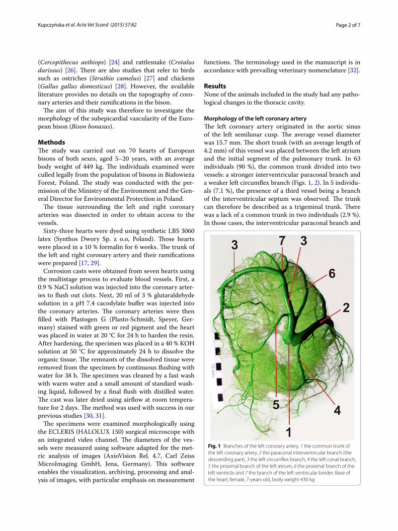
Morphology of the left coronary arteryThe left coronary artery originated in the aortic sinus of the left semilunar cusp. The average vessel diameter was 15.7 mm. The short trunk (with an average length of 4.2 mm) of this vessel was placed between the left atrium and the initial segment of the pulmonary trunk. In 63 individuals (90 %), the common trunk divided into two vessels: a stronger interventricular paraconal branch and a weaker left circumflex branch (Figs. 1, 2). In 5 individu-als (7.1 %), the presence of a third vessel being a branch of the interventricular septum was observed. The trunk can therefore be described as a trigeminal trunk. There was a lack of a common trunk in two individuals (2.9 %). In those cases, the interventricular paraconal branch and
Fig. 1 Branches of the left coronary artery. 1 the common trunk of the left coronary artery, 2 the paraconal interventricular branch (the descending part), 3 the left circumflex branch, 4 the left conal branch, 5 the proximal branch of the left atrium, 6 the proximal branch of the left ventricle and 7 the branch of the left ventricular border. Base of the heart; female, 7‑years‑old, body weight 430 kg
Page 3 of 7Kupczyńska et al. Acta Vet Scand (2015) 57:82
the left circumflex branch divided individually at the level of the aortic sinus of the left semilunar cusp.
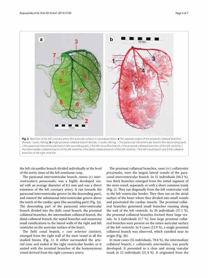
The paraconal interventricular branch, ramus (r.) inter-ventricularis paraconalis, was a highly developed ves-sel with an average diameter of 8.5 mm and was a direct extension of the left coronary artery. It ran towards the paraconal interventricular groove (in the descending part), and entered the subsinuosal interventricular groove above the notch of the cardiac apex (the ascending part) (Fig. 2a). The descending part of the paraconal interventricular branch divided into the (left) conal branch, the proximal collateral branches, the intermediate collateral branch, the distal collateral branch, the septal branches and numerous small ramifications to the lateral walls of the right and left ventricles on the auricular surface of the heart:.
The (left) conal branch, r. coni arteriosi (sinister), emerged from the right wall of the stem vessel in all the studied bisons (Fig. 1). It either surrounded the arte-rial cone and ended at the right ventricular border, or it united with the terminal branches of the homonymous vessel derived from the right coronary artery.
The proximal collateral branches, rami (rr.) collaterales proximales, were the largest lateral vessels of the para-conal interventricular branch. In 31 individuals (44.3 %), two thick branches emerged from the initial segment of the stem vessel, separately or with a short common trunk (Fig. 2). They ran diagonally from the left ventricular wall to the left ventricular border. They then ran on the atrial surface of the heart where they divided into small vessels and penetrated the cardiac muscle. The proximal collat-eral branches generated small branches running along the wall of the left ventricle. In 26 individuals (37.1 %), the proximal collateral branches formed three large ves-sels. In 4 individuals (5.7 %), four large proximal collat-eral branches were present on the entire auricular surface of the left ventricle. In 9 cases (12.9 %), a single proximal collateral branch was observed, which ramified near its origin (Fig. 2b).
In most cases (55 individuals, 78.6 %), the intermediate collateral branch, r. collateralis intermedius, was poorly developed. It presented as a clear and strong vascular trunk in 15 individuals (21.4 %). It originated from the
Fig. 2 Branches of the left coronary artery (the auricular surface) in a European bison. a The separate origins of the proximal collateral branches (female, 7 years, 430 kg), b single proximal collateral branch (female, 15 years, 450 kg). 1 The paraconal interventricular branch (the descending part), 2 the paraconal interventricular branch (the ascending part), 3 the left circumflex branch, 4 the proximal collateral branches of the left ventricle, 5 the intermediate collateral branch of the left ventricle, 6 the distal collateral branch of the left ventricle, 7 the left conal branch and 8 the collateral branches of the right ventricle

Page 4 of 7Kupczyńska et al. Acta Vet Scand (2015) 57:82
left wall of the paraconal interventricular branch, approx-imately half-way along the paraconal interventricular groove and ran along the wall of the left ventricle.
The distal collateral branch, r. collateralis distalis, ran from the left side of the stem vessel. Its ramifications reached the left ventricular border and supplied the walls of the left ventricle close to the cardiac apex.
There were numerous septal branches, rr. septales, along the entire course of the paraconal interventricu-lar branch. They penetrated the interventricular septum where they gave off smaller branches.
Apart from the aforementioned thick vascular trunks, there were numerous small right ventricular branches, rr. ventriculi dextri and left ventricular branches, rr. ventric-uli sinistri, which supplied the walls of both ventricles.
As described above, the ascending part, pars ascend-ens of the paraconal interventricular branch was a direct extension of the paraconal interventricular branch on the atrial surface of the heart. It proceeded dorsally from the notch of the cardiac apex in the subsinuosal interven-tricular groove. It gave off ramifications to lateral walls of both ventricles and the interventricular septum.
The left circumflex branch, r. circumflexus sinister, was the weakest ramification of the left coronary artery. It had a diameter of 5.7 mm measured in its proximal segment.
The circumflex branch initially ran in the coronary groove on the cardiac auricular surface, then passed the left ventricular border and reached the atrial surface of the heart. Most frequently (53 individuals, 75.7 %), it extended in the subsinuosal interventricular groove into the subsinuosal interventricular branch. Along its course, it gave off some small branches to the lateral wall of the left and right ventricle, and penetrated the cardiac mus-cle in the middle of the groove. In the remaining bisons (17 individuals, 24.3 %), the left circumflex branch ended with small ramifications. In those cases, the subsinuosal interventricular branch was supplied by the right coro-nary artery and ran in the subsinuosal interventricular groove. The circumflex branch was surrounded by large amounts of adipose tissue and was covered by the ventral border of the left atrial auricle on the auricular surface of the heart.
The following sub-branches emerged from the circum-flex branch: the proximal branch of the left ventricle, the proximal branch of the left atrium, the branch of the left ventricular border, the intermediate branch of the left atrium, the distal branch of the left ventricle, the distal branch of the left atrium, the subsinuosal interventricular branch and numerous small ramifications to the lateral walls of the left atrium and the left ventricle of the heart.
The proximal branch of the left ventricle (r. proximalis ventriculi sinistri) was correctly developed in 17 individ-uals (24.3 %). It originated at the beginning of the (left)
circumflex branch (Fig. 3), proceeded caudally and ven-trally on the auricular surface of the left ventricle and reached the middle part of the left ventricular border, where it entered the atrial surface and ended as numer-ous ramifications in the lateral wall of the left ventricle or at the cardiac apex. It was poorly developed in 53 indi-viduals (75.7 %).
The proximal branch of the left atrium, r. proximalis atrii sinistri, ran from the medial wall of the (left) cir-cumflex branch towards the dorsal wall of the left atrium covered by a thin layer of cardiac muscle.
The left circumflex branch divided under the left auri-cle to form the branch of the left ventricular border, r. marginis ventricularis sinistri (Fig. 3). This vessel was clearly identifiable in all the individuals. It proceeded ventrally, along the left ventricular border and ended at half of its length. Along its course, that vessel gave off ramifications to the lateral wall of the left ventricle and the left ventricular border.
The intermediate branch of the left atrium, r. interme-dius atrii sinistri, emerged immediately below the branch of the left ventricular border. It ran dorsally and slightly
Fig. 3 Branches of the left circumflex branch in a European bison. 1 the left circumflex branch, 2 the proximal branch of the left ventricle, 3 the branch of the left ventricular border, 4 the distal branch of the left ventricle, 5 the paraconal interventricular branch (the descending part), 6 the paraconal interventricular branch (the ascending part). Left ventricular border, male, 13‑years‑old, body weight 420 kg
Page 5 of 7Kupczyńska et al. Acta Vet Scand (2015) 57:82
cranially across the coronary groove and entered the cav-ity between the left auricle and the left azygos vein.
The (left) circumflex branch gave off the distal branch of the left ventricle, r. distalis ventriculi sinistri, on the atrial surface (Fig. 3). That branch ran ventrally on the wall of the left ventricle, close to the left ventricular bor-der. It ended in the middle of the left ventricle or slightly beneath the ventricular wall.
The distal branch of the left atrium, r. distalis atrii sin-istri, was the least developed branch derived from the circumflex branch. It ran in a groove between the main caudal vein and the pulmonary veins.
Several small branches originating from the left cir-cumflex branch and supplying the left ventricle and atrium, rr. ventriculi sinistri, rr. atrii sinistri, were observed.
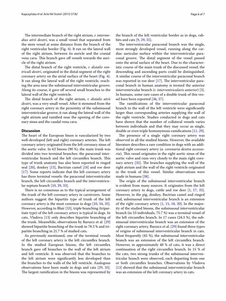
Morphology of the right coronary arteryThe right coronary artery, a. coronaria dextra, was found to be less developed than the left coronary artery. Its average diameter was 7.1 mm. It originated in the right coronary sinus of the aortic valve in all the subjects. At the base of the heart, it ran between the right atrium and the pulmonary trunk. It reached the right ventricu-lar border, entered the coronary groove and was visible in some individuals as the right circumflex branch (r. cir-cumflexus dexter) (Fig. 4). The initial section of the right coronary artery was covered by the auricle of the right atrium. It was surrounded by large amounts of adipose tissue along its entire length. Finally, the right coronary artery reached the subsinuosal interventricular groove, where it typically (53 individuals, 75.7 %) branched off a few small vessels. In the remaining animals (17 individu-als, 24.3 %), the right coronary artery extended into the subsinuosal interventricular branch.
Numerous vessels branched off from the right coronary artery: the (right) conal branch, the proximal branch of the right atrium, the proximal branch of the right ventri-cle, the branch of the right ventricular border, the inter-mediate branch of the right atrium, the distal branch of the right ventricle and the distal branch of the right atrium.
The (right) conal branch, r. coni arteriosi (dextra), origi-nated from the right wall of the right coronary artery at its orifice from the ascending aorta in all individuals and surrounded the arterial cone (Fig. 4). Multiple small ves-sels branched from the main trunk and ran towards the arterial cone.
The proximal branch of the right atrium, r. proxima-lis atrii dextri, was the largest vessel supplying the right atrium. It branched off at various levels of the right coro-nary artery, prior to the origin of the (right) conal branch. It ran on the right on the medial wall of the auricle of
the right atrium, towards the opening of the cranial vena cava. The proximal branch of the right ventricle gave off small branches to the wall of the right atrium and the cra-nial vena cava.
The proximal branch of the right ventricle, r. proxima-lis ventriculi dextri, originated below the (right) conal branch and below the auricle of the right atrium. It ran towards the cardiac auricle on the wall of the right ven-tricle, slightly to the left of the right ventricular border and divided in the middle into two branches. The branch of the right ventricular border, r. marginis ventricularis dextri was a large vessel that ran along the right ven-tricular border and ended at various distances from the notch of the cardiac apex (Fig. 4). It mostly lied under the cardiac muscles, and only its initial short segment was surrounded by adipose tissue. The branch of the right ventricular border vascularized a vast part of the right ventricle in the proximity of the right ventricular border.
Fig. 4 Branches of the right circumflex branch in a European bison. 1 the right circumflex branch, 2 the right conal branch, 3 the branch of the right ventricular border, 4 the intermediate branch of the right atrium, 5 the distal branch of the right ventricle, 6 the paraconal interventricular branch (the descending part) and 7 the intermediate collateral branch of the left ventricle. Right ventricular border; male, 8‑years‑old, body weight 570 kg
Page 6 of 7Kupczyńska et al. Acta Vet Scand (2015) 57:82
The intermediate branch of the right atrium, r. interme-dius atrii dextri, was a small vessel that separated from the stem vessel at some distance from the branch of the right ventricular border (Fig. 4). It ran on the lateral wall of the right atrium, between its auricle and the cranial vena cava. This branch gave off vessels towards the auri-cle of the right atrium.
The distal branch of the right ventricle, r. distalis ven-triculi dextri, originated in the distal segment of the right coronary artery on the atrial surface of the heart (Fig. 4). It ran along the lateral wall of the right ventricle, reach-ing the area near the subsinuosal interventricular groove. Along its course, it gave off several small branches to the lateral wall of the right ventricle.
The distal branch of the right atrium, r. distalis atrii dextri, was a very small vessel. After it stemmed from the right coronary artery in the proximity of the subsinuosal interventricular groove, it ran along the lateral wall of the right atrium and ramified near the opening of the coro-nary sinus and the caudal vena cava.
DiscussionThe heart of the European bison is vascularized by two well-developed (left and right) coronary arteries. The left coronary artery originated from the left coronary sinus of the aortic valve. In 63 bisons (90 %), the main trunk was divided into two terminal branches: the paraconal inter-ventricular branch and the left circumflex branch. This type of trunk anatomy has also been reported in ringed seal [20], donkey [15], Bactrian camel [18] and roe deer [17]. Some reports indicate that the left coronary artery has three terminal vessels: the paraconal interventricular branch, the left circumflex branch and the interventricu-lar septum branch [10, 29, 33].
There is no consensus as to the typical arrangement of the trunk of the left coronary artery in carnivores. Some authors suggest the bipartite type of trunk of the left coronary artery is the most common in dogs [10, 34, 35]. However, according to Blair [33], triple branching (tripar-tiate type) of the left coronary artery is typical in dogs. In cats, Vladova [13] only describes bipartite branching of the trunk. Meanwhile, observations by Barszcz et al. [29] showed bipartite branching of the trunk in 78.3 % and tri-partite branching in 21.7 % of studied cats.
As previously mentioned, one of the terminal vessels of the left coronary artery is the left circumflex branch. In the studied European bisons, the left circumflex branch gave off branches to the wall of the left atrium and left ventricle. It was observed that the branches to the left atrium were significantly less developed than the branches to the walls of the left ventricle. Analogous observations have been made in dogs and cats [29, 35]. The largest ramification in the bisons was represented by
the branch of the left ventricular border as in dogs, rab-bits and cats [9, 29, 35].
The interventricular paraconal branch was the single, most strongly developed vessel, running along the car-diac auricular surface within the interventricular para-conal groove. The distal segment of the vessel passed onto the atrial surface of the heart. Due to the character-istic course of the main trunk of the discussed vessel, the descending and ascending parts could be distinguished. A similar course of the interventricular paraconal branch was reported in roe deer [17]. The interventricular para-conal branch in human anatomy is termed the anterior interventricular branch (r. interventricularis anterior) [3]. In humans, some rare cases of a double trunk of this ves-sel have been reported [36, 37].
The ramifications of the interventricular paraconal branch to the wall of the left ventricle were significantly larger than corresponding arteries supplying the wall of the right ventricle. Studies conducted in dogs and cats have shown that the number of collateral vessels varies between individuals and that they may occur as single, double or even triple homonymous ramifications [11, 29].
The presence of a single right coronary artery was observed in all the studied bisons. However, the available literature describes a rare condition in dogs with an addi-tional right coronary artery (a. coronaria dextra accesso-ria). This vessel originates in the right aortic sinus of the aortic valve and runs very closely to the main right coro-nary artery [35]. The branches supplying the wall of the right atrium and the wall of the right ventricle originated in the trunk of this vessel. Similar observations were made in humans [38].
The origin of the subsinuosal interventricular branch is evident from many sources. It originates from the left coronary artery in dogs, cattle and roe deer [1, 17, 35]. However, in the pig, donkey, Bactrian camel and ringed seal, subsinuosal interventricular branch is an extension of the right coronary artery [1, 15, 18, 20]. In the major-ity of the studied bisons, the subsinuosal interventricular branch (in 53 individuals, 75.7 %) was a terminal vessel of the left circumflex branch. In 17 cases (24.3 %), the sub-sinuosal interventricular branch was an extension of the right coronary artery. Barszcz et al. [29] found three types of origins of subsinuosal interventricular branch in cats. Most frequently (45 %), the subsinuosal interventricular branch was an extension of the left circumflex branch. However, in approximately 40 % of cats, it was a direct continuation of the right circumflex branch. In 15 % of the cats, two strong trunks of the subsinuosal interven-tricular branch were observed, each departing from one or both circumflex branches. Observations by Vladova [13] showed that the subsinuosal interventricular branch was an extension of the left coronary artery in cats.
Page 7 of 7Kupczyńska et al. Acta Vet Scand (2015) 57:82
Two main types of vascularization of the atrial surface of the heart in the bisons were observed. In 53 cases (75.7 %), the subsinuosal interventricular branch was an extension of the left circumflex branch while in 17 cases (24.3 %), it was a direct continuation of the right circumflex branch.
ConclusionsThe blood supply of the heart in the European bison is provided by the left and right coronary arteries, with the left coronary artery being better developed than the right.
Authors’ contributionsMK conceived the study, and participated in its design and coordination. KB, KO, JK carried out the dissection of the cadavers, participated in the data analyses and helped to draft the manuscript. MP, GW, MT performed the cor‑rosive cast, photographic documentation and helped to draft the manuscript. All authors have read and approved the final manuscript.
Author details1 Department of Morphological Sciences, Faculty of Veterinary Medicine, War‑saw University of Life Sciences, SGGW, Nowoursynowska 159, 02‑776 Warsaw, Poland. 2 Department of Angiology, Interfaculty Chair of Anatomy and His‑tology, Medical University of Łódź, Narutowicza 60, 90‑136 Lodz, Poland. 3 Department of Normal and Clinical Anatomy, Interfaculty Chair of Anatomy and Histology, Medical University of Łódź, Narutowicza 60, 90‑136 Lodz, Poland. 4 Department of Animal Physiology and Biostructure, Faculty of Vet‑erinary Medicine, Wroclaw University of Environmental and Life Sciences, Kożuchowska 1/3, 51‑631 Wrocław, Poland.
Competing interestsThe authors declare that they have no competing interests.
Received: 8 July 2015 Accepted: 18 November 2015
References 1. Dyce KM, Sack WO, Wensing CJG. Textbook of Veterinary Anatomy. 3rd ed.
Philadelphia: Saunders; 2010. 2. König HE, Ruberte J, Liebich HG. Organs of the cardiovascular system
(systema cardiovasculare). In: König HE, Liebich HG, editors. Veterinary anatomy of domestic mammals. Stuttgart: Schattauer; 2009. p. 441–74.
3. Standring S, editor. Gray’s anatomy: the anatomical basis of clinical prac‑tice. 40th ed. London: Churchill Livingstone; 2008. p. 978–81.
4. Turkmen S, Yolcu M, Sertcelik A, Ipek E, Dokumaci B, Batyraliev T. Single coronary artery incidence in 215,140 patients undergoing coronary angiography. Folia Morphol. 2014;73:469–74.
5. Turhan H, Duru E, Yetkin E, Atak R, Senen K. Right coronary artery originat‑ing from distal left circumflex: an extremely rare variety of single coronary artery. Int J Cardiol. 2003;88:309–11.
6. Ozdemir V, Cevik‑Demirkna A, Turkmenoglu I. The right coronary artery is absent in the chinchilla (Chinchilla lanigera). Anat Histol Embryol. 2008;37:114–7.
7. Durán AC, Arqué JM, Fernández B, Fernández MC, Rodríguez C, Sans‑Coma V. Rudimentary coronary artery in Syrian hamster (Mesocricetus auratus). Anat Histol Embryol. 2009;38:270–4.
8. Vicentini CA, Oris AM, Dias SM. Anatomical observations of the coronary artery vascularization in the guinea pigs (Cavia porcellus L.). Anat Anz. 1991;172:209–12.
9. Bahar S, Ozdemir V, Eken E, Tipirdamaz S. The distribution of the coronary arteries in the Angora rabbit. Anat Histol Embryol. 2007;36:321–7.
10. Noestelthaller A, Probst A, König HE. Branching patterns of the left main coronary artery in the dog demonstrated by the use of corrosion casting technique. Anat Histol Embryol. 2007;36:33–7.
11. Barszcz K, Kupczyńska M, Wąsowicz M, Czubaj N, Sokołowski W. Pat‑terns of the arterial vascularization of the dog’s heart. Med Weter. 2013;69:531–4.
12. Barszcz K, Kupczyńska M, Klećkowska‑Nawrot J, Skibniewski M, Janczyk P. Morphology of coronary ostia in domestic shorthair cat. Anat Histol Embryol. 2015. doi:10.1111/ahe.12174.
13. Vladova D. Ventricular coronary pattern in the cat. Trakia J Sci. 2005;3:44–9.
14. Ozgel O, Dursun N. The arterial vascularization of septum interventricu‑lare in donkeys (Equus asinus L.). Anat Histol Embryol. 2005;34:80–4.
15. Ozgel O, Haligur A, Dursun N, Karakurum E. The macroanatomy of coronary arteries in donkeys (Equus asinus L.). Anat Histol Embryol. 2004;33:278–83.
16. Besoluk K, Tipirdamaz S. Comparative macroanatomic investigations of the venous drainage of the heart in Akkaraman sheep and Angora goats. Anat Histol Embryol. 2001;30:249–52.
17. Frąckowiak H, Jasiczak K, Pluta K, Godynicki S. Coronary arteries of the roe deer (Capreplus capreolus; Linnaeus 1758) heart. Pol J Vet Sci. 2007;10:105–8.
18. Yuan G, Ma J, Ye W, Bai Z, Wang J. Macroanatomy of coronary arteries in Bactrian camel (Camelus bactrianus). Vet Res Commun. 2009;33:367–77.
19. Ghazi SR, Tadjalli M. Coronary arterial anatomy of the one‑humped camel (Camelus dromedarius). Vet Res Commun. 1993;17:163–70.
20. Smodlaka H, Henry RW, Schumacher J, Reed RB. Macroscopic anatomy of the heart of the Ringed Seal (Phoca hispida). Anat Histol Embryol. 2008;37:30–5.
21. Atalar Ö, Yilmaz S, İlkay E, Burma O. Investigation of coronary arteries in the porcupine (Hystrix cristata) by latex injection and angiography. Ann Anat. 2003;185:373–6.
22. Atalar Ö, Yilmaz S, Dinç G, Özdemir D. The venous drainage of the heart in porcupines (Hystrix cristata). Anat Histol Embryol. 2004;33:233–5.
23. Isomura G. Blood supply to the cranial venae cavae and heart in the laboratory shrew (Suncus murinus). J Anat. 1993;183:537–43.
24. Nikolić V, Teofilovski‑Parapid G, Stanković G, Parapid B, Malobabić S, Stojić V. Third coronary artery in monkey heart. Acta Vet Hung. 2004;52:253–7.
25. Hagensen MK, Abe AS, Falk E, Wang T. Physiological importance of the coronary arterial blood supply to the rattlesnake heart. J Exp Biol. 2008;211:3588–93.
26. Teofilovski‑Parapid G, Kreclović G. Coronary artery distribution in Macaca fascicularis (Cynomolgus). Lab Anim. 1998;32:200–5.
27. Bezuidenhout AJ. The coronary circulation of the heart of the ostrich (Struthio camelus). J Anat. 1984;138:385–97.
28. Bartyzel BJ, Charuta A, Barszcz K, Koleśnik A, Kobryń H. Morphology of the aortic valve of Gallus gallus f. domestica. Bull Vet Inst Pulawy. 2009; 53:147–51.
29. Barszcz K, Kupczyńska M, Klećkowska‑Nawrot J, Janczyk P, Krasucki K, Wąsowicz M. Arterial coronary circulation in cats. Medycyna Weter. 2014;70:373–7.
30. Polguj M, Jędrzejewski KS, Dyl Ł, Topol M. Topographic and morphomet‑ric comparison study of the terminal part of human and bovine testicular arteries. Folia Morphol. 2009;68:271–6.
31. Polguj M, Jedrzejewski KS, Topol M. Angioarchitecture of the bovine spermatic cord. J Morphol. 2011;272:497–502.
32. World Association of Veterinary Anatomist: Nomina Anatomica Veteri‑naria. 2012 Gent, Belgium, p. 74.
33. Blair E. Anatomy of the ventricular coronary arteries in the dog. Circ Res. 1961;9:333–41.
34. Büll ML, Martins MRFB. Study of the arterial coronary circulation in the dog (Canis familiaris). Rev Chil Anat. 2002;2:117–23.
35. Bezuidenhout A. The heart and arteries. In: Evans HE, de Lahunta A, editors. Miller’s Anatomy of the Dog. St. Louis: Elsevier Saunders; 2013. p. 428–504.
36. Kosar F. An unusual case of double anterior descending artery originating from the left and right coronary artery. Heart Vessels. 2006;21:385–7.
37. Turhan H, Atak R, Erbay AR, Senen K, Yetkin E. Double left anterior descending coronary artery arising from the left and right coronary arteries: a rare congenital coronary artery anomaly. Heart Vessels. 2004;19:196–8.
38. Harikrishnan S, Bhat A, Tharakan JM. Double right coronary artery. Int J Cardiol. 2000;77:315–6.