Embed Size (px)
Citation preview
NovaLisa TM
Corynebacterium diphtheriae toxin
IgG - ELISA Enzyme immunoassay for the quantitative determination of IgG-class antibodies against Corynebacterium diphtheriae toxin in human serum or plasma
Enzymimmunoassay zur quantitativen immunenzymatischen Bestimmung von IgG-Antikörpern gegen Corynebacterium diphtheriae Toxin in Humanserum oder Plasma
Dosage immunoenzymatique pour la détermination quantitative des anticorps IgG dirigés contre la toxine du Corynebacterium diphtheriae dans le sérum humain ou plasma
Test immunoenzimatico per la determinazione quantitativa degli anticorpi della classe IgG per la tossina del Corynebacterium diphtheriae nel siero o plasma umano
Enzimoinmunoensayo para la determinación cuantitativa de anticuerpos IgG contra –Corynebacterium difteriae en suero o plasma humano Only for in-vitro diagnostic use English: Page 2 to 7 Deutsch: Seite 8 bis 13 Francais: Page 14 à 18 Italiano: da Pagina 19 a 24 Espanol: Página 25 a 30 For further languages please contact our authorized distributors. Bibliography / Literatur / Bibliographie / Page / Seite / Page / 34 Bibliografia / Bibliografía Pagina / Página Symbols Key / Symbolschlüssel / Page / Seite / Page / 35 Explication des symboles / Legenda / Símbolos Pagina / Página Summary of Test Procedure/ Kurzanleitung Testdurchführung/ Résumé de la procedure de test/ Page / Seite / Page/ 36 Schema della procedura/ Resumen de la técnica / Pagina / Página
_______________________________________________________________________
Product Number: CORG0090 (96 Determinations) _______________________________________________________________________
2
ENGLISH
1. INTRODUCTION
Corynebacteria are aerobic non spore-forming gram-positive rods of irregular shape (0.5 –1 µm thick and 2-6 µm long). They comprise skin commensals, opportunist pathogens and several major pathogens, including Corynebacterium diphtheriae. In general, they are isolated from throat swabs on selective media containing tellurite. The bacterial infection caused by C. diphtheriae, Diphtheria, has two forms. Respiratory diphtheria is typically caused by toxin-producing (toxigenic) strains; cutaneous disease can be caused by either toxigenic or nontoxigenic strains. In the respiratory form of the disease, a membrane is formed; this membrane is usually visible on the throat or tonsils. Persons may die from asphyxiation when the membrane obstructs breathing. Other complications are caused by remote effects of the diphtheria toxin (myocarditis, nerve paralysis) Cutaneous diphtheria is usually mild, typically consisting of non-distinctive sores or shallow ulcers and only rarely involving toxic complications (1-2% of infections with toxigenic strains). Diphtheria was one of the most common causes of death among children during the prevaccine era. Since the introduction and widespread use of diphtheria toxoid vaccine (formalin-inactivated diphtheria toxin) in most industrialized countries the disease is now characterized by sporadic cases and intermittent outbreaks of low intensity. But recent large epidemics of diphtheria in several eastern European countries have again drawn attention to this „forgotten“ disease – and, the majority of these cases have occurred among adolescents and adults instead of children.
Species Disease Symptoms Mechanism of Infection Corynebacterium diphtheriae
Diphtheria (respiratory) sore throat and low-grade fever swelling of the neck (“bull neck”) from inflammation Complications: exotoxin-induced damage to other organs
Transmission from person to person through close physical and respiratory contact Transmission is increased in overcrowded and poor socio-economic conditions
The only effective way to control diphtheria is by prophylactic immunization with diphtheria toxoid. Antibody to the toxoid protects against the action of the toxin; immunized persons can be infected by toxin-producing strains of diphtheria, but the systemic manifestations of diphtheria do not occur. The outcome of the disease improves with early, appropriate treatment. Prompt recognition and reporting of the disease is important to assure early, appropriate treatment with diphtheria anti-toxin. Infection may be identified by � Microscopy: Gram stain
� Serology: Detection of toxin production by ELISA
2. INTENDED USE
The NovaTec Corynebacterium diphtheriae toxin IgG-ELISA is intended for the quantitative determination of IgG class antibodies against Corynebacterium diphtheriae toxin in human serum or plasma (citrate). This allows the determination of the immune status of the patients facilitating individual recommendations about the necessity of a basic immunization or booster injection.
3. PRINCIPLE OF THE ASSAY
The quantitative immunoenzymatic determination of IgG-class antibodies against C. diphtheriae toxin is based on the ELISA (Enzyme-linked Immunosorbent Assay) technique. Microtiterstrip wells are precoated with inactivated specific Corynebacterium diphtheriae toxin (toxoid) antigens to bind corresponding antibodies of the specimen. After washing the wells to remove all unbound sample material horseradish peroxidase (HRP) labelled anti-human IgG conjugate is added. This conjugate binds to the captured C. diphtheriae toxin-specific antibodies. The immune complex formed by the bound conjugate is visualized by adding Tetramethylbenzidine (TMB) substrate which gives a blue reaction product. The intensity of this product is proportional to the amount of C. diphtheriae toxin-specific IgG antibodies in the specimen. Sulphuric acid is added to stop the reaction. This produces a yellow endpoint colour. Absorbance at 450 nm is read using an ELISA microwell plate reader.
4. MATERIALS
4.1. Reagents supplied � C. diphtheriae toxin Coated Wells (IgG): 12 breakapart 8-well snap-off strips coated with C. diphtheriae toxin (toxoid)
antigens; in resealable aluminium foil.
� IgG Sample Diluent***: 1 bottle containing 100 ml of buffer for sample dilution; pH 7.2 ± 0.2; coloured yellow; ready to use; white cap.
� Stop Solution: 1 bottle containing 15 ml sulphuric acid, 0.2 mol/l; ready to use; red cap.
� Washing Solution (20x Concentrate)*: 1 bottle containing 50 ml of a 20-fold concentrated buffer for washing the wells; pH 7.2 ± 0.2; white cap.
3
� C. diphtheriae toxin anti-IgG Conjugate**: 1 bottle containing 20 ml of peroxidase labelled antibodies to human IgG; coloured blue; ready to use; black cap.
� TMB Substrate Solution: 1 bottle containing 15 ml 3,3',5,5'-tetramethylbenzidine (TMB); ready to use; yellow cap.
� C. diphtheriae toxin IgG Standards***: 4 vials, each containing 2ml, coloured yellow; ready to use: Standard A: 0.000 IU/ml; blue cap Standard B: 0.015 IU/ml; green cap Standard C: 0.075 IU/ml; yellow cap Standard D: 0.150 IU/ml; red cap
* contains 0.1 % Bronidox L after dilution ** contains 0.2 % Bronidox L *** contains 0.1 % Kathon
4.2. Materials supplied � 1 Strip holder � 1 Cover foil � 1 Test protocol � 1 distribution and identification plan
4.3. Materials and Equipment needed � ELISA microwell plate reader, equipped for the measurement of absorbance at 450/620nm � Incubator 37°C � Manual or automatic equipment for rinsing wells � Pipettes to deliver volumes between 10 and 1000 µl � Vortex tube mixer � Deionised or (freshly) distilled water � Disposable tubes � Timer
5. STABILITY AND STORAGE
The reagents are stable up to the expiry date stated on the label when stored at 2…8 °C.
6. REAGENT PREPARATION
It is very important to bring all reagents, samples and standards to room temperature (20…25°C) before starting the test run!
6.1. Coated Snap-off Strips The ready to use breakapart snap-off strips are coated with C. diphtheriae toxin (toxoid) antigens. Store at 2...8°C. Immediately after removal of strips, the remaining strips should be resealed in the aluminium foil along with the desiccant supplied and stored at 2…8°C; stability until expiry date.
6.2. C. diphtheriae toxin anti-IgG Conjugate The bottle contains 20 ml conjugate with the components anti-human IgG horseradish peroxidase, buffer, stabilizers, preservatives and an inert blue dye. The solution is ready to use. Store at 2...8°C. After first opening stability until expiry date when stored at 2…8°C.
6.3. Standards The vials labelled with Standard A, B, C and D contain a ready to use standard solution. The concentration of the standards, calibrated in accordance with the “International Standard for Diphtheria Antitoxin (00/496)”, are: Standard A: 0.000 IU/ml Standard B: 0.015 IU/ml Standard C: 0.075 IU/ml Standard D: 0.150 IU/ml The solutions have to be stored at 2...8°C and contain 0.1% Kathon. After first opening stability until expiry date when stored at 2…8°C.
6.4. IgG Sample Diluent The bottle contains 100 ml phosphate buffer, stabilizers, preservatives and an inert yellow dye. It is used for the dilution of the patient specimen. This ready to use solution has to be stored at 2...8°C. After first opening stability until expiry date when stored at 2…8°C.
6.5. Washing Solution (20x conc.) The bottle contains 50 ml of a concentrated buffer, detergents and preservatives. Dilute Washing Solution 1+19; e.g. 10 ml Washing Solution + 190 ml fresh and germ free redistilled water. The diluted buffer is stable for 5 days at room temperature. After first opening stability until expiry date when stored at 2…8°C.
4
6.6. TMB Substrate Solution The bottle contains 15 ml of a Tetramethylbenzidine/hydrogen peroxide system. The reagent is ready to use and has to be stored at 2...8°C, away from the light. The solution should be colourless or could have a slight blue tinge. If the substrate turns into blue, it may have become contaminated and should be thrown away. After first opening stability until expiry date when stored at 2…8°C.
6.7. Stop Solution The bottle contains 15 ml 0.2 M sulphuric acid solution (R 36/38, S 26), ready to use, store at 2...8°C. After first opening stability until expiry date.
7. SPECIMEN COLLECTION AND PREPARATION
Use human serum or plasma (citrate) samples with this assay. If the assay is performed within 5 days after sample collection, the specimen should be kept at 2...8°C; otherwise they should be aliquoted and stored deep-frozen (-20 to -70°C). If samples are stored frozen, mix thawed samples well before testing. Avoid repeated freezing and thawing. Heat inactivation of samples is not recommended.
7.1. Sample Dilution Before assaying, all samples should be diluted 1+100 with IgG Sample Diluent. Dispense 10µl sample and 1ml IgG Sample Diluent into tubes to obtain a 1+100 dilution and thoroughly mix with a Vortex.
For patients with expected antitoxin concentrations greater than Standard D (0.15 IU/ml) a second 1 + 1 dilution of this 1 + 100 diluted patient sample should be performed; e.g. 100 µl of first sample dilution + 100 µl of IgG sample diluent (mix well). Dilution factor: 2
8. ASSAY PROCEDURE
8.1. Test Preparation Please read the test protocol carefully before performing the assay. Result reliability depends on strict adherence to the test protocol as described. The following test procedure is only validated for manual procedure. If performing the test on ELISA automatic systems we recommend to increase the washing steps from three to five and the volume of washing solution from 300µl to 350µl to avoid washing effects. Prior to commencing the assay, the distribution and identification plan for all specimens and controls should be carefully established on the result sheet supplied in the kit. Select the required number of microtiter strips or wells and insert them into the holder.
Please allocate at least:
1 well (e.g. A1) for the substrate blank, 4 wells (e.g. B1, C1, etc.) for Standard A, B, C and D.
It is recommended to determine patient samples in duplicate.
Perform all assay steps in the order given and without any appreciable delays between the steps.
A clean, disposable tip should be used for dispensing each standard and sample.
Adjust the incubator to 37° ± 1°C.
1. Dispense 100µl of each Standard (A, B, C and D) and diluted sample into the respective wells. Leave well A1 for substrate blank.
2. Cover wells with the foil supplied in the kit.
3. Incubate for 1 hour ± 5 min at 37±1°C.
4. When incubation has been completed, remove the foil, aspirate the content off the wells and wash each well three times with 300µl of washing solution. Avoid overflows from the reaction wells. The soak time between each wash cycle should be >5sec. At the end carefully remove remaining fluid by tapping strips on tissue paper prior to the next step!
Note: Washing is critical! Insufficient washing results in poor precision and falsely elevated absorbance values.
5. Dispense 100µl C. diphtheriae toxin anti-IgG Conjugate into all wells except for the blank well (e.g. A1). Cover with foil.
6. Incubate for 30 min at room temperature (20…25°C). Do not expose to direct sunlight.
7. Repeat step 4.
8. Dispense 100µl TMB Substrate Solution into all wells
9. Incubate for exactly 15 min at room temperature (20…25°C) in the dark.
10. Dispense 100µl Stop Solution into all wells in the same order and at the same rate as for the TMB Substrate Solution. Any blue colour developed during the incubation turns into yellow.
Note: Highly positive patient samples can cause dark precipitates of the chromogen! These precipitates have an influence when reading the optical density. Dilute the specimen as mentioned under 7.1. Sample Dilution.
11. Measure the absorbance of the specimen at 450/620nm within 30 min after addition of the Stop Solution.
5
8.2. Measurement Adjust the ELISA Microwell Plate Reader to zero using the substrate blank in well A1.
If - due to technical reasons - the ELISA reader cannot be adjusted to zero using the substrate blank in well A1, subtract the absorbance value of well A1 from all other absorbance values measured in order to obtain reliable results!
Measure the absorbance of all wells at 450 nm and record the absorbance values for each standard and patient sample in the distribution and identification plan.
Dual wavelength reading using 620 nm as reference wavelength is recommended.
Where applicable calculate the mean absorbance values of all duplicates.
9. RESULTS
9.1. Assay Validation Criteria
In order for an assay to be considered valid, the following criteria must be met:
� Substrate blank in A1: Absorbance < 0.100
� Standard A in B1: Absorbance < 0.200 Standard B in C1: Absorbance > 0.100 Standard C in D1: Absorbance > 0.500
� Standard D in E1: Absorbance > 1.000
Standard A < Standard B < Standard C < Standard D If these criteria are not met, the test is not valid and must be repeated.
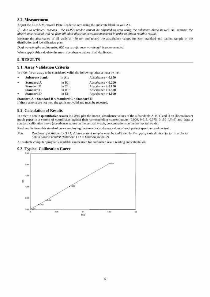
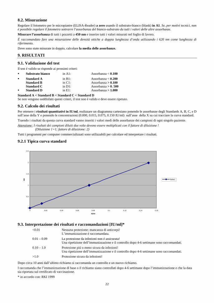
9.2. Calculation of Results In order to obtain quantitative results in IU/ml plot the (mean) absorbance values of the 4 Standards A, B, C and D on (linear/linear) graph paper in a system of coordinates against their corresponding concentrations (0.000, 0.015, 0.075, 0.150 IU/ml) and draw a standard calibration curve (absorbance values on the vertical y-axis, concentrations on the horizontal x-axis).
Read results from this standard curve employing the (mean) absorbance values of each patient specimen and control.
Note: Readings of additionally (1+1) diluted patient samples must be multiplied by the appropriate dilution factor in order to obtain correct results! (Dilution: 1+1 = Dilution factor: 2).
All suitable computer programs available can be used for automated result reading and calculation.
9.3. Typical Calibration Curve
6
9.4. Interpretation of Results and Recommendations [ IU/ml]* Each result should be carefully assessed by a physician.
< 0.01 No protective antibody level! Immediate full course of basic immunization is recommended!
0.01 - 0.09 No reliable protection! Immediate booster injection is recommended.
0.1 – 1.0 Reliable protection! > 1.0 Reliable long term protection! After about 10 years after last booster control and booster injection is recommended.
It is recommended that the basic immunisation or booster is checked 4-6 weeks after immunisation and to record the data on the certificate of vaccination.
* according to: RKI 1999
10. SPECIFIC PERFORMANCE CHARACTERISTICS
10.1. Precision Intraassay n Mean value (OD) CV (%)
Weak pos. 24 0.95 12.1 Pos. 24 3.02 2.1
Interassay n Mean value (IU/ml) CV (%) Weak pos. 12 0.04 11.4 Pos. 12 0.16 2.8
10.2. Diagnostic Specificity The diagnostic specificity is defined as the probability of the assay of scoring negative in the absence of the specific analyte. It is 84.6%
10.3. Diagnostic Sensitivity The diagnostic sensitivity is defined as the probability of the assay of scoring positive in the presence of the specific analyte. It is 100%.
10.4. Analytical sensitivity The analytical sensitivity – defined as the apparent concentration of the analyte that can be distinguished from the zero calibrator –is 0.01 IU/ml.
10.5. Interferences Interferences with hemolytic, lipemic or icteric sera are not observed up to a concentration of 10 mg/ml hemoglobin, 5 mg/ml triglycerides and 0.2 mg/ml bilirubin.
Note: The results refer to the groups of samples investigated; these are not guaranteed specifications.
11. LIMITATIONS OF THE PROCEDURE
Bacterial contamination or repeated freeze-thaw cycles of the specimen may affect the absorbance values. Diagnosis of an infectious disease should not be established on the basis of a single test result. A precise diagnosis should take into consideration clinical history, symptomatology as well as serological data. In immunocompromized patients and newborns serological data only have restricted value.
12. PRECAUTIONS AND WARNINGS
� In compliance with article 1 paragraph 2b European directive 98/79/EC the use of the in vitro diagnostic medical devices is intended by the manufacturer to secure suitability, performances and safety of the product. Therefore the test procedure, the information, the precautions and warnings in the instructions for use have to be strictly followed. The use of the testkits with analyzers and similar equipment has to be validated. Any change in design, composition and test procedure as well as for any use in combination with other products not approved by the manufacturer is not authorized; the user himself is responsible for such changes. The manufacturer is not liable for false results and incidents for these reasons. The manufacturer is not liable for any results by visual analysis of the patient samples.
� Only for in-vitro diagnostic use. � All components of human origin used for the production of these reagents have been tested for anti-HIV antibodies, anti-HCV
antibodies and HBsAg and have been found to be non-reactive. Nevertheless, all materials should still be regarded and handled as potentially infectious.
� Do not interchange reagents or strips of different production lots.
7
� No reagents of other manufacturers should be used along with reagents of this test kit. � Do not use reagents after expiry date stated on the label. � Use only clean pipette tips, dispensers, and lab ware. � Do not interchange screw caps of reagent vials to avoid cross-contamination. � Close reagent vials tightly immediately after use to avoid evaporation and microbial contamination. � After first opening and subsequent storage check conjugate and standard vials for microbial contamination prior to further use. � To avoid cross-contamination and falsely elevated results pipette patient samples and dispense conjugate without splashing
accurately to the bottom of wells. � The NovaLisa™ ELISA is only designed for qualified personnel who are familiar with good laboratory practice.
WARNING: In the used concentration Bronidox L has hardly any toxicological risk upon contact with skin and mucous membranes!
WARNING: Sulphuric acid irritates eyes and skin. Keep out of the reach of children. Upon contact with the eyes, rinse thoroughly with water and consult a doctor!
12.1. Disposal Considerations Residues of chemicals and preparations are generally considered as hazardous waste. The disposal of this kind of waste is regulated through national and regional laws and regulations. Contact your local authorities or waste management companies which will give advice on how to dispose hazardous waste. 13. ORDERING INFORMATION
Prod. No.: CORG0090 Corynebacterium diphtheriae toxin IgG-ELISA (96 Determinations)
8
DEUTSCH
1. EINLEITUNG
Corynebakterien sind grampositive, nicht sporenbildende, unbewegliche, pleomorphe Stäbchenbakterien, die als besonderes Charakteristikum häufig terminale keulenförmige Auftreibungen zeigen. Sie sind in der Umwelt weit verbreitet. Einige Arten sind tier- und pflanzenpathogen. Neben apathogenen Haut- und Schleimhautbewohnern sind für den Menschen die opportunistisch pathogenen Spezies und der Erreger der Diphtherie (C. diphtheriae) von Bedeutung. Die Pathogenität von C. diphtheriae beruht auf der Bildung eines Exotoxins. Die genetische Information zur Bildung dieses Toxins wird durch einen lysogenen Phagen kodiert. Nur Stämme, die diesen oder einen verwandten Prophagen enthalten sind pathogen. Die Erkrankung beginnt nach einer Inkubationszeit von 3-5 Tagen als Lokalinfektion. Je nach Eintrittspforten der Erreger entsteht eine Rachen-, Nasen-, Augen-, Wund-, Haut-, Nabel oder Genitaldiphtherie. Das gebildete Toxin führt lokal zu Nekrosen, die einen typischen Foetor ex ore bedingen. Abgestorbene Epithelzellen, Fibrin und Entzündungszellen bilden einen Belag, der Mukosa ziemlich fest anliegt und deshalb als Pseudomembran bezeichnet wird. Im Rachenraum kann diese die Atemwege verlegen und zu schwerer Atemnot führen. Massives Krankheitsgefühl, Fieber und Schwellen der regionalen Lymphknoten kommen hinzu. Bei der Rachendiphtherie kommt es innerhalb von Stunden zum massiven Anschwellen des Halses (Cäsarenhals: Schwellung der regionalen Halslymphknoten und Ausbildung eines periglandulären Ödems). Das Diphtherie Toxin wird auch in die Zirkulation eingeschwemmt und begründet eine systemische Intoxikation, deren schwere vom jeweiligen Organbefall abhängig ist (Herz, Leber, Nieren, Nerven). Diese Spätfolge der Diphtherie kann den Tod bedeuten (toxisches Kreislaufversagen). Die Keime werden durch Tröpfchen- oder Schmierinfektion übertragen. Gesunde Keimträger sind sehr selten. In Mitteleuropa ist die Rachendiphtherie, in den Tropen die Wunddiphtherie die häufigste Form der Krankheit. Es existiert die Möglichkeit einer aktiven Immunisierung mit einem Totimpfstoff.
Spezies Erkrankung Symptome Infektionsmodus Corynebacterium diphtheriae
Diphtherie Massives Krankheitsgefühl, feste Belege im Rachen, schmerzlos, Lymphknotenschwellung, Cäsarenhals Komplikation: Toxin induzierte Organschäden an Nerven und Herz
Tröpfchen- oder Schmierinfektion, von Mensch zu Mensch
Eine Infektion mit Corynebacterium diphtheriae kann nachgewiesen werden mittels: � Mikroskopie: Gram-Färbung � Serologie: Nachweis der Toxinbildung mittels ELISA
2. VERWENDUNGSZWECK
Der NovaTec Corynebacterium diphtheriae toxin IgG-ELISA ist für den quantitativen Nachweis spezifischer IgG-Antikörper gegen Corynebacterium diphtheriae Toxin in humanem Serum oder Plasma (Citrat) bestimmt. Dies ermöglicht die Bestimmung des Impfstatus und die individuelle Empfehlung einer Grund- bzw. Auffrisch-Immunisierung.
3. TESTPRINZIP
Die quantitative immunenzymatische Bestimmung von spezifischem IgG gegen C. diphtheriae Toxin beruht auf der ELISA (Enzyme-linked Immunosorbent Assay)-Technik. Mikrotiterstreifen als solide Phase sind beschichtet mit inaktiviertem C. diphtheriae Toxin (Toxoid) Antigenen. Vorhandene spezifische Antikörper in der Probe binden an die immobilisierten Antigene der Mikrotiterplatte. Meerrettich-Peroxidase (HRP) -konjugierte anti-human-IgG Antikörper binden an Antigen-Antikörperkomplexe in positiven Proben. Die entstandenen Immunkomplexe werden durch Blaufärbung nach Inkubation mit Tetramethylbenzidin (TMB)-Substratlösung nachgewiesen. Stoppen der enzymatischen Reaktion mit Schwefelsäure führt zu einem Farbumschlag von blau zu gelb, der einfach nachgewiesen und mit einem ELISA-Reader bei 450 nm gemessen werden kann.
4. MATERIALIEN
4.1. Mitgelieferte Reagenzien � C. diphtheriae Toxin beschichtete Mikrotiterstreifen (IgG): 12 teilbare 8er-Streifen, beschichtet mit C. diphtheriae Toxoid
Antigenen; in wieder verschließbarem Aluminiumbeutel.
� IgG-Probenverdünnungspuffer***: 1 Flasche mit 100 ml Puffer zur Probenverdünnung; pH 7.2 ± 0.2; gelb gefärbt; gebrauchsfertig; weiße Verschlusskappe,
� Stopplösung: 1 Flasche mit 15 ml Schwefelsäure, 0.2 mol/l, gebrauchsfertig; rote Verschlusskappe.
� Waschlösung (20x konz.)*: 1 Flasche mit 50 ml eines 20-fach konzentrierten Puffers zum Waschen der Kavitäten; pH 7.2 ± 0.2; weiße Verschlusskappe.
� C. diphtheriae Toxin anti-IgG-Konjugat**: 1 Flasche mit 20 ml Peroxidase-konjugierten Antikörpern gegen humanes IgG; blau gefärbt; gebrauchsfertig. schwarze Verschlusskappe;
� TMB-Substratlösung: 1 Flasche mit 15 ml 3,3`,5,5`-Tetramethylbenzidin (TMB); gebrauchsfertig; gelbe Verschlusskappe.
9
� C. diphtheriae Toxin IgG Standards***: 4 Fläschchen, je 2 ml, gebrauchsfertig,: Standard A: 0.000 IU/ml; blaue Verschlusskappe Standard B: 0.015 IU/ml; grüne Verschlusskappe Standard C: 0.075 IU/ml; gelbe Verschlusskappe Standard D: 0.150 IU/ml; rote Verschlusskappe
* enthält 0.1 % Bronidox L nach Verdünnung ** enthält 0.2 % Bronidox L *** enthält 0.1 % Kathon
4.2. Mitgeliefertes Zubehör � 1 selbstklebende Abdeckfolie � 1 Rahmenhalter � 1 Arbeitsanleitung � 1 Ergebnisblatt
4.3. Erforderliche Materialien und Geräte � Photometer mit Filtern 450/620 nm � Feuchtkammer/Brutschrank mit Thermostat � Manuelle oder automatische Waschvorrichtung � Mikropipetten mit Einmalspitzen (10, 100, 200, 1000 µl) � Vortex-Mischer � Plastikröhrchen für den einmaligen Gebrauch � Röhrchen-Ständer � Aqua dest. � Timer
5. STABILITÄT UND LAGERUNG
Testkit bei 2...8°C lagern. Die Reagenzien nicht nach den angegebenen Verfallsdaten verwenden. Die Verfallsdaten sind jeweils auf den Flaschenetiketten und auf dem Außenetikett angegeben.
6. VORBEREITUNG DER REAGENZIEN
Alle Reagenzien, Proben und Kontrollen sind vor ihrer Verwendung auf Raumtemperatur(20...25°C) zu bringen!
6.1. Beschichtete Streifen Die abbrechbaren Streifen sind mit inaktiviertem C. diphtheriae Toxoid Antigen beschichtet. Die gebrauchsfertigen Vertiefungen sind bei 2...8°C aufzubewahren. Nichtverbrauchte Vertiefungen im Aluminiumbeutel zusammen mit dem Trockenmittel sofort wieder verschließen und bei 2...8°C lagern. Haltbarkeit bis zum angegebenen Verfallsdatum.
6.2. C. diphtheriae Toxin anti-IgG Konjugat Das Fläschchen enthält 20 ml einer Lösung von anti-human IgG-Meerrettichperoxidase, Puffer, Stabilisatoren, Konservierungsmittel und einen inerten blauen Farbstoff. Die gebrauchsfertige Lösung ist bei 2...8°C aufzubewahren. Nach dem ersten Öffnen haltbar bis zum angegebenen Verfallsdatum (bei 2...8°C).
6.3. Standards Die Fläschchen mit Nullstandard A und Standards B, C und D enthalten 2,0 ml gebrauchsfertige Standardlösung. Die Konzentrationen der Standards, gegen den „International Standard for Diphtheria Antitoxin (00/496)“ standardisiert, sind: Standard A: 0.000 IU/ml Standard B: 0.015 IU/ml Standard C: 0.075 IU/ml Standard D: 0.150 IU/ml Die gebrauchsfertigen Lösungen sind bei 2...8°C aufzubewahren und enthalten 0.1 % Kathon. Nach dem ersten Öffnen haltbar bis zum angegebenen Verfallsdatum (bei 2...8°C).
6.4. IgG-Probenverdünnungspuffer Die Flasche enthält 100 ml Phosphatpuffer, Stabilisatoren, Konservierungsmittel und einen inerten gelben Farbstoff. Die gebrauchsfertige Lösung ist bei 2...8°C aufzubewahren. Die Lösung wird für die Verdünnung der Proben eingesetzt. Nach dem ersten Öffnen haltbar bis zum angegebenen Verfallsdatum (bei 2...8°C).
6.5. Waschlösung (20x konz.) Die Flasche enthält 50 ml konzentrierten Puffer, Detergenzien und Konservierungsmittel. Der Inhalt wird auf einen Liter mit Aqua dest. verdünnt (1+19). Der frische Puffer ist mindestens 4 Wochen bei 2...8°C haltbar. Die Waschlösung wird zum Waschen der Streifen eingesetzt. Sollte eine Kristallisation im Konzentrat auftreten, die Waschlösung auf 37°C erwärmen und vor dem Verdünnen gut mischen. Nach dem ersten Öffnen, Konzentrat haltbar bis zum angegebenen Verfallsdatum (bei 2...8°C).
10
6.6. TMB-Substratlösung Das Fläschchen enthält 15 ml eines Tetramethylbenzidin/Wasserstoffperoxidgemisches. Die gebrauchsfertige Lösung ist bei 2...8°C vor Licht geschützt aufzubewahren. Die Lösung ist leicht hellblau. Sollte die TMB-Substratlösung dunkelblau sein, ist sie kontaminiert und kann nicht im Test verwendet werden. Nach dem ersten Öffnen haltbar bis zum Verfallsdatum bei sachgerechter Lagerung von 2…8°C.
6.7. Stopplösung Das Fläschchen enthält 15 ml 0,2 M Schwefelsäure (R36/38, S26). Die gebrauchsfertige Lösung ist bei 2-8 C aufzubewahren. Nach dem ersten Öffnen haltbar bis zum angegebenen Verfallsdatum (bei 2...8°C).
7. ENTNAHME UND VORBEREITUNG DER PROBEN
Es sollten humane Serum- oder Plasmaproben (Citrat) verwendet werden. Werden die Bestimmungen innerhalb von 5 Tagen nach Blutentnahme durchgeführt, können die Proben bei 2...8°C aufbewahrt werden, sonst tiefgefrieren (-70...-20°C). Wiederaufgetaute Proben vor dem Verdünnen gut schütteln. Wiederholtes Tiefgefrieren und Auftauen vermeiden! Hitzeinaktivierung der Proben wird nicht empfohlen.
7.1. Probenverdünnung Proben vor Testbeginn im Verhältnis 1+100 mit IgG-Probenverdünnungspuffer verdünnen, z.B. 10µl Probe und 1 ml IgG-Probenverdünnungspuffer in die entsprechenden Röhrchen pipettieren, um eine Verdünnung von 1+100 zu erhalten; gut mischen (Vortex). Proben mit erwarteten Antitoxinkonzentration oberhalb der Konzentration von Standard D (0.15 IU/ml) sollten nach der oben beschriebenen Verdünnung 1+1 weiter verdünnt werden, z.B. 100µl der ersten Probenverdünnung + 100µl IgG-Probenverdünnungspuffer (Verdünnungsfaktor: 2).
8. TESTDURCHFÜHRUNG
8.1. Testvorbereitung Gebrauchsinformation vor Durchführung des Tests sorgfältig lesen. Für die Zuverlässigkeit der Ergebnisse ist es notwendig, die Arbeitsanleitung genau zu befolgen. Die folgende Testdurchführung ist für die manuelle Methode validiert. Beim Arbeiten mit ELISA Automaten empfehlen wir, um Wascheffekte auszuschließen, die Zahl der Waschschritte von drei auf fünf und das Volumen der Waschlösung von 300 µl auf 350 µl zu erhöhen. Vor Testbeginn auf dem mitgelieferten Ergebnisblatt die Verteilung bzw. Position der Patientenproben und Standards auf den Mikrotiterstreifen genau festlegen. Die benötigte Anzahl von Mikrotiterstreifen (Kavitäten) in den Streifenhalter einsetzen.
Hierbei mindestens
1 Vertiefung (z.B. A1) für den Substratleerwert (Blank), 4 Vertiefungen (z.B. B1,C1, etc) für die Standards A, B, C und D.
Prinzipien der Qualitätssicherung in der Laboratoriumsmedizin erfordern zur höheren Sicherheit für Standards und Patientenproben mindestens Doppelbestimmungen. Es wird empfohlen positive bzw. negative Kontrollproben bei jeder Testdurchführung mitzuführen.
Den Test in der angegebenen Reihenfolge und ohne Verzögerung durchführen.
Für jeden Pipettierschritt der Standards und Proben saubere Einmalspitzen verwenden.
Den Brutschrank auf 37 ± 1°C einstellen.
1. Je 100 µl Standard A, B, C und D und vorverdünnte Proben in die entsprechenden Vertiefungen pipettieren. Vertiefung A1 ist für den Substratleerwert vorgesehen.
2. Die Streifen mit der mitgelieferten Abdeckfolie bedecken.
3. 1 h ± 5 min bei 37°C inkubieren.
4. Am Ende der Inkubationszeit Abdeckfolie entfernen und die Inkubationsflüssigkeit aus den Teststreifen absaugen. Anschließend dreimal mit 300µl Waschlösung waschen. Überfließen von Flüssigkeit aus den Vertiefungen vermeiden. Intervall zwischen Waschen und Absaugen sollte mindestens 5 sec betragen. Nach dem Waschen die Teststreifen mit den Öffnungen nach unten kurz auf Fliesspapier ausklopfen, um die restliche Flüssigkeit zu entfernen.
Beachte: Der Waschvorgang ist wichtig, da unzureichendes Waschen zu schlechter Präzision und falsch erhöhten Messergebnissen führt!
5. 100µl C. diphtheriae Toxin anti-IgG Konjugat in alle Vertiefungen, mit Ausnahme der für die Berechnung des Leerwertes vorgesehenen, pipettieren. Mit Folie abdecken.
6. 30 min bei Raumtemperatur (20…25°C) inkubieren. Nicht dem direkten Sonnenlicht aussetzen.
7. Waschvorgang gemäß Punkt 4 wiederholen.
8. 100µl TMB-Substratlösung in alle Vertiefungen pipettieren.
9. Genau 15 min im Dunkeln bei Raumtemperatur (20…25°C) inkubieren.
10. In alle Vertiefungen 100µl Stopplösung in der gleichen Reihenfolge und mit den gleichen Zeitintervallen wie bei der TMB-Substratlösung Zugabe pipettieren. Während der Inkubation gebildete blaue Farbe schlägt in gelb um.
11
Hinweis: Hochpositive Patientenproben können schwärzliche Präzipitate des Chromogens verursachen! Diese Präzipitate beeinflussen die Messwerte. Es wird empfohlen, die Patientenprobe wie unter 7.1. beschrieben weiter zu verdünnen und den Test zu wiederholen.
11. Die Extinktion der Lösung in jeder Vertiefung bei 450/620 nm innerhalb von 30 min nach Zugabe der Stopplösung messen
8.2. Messung Mit Hilfe des Substratleerwertes (Blank) in A1 den Nullabgleich des Mikrotiterplatten-Photometers (ELISA-Readers) vornehmen.
Falls diese Eichung aus technischen Gründen nicht möglich ist, muss nach der Messung der Extinktionswert der Position A1 von allen anderen Extinktionswerten abgezogen werden, um einwandfreie Ergebnisse zu erzielen!
Extinktion aller Kavitäten bei 450 nm messen und die Messwerte der vier Standards und Proben in das Ergebnisblatt eintragen.
Eine bichromatische Messung mit der Referenzwellenlänge 620 nm wird empfohlen.
Falls Doppel- oder Mehrfachbestimmungen durchgeführt wurden, den Mittelwert der Extinktionswerte berechnen.
9. BERECHNUNG DER ERGEBNISSE
9.1. Testgültigkeitskriterien Der Test wurde richtig durchgeführt, wenn er folgende Kriterien erfüllt:
� Substrat-Leerwert in A1: Extinktion < 0,100
� Standard A in B1: Extinktion < 0,200
� Standard B in C1: Extinktion > 0,100
� Standard C in D1: Extinktion > 0,500
� Standard D in E1: Extinktion > 1,000
Standard A < Standard B < Standard C < Standard D Sind diese Kriterien nicht erfüllt, ist der Testlauf ungültig und muss wiederholt werden.
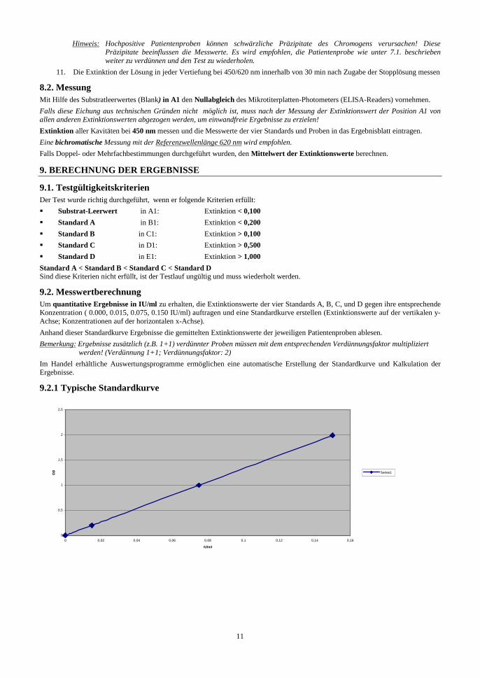
9.2. Messwertberechnung Um quantitative Ergebnisse in IU/ml zu erhalten, die Extinktionswerte der vier Standards A, B, C, und D gegen ihre entsprechende Konzentration ( 0.000, 0.015, 0.075, 0.150 IU/ml) auftragen und eine Standardkurve erstellen (Extinktionswerte auf der vertikalen y-Achse; Konzentrationen auf der horizontalen x-Achse).
Anhand dieser Standardkurve Ergebnisse die gemittelten Extinktionswerte der jeweiligen Patientenproben ablesen.
Bemerkung: Ergebnisse zusätzlich (z.B. 1+1) verdünnter Proben müssen mit dem entsprechenden Verdünnungsfaktor multipliziert werden! (Verdünnung 1+1; Verdünnungsfaktor: 2)
Im Handel erhältliche Auswertungsprogramme ermöglichen eine automatische Erstellung der Standardkurve und Kalkulation der Ergebnisse.
9.2.1 Typische Standardkurve
0
0,5
1
1,5
2
2,5
0 0,02 0,04 0,06 0,08 0,1 0,12 0,14 0,16
IU/ml
OD Series1
12
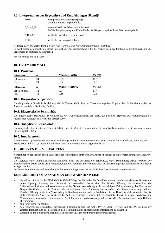
9.3. Interpretation der Ergebnisse und Empfehlungen [IU/ml]* <0,01 Kein protektiver Antikörperspiegel! Grundimmunisierung empfohlen.
0,01 – 0,09 Kein verlässlicher Schutz vor Infektion! Auffrischungsimpfung und Kontrolle der Antikörperspiegel nach 4-6 Wochen empfohlen.
0,10 – 1,0 Verlässlicher Schutz vor Infektion!
>1,0 Verlässlicher Langzeit-Schutz! 10 Jahren nach der letzten Impfung wird eine Kontrolle und Auffrischungsimpfung empfohlen. Es wird empfohlen sowohl die Basis- als auch die Auffrischimpfung 4 bis 6 Wochen nach der Impfung zu kontrollieren und die Ergebnisse im Impfpass zu vermerken.
*In Anlehnung an: RKI 1999
10. TESTMERKMALE
10.1. Präzision Intraassay n Mittelwert (OD) Vk (%)
Schwach pos. 24 0.95 12.1 Pos. 24 3.02 2.1
Interassay n Mittelwert (IU/ml) Vk (%)
Schwach pos. 12 0.04 11.4 Pos. 12 0.16 2.8
10.2. Diagnostische Spezifität Die diagnostische Spezifität ist definiert als die Wahrscheinlichkeit des Tests, ein negatives Ergebnis bei Fehlen des spezifischen Analyten zu liefern. Sie beträgt 84,6%.
10.3. Diagnostische Sensitivität
Die diagnostische Sensitivität ist definiert als die Wahrscheinlichkeit des Tests, ein positives Ergebnis bei Vorhandensein des spezifischen Analyten zu liefern. Sie beträgt 100%.
10.4. Analytische Sensitivität Die analytische Sensitivität des Tests ist definiert als die kleinste Konzentration, die vom Nullstandard unterschieden werden kann. Sie beträgt 0.01 IU/ml.
10.5. Interferenzen Hämolytische,. lipämische und ikterische Proben ergaben bis zu einer Konzentration von 10 mg/ml für Hämoglobin, von 5 mg/ml Triglyceride und von 0,2 mg/ml für Bilirubin keine Interferenzen im vorliegenden ELISA.
11. GRENZEN DES VERFAHRENS
Kontamination der Proben durch Bakterien oder wiederholtes Einfrieren und Auftauen können zu einer Veränderung der Messwerte führen. Die Diagnose einer Infektionskrankheit darf nicht allein auf der Basis des Ergebnisses einer Bestimmung gestellt werden. Die anamnestischen Daten sowie die Symptomatologie des Patienten müssen zusätzlich zu den serologischen Ergebnissen in Betracht gezogen werden. Bei Immunsupprimierten und Neugeborenen besitzen die Ergebnisse der serologischen Tests nur einen begrenzten Wert.
12. SICHERHEITSMASSNAHMEN UND WARNHINWEISE
� Gemäß Art. 1 Abs. 2b der EU-Richtlinie 98/79/EG legt der Hersteller die Zweckbestimmung von In-vitro-Diagnostika fest, um deren Eignung, Leistung und Sicherheit sicherzustellen. Daher sind die Testdurchführung, die Information, die Sicherheitsmaßnahmen und Warnhinweise in der Gebrauchsanweisung strikt zu befolgen. Bei Anwendung des Testkits auf Diagnostika-Geräten ist die Testmethode zu validieren. Jede Änderung am Aussehen, der Zusammensetzung und der Testdurchführung sowie jede Verwendung in Kombination mit anderen Produkten, die der Hersteller nicht autorisiert hat, ist nicht zulässig; der Anwender ist für solche Änderungen selbst verantwortlich. Der Hersteller haftet für falsche Ergebnisse und Vorkommnisse aus solchen Gründen nicht. Auch für falsche Ergebnisse aufgrund von visueller Auswertung wird keine Haftung übernommen.
� Nur für in-vitro-Diagnostik. � Alle verwendeten Bestandteile menschlichen Ursprungs sind auf Anti-HIV-AK, Anti-HCV-AK und HBsAG nicht-reaktiv
getestet. Dennoch sind alle Materialien als potentiell infektiös anzusehen und entsprechend zu behandeln. � Reagenzien und Mikrotiterplatten unterschiedlicher Chargen nicht untereinander austauschen.
13
� Keine Reagenzien anderer Hersteller zusammen mit den Reagenzien dieses Testkits verwenden. � Nicht nach Ablauf des Verfallsdatums verwenden. � Nur saubere Pipettenspitzen, Dispenser und Labormaterialien verwenden. � Verschlusskappen der einzelnen Reagenzien nicht untereinander vertauschen. � Flaschen sofort nach Gebrauch fest verschließen, um Verdunstung und mikrobielle Kontamination zu vermeiden. � Nach dem ersten Öffnen Konjugat- und Standardfläschchen vor weiterem Gebrauch auf mikrobielle Kontamination prüfen. � Zur Vermeidung von Kreuzkontamination und falsch erhöhten Resultaten Patientenproben und Konjugat sorgfältig in die
Kavitäten pipettieren. � Der NovaLisa™ ELISA ist nur für die Anwendung durch Fachpersonal vorgesehen, welches die Arbeitstechniken einwandfrei
beherrscht.
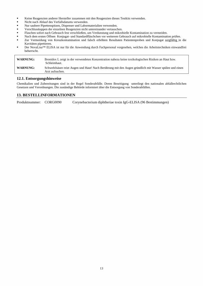
WARNUNG: Bronidox L zeigt in der verwendeten Konzentration nahezu keine toxikologischen Risiken an Haut bzw. Schleimhaut.
WARNUNG: Schwefelsäure reizt Augen und Haut! Nach Berührung mit den Augen gründlich mit Wasser spülen und einen Arzt aufsuchen.
12.1. Entsorgungshinweise Chemikalien und Zubereitungen sind in der Regel Sonderabfälle. Deren Beseitigung unterliegt den nationalen abfallrechtlichen Gesetzen und Verordnungen. Die zuständige Behörde informiert über die Entsorgung von Sonderabfällen.
13. BESTELLINFORMATIONEN
Produktnummer: CORG0090 Corynebacterium diphtheriae toxin IgG-ELISA (96 Bestimmungen)
14
FRANCAIS
1. INTRODUCTION
Les corynébactéries sont des bacilles Gram-positifs, aérobies, non sporulés et de forme irrégulière (0.5-1 µm d’épaisseur et 2-6 µm de longueur). Les corynébactéries incluent des bactéries commensales cutanées, des pathogènes opportunistes et plusieurs pathogènes majeurs, y compris Corynebacterium diphtheriae. En général, elles sont isolées dans les prélèvements de gorge sur des milieux sélectifs qui contiennent du tellurite. La diphtérie, infection bactérienne provoquée par Corynebacterium diphtheriae, a deux formes. La diphtérie respiratoire est typiquement causée par des souches productrices de toxine ; la maladie cutanée peut être causée par des souches toxinogènes ou non toxinogènes. Dans la forme respiratoire de la maladie, une membrane se développe ; celle-ci est généralement visible sur la gorge ou les amygdales. La membrane peut être mortelle si elle obstrue la respiration. D'autres complications sont provoquées par des effets à distance de la toxine diphtérique (myocardite, lésions nerveuses -paralysie,). D’habitude, la diphtérie cutanée est bénigne, et comprend des blessures superficielles et des petits ulcères. Des complications toxiques n’apparaissent que rarement (1-2% des infections avec des souches productrices de toxine). La diphtérie était l'une des causes les plus communes de mort parmi les enfants avant l’arrivée des vaccinations. Depuis l'introduction et l'utilisation répandue de l’anatoxine diphtérique (toxine diphtérique inactivée au formol), la maladie est actuellement caractérisée par des cas sporadiques et des manifestations intermittentes faibles, dans la plupart des pays industrialisés. Mais des grandes épidémies récentes de la diphtérie dans plusieurs pays de l’Europe de l'Est ont de nouveau tiré l'attention sur cette maladie "oubliée" – et la majorité de ces cas se sont produits chez des adolescents et des adultes au lieu des enfants.
Espèce La maladie Symptômes Mécanisme de l'infection Corynebacterium diphtheriae
Diphtérie (respiratoire) Maux de gorge et fièvre légère, gonflement du cou par inflammation ("cou proconsulaire") Complications: dommages à d’autres organes, provoqués par l’exotoxine
Transmission d’individu à individu par contact étroit (physique ou respiratoire) La transmission augmente dans des conditions socio-économiques défavorables
La seule manière efficace de contrôler la diphtérie est une immunisation prophylactique avec l’anatoxine diphtérique. Des anticorps anti-anatoxine protègent contre la toxine ; des personnes immunisées peuvent être infectées par des souches toxinogènes de la diphtérie, mais dans ces cas-là, les manifestations systémiques de la diphtérie ne se produisent pas. Les chances de guérison s'améliorent avec un traitement précoce et approprié. Un diagnostic immédiat est important pour assurer un traitement précoce et approprié avec l'antitoxine diphtérique.
L'infection peut être identifiée par :
� Microscopie : Coloration Gram � Sérologie : Détection de la production de toxine par ELISA
2. INDICATION D’UTILISATION
La trousse Corynebacterium diphtheriae toxin IgG ELISA de NovaTec est prévue pour la détermination quantitative des anticorps IgG anti-toxine de Corynebacterium diphtheriae dans le sérum humain ou plasma (citrate). Elle permet la détermination du statut immun des patients et déterminer ainsi individuellement la nécessité d'une immunisation ou d’un rappel.
3. PRINCIPE DU DOSAGE
La détermination immunoenzymatique quantitative des anticorps IgG anti-toxine de Corynebacterium diphtheriae est basée sur la technique ELISA (Enzyme-Linked Immunosorbent Assay). Les puits des barrettes de microtitration sont revêtus d’antigènes de toxine pour lier les anticorps correspondants de l’échantillon. Après le lavage des puits pour éliminer l’échantillon non lié, le conjugué anti-IgG humaines à la peroxydase du raifort (HRP) est ajouté. Ce conjugué se lie aux anticorps capturés spécifiques de la toxine de Corynebacterium diphtheria. Le complexe immun constitué par le conjugué lié est visualisé en ajoutant le substrat de Tétraméthylbenzidine (TMB) qui donne un produit de réaction bleu. L'intensité de ce produit est proportionnelle à la quantité d'anticorps IgG spécifiques de la toxine de Corynebacterium diphtheriae dans l’échantillon de patient. De l'acide sulfurique est ajouté pour arrêter la réaction. Ceci produit une couleur jaune. L'absorbance à 450 nm est lue en utilisant un lecteur de microplaques ELISA.
4. MATERIEL
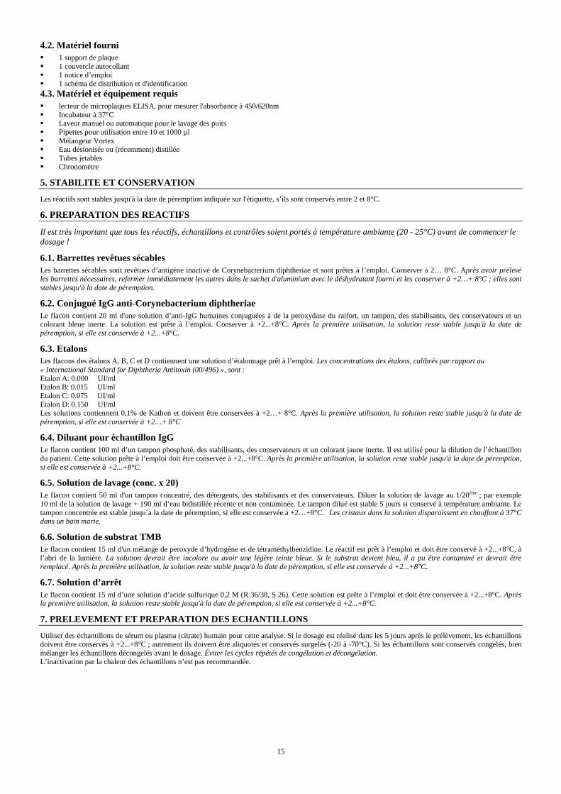
4.1. Réactifs fournis � Puits revêtus de toxine de Corynebacterium diphtheriae (IgG) : 12 barrettes de 8 puits sécables revêtus d’antigène de toxine de Corynebacterium
diphtheriae; en sachets d'aluminium refermables.
� Diluant pour échantillon IgG *** : 1 flacon contenant 100 ml de tampon pour la dilution de l'échantillon ; pH 7.2 ± 0.2 ; prêt à l’emploi ; couleur jaune ; bouchon blanc.
� Solution d'arrêt : 1 flacon contenant 15 ml d'acide sulfurique, 0.2 mol/l ; prêt à l’emploi ; couvercle rouge.
� Solution de lavage (concentrée x 20.) * : 1 flacon contenant 50 ml d'un tampon concentré 20 fois (pH 7.2 ± 0.2) pour laver les puits ; bouchon blanc.
� Conjugué IgG anti-toxine de Corynebacterium diphtheriae ** : 1 flacon contenant 20 ml d'anticorps de lapin conjugués à de la peroxydase du raifort ; prêt à l’emploi ; couleur bleue, bouchon noir.
� Solution de substrat TMB : 1 flacon contenant 15 ml de 3,3',5,5'-tétraméthylbenzidine (TMB) ; prêt à l’emploi ; bouchon jaune.
� Etalons IgG toxine de Corynebacterium diphtheriae ***: 4 flacons contenant chacun 2ml; prêt à l’emploi couleur jaune : Etalon A: 0.000 UI/ml; bouchon bleu Etalon B: 0.015 UI/ml; bouchon vert Etalon C: 0.075 UI/ml; bouchon jaune
Etalon D: 0.150 UI/ml; bouchon rouge
* contient 0,1 % de Bronidox L après dilution ** contient 0,2 % de Bronidox L *** contient 0,1 % de Kathon
15
4.2. Matériel fourni � 1 support de plaque � 1 couvercle autocollant � 1 notice d’emploi � 1 schéma de distribution et d'identification
4.3. Matériel et équipement requis � lecteur de microplaques ELISA, pour mesurer l'absorbance à 450/620nm � Incubateur à 37°C � Laveur manuel ou automatique pour le lavage des puits � Pipettes pour utilisation entre 10 et 1000 µl � Mélangeur Vortex � Eau désionisée ou (récemment) distillée � Tubes jetables � Chronomètre
5. STABILITE ET CONSERVATION
Les réactifs sont stables jusqu'à la date de péremption indiquée sur l'étiquette, s’ils sont conservés entre 2 et 8°C.
6. PREPARATION DES REACTIFS
Il est très important que tous les réactifs, échantillons et contrôles soient portés à température ambiante (20 - 25°C) avant de commencer le dosage !
6.1. Barrettes revêtues sécables Les barrettes sécables sont revêtues d’antigène inactivé de Corynebacterium diphtheriae et sont prêtes à l’emploi. Conserver à 2… 8°C. Après avoir prélevé les barrettes nécessaires, refermer immédiatement les autres dans le sachet d'aluminium avec le déshydratant fourni et les conserver à +2…+ 8°C ; elles sont stables jusqu'à la date de péremption.
6.2. Conjugué IgG anti-Corynebacterium diphtheriae Le flacon contient 20 ml d'une solution d’anti-IgG humaines conjuguées à de la peroxydase du raifort, un tampon, des stabilisants, des conservateurs et un colorant bleue inerte. La solution est prête à l’emploi. Conserver à +2...+8°C. Après la première utilisation, la solution reste stable jusqu'à la date de péremption, si elle est conservée à +2...+8°C. 6.3. Etalons Les flacons des étalons A, B, C et D contiennent une solution d’étalonnage prêt à l’emploi. Les concentrations des étalons, calibrés par rapport au « International Standard for Diphtheria Antitoxin (00/496) », sont : Etalon A: 0.000 UI/ml Etalon B: 0.015 UI/ml Etalon C: 0.075 UI/ml Etalon D: 0.150 UI/ml Les solutions contiennent 0,1% de Kathon et doivent être conservées à +2…+ 8°C. Après la première utilisation, la solution reste stable jusqu'à la date de péremption, si elle est conservée à +2…+ 8°C
6.4. Diluant pour échantillon IgG Le flacon contient 100 ml d’un tampon phosphaté, des stabilisants, des conservateurs et un colorant jaune inerte. Il est utilisé pour la dilution de l’échantillon du patient. Cette solution prête à l’emploi doit être conservée à +2...+8°C. Après la première utilisation, la solution reste stable jusqu'à la date de péremption, si elle est conservée à +2...+8°C. 6.5. Solution de lavage (conc. x 20) Le flacon contient 50 ml d'un tampon concentré, des détergents, des stabilisants et des conservateurs. Diluer la solution de lavage au 1/20ème ; par exemple 10 ml de la solution de lavage + 190 ml d’eau bidistillée récente et non contaminée. Le tampon dilué est stable 5 jours si conservé à température ambiante. Le tampon concentrée est stable jusqu´a la date de péremption, si elle est conservée à +2…+8°C. Les cristaux dans la solution disparaissent en chauffant à 37°C dans un bain marie.
6.6. Solution de substrat TMB Le flacon contient 15 ml d'un mélange de peroxyde d’hydrogène et de tétraméthylbenzidine. Le réactif est prêt à l’emploi et doit être conservé à +2...+8°C, à l’abri de la lumière. La solution devrait être incolore ou avoir une légère teinte bleue. Si le substrat devient bleu, il a pu être contaminé et devrait être remplacé. Après la première utilisation, la solution reste stable jusqu'à la date de péremption, si elle est conservée à +2...+8°C.
6.7. Solution d’arrêt Le flacon contient 15 ml d’une solution d’acide sulfurique 0,2 M (R 36/38, S 26). Cette solution est prête à l’emploi et doit être conservée à +2...+8°C. Après la première utilisation, la solution reste stable jusqu'à la date de péremption, si elle est conservée à +2...+8°C.
7. PRELEVEMENT ET PREPARATION DES ECHANTILLONS
Utiliser des échantillons de sérum ou plasma (citrate) humain pour cette analyse. Si le dosage est réalisé dans les 5 jours après le prélèvement, les échantillons doivent être conservés à +2...+8°C ; autrement ils doivent être aliquotés et conservés surgelés (-20 à -70°C). Si les échantillons sont conservés congelés, bien mélanger les échantillons décongelés avant le dosage. Éviter les cycles répétés de congélation et décongélation. L’inactivation par la chaleur des échantillons n’est pas recommandée.
16
7.1. Dilution de l’échantillon Avant le dosage, tous les échantillons doivent être dilués au 1/101ème avec le diluant pour échantillon IgG. Diluer 10 µl d’échantillon avec 1 ml du diluant pour échantillon IgG dans des tubes pour obtenir une dilution au 1/101ème et mélanger soigneusement sur un Vortex. Pour des patients avec des concentrations d’antitoxine prévues plus élevées que l’étalon D (0.15 UI/ml), une deuxième dilution au 1/2ème de cet échantillon dilué au 1/101ème devrait être utilisée; par exemple 100 µl de la première dilution d'échantillon + 100 µl du diluant pour échantillon IgG (mélanger soigneusement). Facteur de dilution : 2
8. PROCEDE DE DOSAGE
8.1. Préparation du dosage Lire attentivement la notice d’emploi avant de réaliser le dosage. La fiabilité des résultats dépend du suivi strict du protocole. La technique de dosage suivante a été validée uniquement pour une procédure manuelle. Si le dosage doit être effectué sur un automate, nous conseillons d’augmenter le nombre d’étapes de lavage de trois à cinq et le volume de la solution de lavage de 300 à 350 µl. Avant de commencer le dosage, déterminer, sur le formulaire fourni dans la trousse, le plan de distribution et d’identification des échantillons et des étalons. Sélectionner le nombre de barrettes ou de puits nécessaires et les placer sur le support.
Veuillez assigner au moins : 1 puits (par exemple A1) pour le blanc substrat, 4 puits (par exemple B1, C1, etc.) pour les étalons A, B, C et D.
Il est conseillé de déterminer les échantillons du patient en doublets si nécessaire. Réaliser toutes les étapes du dosage dans l'ordre donné et sans délai entre les étapes. Un embout de pipette propre et jetable doit être utilisé pour distribuer chaque étalon et échantillon. Régler l'incubateur à 37° ± 1°C.
1. Pipeter 100 µl de chaque étalon (A, B, C et D) et d’échantillons dilués dans leurs puits respectifs. Gardez le puits A1 pour le blanc substrat. 2. Couvrir les puits avec le couvercle. 3. Incuber pendant 1 heure ± 5 minutes à 37±1°C. 4. A la fin de l'incubation, enlever le couvercle, aspirer le contenu des puits et laver chaque puits trois fois avec 300 µl de solution de lavage. Éviter
les débordements des puits de réaction. Le temps de trempage entre chaque cycle de lavage devrait être > 5 sec. À la fin, enlever soigneusement le liquide restant en tapotant les barrettes sur du papier absorbant avant la prochaine étape !
Note : L‘étape de lavage est très importante ! Un lavage insuffisant peut conduire à une précision faible et à des valeurs d'absorbance faussement élevées.
5. Pipeter 100 µl du conjugué IgG anti-Corynebacterium diphtheriae dans tous les puits sauf le puits blanc (par exemple A1). Fermer avec le couvercle.
6. Incuber pendant 30 minutes à température ambiante (20…25°C). Ne pas exposer à la lumière directe du soleil. 7. Répéter l'étape numéro 4. 8. Pipeter 100 µl de la solution de substrat TMB dans tous les puits.
9. Incuber pendant exactement 15 minutes à température ambiante (20…25°C) dans l'obscurité.
10. Pipeter 100 µl de la solution d'arrêt dans tous les puits dans le même ordre et à la même vitesse que pour la solution de substrat TMB. La couleur bleue développée pendant l'incubation vire au jaune. Note : Des échantillons de patients fortement positifs peuvent causer des précipités foncés du chromogène ! Ces précipités peuvent influencer
les valeurs mesurées de densité optique. Diluer l’échantillon comme indiqué au chapitre 7.1 Dilution de l’échantillon. 11. Mesurer l'absorbance de l’échantillon à 450/620nm dans les 30 minutes après l'addition de la solution d'arrêt.
8.2. Mesure Faire le zéro du lecteur ELISA à l’aide du blanc substrat dans le puits A1.
Si - pour des raisons techniques - le lecteur d'ELISA ne peut pas être ajusté à zéro en utilisant le blanc substrat dans le puits A1, soustraire la valeur d'absorbance du puits A1 de toutes les autres valeurs d’absorbance mesurées afin d'obtenir des résultats fiables !
Mesurer l'absorbance de tous les puits à 450 nm et enregistrer les valeurs d'absorbance pour chaque contrôle et échantillon de patient dans le plan de distribution et d'identification.
Une lecture en double longueur d'onde employant 620 nm comme longueur d'onde de référence est conseillée.
Calculer les valeurs moyennes d'absorbance pour tous les doublets, si nécessaire.
9. RESULTATS
9.1. Critères de validation Afin qu’une analyse soit considérée valide, les critères suivants doivent être respectés :
� Blanc Substrat dans A1 : Valeur d’absorbance < 0,100. Etalon A dans B1 : Valeur d’absorbance < 0,200. Etalon B dans C1: Valeur d’absorbance > 0.100 Etalon C dans D1: Valeur d’absorbance > 0.500
� Etalon D dans E1: Valeur d’absorbance > 1.000
Etalon A < Etalon B < Etalon C < Etalon D Lorsque ces critères ne sont pas remplis, le test n’est pas valide et doit être recommencé.
9.2. Calcul des résultats Afin d'obtenir des résultats quantitatifs en UI/ml, inscrire les valeurs (moyennes) d'absorbance des 4 étalons A, B, C et D sur du papier millimétré bilinéaire. Inscrire les valeurs d’absorbance en ordonnées et leurs concentrations correspondantes (0.000, 0.015, 0.075 et 0.150 UI/ml ) en abscisses, et dessiner une courbe d'étalonnage. Lire les résultats sur cette courbe d’étalonnage en utilisant les valeurs (moyennes) d'absorbance de chaque échantillon patient et de chaque étalon.
NOTE : Les résultats des échantillons dilués une deuxième fois (1/2ème) doivent être multipliés par le facteur de dilution approprié afin d'obtenir des résultats corrects ! (dilution : 1/2ème = facteur de dilution : 2).
Tous les programmes informatiques appropriés peuvent être utilisés pour la lecture et le calcul automatiques des résultats.
17
9.3. Courbe d’étalonnage typique
0,000
0,345
0,601
1,136
0,000
0,200
0,400
0,600
0,800
1,000
1,200
0 50 100 150 200 250 IU/ml
OD
9.4 Interprétation des résultats du test [IU/ml]*
< 0,01 Absence d’anticorps et de protection ! Il est recommandé de procéder immédiatement à une vaccination complète !
0,01–0,09 Taux d’anticorps faible, aucune protection fiable ! Il est recommandé d’effectuer immédiatement une injection de rappel
0,1 –1,0 Protection fiable !
> 1,0 Protection fiable à long terme ! Un contrôle et une injection de rappel sont conseillés au bout de 10 ans environ.
Il est recommandé de vérifier l’effacité de la vaccination ou du rappel 4 à 6 semaines après l’injection et de consigner l’information dans le carnet de vaccination.
* En reference à: RKI 1999
10. PERFORMANCES DU DOSAGE
10.1. Précision Intra-essai n moyenne (OD) CV (%)
Tangent pos. 24 0.95 12.1 Pos. 24 3.02 2.1
Inter-essais n moyenne (UI/ml) cv (%) Tangent pos. 12 0.04 11.4 Pos. 12 0.16 2.8
10.2. Spécificité diagnostique La spécificité diagnostique est définie comme la probabilité d’obtenir un résultat négatif en l'absence d'un analyte spécifique. Elle est de 84.6 %.
10.3. Sensibilité diagnostique La sensibilité diagnostique est définie comme la probabilité d’obtenir un résultat positif en présence d'un analyte spécifique. Elle est de 100 %.
10.4. Sensibilité Analytique La sensibilité analytique définie comme la concentration apparente de l'analyte qui peut être distinguée de l’étalon zéro est 0.01 UI/ml.
10.5. Interférences Des sérums hémolytiques ou lipémiques ou ictériques n’ont pas montré d’interférences, avec des concentrations jusqu’à 10 mg/ml de hémoglobine, 5 mg/ml de triglycérides et 0,2 mg/ml de bilirubine.
Attention: Les résultats concernent des groupes d’échantillons testés ; ces spécifications ne sont pas garanties.
11. LIMITES DE LA TECHNIQUE
Une contamination bactérienne ou des cycles de congélation-décongélation répétés de l’échantillon peuvent affecter les valeurs d'absorbance. Le diagnostic d'une maladie infectieuse ne devrait pas être établi sur la base du résultat d’une seule analyse. Un diagnostic précis devrait tenir compte de l'historique clinique, de la symptomatologie ainsi que des données sérologiques. Les données sérologiques sont de valeur restreinte dans le cas des patients immunodéficients et des nouveaux-nés.
12. PRECAUTIONS ET AVERTISSEMENTS
� En accord avec l’article 1 paragraphe 2b de la directive européenne 98/79/EC, l’utilisation des dispositifs médicaux de diagnostic in vitro est destinée par le fabricant à garantir le bien-fondé, les performances et la sécurité du produit. Par conséquent, la procédure de dosage, l’information, les précautions et mises en garde de la notice d’emploi, doivent être suivies de façon stricte. L’utilisation de ces trousses avec des automates ou dispositifs similaires doit être validée. Aucun changement de la conception, composition et procédure de dosage, ainsi que l’utilisation avec d’autres produits non approuvés par le fabricant, ne sont autorisés ; seul l’utilisateur est responsable de tels changements. Le fabricant n’est pas responsable des faux résultats et des incidents dus à ces motifs. Le fabricant n’est pas responsable des résultats fournis par analyse visuelle des échantillons des patients.
� Uniquement pour diagnostic in vitro. � Tous les composants d'origine humaine utilisés pour la fabrication de ces réactifs ont été analysés et ont été trouvés non réactifs en Ag HBs, en anticorps
anti-HIV 1 et 2 et en anticorps anti-VHC. Néanmoins, tous les produits doivent être considérés et traités comme étant potentiellement infectieux. � Ne pas échanger les réactifs ou les barrettes provenant de différents lots de production. � Ne pas utiliser de réactifs provenant d'autres fabricants avec les réactifs de cette trousse.

18
� Ne pas utiliser les réactifs après la date de péremption indiquée sur l'étiquette. � Utiliser seulement des embouts de pipette, des distributeurs et du matériel de laboratoire propres. � Ne pas échanger les bouchons des flacons, pour éviter la contamination croisée. � Fermer soigneusement les flacons après utilisation pour éviter l'évaporation et la contamination microbienne. � Avant une nouvelle utilisation, vérifier les flacons de conjugué et de contrôle, déjà utilisés, pour exclure une contamination microbienne. � Pour éviter la contamination croisée et des résultats faussement élevés, introduire les échantillons de patients et le conjugué exactement au fond des puits
sans éclabousser. � Le NovaLisa ELISA est uniquement destiné à l’utilisation par un personnel compétent, maîtrisant parfaitement les techniques de travail.
AVERTISSEMENT : A la concentration utilisée, Bronidox L ne présente pratiquement aucun risque toxicologique en cas de contact avec la peau et les membranes muqueuses !
AVERTISSEMENT : L'acide sulfurique est irritant pour les yeux et la peau. Garder hors de la portée des enfants. En cas de contact avec les yeux, rincer soigneusement avec de l'eau et consulter un médecin !
12.1. Elimination des déchets Les résidus des produits chimiques et des préparations sont considérés en général comme des déchets dangereux. L’élimination de ce type de déchet est réglementée par des lois et réglementations nationales et régionales. Contacter les autorités compétentes ou les sociétés de gestion des déchets pour obtenir des renseignements sur l’élimination des déchets dangereux. 13. INFORMATION POUR LES COMMANDES
Référence : CORG0090 Corynebacterium diphtheriae toxin IgG-ELISA (96 déterminations)
19
ITALIANO
1. INTRODUZIONE
Il Corynebacterium diphtheriae, appartenente alla famiglia delle Corynebacteriaceae, è un bacillo Gram-positivo, asporigeno, aerobio e non mobile. È causa della difterite, un'infezione principalmente a trasmissione aerea che si sviluppa a livello delle prime vie respiratorie. Il batterio penetra a livello di faringe o laringe dove causa una reazione infiammatoria, fibrinoso-necrotica, con caratteristiche pseudomembrane; confinato nelle pseudomembrane, il batterio produce una tossina, che va in circolo agendo sul sistema nervoso, sul cuore, sulle ghiandole surrenali e sugli altri organi. A seconda della localizzazione delle pseudomembrane si possono avere forme diverse di difterite: angina difterica, con membrane in sede faringea; laringite difterica, più rara, in cui le membrane sono localizzate sulle corde vocali e possono portare a ostruzione laringea con soffocamento. La prognosi può essere più o meno sfavorevole con l'exitus che si verifica nel 5-10% dei casi. In caso di decesso, esso è dovuto a insufficienza respiratoria, insufficienza cardiaca o ad un accumulo di tossina nel sistema nervoso. La malattia, un tempo molto diffusa, è ormai rarissima in tutti i paesi dove è stata resa obbligatoria la vaccinazione. Nei paesi tropicali esiste anche una forma di difterite cutanea dovuta a ferite o morsi di insetti.
Specie Malattia Sintomi Modo d’infezione Corynebacterium diphtheriae
Difterite
Malessere generale, gonfiore della gola e dei linfonodi Complicanze: danni su altri organi causati dalla tossina
Trasmissione: aerea; contatto diretto con una persona infetta
Diagnosi � Microscopia: colorazione di Gram � Sierologia: ELISA
2. USO PREVISTO
Il NovaTec Corynebacterium diphtheriae toxin IgG ELISA è un kit per la determinazione quantitativa degli anticorpi specifici della classe IgG per la tossina del Corynebacterium diphtheriae nel siero o plasma (citrato) umano. . Si determina lo stato dell’immunità e verifica la necessità di ulteriori immunizzazioni.
3. PRINCIPIO DEL TEST
La determinazione quantitativa degli anticorpi IgG per la tossina del Corynebacterium diphtheriae si basa sul principio ELISA. I pozzetti delle micropiastre contengono una fase solida con la tossina inattivata del Corynebacterium diphtheriae (antigeni). Anticorpi specifici nel campione si legano agli antigeni immobilizzati nei pozzetti. Gli anticorpi del coniugato (perossidasi di rafano-anticorpi anti-IgG umane) si legano ai complessi antigene (fase solida)-anticorpo (paziente) nei campioni positivi. Questi complessi vengono evidenziati da una colorazione blu dopo l’incubazione con la soluzione TMB. L’intensità di questa colorazione è direttamente proporzionale alla quantità di anticorpi specifici per il Corynebacterium diphtheriae di classe IgG presenti nel campione. Fermando la reazione enzimatica con acido solforico si causa un cambiamento di colore dal blu al giallo che può essere misurato facilmente con un fotometro per l’ELISA a 450 nm.
4. MATERIALI
4.1. Reagenti forniti � Micropiastre con la tossina del Corynebacterium diphtheriae (IgG): 12 strisce divisibili in 8 pozzetti, con adesi la tossina
del Corynebacterium diphtheriae; dentro una busta d’alluminio richiudibile.
� Tampone diluente IgG***: 1 flacone contenente 100 ml di diluente per campioni; pH 7.2 ± 0.2; color giallo; pronto all’uso; tappo bianco.
� Soluzione stop: 1 flacone contenente 15 ml di acido solforico, 0.2 mol/l, pronto all’uso; tappo rosso.
� Tampone di lavaggio (20x conc.)*: 1 flacone contenente 50 ml di un tampone concentrato 20 volte per il lavaggio dei pozzetti; pH 7.2 ± 0.2; tappo bianco.
� Coniugato Corynebacterium diphtheriae tossina anti IgG**: 1 flacone contenente 20 ml di anticorpi anti-IgG umane, coniugati a perossidasi; color azzurro; pronto all’uso; tappo nero.
� Soluzione TMB: 1 flacone contenente 15 ml di 3,3`,5,5`-Tetrametilbenzidina (TMB); pronto all’uso; tappo giallo.
20
� Corynebacterium diphtheriae tossina IgG Standards***: 4 flaconi, contenenti 2 ml, pronti all’uso: Standard A: 0.000 IU/ml; tappo blu Standard B: 0.015 IU/ml; tappo verde Standard C: 0.075 IU/ml; tappo giallo Standard D: 0.150 IU/ml; tappo rosso
* contiene 0.1 % Bronidox L dopo diluizione ** contiene 0.2 % Bronidox L *** contiene 0.1 % Kathon
4.2. Accessori forniti � 1 pellicola adesiva � 1 supporto per micropiastre � 1 istruzione per l’uso � 1 foglio di controllo
4.3. Materiali e attrezzature necessari � Fotometro per micropiastre con filtri da 450/620 nm � Incubatore a 37°C � Lavatore di micropiastre � Micropipette con punte monouso (10, 100, 200, 1000 µl) � Vortex-Mixer � Provette monouso � Supporto per provette � Acqua deionizzata o distillata. � Timer
5. MODALITÀ DI CONSERVAZIONE
I reagenti devono essere conservati tra 2-8°C. Non usare i reagenti dopo la scadenza. La data di scadenza è stampata sull’etichetta di ogni componente e sull’etichetta esterna della confezione.
6. PREPARAZIONE DEI REAGENTI
Portare tutti i reagenti a temperatura ambiente (20-25°C) prima dell’uso!
6.1. Micropiastre I pozzetti sono separabili. Contengono adesi la tossina inattivata del Corynebacterium diphtheriae. I pozzetti, pronti all’uso, devono essere conservati tra 2-8°C. Riporre i pozzetti non utilizzati nel sacchetto con il gel essiccante di silice. Il prodotto è stabile fino alla data di scadenza se conservato tra 2-8°C.
6.2. Coniugato Corynebacterium diphtheriae tossina IgG
Il flacone contiene 20 ml di anticorpi anti-IgG umane coniugati a perossidasi di rafano, stabilizzanti, conservanti e un colorante inerte azzurro. Una volta aperto, il prodotto é stabile fino alla data di scadenza se conservato tra 2-8°C.
6.3. Standards I flaconi contengono 2.0 ml di soluzione Standard pronta all’uso con le seguenti concentrazioni (La concentrazione degli standards si riferiscono al ”International Standard for Diphtheria Antitoxin (00/496)“): Standard A: 0.000 IU/ml Standard B: 0.015 IU/ml Standard C: 0.075 IU/ml Standard D: 0.150 IU/ml Contengono 0,1% Kathon. Una volta aperto, il prodotto é stabile fino alla data di scadenza se conservato tra 2-8°C.
6.4. Tampone diluente IgG Il flacone contiene 100 ml di tampone fosfato, stabilizzanti, conservanti e un colorante giallo inerte. La soluzione viene usata per diluire i campioni . Una volta aperto, il prodotto é stabile fino alla data di scadenza se conservato tra 2-8°C.
6.5. Tampone di lavaggio (20x conc.) Il flacone contiene 50 ml di un tampone concentrato, detergenti e conservanti. Il contenuto viene diluito con acqua deionizzata o distillata (1 + 19). Il tampone diluito é stabile fino 5 giorni se conservato a temperatura ambiente. Se sono presenti cristalli, scioglierli a 37°C prima di diluire. Una volta aperto, il prodotto é stabile fino alla data di scadenza se conservato tra 2-8°C.
6.6. Soluzione TMB Il flacone contiene 15 ml di 3,3`,5,5`-Tetrametilbenzidina (TMB) e perossido di idrogeno pronto all’uso. Conservare al buio. La soluzione é incolore o celeste chiaro. Nel caso in cui diventasse blu significa che é contaminata e non può essere più usata. Una volta aperto, il prodotto é stabile fino alla data di scadenza se conservato tra 2-8°C.
21
6.7. Soluzione Stop Il flacone contiene 15 ml di acido solforico, 0.2 mol/l (R36/38, S26), pronto all’uso. Una volta aperto, il prodotto é stabile fino alla data di scadenza se conservato tra 2-8°C.
7. PRELIEVO E PREPARAZIONE DEI CAMPIONI
Usare campioni di siero o plasma (citrato) umano. Se il test viene fatto entro 5 giorni dal prelievo i campioni possono essere conservati tra 2-8°C. Altrimenti devono essere aliquotati e congelati tra -70…-20°C. Agitare bene i campioni scongelati prima di diluirli. Evitare cicli ripetuti di congelamento/scongelamento. L’inattivazione dei campioni per mezzo del calore non è raccomandata.
7.1. Diluizione dei campioni Prima del test, diluire i campioni 1+100 con tampone diluente IgG. Per esempio, pipettare nelle provette 10 µl di campione + 1 ml di tampone e mescolare bene (Vortex). Per pazienti con probabili concentrazioni superiori allo Standard D (0.15 IU/ml) i campioni devono essere ulteriormente diluiti dopo la diluizione discritta sopra 1+1. Es. 100µl della prima diluizione + 100µl tampone diluente IgG (Fattore di diluizione: 2).
8. PROCEDIMENTO
8.1. Preparazione del test Leggere bene le istruzioni prima di iniziare il dosaggio. Per ottenere risultati validi é indispensabile seguire esattamente le istruzioni. La seguente procedura è stata validata per l’esecuzione manuale. Per una esecuzione su strumentazione automatica si consiglia di incrementare il numero di lavaggi de 3 a 5 volte e il volume della soluzione di lavaggio da 300 a 350µl per evitare interferenze. Stabilire innanzitutto il piano di distribuzione ed identificazione dei campioni e controlli sul foglio di lavoro fornito con il kit. Inserire i pozzetti necessari nel supporto micropiastre
Utilizzare almeno:
1 pozzetto (es. A1) per il bianco-substrato (blank) 4 pozzetti (z.B. B1, C1, etc) per gli Standards A, B, C e D.
È consigliato effettuare ogni analisi in duplicato.
Eseguire il test nell’ordine stabilito dalle istruzioni, senza pause.
Utilizzare puntali nuovi e puliti per ogni campione e standard.
Regolare l’incubatore a 37° ± 1°C
1. Pipettare 100 µl degli standards A B C D e di campione diluito nei relativi pozzetti. Usare il pozzetto A1 per il bianco-substrato.
2. Coprire i pozzetti con la pellicola adesiva.
3. Incubare 1 ora ± 5 min a 37° ±±±± 1°C.
4. Al termine dell’incubazione, togliere la pellicola ed aspirare il liquido dai pozzetti. Successivamente lavare i pozzetti tre volte con 300 µl di tampone di lavaggio. Evitare che la soluzione trabocchi dai pozzetti. L’intervallo tra il lavaggio e l’aspirazione deve essere almeno di 5 sec. Dopo il lavaggio picchiettare delicatamente i pozzetti con l’apertura verso il basso su una carta assorbente per togliere completamente il liquido.
Attenzione: Il lavaggio é una fase critica. Un lavaggio non accurato determina una cattiva precisione del test ed un innalzamento falsato delle densità ottiche.
5. Pipettare 100µl di Coniugato Corynebacterium diphtheriae tossina anti-IgG in tutti i pozzetti, escludendo quello con il bianco-substrato (blank). Coprire i pozzetti con la pellicola adesiva.
6. Incubare 30 min a temperatura ambiente (20°...25°C). Non esporre a fonti di luce diretta.
7. Ripetere il lavaggio secondo punto 4.
8. Pipettare 100µl di Soluzione TMB in tutti i pozzetti.
9. Incubare precisamente per 15 min a temperatura ambiente (20°...25°C) al buio.
10. Pipettare 100µl di Soluzione Stop in tutti i pozzetti, nello stesso ordine della soluzione TMB. Durante l’incubazione il colore cambia dal blu al giallo.
Attenzione: Campioni con un risultato positivo molto alto possono causare precipitati scuri del cromogeno! Questi precipitati influenzano la lettura delle densità ottiche. È consigliato diluire i campioni un’altra volta come discritto in 7.1 e di ripetere il test.
11. Misurare l’assorbanza di tutti i pozzetti a 450/620 nm entro 30 min dopo l’aggiunta della soluzione stop.
22
8.2. Misurazione Regolare il fotometro per le micropiastre (ELISA-Reader) a zero usando il substrato-bianco (blank) in A1. Se, per motivi tecnici, non é possibile regolare il fotometro sottrarre l’assorbanza del bianco-substrato da tutti i valori delle altre assorbanze.
Misurare l’assorbanza di tutti i pozzetti a 450 nm e inserire tutti i valori misurati nel foglio di lavoro.
É raccomandato fare una misurazione delle densità ottiche a doppia lunghezza d’onda utilizzando i 620 nm come lunghezza di riferimento.
Dove sono state misurate in doppio, calcolare la media delle assorbanze.
9. RISULTATI
9.1. Validazione del test Il test é valido se risponde ai prossimi criteri:
� Substrato bianco in A1: Assorbanza < 0.100
� Standard A in B1: Assorbanza < 0.200 Standard B in C1: Assorbanza > 0.100 Standard C in D1: Assorbanza > 0. 500
� Standard D in E1: Assorbanza > 1.000
Standard A < Standard B < Standard C < Standard D Se non vengono soddisfatti questi criteri, il test non è valido e deve essere ripetuto.
9.2. Calcolo dei risultati Per ottenere i risultati quantitativi in IU/ml , realizzare un diagramma cartesiano ponendo le assorbanze degli Standards A, B, C, e D sull’asse della Y e ponendo le concentrazioni (0.000, 0.015, 0.075, 0.150 IU/ml) sull’asse della X su cui tracciare la curva standard.
Traendo i risultati da questa curva standard vanno inseriti i valori medi delle assorbanze dei campioni di ogni singolo paziente.
Attenzione: I risultati dei campioni diluiti due volte devono essere moltiplicati con il fattore di diluizione ! (Diluizione 1+1; fattore di diluizione: 2)
Tutti i programmi per computer commercializzati sono utilizzabili per calcolare ed interpretare i risultati.
9.2.1 Tipica curva standard
0
0,5
1
1,5
2
2,5
0 0,02 0,04 0,06 0,08 0,1 0,12 0,14 0,16
IU/ml
OD Reihe1
9.3. Interpretazione dei risultati e raccomandazioni [IU/ml]* <0.01 Nessuna protezione; mancanza di anticorpi! L’immunizzazione é raccomandata.
0.01 – 0.09 La protezione da infezioni non é assicurata! Una ripetizione dell’immunizzazione e il controllo dopo 4-6 settimane sono raccomandati.
0.10 – 1.0 Protezione piú o meno sicura da infezioni! Una ripetizione dell’immunizzazione e il controllo dopo 4-6 settimane sono raccomandati.
>1.0 Protezione sicura da infezioni!
Dopo circa 10 anni dall’ultimo richiamo si raccomanda un controllo e un nuovo richiamo.
I raccomanda che l’immunizzazione di base o il richiamo siano controllati dopo 4-6 settimane dopo l’immunizzazione e che la data sia riportata sul certificato di vaccinazione.
* in accordo con: RKI 1999
23
10. CARATTERISTICHE DEL TEST
10.1. Precisione Interdosaggio n Media (IU/ml) Cv (%)
Lievemente pos. 24 0.95 12.1 Pos. 24 3.02 2.1
Intradosaggio n Media (OD) CV (%)
Lievemente pos. 12 0.04 11.4 Pos. 12 0.16 2.8
10.2. Specificità diagnostica La specificità diagnostica é la probabilità del test di fornire un risultato negativo in assenza di anticorpi specifici. La specificità diagnostica é pari a 84.6%.
10.3. Sensibilità diagnostica La sensibilità diagnostica é la probabilità del test di fornire un risultato positivo in presenza di anticorpi specifici. La sensibilità diagnostica é pari a 100%.
10.4. Sensitività analitica La sensitività analitica del test é definita dalla concentrazione più piccola che può essere distinta dallo standard A. Essa equivale a 0.01 IU/ml.
10.5. Possibili interferenze Campioni emolitici, lipidici ed itterici contenenti fino a 10 mg/mL di emoglobina, 5 mg/mL di trigliceridi e 0,2 mg/mL di bilirubina non hanno presentato fenomeni di interferenza nel presente test.
Nota: I risultati si riferiscono al gruppo di campioni realizzati, questi non sono specifiche garantite.
11. LIMITAZIONI
Una contaminazione da microorganismi o ripetuti cicli di congelamento-scongelamento possono alterare i valori delle assorbanze. La diagnosi di una malattia infettiva non deve essere fatta soltanto sulla risultanza di un unico test. È importante considerare anche l’anamnesi ed i sintomi del paziente. I risultati del test da pazienti immunosoppressi e neonati hanno un valore limitato.
12. PRECAUZIONI E AVVERTENZE
� In ottemperanza all’articolo 1, paragrafo 2 della direttiva Europea 98/79/EC, l’uso dei diagnostici medici in vitro è inteso da parte del produttore ad assicurare la congruenza, le prestazioni e la sicurezza del prodotto. Di conseguenza la procedura analitica, le informazioni, le precauzioni e le avvertenze contenute nelle istruzioni per l’uso devono essere seguite scrupolosamente. L’uso dei kit con analizzatori e attrezzature similari deve essere previamente convalidato. Qualunque cambiamento nello scopo, nel progetto, nella composizione o struttura e nella procedura analitica, così come qualunque uso dei kit in associazione ad altri prodotti non approvati dal produttore non è autorizzato; l’utilizzatore stesso è responsabile di questi eventuali cambiamenti. Il produttore non è responsabile per falsi risultati e incidenti che possano essere causati da queste ragioni. Il produttore non è responsabile per qualunque risultato ottenuto attraverso esame visivo dei campioni dei pazienti.
� Solo per uso diagnostico in-vitro. � Tutti i componenti di origine umana sono stati trovati non reattivi con Anti-HIV-Ab, Anti-HCV-Ab e HBsAg. Nonostante ciò e
tutti i materiali devono comunque essere considerati potenzialmente contagiosi e infettivi. � Non scambiare reagenti e micropiastre di lotti diversi. � Non utilizzare reagenti di altri produttori insieme con i reagenti di questo kit. � Non usare dopo la data di scadenza. � Utilizzare soltanto attrezzatura pulita. � Non scambiare i tappi dei flaconi. � Richiudere i flaconi immediatamente dopo l’uso per evitare la vaporizzazione e contaminazione. � Una volta aperti e dopo relativo stoccaggio verificare i reagenti per una loro eventuale contaminazione prima dell’uso. � Per evitare contaminazioni crociate e risultati erroneamente alti pipettare i campioni e reagenti con molta precisione nei
pozzetti. � Il NovaLisa™ ELISA è previsto soltanto per essere impiegato da parte di personale specializzato che conosce perfettamente le
tecniche di lavoro.
ATTENZIONE: Bronidox L, nella concentrazione usata, mostra quasi assenza di tossicità sulla pelle e sulle mucose. ATTENZIONE: L’acido solforico irrita occhi e pelle! Dopo il contatto sciacquare immediatamente e abbondantemente. Contattare un medico.

24
12.1. Smaltimento In genere tutte le sostanze chimiche vengono considerate rifiuti tossici. Lo smaltimento viene regolato da leggi nazionali. Per ulteriori informazioni contattare l’autorità locale.
13. INFORMAZIONI PER GLI ORDINI
Numero del prodotto: CORG0090 Corynebacterium diphtheriae toxin IgG-ELISA (96 determinazioni)
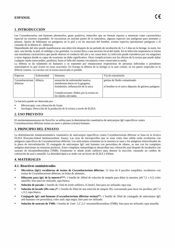
ESPANOL
1. INTRODUCCIÓN
Las Corynebacterias son bastones pleomorfos, gram positivos, inmoviles que no forman esporas y muestran como caracteristica especial un extremo expandido. Se encuentran en muchas partes de la naturaleza, algunas especies son patógenas para animales y plantas. Aparte de habitantes no patógenos en la piel y en las mucosas del hombre, existen especies oportunistas patógenas y el causante de la difetria (C. difteriae). Dependiendo del sitio puede manifiestarse una infección después de un período de incubación de 3 a 5 dias en la faringe, la nariz, los ojos, una herida, la piel, el ombligo o los genitales. La toxina lleva a una necrosis local del tejido. En la infección respiratoria se forma una membrana característica que puede obstruir el conducto del aire y ser causa letal. La infección puede extenderse por vía sanguínea a otros órganos donde es capaz de ocasionar un daño significativo. Otras complicaciones son los efectos de la toxina que puede dañar cualquier tejido (miocarditis, parálisis), hasta el fallo del sistema circulatorio como consecuencia tardía. La difteria se da solamente en humanos y se transmite por emanaciones respiratorias de personas infectadas o portadores asintomáticos lo que ocurre en raras ocaciones. En Europa la difteria de la faringe es la más común, en los paises tropicales es la difteria cutanea. La vacuna con la toxina inactivada es posible.
Especies Enfermedad Síntomas Vía de transmisión
Corynebacterium difteriae
difteria
sensación de enfermedad masiva, membranas duras en la garganta, linfadenitis, inflamación de la nuca Complicaciones: Daños por la toxina en los tejidos afectados
gotitas de fluido contaminado el hombre es el unico déposito de gérmen patógeno
La bacteria puede ser detectada por:
� Microscopia: con coloración de Gram � Serología: Detección de la produción de la toxina a través de ELISA
2. USO PREVISTO
El enzimoinmunoensayo de NovaTec se utiliza para la determinación cuantitativa de anticuerpos IgG específicos contra Corynebacterium difteriae toxina en suero o plasma (citrato) humano.
3. PRINCIPIO DEL ENSAYO
La determinación inmunoenzimatica cuantitativa de anticuerpos específicos contra Corynebacterium difteriae se basa en la técnica ELISA (Enzyme-linked Immunosorbent Assay). Las tiras de micropocillos que se usan como fase sólida están recubiertas con antígenos específicos de Corynebacterium difteriae. Los anticuerpos existentes en la muestra se usan a los antígenos inmovilizados de la placa de microtítulación. El conjugado de anticuerpos IgG anti humano con peroxidasa de rábano, se une con los complejos antígeno-anticuerpo en muestras positivas. Estos complejos inmunológicos desarrollan una coloración azul después de incubarlos con sustrato de tetrametilbenzidina (TMB). Finalmente se añade ácido sulfúrico para detener la reacción, causando un cambio de coloración de azul a amarillo. La densidad óptica se mide con un lector de ELISA a 450nm.
4. MATERIALES
4.1. Reactivos suministrados � Microtiras (IgG) recubiertas de toxina de Corynebacterium difteriae: 12 tiras de 8 pocillos rompibles, recubiertos con
toxina de Corynebacterium difteriae, en bolsa de aluminio.
� Diluyente para IgG de la muestra***: 1 botella de 100ml de solución de tampón para diluir la muestra; pH 7.2 ± 0.2; color amarillo; listo para ser utilizado; tapa blanca.
� Solución de parada: 1 botella de 15ml de ácido sulfúrico, 0.2mol/l, listo para ser utilizado; tapa roja.
� Solución de lavado (20x conc.)*: 1 botella de 50ml de una solución de tampón 20x concentrado para lavar los pocillos; pH 7.2 ± 0.2; tapa blanca.
� Conjugado IgG anti-humano (Corynebacterium difteriae toxina)**: 1 botella de 20ml de conjugado de anticuerpos IgG anti-humano con peroxidasa; color azul; tapa negra; listo para ser utilizado.
� Solución de sustrato de TMB: 1 botella de 15ml 3,3’,5,5’-tetrametilbenzindina (TMB); listo para ser utilizado; tapa amarilla.
26
� Soluciónes IgG de estandar (Corynebacterium difteriae toxina)***: 4 botellas de 2 ml, listas para ser utilizadas: Estandar A: 0.000 IU/ml; tapa azul Estandar B: 0.015 IU/ml; tapa verde Estandar C: 0.075 IU/ml; tapa amarilla Estandar D: 0.150 IU/ml; tapa roja
* contiene 0.1% de Bronidox L después de diluir ** contiene 0.2% Bronidox L *** contiene 0.1% Catón
4.2. Accesorios suministrados � 1 lámina autoadhesiva � 1 soporte � 1 hoja de instrucciones � 1 hoja de resultados
4.3. Materiales e instrumentos necesarios � Fotómetro con filtros de 450/620 nm � Incubadora/cámara húmeda con termostato � Dispositivo de lavado manual o automático � Micropipetas con jeringuillas desechables (10, 100, 200, 1000 µl) � Mezcladora Vortex � Tubos de plástico desechables � Gradilla para los tubos � Agua destilada � Cronómetro
5. ESTABILIDAD Y ALMACENAJE
El test tiene que estar almacenado de 2...8°C. No usar los reactivos después de la fecha de caducidad indicada en la etiqueta de las botellas y en el exterior.
6. PREPARACIÓN DE LOS REACTIVOS
Todos los reactivos, las muestras y los controles tienen que estar a la temperatura ambiente (20...25°C) antes de ser utilizados!
6.1. Tiras reactivas Las tiras separables recubiertas con antígeno de Corynebacterium difteriae. Los pocillos listos para ser utilizados tienen que estar almacenados de 2...8°C. Mantener los pocillos no utilizados en la bolsa de aluminio junto con el desecante y conservar de 2...8°C. El producto se conserva hasta la fecha de caducidad indicada.
6.2. Conjugado de IgG anti-humano (Corynebacterium difteriae toxina) La botella contiene 20ml de una solución de IgG anti-humano conjugada con peroxidasa de rábano, tampón, estabilizadores, conservante y un colorante azul inerte. La solución está lista para ser utilizada y tiene que estar almacenada de 2...8°C. Después de la primera abertura, el producto se conserva hasta la fecha de caducidad si esta almacenado de 2...8°C.
6.3. Estandar Las botellas de estándar cero A y estándar B, C, y D contienen 2.0 ml solución de estándar lista para ser utilizada. Las concentraciones en unidades internacionales (UI) de los estandares (calibrados de acuerdo con el "International Standard for Diphtheria Antitoxin (00/496)”) son: Estandar A: 0.000 IU/ml Estandar B: 0.015 IU/ml Estandar C: 0.075 IU/ml Estandar D: 0.150 IU/ml Las soluciones tienen que estar almacendas de 2...8°C y contienen 0.1% de Catón. Después de la primera apertura, el producto se conserva hasta la fecha de caducidad si esta almacenado entre 2...8°C.
6.4. Tampón de dilución de IgG para la muestra La botella contiene 100ml de tampón de fosfato, estabilizadores, conservantes y un colorante amarillo inerte. La solución lista para ser utilizada ha de almacenarse entre 2...8°C. La solución se usa para diluir las muestras. Después de la primera abertura, el producto se conserva hasta la fecha de caducidad si esta almacenado de 2...8°C.
6.5. Solución para lavar (20x conc.) La botella contiene 50ml de tampón concentrado, detergentes y conservantes. El contenido se diluye con un litro de agua destilada (1+19). La solucíon diluida es estable 5 días a temperatura ambiente. La cristalización en el concentrado desaparece al calentarla a 37°C y mezclarla bien antes de usarla. Después de la primera abertura, el producto se conserva hasta la fecha de caducidad si esta almacenado de 2...8°C.
27
6.6. Solución de TMB La botella contiene 15ml de una mezcla de tetrametilbenzidina con peróxido de hidrógeno. La solución lista para ser utilizada se tiene que almacenar entre 2...8°C protegida de la luz. La solución es levemente azulada. En caso de contaminación cambia a una coloración azul más intensa no pudiendo ser utilizada en el ensayo.
6.7. Solución de parada La botella contiene 15ml de 0.2 M de ácido sulfúrico (R36/38, S26). La solución lista para ser utilizada se tiene que almacenar entre 2...8°C. Después de la primera abertura, el producto se conserva hasta la fecha de caducidad si esta almacenado de 2...8°C.
7. TOMA Y PREPARACIÓN DE LAS MUESTRAS
Usar muestras de suero o plasma (citrato) humano. Si el ensayo se realiza dentro de 5 días después de la toma de sangre, las muestras pueden ser almacenadas de 2...8°C, en caso contrario hay que congelarlas (-20°C). Agitar bien las muestras descongeladas antes de diluirlas. Evitar congelaciones y descongelaciones repetidas. No se recomienda la inactivación por calor de las muestras.
7.1. Dilución de las muestras Antes del ensayo, las muestras tienen que estar diluidas en relación 1+100 con el tampón de dilución para la muestra de IgG, p.e. 10µl de la muestra con 1ml de tampón, mezclar bien con la mezcladora Vortex. Muestras donde se espara una concentración de antitoxinas más alta de la concentración del estándar D (0.150 IU/ml) se tendra que diluir como descrito arriba 1+1, en general 100 µl de la muestra diluida + 100µl de diluente IgG para la muestra (factor de dilución: 2).
8. PROCEDIMIENTO
8.1. Preparación del ensayo Por favor, leer cuidadosamente las instrucciones del ensayo antes de realizarlo. Para el buen funcionamiento de la técnica es necesario seguir las instrucciones. El siguiente procedimiento es válido para el método manual. Para excluir efectos de lavado en caso de utilizar los automáticos ELISA elevas el número de lavado de 3 a 5veces y el volumen de solución de lavado de 300 µl a 350 µl. Antes de comenzar, especificar exactamente la repartición y posición de las muestras y de los controles en la hoja de resultados suministrada. Usar la cantidad necesaria de tiras o pocillos en el soporte.
En este caso por lo menos
1 pocillo (z.B. A1) para el blanco, 4 pocillos (z.B. B1, C1, D1, E1) para los estandardes A, B, C, D
Para mayor seguridad es necesario hacer doble ensayo de controles y muestras del paciente.
Realizar el ensayo en el orden indicado y sin retraso.
Para cada paso de pipeteado en los estandardes y en las muestras, usar siempre puntas de pipeta de un solo uso.
Graduar la incubadora a 37 ± 1°C
1. Pipetear 100 µl de estandardes y muestras en los pocillos respectivos. Dejar el pocillo A1 para el blanco.
2. Recubrir las tiras con los autoadhesivos suministrados.
3. Incubar 1 h ± 5 min a 37°C.
4. Después de la incubación, retirar el autoadhesivo, aspirar el líquido de la tira y lavarla tres veces con 300µl de la solución de lavado. Evitar el rebosamiento de los pocillos. El tiempo entre cada lavado y cada aspiración tiene que ser por lo menos de 5 segundos. Para sacar el líquido restante de las tiras, es conveniente sacudirlas sobre papel absorbente.
Nota: El lavado es muy importante! Un mal lavado provoca una mala precisión y resultados erróneamente aumentados!
5. Pipetar 100µl de conjugado anti-IgG (Corynebacterium difteriae) en cada pocillo con excepción del blanco. Cubrir con una lámina adhesiva.
6. Incubar 30 min a la temperatura ambiente (20…25°C). Evitar la luz solar directa.
7. Repetir el lavado como en el paso numero 4.
8. Pipetar 100µl de sustrato de TMB en todos los pocillos.
9. Incubar exactamente 15 min en oscuridad a temperatura ambiente (20...25 C).
10. Pipetear en todos los pocillos 100µl de la solución de parada en el mismo orden y mismo intervalo de tiempo como con el sustrato de TMB. Toda coloración azul formada durante la incubación se convierte en amarilla.
Nota: Muestras que son altamente positivas pueden causar precipitados negros del cromógeno! Estos precipitados influyen en los valores de las mediciones. Se recomienda diluir las muestras del paciente con solución salina 1+1. Después, preparar la muestra diluida con el tampón de dilución para la prueba de IgG 1+100. En este caso, el resultado se multiplica por 2.
11. Medir la extinción de la solución en cada pocillo con 450/620nm en un periodo de 30 min después de añadir la solución de parada.
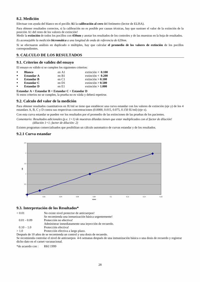
28
8.2. Medición Efectuar con ayuda del blanco en el pocillo A1 la calibración al cero del fotómetro (lector de ELISA).
Para obtener resultados correctos, si la calibración no es posible por causas técnicas, hay que sustraer el valor de la extinción de la posición A1 del resto de los valores de extinción! Medir la extinción de todos los pocillos con 450nm y anotar los resultados de los controles y de las muestras en la hoja de resultados.
Es aconsejable la medición bicromática a una longitud de onda de referencia de 620nm.
Si se efectuaron análisis en duplicado o múltiples, hay que calcular el promedio de los valores de extinción de los pocillos correspondientes.
9. CALCULO DE LOS RESULTADOS
9.1. Criterios de validez del ensayo El ensayo es válido si se cumplen los siguientes criterios:
� Blanco en A1 extinción < 0.100 � Estandar A en B1 extinción < 0.200 � Estandar B en C1 extinción > 0.100 � Estandar C en D1 extinción > 0.500 � Estandar D en E1 extinción > 1.000
Estandar A < Estandar B < Estandar C < Estandar D Si estos criterios no se cumplen, la prueba no es váida y deberá repetirse.
9.2. Calculo del valor de la medición Para obtener resultados cuantitativos en IU/ml se tiene que establecer una curva estandar con los valores de extinción (eje y) de los 4 estandars A, B, C y D contra sus respectivas concentraciones (0.0000, 0.015, 0.075, 0.150 IU/ml) (eje x).
Con esta curva estandar se pueden ver los resultados por el promedio de las extinciones de las pruebas de los pacientes.
Comentario: Resultados adicionales (p.e. 1+1) de muestras diluidas tienen que estar multiplicados con el factor de dilución! (dilución 1+1; factor de dilución: 2)
Existen programas comercialisados que posibilitan un cálculo automatico de curvas estandar y de los resultados.
9.2.1 Curva estandar
0
0,5
1
1,5
2
2,5
0 0,02 0,04 0,06 0,08 0,1 0,12 0,14 0,16
IU/ml
OD
9.3. Interpretación de los Resultados* < 0.01 No existe nivel protector de anticuerpos! Se recomienda una inmunización básica urgentemente! 0.01 - 0.09 Protección no efectiva!
Administrar inmediatamente una inyección de recuerdo. 0.10 – 1.0 Protección efectiva! > 1.0 Protección efectiva a largo plazo. Después de 10 años de se recomienda un control y una dosis de recuerdo. Se recomienda controlar el nivel de anticuerpos 4-6 semanas después de una inmunización básica o una dosis de recuerdo y registrar dicho dato en el carnet vacunacional.
*de acuerdo con : RKI 1999
29
10. CARACTERÍSTICAS DEL ENSAYO
10.1. Precisión Intraensayo n Media (OD) CV (%)
Pos. Leve 24 0.95 12.1 Pos. 24 3.02 2.1
Interensayo n Media (IU/ml) CV (%)
Pos. Leve 12 0.04 11.4 Pos. 12 0.16 2.8
10.2. Especificad del ensayo La especificidad del ensayo se define como la probabilidad que tiene el ensayo de dar un resultado negativo en ausencia de la sustancia a analizar específicamente (84.6 %).
10.3. Sensibilidad del ensayo La sensibilidad del ensayo se define como la probabilidad que tiene el ensayo de dar un resultado positivo en presencia del analítico específico (100 %).
10.4. Sensibilidad analítica La sensibilidad analítica se define como la menor concentración que se puede distinguir del estándar cero. Es de ≤ 0.01 IU/ml.
10.5. Interferencias Las muestras lipémicas e ictéricas no mostraron interferencias con este equipo ELISA hasta una concentración de 5 mg/ml para triglicéridos y de 0,2 mg/ml para bilirrubina.
Los resultados están basados en pruebas de ensayos querales: No se trata de especificaciones garantizadas.
11. LIMITACIONES DEL ENSAYO
Una contaminación de las muestras con bacterias, o una congelación y descongelación repetida pueden producir cambios en los valores de la extinción. El diagnóstico de una infección no solamente se debe basar en el resultado del ensayo. Es necesario considerar la anamnésis y la sintomatología del paciente junto al resultado serológico. Estos resultados sólo tienen valor restringido en personas inmunodeprimidas o en neonatos.
12. PRECAUCIONES Y ADVERTENCIAS
� En cumplimiento con el artículo 1 párrafo 2b de la directiva europea 98/79/EC, la utilización de sistemas médicos para diagnóstico in vitro tiene la intención por parte del fabricante de asegurar la adecuación, realizaciones y seguridad del producto. Por lo tanto, el procedimiento, la información, las precauciones y los avisos de las instrucciones de uso han de ser seguidas estrictamente. La utilización de equipos con analizadores y equipamiento similar tiene que ser validada. No se autorizan cambios en el diseño, composición y procedimiento, así como cualquier utilización en combinación con otros productos no aprobados por el fabricante; el usuario debe hacerse responsable de estos cambios. El fabricante no responderá ante falsos resultados e incidentes debidos a estas razones. El fabricante no responderá ante cualquier resultado por análisis visual de las muestras de los pacientes.
� Solo para diagnostico in vitro. � Todos los componentes de origen humano han sido examinados y resultaron no reactivos a anticuerpos contra el VIH, VHC y
HbsAG. No obstante, todos los materiales se deben considerar y tratar como potencialmente infecciosos. � No intercambiar reactivos y placas de microtítulo de cargas diferentes. � No usar reactivos de otro fabricante para este ensayo. � No usar después de la fecha de caducidad. � Sólo usar recambios de pipetas, dispensadores y materiales de laboratorio limpios. � No intercambiar las tapas de los diferentes reactivos. � Para evitar la evaporación y una contaminación microbiana, cierre inmediatamente las botellas después de usarlas. � Después de abrirlas y posterior almacenaje, asegurarse de que no existe contaminación microbiana antes de seguir usándolas. � Pipetear cuidadosamente las muestras y el conjugado en los pocillos para evitar contaminaciones cruzadas y resultados
erróneamente aumentados. � El NovaLisa™ ELISA está pensado exclusivamente para su uso por personal especializado que domine perfectamente las
técnicas de trabajo.
ADVERTENCIA: Bronidox L, en la concentración utilizada, casi no muestra riesgos tóxicos en la piel y en las mucosas.
ADVERTENCIA: El ácido sulfúrico irrita los ojos y la piel! En caso de contacto con los ojos lavar abundantemente con agua y consultar a un médico.

30
12.1. Indicaciones para la eliminación de residuos Por regla general, los productos químicos y las preparaciones son residuos peligrosos. Su eliminación esta sometida a las leyes y los decretos nacionales sobre la eliminación de residuos. Las autoridades informan sobre la eliminación de residuos peligroso.
13. INFORMACIONES PARA PEDIDOS
N˚ del producto: CORG0090 Corynebacterium difteriae toxin IgG-ELISA (96 determinaciones)

31

32

33

34
BIBLIOGRAPHY / LITERATUR / BIBLIOGRAPHIE / BIBLIOGR AFIA / BIBLIOGRAFÍA
BGA, Impfempfehlung der Ständigen Impfkommission (STIKO)des Bundesgesundheitsamtes vom 22.2.1994, Bundesgesundheitsblatt 8/94
CDC, International Notes (1993): Diphtheria Outbreak-Russian Federation, 1990-1993:Morbidity and Mortality Weekly Report 42:840
Hofmann,F.,F.Schuh,M.Michaelis,U.Stößel (1994):Zur Akzetanz von Schutzimpfungen bei Ärzten und bei der Allgemein-bevölkerung,Ges.Wes.56,371-376
WHO (7.5.1993):Expanded Programme on Immunization-Outbreak of diphtheria,update Wkly Epid Rec No.19,134-138
WHO (26.8.1994):Expanded Programme on Immunization-Diphtheria Epidemic.Wkly Epid Rec Nr.34 RKI (1999) Populationsimmunität gegen Diphtherie und Pertussis. Epidemiologisches Bulletin 1/99, 1-4
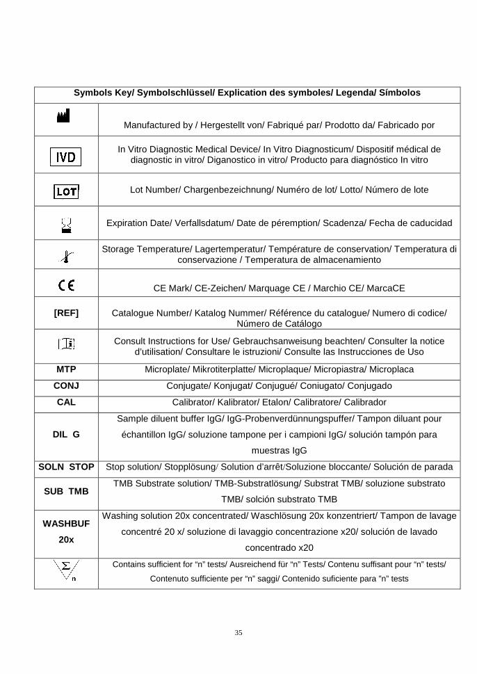
35
Symbols Key/ Symbolschlüssel/ Explication des symbo les/ Legenda/ Símbolos
Manufactured by / Hergestellt von/ Fabriqué par/ Prodotto da/ Fabricado por
In Vitro Diagnostic Medical Device/ In Vitro Diagnosticum/ Dispositif médical de diagnostic in vitro/ Diganostico in vitro/ Producto para diagnóstico In vitro
Lot Number/ Chargenbezeichnung/ Numéro de lot/ Lotto/ Número de lote
Expiration Date/ Verfallsdatum/ Date de péremption/ Scadenza/ Fecha de caducidad
Storage Temperature/ Lagertemperatur/ Température de conservation/ Temperatura di
conservazione / Temperatura de almacenamiento
CE Mark/ CE-Zeichen/ Marquage CE / Marchio CE/ MarcaCE
[REF]
Catalogue Number/ Katalog Nummer/ Référence du catalogue/ Numero di codice/
Número de Catálogo
Consult Instructions for Use/ Gebrauchsanweisung beachten/ Consulter la notice d’utilisation/ Consultare le istruzioni/ Consulte las Instrucciones de Uso
MTP Microplate/ Mikrotiterplatte/ Microplaque/ Micropiastra/ Microplaca
CONJ Conjugate/ Konjugat/ Conjugué/ Coniugato/ Conjugado
CAL Calibrator/ Kalibrator/ Etalon/ Calibratore/ Calibrador
DIL G
Sample diluent buffer IgG/ IgG-Probenverdünnungspuffer/ Tampon diluant pour
échantillon IgG/ soluzione tampone per i campioni IgG/ solución tampón para
muestras IgG
SOLN STOP Stop solution/ Stopplösung/ Solution d’arrêt/Soluzione bloccante/ Solución de parada
SUB TMB TMB Substrate solution/ TMB-Substratlösung/ Substrat TMB/ soluzione substrato
TMB/ solción substrato TMB
WASHBUF
20x
Washing solution 20x concentrated/ Waschlösung 20x konzentriert/ Tampon de lavage
concentré 20 x/ soluzione di lavaggio concentrazione x20/ solución de lavado
concentrado x20
Contains sufficient for “n” tests/ Ausreichend für “n” Tests/ Contenu suffisant pour “n” tests/
Contenuto sufficiente per “n” saggi/ Contenido suficiente para ”n” tests
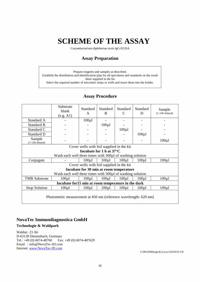
36
SCHEME OF THE ASSAY Corynebacterium diphtheriae toxin IgG-ELISA
Assay Preparation
Prepare reagents and samples as described. Establish the distribution and identification plan for all specimens and standards on the result
sheet supplied in the kit Select the required number of microtiter strips or wells and insert them into the holder.
Assay Procedure
Substrate
blank (e.g. A1)
Standard A
Standard B
Standard C
Standard D
Sample (1+100 diluted)
Standard A - 100µl - - - - Standard B - - 100µl - - - Standard C - - - 100µl - - Standard D - - - - 100µl -
Sample (1+100 diluted)
- - - - - 100µl
Cover wells with foil supplied in the kit Incubate for 1 h at 37°C
Wash each well three times with 300µl of washing solution Conjugate - 100µl 100µl 100µl 100µl 100µl
Cover wells with foil supplied in the kit Incubate for 30 min at room temperature
Wash each well three times with 300µl of washing solution TMB Substrate 100µl 100µl 100µl 100µl 100µl 100µl
Incubate for15 min at room temperature in the dark Stop Solution 100µl 100µl 100µl 100µl 100µl 100µl
Photometric measurement at 450 nm (reference wavelength: 620 nm)
NovaTec Immundiagnostica GmbH
Technologie & Waldpark
Waldstr. 23 A6 D-63128 Dietzenbach, Germany Tel.: +49 (0) 6074-48760 Fax: +49 (0) 6074-487629 Email : [email protected] Internet: www.NovaTec-ID.com
CORG0090engl,dt,fr,it,es12032010-CR