Embed Size (px)
Citation preview
8/8/2019 Crypto Benchaid
http://slidepdf.com/reader/full/crypto-benchaid 1/2
Basic guidelines
A. Multiple stool specimens (at least 3) should be tested before a negative result is reported.B. To maximize recovery of oocysts, stool specimens in formalin, or other fixatives, should be con-
centrated prior to microscopic examination (e.g.,10 min at 500 × g when using the formalin-ethyl-acetate concentration procedure). Exception: Specimens to be used for EIA or rapid car- tridge assays should NOT be concentrated because antigens are lost during the procedure!
C. Choice of diagnostic techniques depends on available equipment and reagents, experience,and considerations of time and cost.
Laboratory diagnosis of cryptosporidiosis
Cryptosporidium spp.
1. Wet mount
In bright-field microscopy using differential interference contrast (DIC), oocysts appear as small round struc-tures (4 to 6 μm) similar to yeasts. They do not autofluoresce.
2. Modified acid-fast stain
Oocysts (4 to 6 μm) often have distinct oocyst walls and stain from light pink to bright red. However, staining maybe variable. In particular, infections that are resolving can have colorless oocyst “ghosts.” Mature oocysts may
have discernible sporozoites (up to 4).
8/8/2019 Crypto Benchaid
http://slidepdf.com/reader/full/crypto-benchaid 2/2
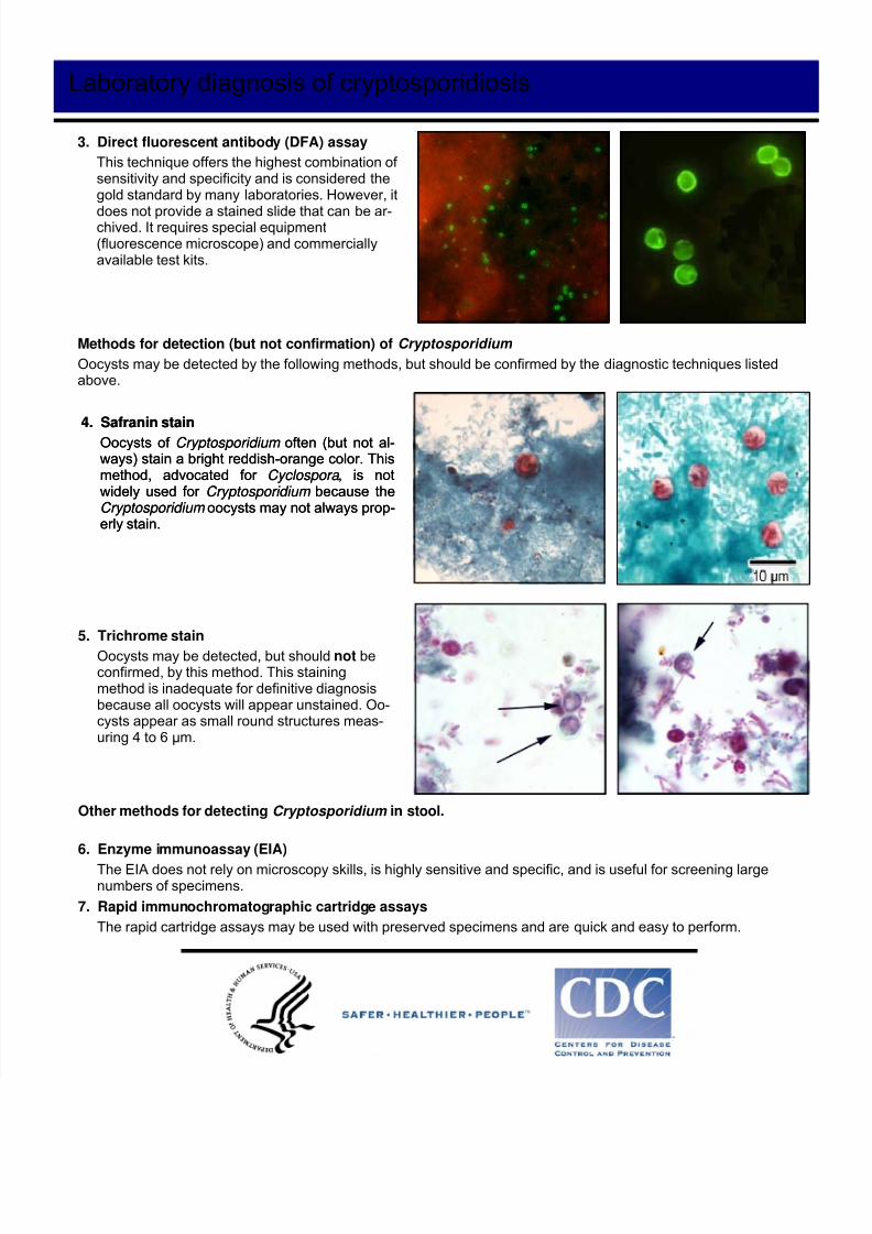
4. Safranin stain
Oocysts of Cryptosporidium often (but not al-ways) stain a bright reddish-orange color. Thismethod, advocated for Cyclospora , is notwidely used for Cryptosporidium because theCryptosporidium oocysts may not always prop-erly stain.
Laboratory diagnosis of cryptosporidiosis
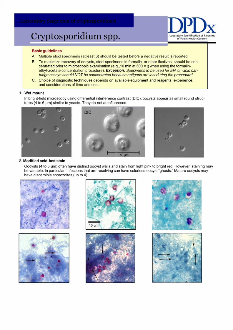
3. Direct fluorescent antibody (DFA) assay
This technique offers the highest combination of sensitivity and specificity and is considered thegold standard by many laboratories. However, it
does not provide a stained slide that can be ar-chived. It requires special equipment(fluorescence microscope) and commerciallyavailable test kits.
Methods for detection (but not confirmation) of Cryptosporidium
Oocysts may be detected by the following methods, but should be confirmed by the diagnostic techniques listedabove.
4. Safranin stain
Oocysts of Cryptosporidium often (but not al-ways) stain a bright reddish-orange color. Thismethod, advocated for Cyclospora , is notwidely used for Cryptosporidium because theCryptosporidium oocysts may not always prop-erly stain.
5. Trichrome stain
Oocysts may be detected, but should not beconfirmed, by this method. This stainingmethod is inadequate for definitive diagnosisbecause all oocysts will appear unstained. Oo-cysts appear as small round structures meas-uring 4 to 6 μm.
Other methods for detecting Cryptosporidium in stool.
6. Enzyme immunoassay (EIA)
The EIA does not rely on microscopy skills, is highly sensitive and specific, and is useful for screening largenumbers of specimens.
7. Rapid immunochromatographic cartridge assays
The rapid cartridge assays may be used with preserved specimens and are quick and easy to perform.