Embed Size (px)
Citation preview
CT Scanning: Principles, Patient Doses, Benefits &Potential Risks
If you are in a group, designate one person to be at the
computer.
If you are in a group, let us know the number of people that are
watching.
Today’s session is being recorded. This recording will include
audio from the conference call.
Questions will be answered at the end of presentation.
CME credits, Posttest, evaluation, certificate, handouts.
Partners and Sponsors
• New York State Department of Health
• University at Albany, School of Public Health, Center for Public Health Continuing Education
The following faculty have indicated a relationship with the following: Keith Strauss, MSc, FAAPM, FACR, is part of the Speaker’s Bureau and Consultant for Philips Healthcare, Inc.
No commercial funding has been accepted for this activity.
http://www.ualbanycphp.org/eval/SPHeval.cfm?ID=243
Evaluation:
Website (handouts, recording, evaluation link):
http://www.nyimagesafe.org/
CT Scanning: Principles, Patient Doses, Benefits &Potential Risks
Keith Strauss, MSc, FAAPM, FACR
Cincinnati Children’s Hospital
University of Cincinnati College of Medicine
CT SCANNING; PRINCIPLES, PATIENT DOSES, BENEFITS & POTENTIAL RISKS
Acknowledgement
Bushberg JT, Seibert JA, Leidholdt EM, Boone JM. The Essential Physics of Medical Imaging. 2012, 3rd Edition, Lippincott Williams & Wilkins, Philadelphia
• provided many of the tables and figures used in this presentation.
Introduction
• Basic principles of CT scanning
• Strengths of CT
• Comparison to other imaging modalities
• Patient dose
• Dose indices
• Comparison to radiation doses of other imaging modalities
• Benefits and potential risks
• Potential risks: deterministic vs stochastic
• Message of popular press must be interpreted
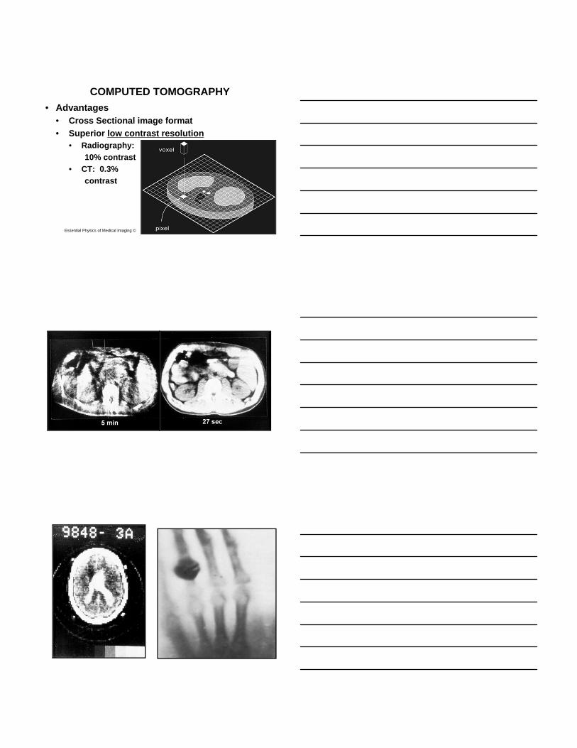
COMPUTED TOMOGRAPHY
• Advantages• Cross Sectional image format
• Superior low contrast resolution• Radiography:
10% contrast
• CT: 0.3%
contrast
Essential Physics of Medical Imaging ©
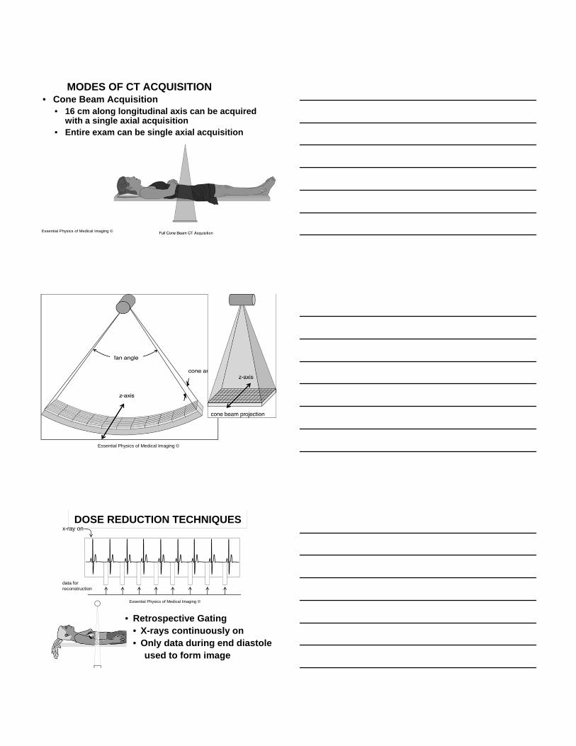
MODES OF CT ACQUISITION• Cone Beam Acquisition
• 16 cm along longitudinal axis can be acquired with a single axial acquisition
• Entire exam can be single axial acquisition
Essential Physics of Medical Imaging ©
Essential Physics of Medical Imaging ©
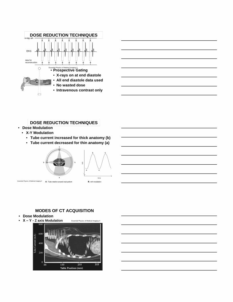
DOSE REDUCTION TECHNIQUES
• Retrospective Gating• X-rays continuously on• Only data during end diastole
used to form image
x-ray on
data for reconstruction
Essential Physics of Medical Imaging ©
DOSE REDUCTION TECHNIQUES
• Prospective Gating• X-rays on at end diastole• All end diastole data used• No wasted dose• Intravenous contrast only
x-ray on
EKG
data for reconstruction
Essential Physics of Medical Imaging ©
DOSE REDUCTION TECHNIQUES• Dose Modulation
• X-Y Modulation• Tube current increased for thick anatomy (b)• Tube current decreased for thin anatomy (a)
Essential Physics of Medical Imaging ©
MODES OF CT ACQUISITION• Dose Modulation• X – Y - Z axis Modulation Essential Physics of Medical Imaging ©
Essential Physics of Medical Imaging ©
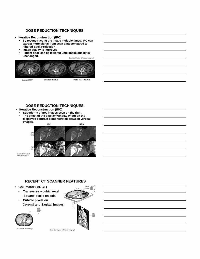
DOSE REDUCTION TECHNIQUES
• Iterative Reconstruction (IRC)• By reconstructing the image multiple times, IRC can
extract more signal from scan data compared to Filtered Back Projection
• Image quality is improved• Patient dose can be lowered until image quality is
unchanged.
Essential Physics of Medical Imaging ©
DOSE REDUCTION TECHNIQUES• Iterative Reconstruction (IRC)
• Superiority of IRC images seen on the right• The effect of the display Window Width on the
displayed contrast demonstrated between vertical images.
RECENT CT SCANNER FEATURES
• Collimator (MDCT)• Transverse ~ cubic voxel
‘Square’ pixels on axial
• Cubicle pixels on
Coronal and Sagittal images
Essential Physics of Medical Imaging ©
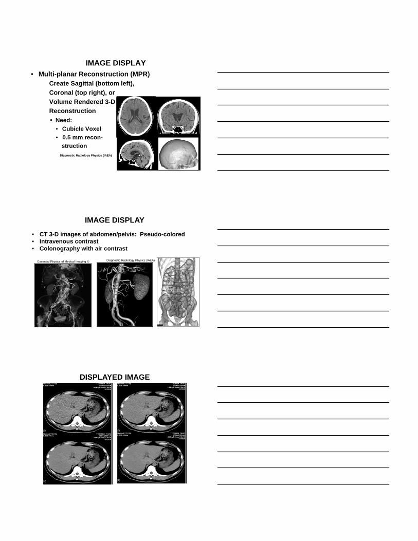
IMAGE DISPLAY
• Multi-planar Reconstruction (MPR)Create Sagittal (bottom left),
Coronal (top right), or
Volume Rendered 3-D
Reconstruction
• Need:
• Cubicle Voxel
• 0.5 mm recon-
struction
Diagnostic Radiology Physics (IAEA)
Essential Physics of Medical Imaging ©
IMAGE DISPLAY
• CT 3-D images of abdomen/pelvis: Pseudo-colored • Intravenous contrast• Colonography with air contrast
Diagnostic Radiology Physics (lAEA)
600 mAs
150 mAs 300 mAs
900 mAs
DISPLAYED IMAGE
Essential Physics of Medical Imaging ©
Advantages/Disadvantage of Other Imaging Modalities vs CTMRI: Great Tissue Discrimination
• Image of chemistrya. Sagittal T1-weightedb. Axial FLAIR: infarctc. T1-weighted contrastd. T1-weighted with fat
saturatione. MIP: time-of-flight MR
angiogramf. Gadolinium enhanced
• No Ionizing Radiation• Energy deposition occurs• Radio waves
Essential Physics of Medical Imaging ©
Advantages/Disadvantage of Other Imaging Modalities vs CT• Ultrasound: poor sharpness
• Image from deposition of mechanical waves (sound)a. Sagittal obstetrical US image of 5.5 month fetusb. Diameter of fetal head measurementc. 3-D image rendering of
fetus with cleft palated. Doppler color-flow
imaging: aneurysmseen in carotid artery
• No Ionizing Radiation• Low levels of mechanical
energy believed to haveno biological effect.
Essential Physics of Medical Imaging ©
Advantages/Disadvantage of Other Imaging Modalities vs CT• Radiography: Best sharpness• Projection radiographs require relatively small doses of
ionizing radiationa & b. AP and LAT chest x-ray: one of smallest doses
for one of the most common exams.c. LAT cervical spine: neck injury after trauma
Essential Physics of Medical Imaging ©
Advantages/Disadvantage of Other Imaging Modalities vs CT
• Radiography: Poor soft tissue discrimination• Projection radiographs of extremities require smal-ler
doses of ionizing radiation than images of trunkd, e, f, g. Wrist, Foot, Knee, Hipg. Metal objects are well seen on radiographs
Essential Physics of Medical Imaging ©
Advantages/Disadvantage of Other Imaging Modalities vs CTMammography: best radiographic images
• Projection radiographs of breasts require larger doses of ionizing radiation due to low energy xrays
a. Digital image of glandularfatty tissues
b. Tomogram of breast ellim-inates tissue superpositionImproves cancer detection
c. Preferred screening test for early detection ofcancer.
Essential Physics of Medical Imaging ©
Advantages/Disadvantage of Other Imaging Modalities vs CTNuclear Medicine: image of physiology
• Planar 2-D maps of 3-D radioisotope distribution.
• Multiple metastatic lesionsnot detectable by radiographyare seen.
• Ionizing radiation is used.• Patient dose determined by
biological and physical half life of radioisotope and howmuch activity is injected.
Essential Physics of Medical Imaging ©
Advantages/Disadvantage of Other Imaging Modalities vs CT• Nuclear Medicine: high contrast but poor
indication of diseased organ• Hybrid Imaging: combine nuclear camera with higher
resolution modality: CT or MRIa. Small cell lung cancerb. Images before and after
chemotherapyC & d. PET/CT fusion images
Essential Physics of Medical Imaging ©
Advantages/Disadvantage of Other Imaging Modalities vs CT• Sharpness:
1. Mammography S/F2. Mammography Dig3. Radiography: S/F4. Fluoroscopy:5. Radiography: Dig6. Ultrasound7. CT8. MRI9. Nuclear Medicine
Air Kerma
• Dose to air at the entrance plane of the patient.
• Ability of x-ray machine to produce energy (radiation).
• Energy carried by x-rays.
• Must specify a location from the source
• Units: Gray, mGy, µGy, nGy . . .
• Replaces exposure (Roentgens)
• 1 mR = 8.7 µGy

Dose
• Dose occurs when x-rays pass through the patient’s body and interact by:
• Photoelectric effect
• Compton scattering
• Energy of x-ray is transferred (unloaded) to KE of charged particles in tissue.
• Units: Gray, mGy, µGy, nGy . . .
• Replaces rad
• 1 mrad = 10 µGy
Equivalent Dose
• Dose adjusted for the type of carrier of the energy
H = Quality Factor x Dose
where Q = 1 for x & beta (electrons) rays
10 – 20 for protons, Alpha particles
•Units: Sievert, mSv, µSv, nSv . . .
Effective Dose
Effective Absorbed Dose is the absorbed dosegiven to the whole body of the patient that
wouldresult in the same biological damage as the actual clinical dose given to a fraction of thepatient’s whole body.
•Units: Sievert (Sv) or milliSievert (mSv)• For x-rays:
1 rem = 10 mSv; 1 Sv = 100 rem
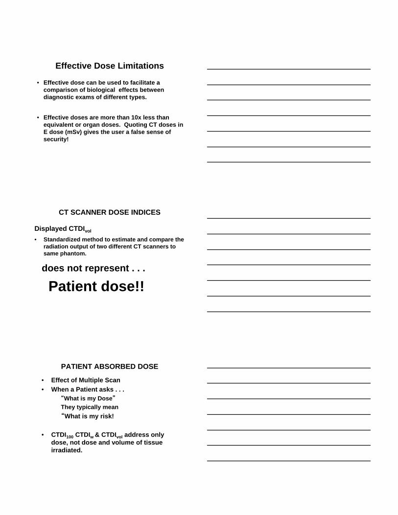
Effective Dose Limitations
• Effective dose can be used to facilitate a comparison of biological effects between diagnostic exams of different types.
• Effective doses are more than 10x less than equivalent or organ doses. Quoting CT doses in E dose (mSv) gives the user a false sense of security!
CT SCANNER DOSE INDICES
Displayed CTDIvol
• Standardized method to estimate and compare the radiation output of two different CT scanners to same phantom.
does not represent . . .
Patient dose!!
PATIENT ABSORBED DOSE
• Effect of Multiple Scan
• When a Patient asks . . .“What is my Dose”
They typically mean
“What is my risk!
• CTDI100 CTDIw & CTDIvol address only dose, not dose and volume of tissue irradiated.
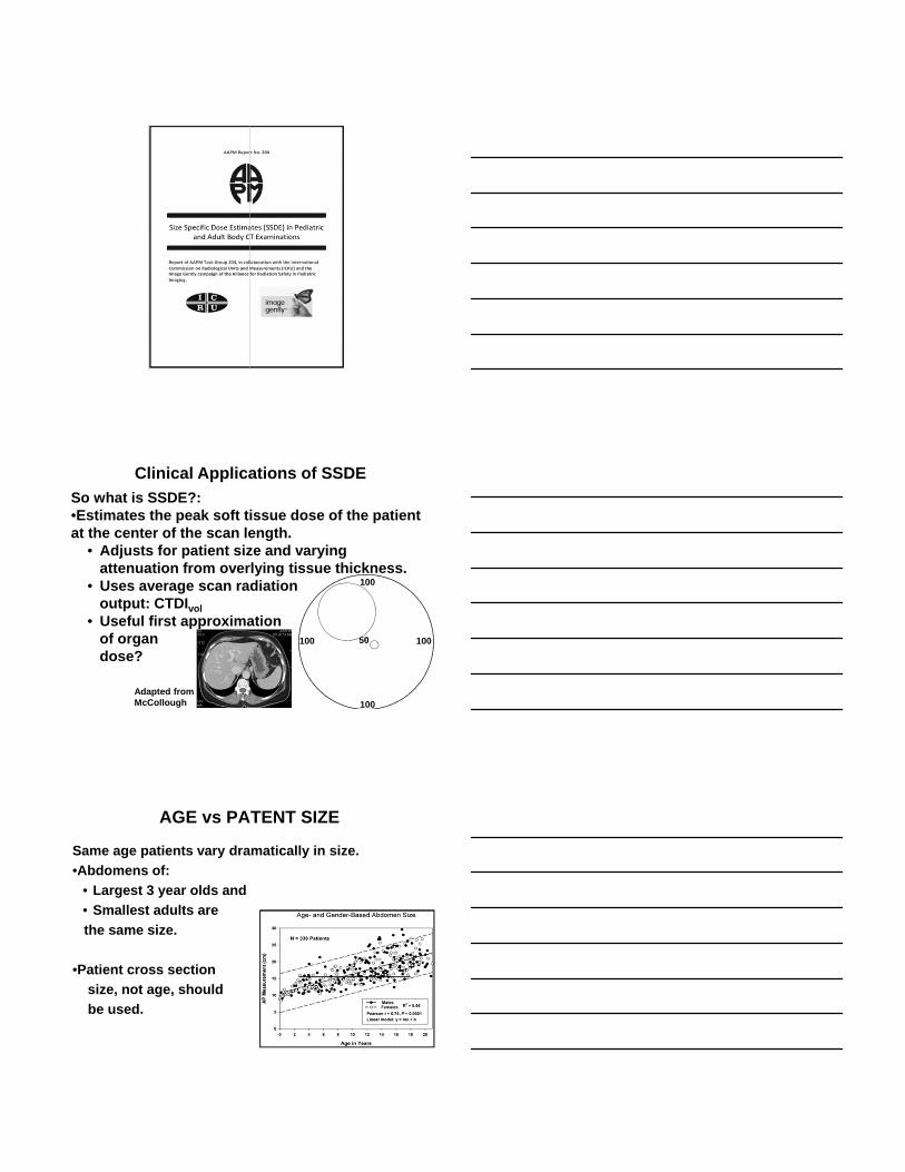
So what is SSDE?:•Estimates the peak soft tissue dose of the patient at the center of the scan length.
• Adjusts for patient size and varying attenuation from overlying tissue thickness.
• Uses average scan radiationoutput: CTDIvol
• Useful first approximationof organdose?
Clinical Applications of SSDE
50
100
100
100
100
Adapted from McCollough
AGE vs PATENT SIZE
Same age patients vary dramatically in size.
•Abdomens of:
• Largest 3 year olds and
• Smallest adults are
the same size.
•Patient cross section
size, not age, should
be used.
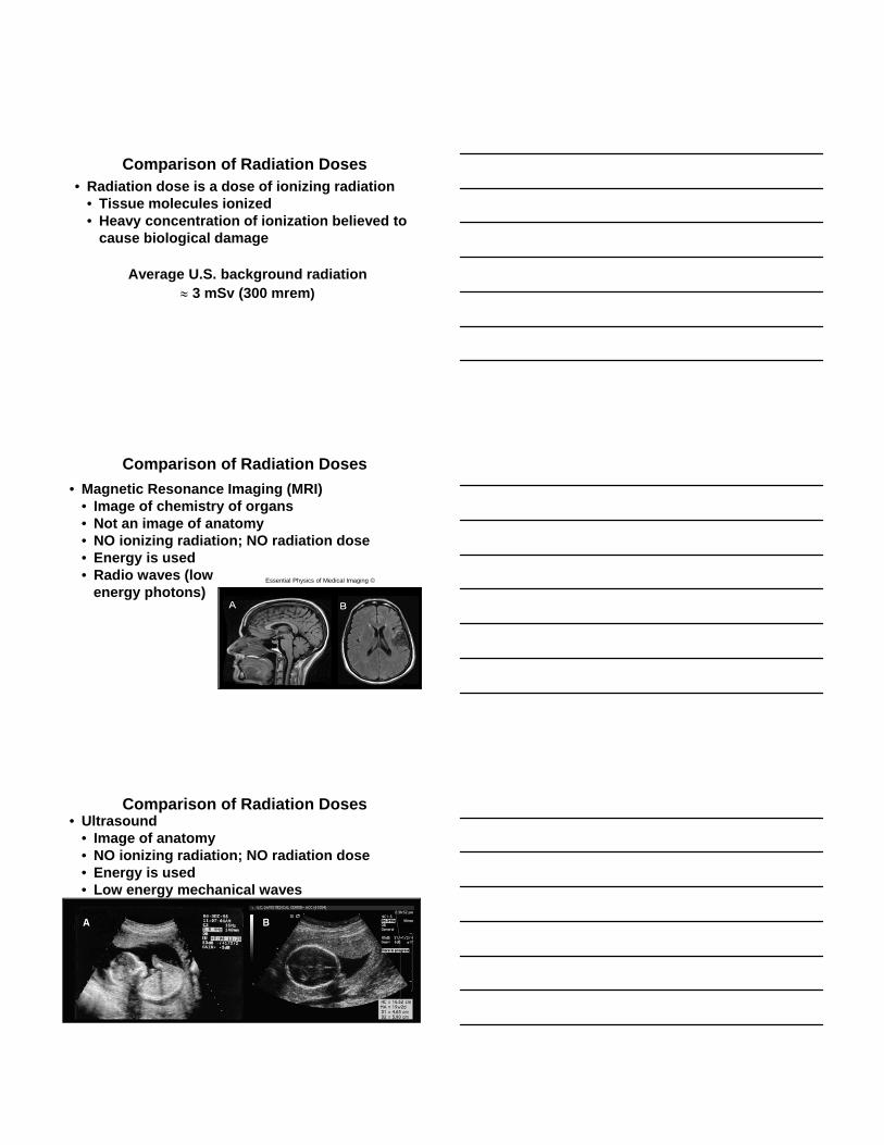
Comparison of Radiation Doses• Radiation dose is a dose of ionizing radiation
• Tissue molecules ionized• Heavy concentration of ionization believed to
cause biological damage
Average U.S. background radiation 3 mSv (300 mrem)
Comparison of Radiation Doses
• Magnetic Resonance Imaging (MRI)• Image of chemistry of organs• Not an image of anatomy• NO ionizing radiation; NO radiation dose• Energy is used• Radio waves (low
energy photons)Essential Physics of Medical Imaging ©
Comparison of Radiation Doses• Ultrasound
• Image of anatomy• NO ionizing radiation; NO radiation dose• Energy is used• Low energy mechanical waves
Essential Physics of Medical Imaging ©
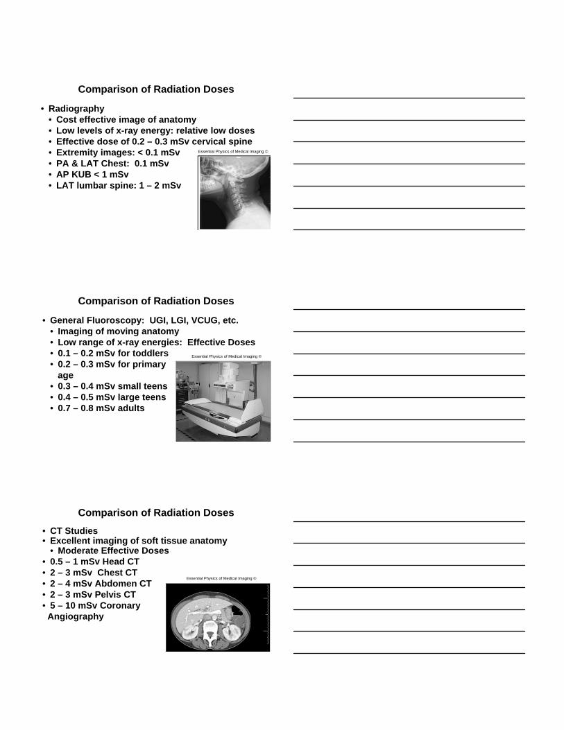
Comparison of Radiation Doses
• Radiography• Cost effective image of anatomy• Low levels of x-ray energy: relative low doses• Effective dose of 0.2 – 0.3 mSv cervical spine• Extremity images: < 0.1 mSv• PA & LAT Chest: 0.1 mSv• AP KUB < 1 mSv • LAT lumbar spine: 1 – 2 mSv
Essential Physics of Medical Imaging ©
Comparison of Radiation Doses
• General Fluoroscopy: UGI, LGI, VCUG, etc.• Imaging of moving anatomy• Low range of x-ray energies: Effective Doses• 0.1 – 0.2 mSv for toddlers• 0.2 – 0.3 mSv for primary
age• 0.3 – 0.4 mSv small teens• 0.4 – 0.5 mSv large teens• 0.7 – 0.8 mSv adults
Essential Physics of Medical Imaging ©
Comparison of Radiation Doses
• CT Studies• Excellent imaging of soft tissue anatomy
• Moderate Effective Doses• 0.5 – 1 mSv Head CT • 2 – 3 mSv Chest CT• 2 – 4 mSv Abdomen CT• 2 – 3 mSv Pelvis CT• 5 – 10 mSv Coronary
Angiography
Essential Physics of Medical Imaging ©
Comparison of Radiation Doses
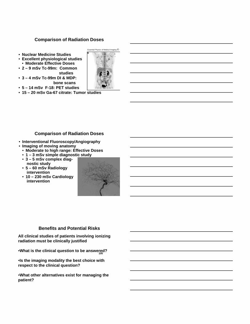
• Nuclear Medicine Studies• Excellent physiological studies
• Moderate Effective Doses• 2 – 9 mSv Tc-99m: Common
studies• 3 – 4 mSv Tc-99m DI & MDP:
bone scans• 5 – 14 mSv F-18: PET studies• 15 – 20 mSv Ga-67 citrate: Tumor studies
Essential Physics of Medical Imaging ©
Comparison of Radiation Doses
• Interventional Fluoroscopy/Angiography• Imaging of moving anatomy
• Moderate to high range: Effective Doses• 1 – 3 mSv simple diagnostic study• 3 – 5 mSv complex diag-
nostic study• 5 – 60 mSv Radiology
intervention• 10 – 230 mSv Cardiology
intervention
All clinical studies of patients involving ionizing radiation must be clinically justified
•What is the clinical question to be answered?
•Is the imaging modality the best choice with respect to the clinical question?
•What other alternatives exist for managing the patient?
Benefits and Potential Risks
100
• Identified benefits are:• Real• Immediate• Small child who has fallen and struck their head
could die in 24 hours from a brain bleed.
• Potential risks are:• Hypothetical• Delayed in some cases up to 30 years
Benefits and Potential Risks
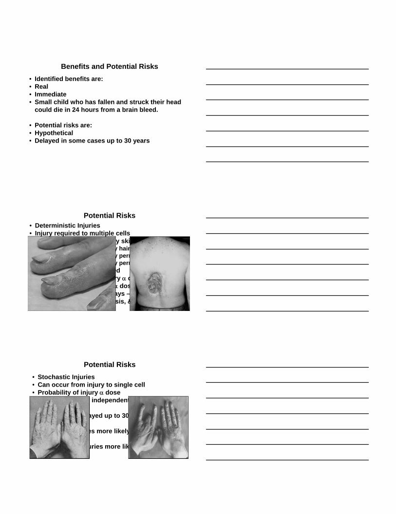
• Deterministic Injuries• Injury required to multiple cells• Threshold Dose: 2,000 mGy skin erythema
3,000 mGy hair loss6,000 mGy permanent erythema7,000 mGy permanent hair loss
• Below: no injury expressed• Above: Probability of injury dose
Severity of injury dose• First symptoms delayed days – weeks• Cataracts, epilation, necrosis, & erythema
Potential Risks
• Stochastic Injuries• Can occur from injury to single cell• Probability of injury dose• Severity of injury independent of dose• Neoplasms• Solid tumors delayed up to 30 years
• Stochastic injuries more likely in children
• Deterministic injuries more likely in adults
Potential Risks
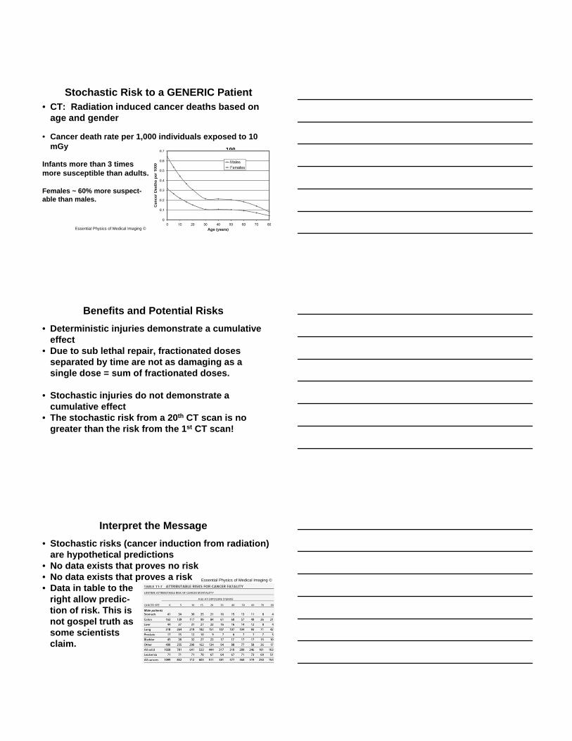
• CT: Radiation induced cancer deaths based on age and gender
• Cancer death rate per 1,000 individuals exposed to 10 mGy
Infants more than 3 times more susceptible than adults.
Females ~ 60% more suspect-able than males.
Stochastic Risk to a GENERIC Patient
100
Essential Physics of Medical Imaging ©
• Deterministic injuries demonstrate a cumulative effect
• Due to sub lethal repair, fractionated doses separated by time are not as damaging as a single dose = sum of fractionated doses.
• Stochastic injuries do not demonstrate a cumulative effect
• The stochastic risk from a 20th CT scan is no greater than the risk from the 1st CT scan!
Benefits and Potential Risks
• Stochastic risks (cancer induction from radiation) are hypothetical predictions
• No data exists that proves no risk• No data exists that proves a risk• Data in table to the
right allow predic-tion of risk. This isnot gospel truth assome scientistsclaim.
Interpret the Message
Essential Physics of Medical Imaging ©

When the media like the NY Times publishes this type of sensationalism, patients read the article and become fearful of radiation!
Sensational scare tactics sell
Radiation must be respectedand properly used, not feared
Interpret the Message
What is More Realistic?
Essential Physics of Medical Imaging ©
• Imaging plays an important role in the diagnosis and treatment of diseases today.
• Today’s production of clinical images is very sophisticated.
• Remain abreast of changes in imaging; consult with radiologists and ask questions.
• Order the best exam for your patient, it may be different today than in the past.
Conclusions

• Seek the diagnostic examination that can best answer the clinical question about your patient.
• Do not avoid, out of fear, the use of ionizing radiation when it is clinically justified.
• Interpret carefully the media’s message about imaging risks.
• Potential risks of ionizing radiation are much lower than portrayed in the media.
Conclusions
Please join us for our next webinar!
Creating a Culture of Safety in Radiology
July 22nd, 2015 12:00pm – 1:00pm
Dr. Kimberly Applegate
Register here: http://www.nyimagesafe.org/
Did you miss the last one?
Image Gently: Radiation Exposure to Children From Medical Imaging- Is there a Problem?
Presenter: Dr. Marilyn Goske
Register to watch the recording here: http://www.nyimagesafe.org/
Public Health Live!
Pediatric Computed Tomography (CT) Scans, Radiation, and Risks: Having an Informed Dialogue
June 18, 2015 9:00 am- 10:00 am
Presenter: Donald P. Frush, MD, FACR, FAAP
Register to watch here:
http://www.nyimagesafe.org
http://www.ualbanycphp.org/eval/SPHeval.cfm?ID=243
Evaluation:
Website (handouts, recording, evaluation link):
http://www.nyimagesafe.org/
CT Scanning: Principles, Patient Doses, Benefits &Potential Risks