Embed Size (px)
Citation preview
ARTICLE
Cysteinyl-tRNA synthetase governs cysteinepolysulfidation and mitochondrial bioenergeticsTakaaki Akaike1, Tomoaki Ida1, Fan-Yan Wei2, Motohiro Nishida3,4, Yoshito Kumagai5, Md. Morshedul Alam6,
Hideshi Ihara7, Tomohiro Sawa8, Tetsuro Matsunaga1, Shingo Kasamatsu1, Akiyuki Nishimura3,
Masanobu Morita1, Kazuhito Tomizawa2, Akira Nishimura1, Satoshi Watanabe 9, Kenji Inaba9,
Hiroshi Shima10, Nobuhiro Tanuma10, Minkyung Jung1, Shigemoto Fujii1, Yasuo Watanabe11,
Masaki Ohmuraya12, Péter Nagy13, Martin Feelisch14, Jon M. Fukuto15 & Hozumi Motohashi 6
Cysteine hydropersulfide (CysSSH) occurs in abundant quantities in various organisms, yet
little is known about its biosynthesis and physiological functions. Extensive persulfide
formation is apparent in cysteine-containing proteins in Escherichia coli and mammalian cells
and is believed to result from post-translational processes involving hydrogen sulfide-related
chemistry. Here we demonstrate effective CysSSH synthesis from the substrate L-cysteine, a
reaction catalyzed by prokaryotic and mammalian cysteinyl-tRNA synthetases (CARSs).
Targeted disruption of the genes encoding mitochondrial CARSs in mice and human cells
shows that CARSs have a crucial role in endogenous CysSSH production and suggests that
these enzymes serve as the principal cysteine persulfide synthases in vivo. CARSs also
catalyze co-translational cysteine polysulfidation and are involved in the regulation of
mitochondrial biogenesis and bioenergetics. Investigating CARS-dependent persulfide
production may thus clarify aberrant redox signaling in physiological and pathophysiological
conditions, and suggest therapeutic targets based on oxidative stress and mitochondrial
dysfunction.
DOI: 10.1038/s41467-017-01311-y OPEN
1 Department of Environmental Health Sciences and Molecular Toxicology, Tohoku University Graduate School of Medicine, Sendai 980-8575, Japan.2 Department of Molecular Physiology, Graduate School of Medical Sciences, Kumamoto University, Kumamoto 860-8556, Japan. 3 Division ofCardiocirculatory Signaling, Okazaki Institute for Integrative Bioscience, National Institute for Physiological Sciences, National Institutes of Natural Sciences,Okazaki 444-8787, Japan. 4 Department of Translational Pharmaceutical Sciences, Graduate School of Pharmaceutical Sciences, Kyushu University, Fukuoka812-8582, Japan. 5 Environmental Biology Section, Faculty of Medicine, University of Tsukuba, Tsukuba 305-8575, Japan. 6Department of Gene ExpressionRegulation, Institute of Development, Aging and Cancer, Tohoku University, Sendai 980-8575, Japan. 7 Department of Biological Science, Graduate School ofScience, Osaka Prefecture University, Osaka 599-8531, Japan. 8Department of Microbiology, Graduate School of Medical Sciences, Kumamoto University,Kumamoto 860-8556, Japan. 9 Institute of Multidisciplinary Research for Advanced Materials, Tohoku University, Sendai 980-8577, Japan. 10 Division ofCancer Chemotherapy, Miyagi Cancer Center Research Institute, Natori 981-1293, Japan. 11 Laboratory of Pharmacology, Showa Pharmaceutical University,Tokyo 194-8543, Japan. 12 Department of Genetics, Hyogo College of Medicine, Nishinomiya, Hyogo 663-8501, Japan. 13 Department of MolecularImmunology and Toxicology, National Institute of Oncology, Budapest 1122, Hungary. 14 Clinical and Experimental Sciences, Faculty of Medicine, University ofSouthampton, Southampton General Hospital and Institute for Life Sciences, Southampton SO16 6YD, UK. 15 Department of Chemistry, Sonoma StateUniversity, Rohnert Park, CA 94928, USA. Tomoaki Ida, Fan-Yan Wei, Motohiro Nishida and Yoshito Kumagai contributed equally to this work.Correspondence and requests for materials should be addressed to T.A. (email: [email protected])
NATURE COMMUNICATIONS |8: 1177 |DOI: 10.1038/s41467-017-01311-y |www.nature.com/naturecommunications 1
1234
5678
90
Cysteine hydropersulfide (CysSSH) is found physiologicallyin prokaryotes, eukaryotic cells, and mammalian tissues1,2.Previously, we unequivocally verified the presence of
remarkable amounts of CysSSH, glutathione persulfide (GSSH),and longer chain sulfur compounds (polysulfides, including CysS/GS–(S)n–H) in cultured cells and tissues in vivo in mice andhumans3–6. The chemical properties and abundance of thesespecies suggest a pivotal role for reactive persulfides (i.e., com-pounds containing an—SSH group) in cell-regulatory processes.Researchers proposed that CysSSH and related species can behaveas potent antioxidants and cellular protectants, and may functionas redox signaling intermediates3–10. Persulfides are also essentialstructural components of several proteins and enzymes, e.g. ser-ving as metal ligands in iron-sulfur clusters (or sulfide donors)and in iron-cysteine and zinc-cysteine complexes11–15. In fact, theexistence of a cell reservoir for sulfane sulfur (sulfur-bondedsulfur atoms with six electrons), including low-molecular-weight(LMW) and protein-bound cysteine polysulfides, has long beenknown1,3–7,15,16. Thus, although the prevalence of endogenouspolysulfides is clearly established and their biological relevanceincreasingly being recognized, the chemical biology and physio-logical functions of these species are not known with any cer-tainty. Current dogma holds that persulfide/polysulfide formationarises as a result of hydrogen sulfide (H2S) oxidation3,4,7–9 orchemical reaction with nitric oxide3,17. Two H2S-generatingenzymes involved in sulfur-containing amino acid metabolism—cystathionine γ-lyase (cystathionase, CSE) and cystathionine β-synthase (CBS)—can catalyze CysSSH biosynthesis using cystine(CysSSCys) as a substrate3,4,6–10,18–21. However, the observed Km
is high, and both cells and mice lacking CSE and/or CBS stilldisplay appreciable levels of CysSSH20–24, which suggests thepossibility that alternative processes may be responsible forendogenous persulfide production. Thus, it appears that otherbiosynthetic routes of CysSSH formation exist that have yet to beidentified.
This study reveals that cysteinyl-tRNA synthetases (CARSs), inaddition to their canonical role in protein translation, act as theprincipal cysteine persulfide synthases (CPERSs) in vivo. CARSsplay a novel and prominent role in endogenous production ofboth LMW polysulfides and polysulfidated proteins that areabundantly detected in cells and in mice. Notably, CARS2, amitochondrial isoform of CARS, is involved in mitochondrialbiogenesis and bioenergetics via CysSSH production.
ResultsRedox property of cysteine and protein polysulfides. CysSSHhas unique redox-active properties that distinguishes it from thecysteine (CysSH) thiol. In evaluating the physiological rationalefor biological CysSSH production, our present study confirmedthat cysteine persulfide/polysulfides (CysSSH/CysS–(S)n–H) pos-sess mixed sulfur reactivity—both nucleophilic and electrophilic(Supplementary Figs 1 and 2)—a property that is unique anddistinct from that of other simple biologically relevant thiols. Thedual electrophilic-nucleophilic character of hydropersulfides iswell documented (the anionic RSS− species being nucleophilicand the protonated RSSH species possessing electrophilic prop-erties akin to disulfides, RSSR)25–27. Moreover, dialkylpolysulfidescan also be nucleophilic and electrophile-mediated cleavage of S-Sbonds is established28. The unique properties and reactivity ofpolysulfides allowed us to develop several analytical techniquesaimed at determining endogenous production of LMW andprotein-bound polysulfides (Supplementary Fig. 3). We firstdeveloped a convenient method for selective detection of poly-sulfidated proteins: the biotin-polyethylene glycol (PEG)-con-jugated maleimide (biotin-PEG-MAL) labeling gel shift assay
(PMSA; Supplementary Fig. 3a)15. PMSA demonstrated extensiveprotein-bound cysteine polysulfidation (Supplementary Fig. 4),not only for recombinant proteins, prepared in an Escherichia colicell expression system (Supplementary Table 1) but also forendogenous proteins expressed in mammalian cells.
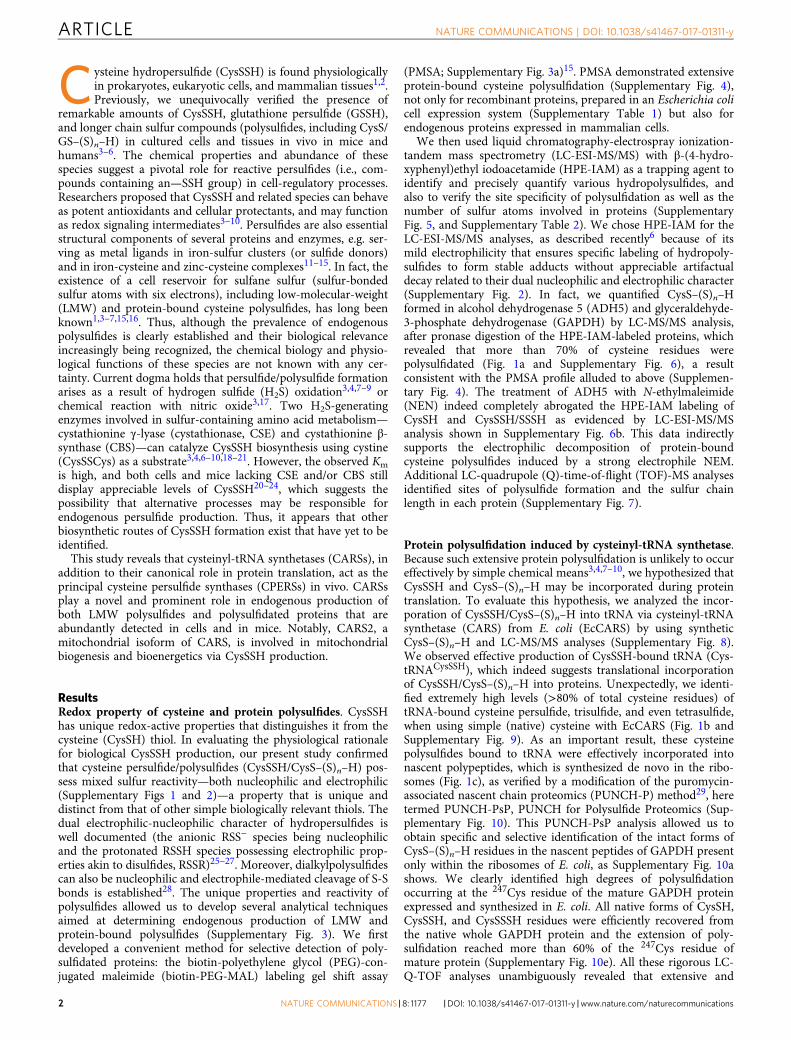
We then used liquid chromatography-electrospray ionization-tandem mass spectrometry (LC-ESI-MS/MS) with β-(4-hydro-xyphenyl)ethyl iodoacetamide (HPE-IAM) as a trapping agent toidentify and precisely quantify various hydropolysulfides, andalso to verify the site specificity of polysulfidation as well as thenumber of sulfur atoms involved in proteins (SupplementaryFig. 5, and Supplementary Table 2). We chose HPE-IAM for theLC-ESI-MS/MS analyses, as described recently6 because of itsmild electrophilicity that ensures specific labeling of hydropoly-sulfides to form stable adducts without appreciable artifactualdecay related to their dual nucleophilic and electrophilic character(Supplementary Fig. 2). In fact, we quantified CysS–(S)n–Hformed in alcohol dehydrogenase 5 (ADH5) and glyceraldehyde-3-phosphate dehydrogenase (GAPDH) by LC-MS/MS analysis,after pronase digestion of the HPE-IAM-labeled proteins, whichrevealed that more than 70% of cysteine residues werepolysulfidated (Fig. 1a and Supplementary Fig. 6), a resultconsistent with the PMSA profile alluded to above (Supplemen-tary Fig. 4). The treatment of ADH5 with N-ethylmaleimide(NEN) indeed completely abrogated the HPE-IAM labeling ofCysSH and CysSSH/SSSH as evidenced by LC-ESI-MS/MSanalysis shown in Supplementary Fig. 6b. This data indirectlysupports the electrophilic decomposition of protein-boundcysteine polysulfides induced by a strong electrophile NEM.Additional LC-quadrupole (Q)-time-of-flight (TOF)-MS analysesidentified sites of polysulfide formation and the sulfur chainlength in each protein (Supplementary Fig. 7).
Protein polysulfidation induced by cysteinyl-tRNA synthetase.Because such extensive protein polysulfidation is unlikely to occureffectively by simple chemical means3,4,7–10, we hypothesized thatCysSSH and CysS–(S)n–H may be incorporated during proteintranslation. To evaluate this hypothesis, we analyzed the incor-poration of CysSSH/CysS–(S)n–H into tRNA via cysteinyl-tRNAsynthetase (CARS) from E. coli (EcCARS) by using syntheticCysS–(S)n–H and LC-MS/MS analyses (Supplementary Fig. 8).We observed effective production of CysSSH-bound tRNA (Cys-tRNACysSSH), which indeed suggests translational incorporationof CysSSH/CysS–(S)n–H into proteins. Unexpectedly, we identi-fied extremely high levels (>80% of total cysteine residues) oftRNA-bound cysteine persulfide, trisulfide, and even tetrasulfide,when using simple (native) cysteine with EcCARS (Fig. 1b andSupplementary Fig. 9). As an important result, these cysteinepolysulfides bound to tRNA were effectively incorporated intonascent polypeptides, which is synthesized de novo in the ribo-somes (Fig. 1c), as verified by a modification of the puromycin-associated nascent chain proteomics (PUNCH-P) method29, heretermed PUNCH-PsP, PUNCH for Polysulfide Proteomics (Sup-plementary Fig. 10). This PUNCH-PsP analysis allowed us toobtain specific and selective identification of the intact forms ofCysS–(S)n–H residues in the nascent peptides of GAPDH presentonly within the ribosomes of E. coli, as Supplementary Fig. 10ashows. We clearly identified high degrees of polysulfidationoccurring at the 247Cys residue of the mature GAPDH proteinexpressed and synthesized in E. coli. All native forms of CysSH,CysSSH, and CysSSSH residues were efficiently recovered fromthe native whole GAPDH protein and the extension of poly-sulfidation reached more than 60% of the 247Cys residue ofmature protein (Supplementary Fig. 10e). All these rigorous LC-Q-TOF analyses unambiguously revealed that extensive and
ARTICLE NATURE COMMUNICATIONS | DOI: 10.1038/s41467-017-01311-y
2 NATURE COMMUNICATIONS |8: 1177 |DOI: 10.1038/s41467-017-01311-y |www.nature.com/naturecommunications
prevalent cysteine polysulfidation is introduced co-translationallyand sustained in the mature protein physiologically present evenin the post-translational processes of the cells.
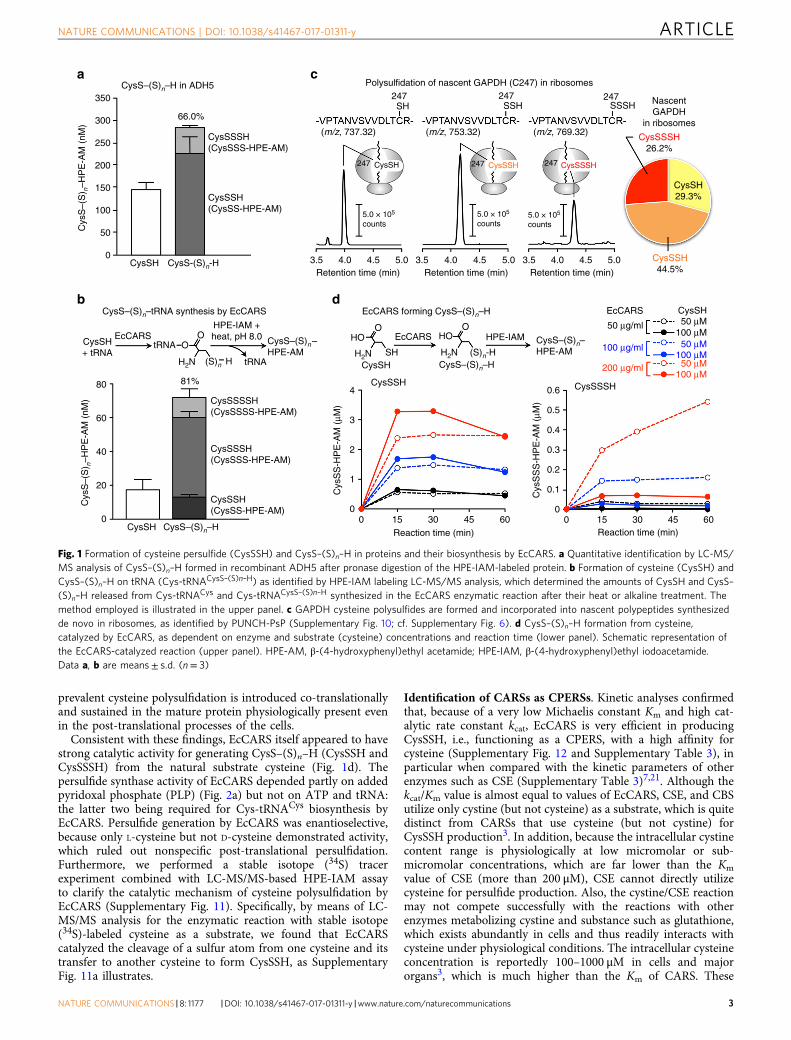
Consistent with these findings, EcCARS itself appeared to havestrong catalytic activity for generating CysS–(S)n–H (CysSSH andCysSSSH) from the natural substrate cysteine (Fig. 1d). Thepersulfide synthase activity of EcCARS depended partly on addedpyridoxal phosphate (PLP) (Fig. 2a) but not on ATP and tRNA:the latter two being required for Cys-tRNACys biosynthesis byEcCARS. Persulfide generation by EcCARS was enantioselective,because only L-cysteine but not D-cysteine demonstrated activity,which ruled out nonspecific post-translational persulfidation.Furthermore, we performed a stable isotope (34S) tracerexperiment combined with LC-MS/MS-based HPE-IAM assayto clarify the catalytic mechanism of cysteine polysulfidation byEcCARS (Supplementary Fig. 11). Specifically, by means of LC-MS/MS analysis for the enzymatic reaction with stable isotope(34S)-labeled cysteine as a substrate, we found that EcCARScatalyzed the cleavage of a sulfur atom from one cysteine and itstransfer to another cysteine to form CysSSH, as SupplementaryFig. 11a illustrates.
Identification of CARSs as CPERSs. Kinetic analyses confirmedthat, because of a very low Michaelis constant Km and high cat-alytic rate constant kcat, EcCARS is very efficient in producingCysSSH, i.e., functioning as a CPERS, with a high affinity forcysteine (Supplementary Fig. 12 and Supplementary Table 3), inparticular when compared with the kinetic parameters of otherenzymes such as CSE (Supplementary Table 3)7,21. Although thekcat/Km value is almost equal to values of EcCARS, CSE, and CBSutilize only cystine (but not cysteine) as a substrate, which is quitedistinct from CARSs that use cysteine (but not cystine) forCysSSH production3. In addition, because the intracellular cystinecontent range is physiologically at low micromolar or sub-micromolar concentrations, which are far lower than the Km
value of CSE (more than 200 μM), CSE cannot directly utilizecysteine for persulfide production. Also, the cystine/CSE reactionmay not compete successfully with the reactions with otherenzymes metabolizing cystine and substance such as glutathione,which exists abundantly in cells and thus readily interacts withcysteine under physiological conditions. The intracellular cysteineconcentration is reportedly 100–1000 μM in cells and majororgans3, which is much higher than the Km of CARS. These
350CysS–(S)n–H in ADH5
300 66.0%
Polysulfidation of nascent GAPDH (C247) in ribosomes247 247 247SH
247
5.0 × 10⁵
counts5.0 × 105
counts5.0 × 105
counts
CysSH
3.5 4.0 4.5 5.0Retention time (min)
3.5 4.0 4.5 5.0Retention time (min)
3.5 4.0 4.5 5.0Retention time (min)
247 247CysSSH CysSSSH
(m/z, 737.32) (m/z, 753.32) (m/z, 769.32)
NascentGAPDH
in ribosomes
CysSH29.3%
CysSSH44.5%
CysSSSH26.2%
SSH SSSH
CysSSSH(CysSSS-HPE-AM)
CysSSH(CysSS-HPE-AM)
250
200
Cys
S–(
S) n
–HP
E-A
M (
nM)
150
100
50
CysSH
EcCARSCysSH+ tRNA
tRNA
81%80
60
40
Cys
S–(
S) n
–HP
E-A
M (
nM)
20
CysSH CysS–(S)n–H0
OO
H2N (S)n H tRNA
CysSSSSH(CysSSSS-HPE-AM)
CysSSSH(CysSSS-HPE-AM)
CysSSH(CysSS-HPE-AM)
CysSSH CysSSSH
Cys
SS
S-H
PE
-AM
(μM
)
0.6
0.5
0.4
0.3
0.2
0.1
00 15 30 45 60
Reaction time (min)
EcCARS forming CysS–(S)n–H
EcCARSHO HO HPE-IAM
EcCARS CysSH
50 μg/ml 50 μM100 μM
100 μM50 μM100 μg/ml
200 μg/ml100 μM50 μM
O O
H2N H2NSHCysSH CysS–(S)n–H
CysS–(S)n–HPE-AM(S)n-H
4
3
Cys
SS
-HP
E-A
M (
μM)
2
1
00 15 30 45 60
Reaction time (min)
HPE-IAM +heat, pH 8.0 CysS–(S)n–
HPE-AM
CysS–(S)n–tRNA synthesis by EcCARS
CysS-(S)n-H0
a c
b d
Fig. 1 Formation of cysteine persulfide (CysSSH) and CysS–(S)n–H in proteins and their biosynthesis by EcCARS. a Quantitative identification by LC-MS/MS analysis of CysS–(S)n–H formed in recombinant ADH5 after pronase digestion of the HPE-IAM-labeled protein. b Formation of cysteine (CysSH) andCysS–(S)n–H on tRNA (Cys-tRNACysS–(S)n–H) as identified by HPE-IAM labeling LC-MS/MS analysis, which determined the amounts of CysSH and CysS–(S)n–H released from Cys-tRNACys and Cys-tRNACysS–(S)n–H synthesized in the EcCARS enzymatic reaction after their heat or alkaline treatment. Themethod employed is illustrated in the upper panel. c GAPDH cysteine polysulfides are formed and incorporated into nascent polypeptides synthesizedde novo in ribosomes, as identified by PUNCH-PsP (Supplementary Fig. 10; cf. Supplementary Fig. 6). d CysS–(S)n–H formation from cysteine,catalyzed by EcCARS, as dependent on enzyme and substrate (cysteine) concentrations and reaction time (lower panel). Schematic representation ofthe EcCARS-catalyzed reaction (upper panel). HPE-AM, β-(4-hydroxyphenyl)ethyl acetamide; HPE-IAM, β-(4-hydroxyphenyl)ethyl iodoacetamide.Data a, b are means± s.d. (n= 3)
NATURE COMMUNICATIONS | DOI: 10.1038/s41467-017-01311-y ARTICLE
NATURE COMMUNICATIONS |8: 1177 |DOI: 10.1038/s41467-017-01311-y |www.nature.com/naturecommunications 3
biochemical reports, therefore, strongly suggest that CARS canfunction as a major source of CysS–(S)n–H generation underphysiological conditions.
Investigation of EcCARS PLP-binding sites with LC-Q-TOF-MS analysis and Mascot data searches indeed revealed that lysine(K) residues, including 73KIIK76 and 266KMSK269 motifs, boundto PLP (Supplementary Fig. 13). The sequence data showed thatseveral Lys residues, especially at the KIIK and KMSK motifs, areconserved in EcCARS and other homologues from differentorganisms, including mammals (Fig. 2b and SupplementaryFig. 14). Also, conserved two cysteine residues bound to the activecenter Zn2+ (Fig. 2b and Supplementary Fig. 14). To clarify thefunction of PLP bound to EcCARS, we constructed a series of Lysmutants of this enzyme (Supplementary Table 4) and measuredenzyme activities in terms of persulfide, i.e., CysS–(S)n–H,formation and protein synthesis or translation. We observed,via the HPE-IAM labeling LC-MS/MS analysis, a markeddecrease in CysSSH and CysSSSH synthesis, compared with thewild type (WT), for various Lys to Ala mutants at K73A, K76A,K266A, K269A, and double mutants K73/76A and K266/269A ofEcCARS (Fig. 2c), all of which had intact protein synthesis
potential as assessed by the PUREfrex cell-free protein synthesisassay (Fig. 2d). We also quantified the amounts of PLP bound toEcCARS by LC-ESI-MS/MS using 2,4-dinitrophenylhydrazine(DNPH). The DNPH-labeling LC-MS/MS analysis indicated thatthe amounts of PLP bound to WT EcCARS and four different Lysmutants correlated well with their CPERS (persulfide producing)activities (Supplementary Fig. 13b). In contrast, cysteine toaspartate mutants such as C28D (also C28S) and the double C28/209D mutant still maintained high persulfide production, similarto that of the WT cells (Fig. 2e), albeit their protein synthesis andtranslational activity were strongly attenuated (Fig. 2f).
Our computational modeling of the three-dimensional struc-ture of EcCARS supported PLP binding to the particular Lysresidues at the 73KIIK76 and 266KMSK269 motifs of EcCARS(Fig. 3a). The present computational simulation predicts twopotential PLP-binding sites at K73 and K269 of KIIK and KMSKmotifs. Also, this modeling revealed that PLP-bound motifs havea vicinal location within 10–20 Å distance but apparently distinctfrom both the ATP-binding HIGH motif and the Zn2+-bindingactive site of the EcCARS for Cys-tRNACys biosynthesis. Acommensurate change in the binding capacity and/or stability of
3.5
**
PLP (–)2.0
1.5
Cys
SS
-HP
E-A
M (
μM)
Cys
SS
-HP
E-A
M (
μM)
1.0
******
*** ******
***0.5
0
2.0
1.5
1.0
0.5
0
WT
WT
K73
A
C28
S
C20
9S
C28
/209
S
C28
D
C20
9D
C28
/209
D
K76
A
K26
6A
K26
9A
K73
/76A
K26
6/26
9A
PLP (+)
PLP (–)PLP (+)
CysSSH
CysSSH from EcCARS(WT/mutants)
CysSSH from EcCARS(WT/mutants)
EcCARS
3.0
2.5
Cys
SS
-HP
E-A
M (
μM)
2.0
1.5
1.0
0.5
030
300
(kDa)E
cCA
RS
(–)
WT
K73
A
K76
A
K26
6A
K26
9AK
73/7
6A
K26
6/26
9A
EcC
AR
S (
–)
WT
C28
S
C20
9SC
28/2
09S
C28
DC
209D
C28
/209
D
ALDH1A1ADH5GAPDH
ETHE1
ALDH1A1ADH5GAPDH
ETHE1
72
53
43
3229
(kDa)
7253
43
3229
250
Cys
SS
S-H
PE
-AM
(nM
)
200
150
100
50
30
C28
Zn2+ PLP Zn2+ PLP
Anticodon bindingCatalytic
C209 K266/269 (KMSK)K73/76 (KIIK)
KIIK
7328 76 209 266 269
KMSK
60Reaction time (min)
0
CysSSSH
**
60Reaction time (min)
a c e
d f
b
Fig. 2 CysS–(S)n–H biosynthesis catalyzed by EcCARS and its various mutant EcCARSs. a CysS–(S)n–H (CysSSH and CysSSSH) biosynthesis from cysteinecatalyzed by EcCARS as a function of reaction time and the presence or absence of PLP. CysS–(S)n–H production was analyzed by using the HPE-IAMlabeling with LC-MS/MS analysis for the reaction of recombinant EcCARS (200 μg/ml) with 100 μM cysteine in the presence or absence of 50 μM PLP.The data are means± s.d. (n= 3). *P< 0.05. b General structure (upper panel) and conserved amino acid alignments (lower panel) of bacterial, human,and rodent CARSs. c, e Enzyme activities of EcCARS lysine (K) mutants c and cysteine (C) mutants e to form CysSSH. WT and EcCARS K and C mutants,200 μg/ml each, reacted with 25 μM cysteine at 37 °C for 30min. Data represent means± s.d. (n= 3). ***P< 0.001. The enzyme activity of EcCARS Lys(d) and Cys (f) mutants was assessed by the PUREfrex assay with the cell-free translational reactions for ALDH1A1 (55 kDa), ADH5 (40 kDa), GAPDH(36 kDa), and ETHE1 (28 kDa), with protein syntheses being identified by western blotting
ARTICLE NATURE COMMUNICATIONS | DOI: 10.1038/s41467-017-01311-y
4 NATURE COMMUNICATIONS |8: 1177 |DOI: 10.1038/s41467-017-01311-y |www.nature.com/naturecommunications
PLP seems to exist, caused by the mutation of any one of four Lysresidue among four Lys residues because each single Lys mutationat the KIIK and KMSK motifs greatly affected all CysS–(S)n–Hsynthesis activity of EcCARS (Fig. 2c). One possible explanationfor the commensurate effect is that PLP may need multiple Lysresidues, rather than a single Lys binding, to exhibit stablebinding and full catalytic activity of CARS to function as CPERSduring CysS–(S)n–H formation. That is, for their stable bindingand catalytic activity, PLP-dependent catalytic activity may needstabilization by a multiple Lys binding, because CysSSH producedby CARS, due to its highly nucleophilic nature, may readilyinterfere with the electrophilic aldehyde group of PLP to form animine (Schiff base) linkage on the Lys residues, which wouldcause instability of the catalytic activity of PLP bound to theseparticular Lys residues of CARS. This interpretation receivessupport from by the aforementioned computational structuralanalysis showing the close localization (in 20 Å) of these Lysresidues at KIIK and KMSK motifs (Fig. 3a). Together these datasuggest that EcCARS is indeed an efficient CPERS enzyme withindependent catalytic functions in aminoacyl-tRNA biosynthesis.
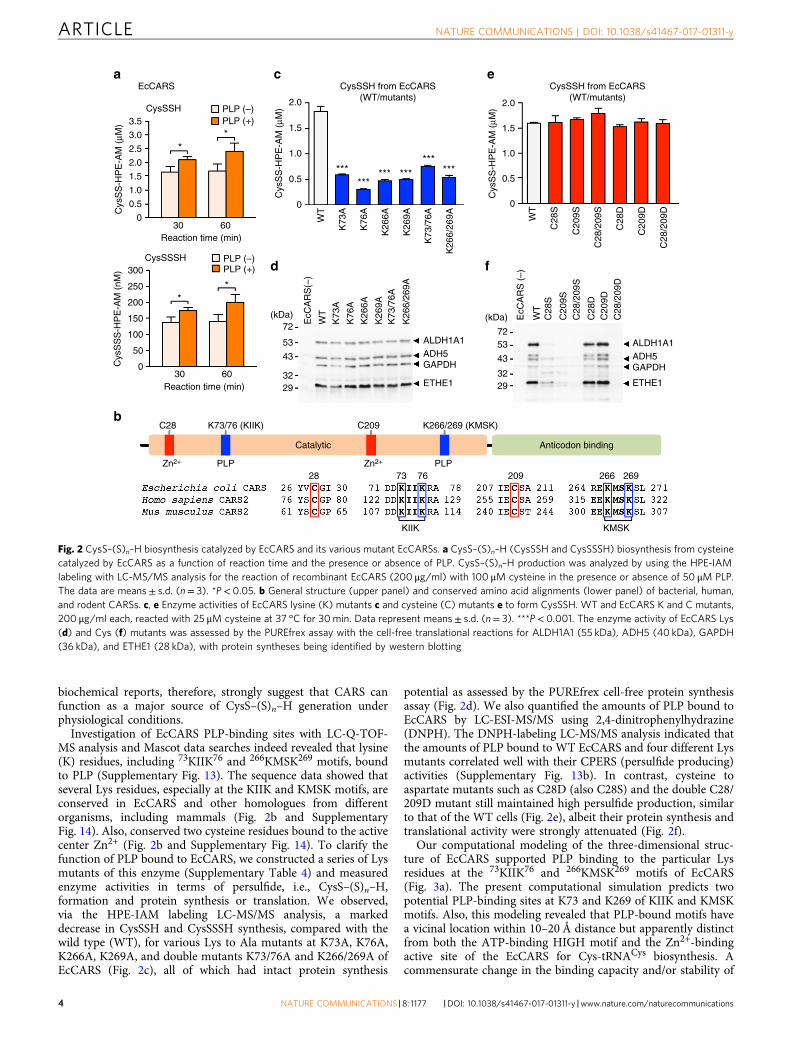
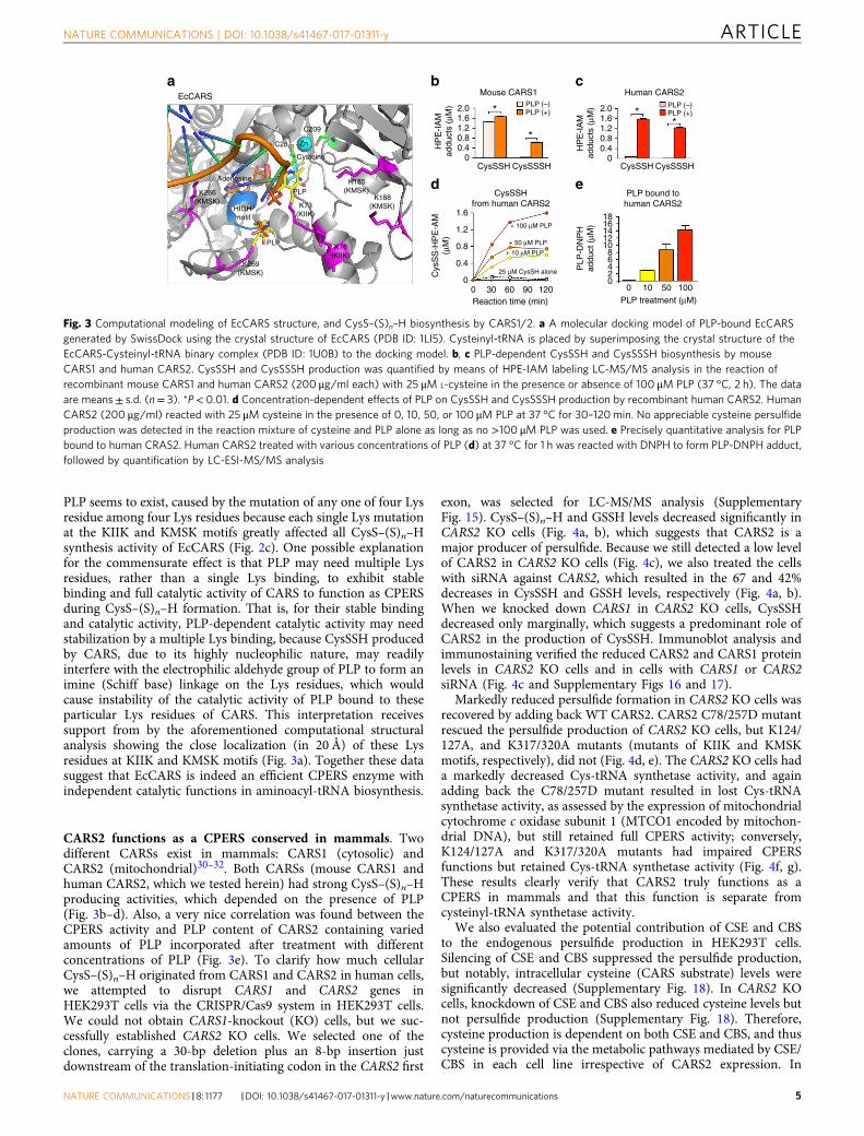
CARS2 functions as a CPERS conserved in mammals. Twodifferent CARSs exist in mammals: CARS1 (cytosolic) andCARS2 (mitochondrial)30–32. Both CARSs (mouse CARS1 andhuman CARS2, which we tested herein) had strong CysS–(S)n–Hproducing activities, which depended on the presence of PLP(Fig. 3b–d). Also, a very nice correlation was found between theCPERS activity and PLP content of CARS2 containing variedamounts of PLP incorporated after treatment with differentconcentrations of PLP (Fig. 3e). To clarify how much cellularCysS–(S)n–H originated from CARS1 and CARS2 in human cells,we attempted to disrupt CARS1 and CARS2 genes inHEK293T cells via the CRISPR/Cas9 system in HEK293T cells.We could not obtain CARS1-knockout (KO) cells, but we suc-cessfully established CARS2 KO cells. We selected one of theclones, carrying a 30-bp deletion plus an 8-bp insertion justdownstream of the translation-initiating codon in the CARS2 first
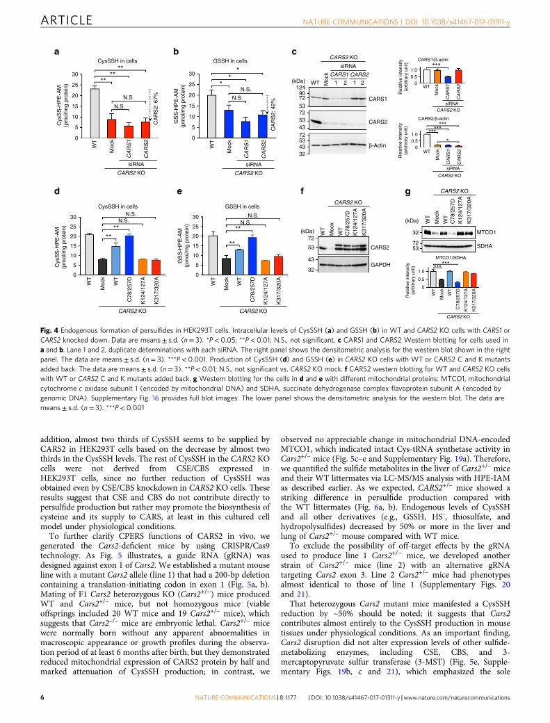
exon, was selected for LC-MS/MS analysis (SupplementaryFig. 15). CysS–(S)n–H and GSSH levels decreased significantly inCARS2 KO cells (Fig. 4a, b), which suggests that CARS2 is amajor producer of persulfide. Because we still detected a low levelof CARS2 in CARS2 KO cells (Fig. 4c), we also treated the cellswith siRNA against CARS2, which resulted in the 67 and 42%decreases in CysSSH and GSSH levels, respectively (Fig. 4a, b).When we knocked down CARS1 in CARS2 KO cells, CysSSHdecreased only marginally, which suggests a predominant role ofCARS2 in the production of CysSSH. Immunoblot analysis andimmunostaining verified the reduced CARS2 and CARS1 proteinlevels in CARS2 KO cells and in cells with CARS1 or CARS2siRNA (Fig. 4c and Supplementary Figs 16 and 17).
Markedly reduced persulfide formation in CARS2 KO cells wasrecovered by adding back WT CARS2. CARS2 C78/257D mutantrescued the persulfide production of CARS2 KO cells, but K124/127A, and K317/320A mutants (mutants of KIIK and KMSKmotifs, respectively), did not (Fig. 4d, e). The CARS2 KO cells hada markedly decreased Cys-tRNA synthetase activity, and againadding back the C78/257D mutant resulted in lost Cys-tRNAsynthetase activity, as assessed by the expression of mitochondrialcytochrome c oxidase subunit 1 (MTCO1 encoded by mitochon-drial DNA), but still retained full CPERS activity; conversely,K124/127A and K317/320A mutants had impaired CPERSfunctions but retained Cys-tRNA synthetase activity (Fig. 4f, g).These results clearly verify that CARS2 truly functions as aCPERS in mammals and that this function is separate fromcysteinyl-tRNA synthetase activity.
We also evaluated the potential contribution of CSE and CBSto the endogenous persulfide production in HEK293T cells.Silencing of CSE and CBS suppressed the persulfide production,but notably, intracellular cysteine (CARS substrate) levels weresignificantly decreased (Supplementary Fig. 18). In CARS2 KOcells, knockdown of CSE and CBS also reduced cysteine levels butnot persulfide production (Supplementary Fig. 18). Therefore,cysteine production is dependent on both CSE and CBS, and thuscysteine is provided via the metabolic pathways mediated by CSE/CBS in each cell line irrespective of CARS2 expression. In
K266(KMSK)
K269(KMSK)
K73(KIIK)
K76(KIIK)
K185(KMSK)
K188(KMSK)
Zn
PLP
Adenosine
C209
C28
HIGHmotif
Cysteine
PLP
EcCARS Mouse CARS1
CysSSHfrom human CARS2
Human CARS2
2.0 2.01.61.20.80.4
1.6 181614121086420
0 10 50 100PLP treatment (μM)
PLP
-DN
PH
addu
ct (
μM)1.2
Cys
SS
-HP
E-A
M(μ
M)
0.8
0.4
0
* **
*
HP
E-I
AM
addu
cts
(μM
)
HP
E-I
AM
addu
cts
(μM
)
0
1.61.20.80.4
0CysSSH
0 30 60 90 120Reaction time (min)
CysSSSH
+ 100 μM PLP
+ 50 μM PLP
+ 10 μM PLP
25 μM CysSH alone
CysSSH
PLP bound tohuman CARS2
CysSSSH
PLP (–)PLP (+)
PLP (–)PLP (+)
a b c
d e
Fig. 3 Computational modeling of EcCARS structure, and CysS–(S)n–H biosynthesis by CARS1/2. a A molecular docking model of PLP-bound EcCARSgenerated by SwissDock using the crystal structure of EcCARS (PDB ID: 1LI5). Cysteinyl-tRNA is placed by superimposing the crystal structure of theEcCARS-Cysteinyl-tRNA binary complex (PDB ID: 1U0B) to the docking model. b, c PLP-dependent CysSSH and CysSSSH biosynthesis by mouseCARS1 and human CARS2. CysSSH and CysSSSH production was quantified by means of HPE-IAM labeling LC-MS/MS analysis in the reaction ofrecombinant mouse CARS1 and human CARS2 (200 μg/ml each) with 25 μM L-cysteine in the presence or absence of 100 μM PLP (37 °C, 2 h). The dataare means± s.d. (n= 3). *P< 0.01. d Concentration-dependent effects of PLP on CysSSH and CysSSSH production by recombinant human CARS2. HumanCARS2 (200 μg/ml) reacted with 25 μM cysteine in the presence of 0, 10, 50, or 100 μM PLP at 37 °C for 30–120min. No appreciable cysteine persulfideproduction was detected in the reaction mixture of cysteine and PLP alone as long as no >100 μM PLP was used. e Precisely quantitative analysis for PLPbound to human CRAS2. Human CARS2 treated with various concentrations of PLP (d) at 37 °C for 1 h was reacted with DNPH to form PLP-DNPH adduct,followed by quantification by LC-ESI-MS/MS analysis
NATURE COMMUNICATIONS | DOI: 10.1038/s41467-017-01311-y ARTICLE
NATURE COMMUNICATIONS |8: 1177 |DOI: 10.1038/s41467-017-01311-y |www.nature.com/naturecommunications 5
addition, almost two thirds of CysSSH seems to be supplied byCARS2 in HEK293T cells based on the decrease by almost twothirds in the CysSSH levels. The rest of CysSSH in the CARS2 KOcells were not derived from CSE/CBS expressed inHEK293T cells, since no further reduction of CysSSH wasobtained even by CSE/CBS knockdown in CARS2 KO cells. Theseresults suggest that CSE and CBS do not contribute directly topersulfide production but rather may promote the biosynthesis ofcysteine and its supply to CARS, at least in this cultured cellmodel under physiological conditions.
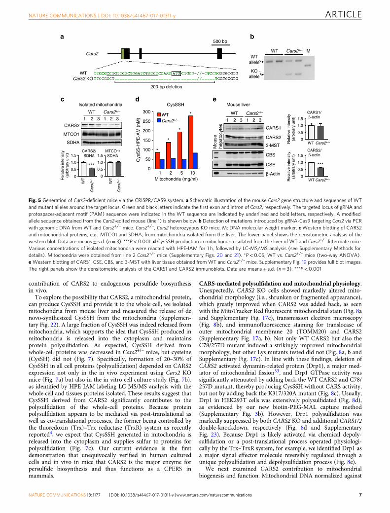
To further clarify CPERS functions of CARS2 in vivo, wegenerated the Cars2-deficient mice by using CRISPR/Cas9technology. As Fig. 5 illustrates, a guide RNA (gRNA) wasdesigned against exon 1 of Cars2. We established a mutant mouseline with a mutant Cars2 allele (line 1) that had a 200-bp deletioncontaining a translation-initiating codon in exon 1 (Fig. 5a, b).Mating of F1 Cars2 heterozygous KO (Cars2+/−) mice producedWT and Cars2+/– mice, but not homozygous mice (viableoffsprings included 20 WT mice and 19 Cars2+/– mice), whichsuggests that Cars2–/– mice are embryonic lethal. Cars2+/– micewere normally born without any apparent abnormalities inmacroscopic appearance or growth profiles during the observa-tion period of at least 6 months after birth, but they demonstratedreduced mitochondrial expression of CARS2 protein by half andmarked attenuation of CysSSH production; in contrast, we
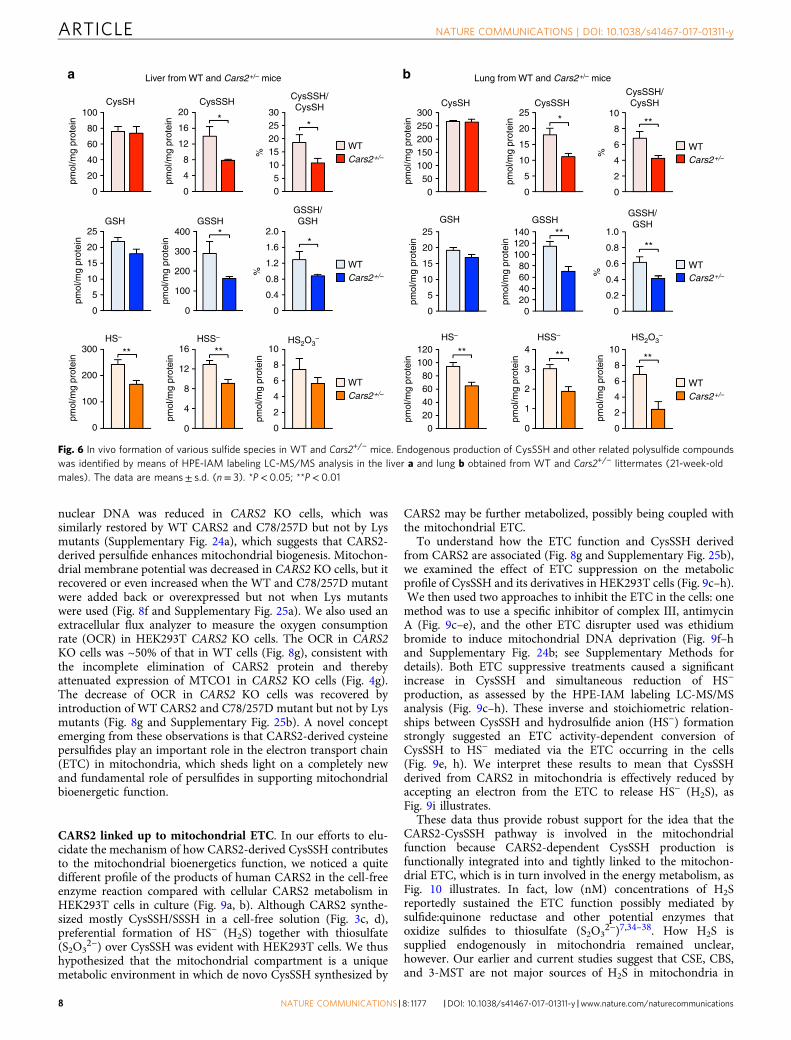
observed no appreciable change in mitochondrial DNA-encodedMTCO1, which indicated intact Cys-tRNA synthetase activity inCars2+/– mice (Fig. 5c–e and Supplementary Fig. 19a). Therefore,we quantified the sulfide metabolites in the liver of Cars2+/– miceand their WT littermates via LC-MS/MS analysis with HPE-IAMas described earlier. As we expected, CARS2+/– mice showed astriking difference in persulfide production compared withthe WT littermates (Fig. 6a, b). Endogenous levels of CysSSHand all other derivatives (e.g., GSSH, HS-, thiosulfate, andhydropolysulfides) decreased by 50% or more in the liver andlung of Cars2+/– mouse compared with WT mice.
To exclude the possibility of off-target effects by the gRNAused to produce line 1 Cars2+/− mice, we developed anotherstrain of Cars2+/– mice (line 2) with an alternative gRNAtargeting Cars2 exon 3. Line 2 Cars2+/– mice had phenotypesalmost identical to those of line 1 (Supplementary Figs. 20and 21).
That heterozygous Cars2 mutant mice manifested a CysSSHreduction by ~50% should be noted; it suggests that Cars2contributes almost entirely to the CysSSH production in mousetissues under physiological conditions. As an important finding,Cars2 disruption did not alter expression levels of other sulfide-metabolizing enzymes, including CSE, CBS, and 3-mercaptopyruvate sulfur transferase (3-MST) (Fig. 5e, Supple-mentary Figs. 19b, c and 21), which emphasized the sole
30
25
Cys
SS
-HP
E-A
M(p
mol
/mg
prot
ein)
20
**
N.S.N.S.
1 12 2
N.S.W
T
Moc
k
WT
CARS1
CARS1/β-actin
CARS2/β-actin
Rel
ativ
e in
tens
ity(a
rbitr
ary
unit)
CARS2
β-Actin
(kDa)124907253
5343
72
534332
72
Moc
k
CA
RS
2: 6
7%
CA
RS
2: 4
2%
CA
RS
1
CARS2 KO
CARS2 KO
CARS2 KO
CARS2 KO
1.00.5
0
Rel
ativ
e in
tens
ity(a
rbitr
ary
unit)
1.00.5
0
Rel
ativ
e in
tens
ity(a
rbitr
ary
unit)
1.0
0.5
0
WT
***
*********
*
Moc
k
CA
RS
1
CA
RS
2
CA
RS
2
CARS1 CARS2
siRNA
WT
Moc
k
CA
RS
1
CARS2 KO
CA
RS
2
siRNA
CysSSH in cells
siRNA
siRNA
CARS2 KO
WT
Moc
k
CA
RS
1
CA
RS
2
siRNA
N.S.
N.S.N.S.
****
****
CysSSH in cells GSSH in cells
15
10
5
0
30
25
Cys
SS
-HP
E-A
M(p
mol
/mg
prot
ein)
20
15
10
5
0
WT
WT
C78
/257
D
K12
4/12
7A
K31
7/32
0A
Moc
k
CARS2 KO
CARS2 KO
CARS2 KO
GSSH in cells
N.S.N.S.
**
**
30
25
GS
S-H
PE
-AM
(pm
ol/m
g pr
otei
n)
20
15
10
5
0
CARS2 KO
WT
WT
C78
/257
D
K12
4/12
7A
K31
7/32
0A
Moc
k
WT(kDa)
(kDa)
7272
CARS2
MTCO1
MTCO1/SDHA
******
SDHA
GAPDH
53 53
43
32
32WT
C78
/257
DK
124/
127A
K31
7/32
0A
Moc
k WT
WT
C78
/257
DK
124/
127A
K31
7/32
0A
Moc
kW
T
WT
C78
/257
D
K12
4/12
7A
K31
7/32
0A
Moc
k
30 **
*25
GS
S-H
PE
-AM
(pm
ol/m
g pr
otei
n)
20
15
10
5
0
a b c
d e f g
Fig. 4 Endogenous formation of persulfides in HEK293T cells. Intracellular levels of CysSSH (a) and GSSH (b) in WT and CARS2 KO cells with CARS1 orCARS2 knocked down. Data are means± s.d. (n= 3). *P< 0.05; **P< 0.01; N.S., not significant. c CARS1 and CARS2 Western blotting for cells used ina and b. Lane 1 and 2, duplicate determinations with each siRNA. The right panel shows the densitometric analysis for the western blot shown in the rightpanel. The data are means± s.d. (n= 3). ***P< 0.001. Production of CysSSH (d) and GSSH (e) in CARS2 KO cells with WT or CARS2 C and K mutantsadded back. The data are means± s.d. (n= 3). **P< 0.01; N.S., not significant vs. CARS2 KO mock. f CARS2 western blotting for WT and CARS2 KO cellswith WT or CARS2 C and K mutants added back. g Western blotting for the cells in d and e with different mitochondrial proteins: MTCO1, mitochondrialcytochrome c oxidase subunit 1 (encoded by mitochondrial DNA) and SDHA, succinate dehydrogenase complex flavoprotein subunit A (encoded bygenomic DNA). Supplementary Fig. 16 provides full blot images. The lower panel shows the densitometric analysis for the western blot. The data aremeans± s.d. (n= 3). ***P< 0.001
ARTICLE NATURE COMMUNICATIONS | DOI: 10.1038/s41467-017-01311-y
6 NATURE COMMUNICATIONS |8: 1177 |DOI: 10.1038/s41467-017-01311-y |www.nature.com/naturecommunications
contribution of CARS2 to endogenous persulfide biosynthesisin vivo.
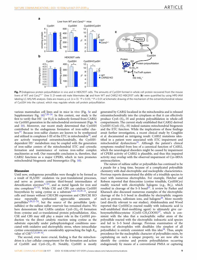
To explore the possibility that CARS2, a mitochondrial protein,can produce CysSSH and provide it to the whole cell, we isolatedmitochondria from mouse liver and measured the release of denovo-synthesized CysSSH from the mitochondria (Supplemen-tary Fig. 22). A large fraction of CysSSH was indeed released frommitochondria, which supports the idea that CysSSH produced inmitochondria is released into the cytoplasm and maintainsprotein polysulfidation. As expected, CysSSH derived fromwhole-cell proteins was decreased in Cars2+/– mice, but cysteine(CysSH) did not (Fig. 7). Specifically, formation of 20–30% ofCysSSH in all cell proteins (polysulfidation) depended on CARS2expression not only in the in vivo experiment using Cars2 KOmice (Fig. 7a) but also in the in vitro cell culture study (Fig. 7b),as identified by HPE-IAM labeling LC-MS/MS analysis with thewhole cell and tissues proteins isolated. These results suggest thatCysSSH derived from CARS2 significantly contributes to thepolysulfidation of the whole-cell proteins. Because proteinpolysulfidation appears to be mediated via post-translational aswell as co-translational processes, the former being controlled bythe thioredoxin (Trx)–Trx reductase (TrxR) system as recentlyreported4, we expect that CysSSH generated in mitochondria isreleased into the cytoplasm and supplies sulfur to proteins forpolysulfidation (Fig. 7c). Our current evidence is the firstdemonstration that unequivocally verified in human culturedcells and in vivo in mice that CARS2 is the major enzyme forpersulfide biosynthesis and thus functions as a CPERS inmammals.
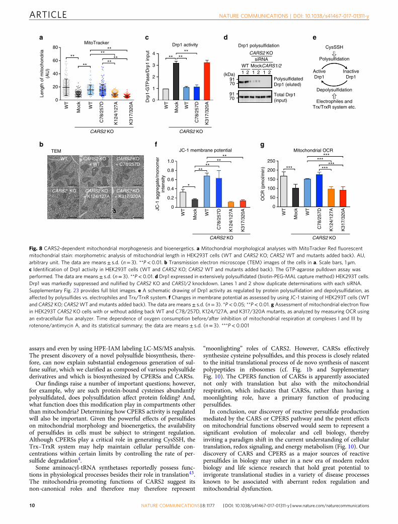
CARS-mediated polysulfidation and mitochondrial physiology.Unexpectedly, CARS2 KO cells showed markedly altered mito-chondrial morphology (i.e., shrunken or fragmented appearance),which greatly improved when CARS2 was added back, as seenwith the MitoTracker Red fluorescent mitochondrial stain (Fig. 8aand Supplementary Fig. 17c), transmission electron microscopy(Fig. 8b), and immunofluorescence staining for translocase ofouter mitochondrial membrane 20 (TOMM20) and CARS2(Supplementary Fig. 17a, b). Not only WT CARS2 but also theC78/257D mutant induced a strikingly improved mitochondrialmorphology, but other Lys mutants tested did not (Fig. 8a, b andSupplementary Fig. 17c). In line with these findings, deletion ofCARS2 activated dynamin-related protein (Drp1), a major med-iator of mitochondrial fission33, and Drp1 GTPase activity wassignificantly attenuated by adding back the WT CARS2 and C78/257D mutant, thereby producing CysSSH without CARS activity,but not by adding back the K317/320A mutant (Fig. 8c). Usually,Drp1 in HEK293T cells was extensively polysulfidated (Fig. 8d),as evidenced by our new biotin-PEG-MAL capture method(Supplementary Fig. 3b). However, Drp1 polysulfidation wasmarkedly suppressed by both CARS2 KO and additional CARS1/2double-knockdown, respectively (Fig. 8d and SupplementaryFig. 23). Because Drp1 is likely activated via chemical depoly-sulfidation or a post-translational process operated physiologi-cally by the Trx–TrxR system, for example, we identified Drp1 asa major signal effector molecule reversibly regulated through aunique polysulfidation and depolysulfidation process (Fig. 8e).
We next examined CARS2 contribution to mitochondrialbiogenesis and function. Mitochondrial DNA normalized against
Cars2
Cars2 KOWT
Isolated mitochondria
200-bp deletion
500 bp
WT MCars2+/–
WT
CysSSH Mouse liver
CARS1/β-actin
CARS2/β-actin
***
Mou
sehe
pato
cyte
sCARS2
MTCO1
SDHA
1.5
***1.0
Rel
ativ
e in
tens
ity(a
rbitr
ary
unit)
0.5
0
1.5
1.0
0.5
0
WT
CARS2/SDHA
MTCO1/SDHA
1 12 23 3
Cars2+/– WT
1 12 23 3
CARS1
1.5
1.0
0.5
Rel
ativ
e in
tens
ity(a
rbitr
ary
unit)
0
1.5
1.0
0.5
Rel
ativ
e in
tens
ity(a
rbitr
ary
unit)
0
CARS2
3-MST
CBS
CSE
β-Actin
Cars2+/–
WT Cars2+/–
WT Cars2+/–
WT300
250
200
150
100
*
*
*
*
50
1 2 5 10Mitochondria (mg/ml)
Cys
SS
-HP
E-A
M (
nM)
0
Cars2+/–
Car
s2+
/–
WT
Car
s2+
/–
a
c d e
b
WTallele
KOallele
Fig. 5 Generation of Cars2-deficient mice via the CRISPR/CAS9 system. a Schematic illustration of the mouse Cars2 gene structure and sequences of WTand mutant alleles around the target locus. Green and black letters indicate the first exon and intron of Cars2, respectively. The targeted locus of gRNA andprotospacer-adjacent motif (PAM) sequence were indicated in the WT sequence are indicated by underlined and bold letters, respectively. A modifiedallele sequence obtained from the Cars2-edited mouse (line 1) is shown below. b Detection of mutations introduced by gRNA-Cas9 targeting Cars2 via PCRwith genomic DNA from WT and Cars2+/− mice. Cars2+/−, Cars2 heterozygous KO mice, M: DNA molecular weight marker. c Western blotting of CARS2and mitochondrial proteins, e.g., MTCO1 and SDHA, from mitochondria isolated from the liver. The lower panel shows the densitometric analysis of thewestern blot. Data are means± s.d. (n= 3). ***P< 0.001. d CysSSH production in mitochondria isolated from the liver of WT and Cars2+/− littermate mice.Various concentrations of isolated mitochondria were reacted with HPE-IAM for 1 h, followed by LC-MS/MS analysis (see Supplementary Methods fordetails). Mitochondria were obtained from line 2 Cars2+/– mice (Supplementary Figs. 20 and 21). *P< 0.05, WT vs. Cars2+/– mice (two-way ANOVA).e Western blotting of CARS1, CSE, CBS, and 3-MST with liver tissue obtained from WT and Cars2+/– mice. Supplementary Fig. 19 provides full blot images.The right panels show the densitometric analysis of the CARS1 and CARS2 immunoblots. Data are means± s.d. (n= 3). ***P< 0.001
NATURE COMMUNICATIONS | DOI: 10.1038/s41467-017-01311-y ARTICLE
NATURE COMMUNICATIONS |8: 1177 |DOI: 10.1038/s41467-017-01311-y |www.nature.com/naturecommunications 7
nuclear DNA was reduced in CARS2 KO cells, which wassimilarly restored by WT CARS2 and C78/257D but not by Lysmutants (Supplementary Fig. 24a), which suggests that CARS2-derived persulfide enhances mitochondrial biogenesis. Mitochon-drial membrane potential was decreased in CARS2 KO cells, but itrecovered or even increased when the WT and C78/257D mutantwere added back or overexpressed but not when Lys mutantswere used (Fig. 8f and Supplementary Fig. 25a). We also used anextracellular flux analyzer to measure the oxygen consumptionrate (OCR) in HEK293T CARS2 KO cells. The OCR in CARS2KO cells was ~50% of that in WT cells (Fig. 8g), consistent withthe incomplete elimination of CARS2 protein and therebyattenuated expression of MTCO1 in CARS2 KO cells (Fig. 4g).The decrease of OCR in CARS2 KO cells was recovered byintroduction of WT CARS2 and C78/257D mutant but not by Lysmutants (Fig. 8g and Supplementary Fig. 25b). A novel conceptemerging from these observations is that CARS2-derived cysteinepersulfides play an important role in the electron transport chain(ETC) in mitochondria, which sheds light on a completely newand fundamental role of persulfides in supporting mitochondrialbioenergetic function.
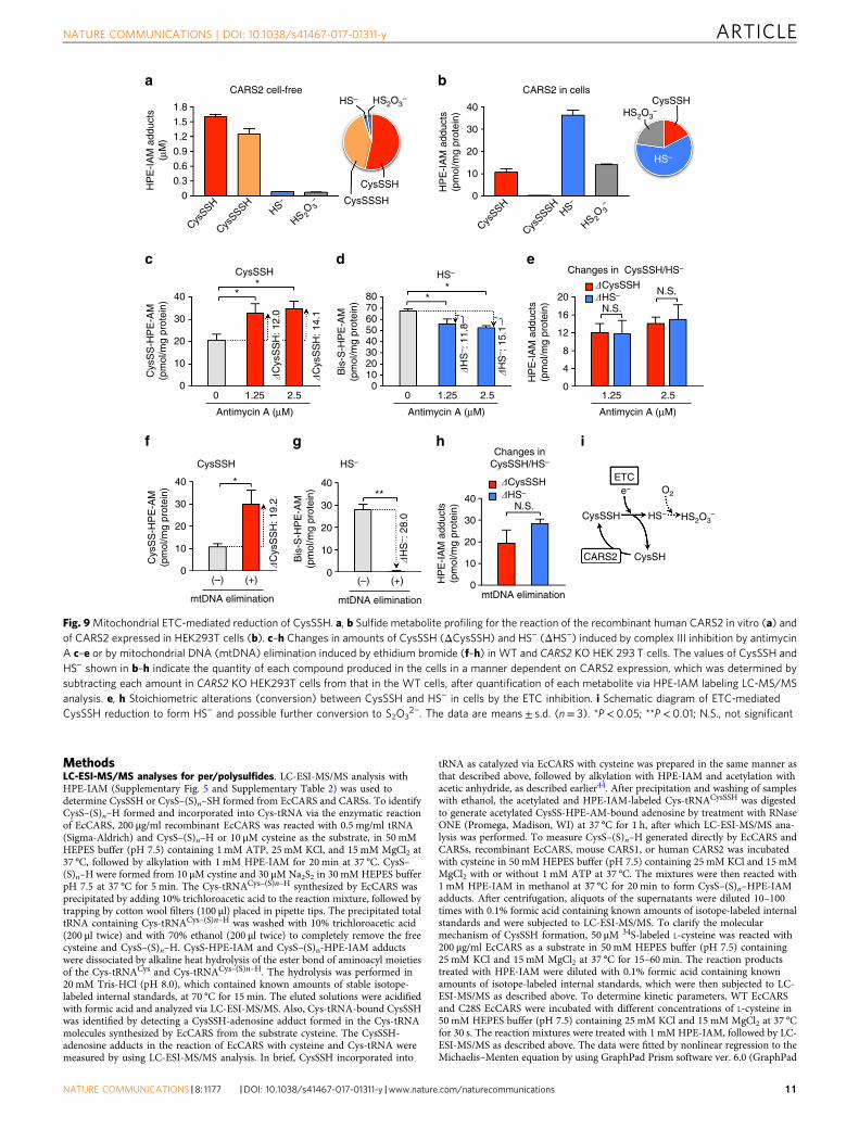
CARS2 linked up to mitochondrial ETC. In our efforts to elu-cidate the mechanism of how CARS2-derived CysSSH contributesto the mitochondrial bioenergetics function, we noticed a quitedifferent profile of the products of human CARS2 in the cell-freeenzyme reaction compared with cellular CARS2 metabolism inHEK293T cells in culture (Fig. 9a, b). Although CARS2 synthe-sized mostly CysSSH/SSSH in a cell-free solution (Fig. 3c, d),preferential formation of HS− (H2S) together with thiosulfate(S2O3
2−) over CysSSH was evident with HEK293T cells. We thushypothesized that the mitochondrial compartment is a uniquemetabolic environment in which de novo CysSSH synthesized by
CARS2 may be further metabolized, possibly being coupled withthe mitochondrial ETC.
To understand how the ETC function and CysSSH derivedfrom CARS2 are associated (Fig. 8g and Supplementary Fig. 25b),we examined the effect of ETC suppression on the metabolicprofile of CysSSH and its derivatives in HEK293T cells (Fig. 9c–h).We then used two approaches to inhibit the ETC in the cells: onemethod was to use a specific inhibitor of complex III, antimycinA (Fig. 9c–e), and the other ETC disrupter used was ethidiumbromide to induce mitochondrial DNA deprivation (Fig. 9f–hand Supplementary Fig. 24b; see Supplementary Methods fordetails). Both ETC suppressive treatments caused a significantincrease in CysSSH and simultaneous reduction of HS−
production, as assessed by the HPE-IAM labeling LC-MS/MSanalysis (Fig. 9c–h). These inverse and stoichiometric relation-ships between CysSSH and hydrosulfide anion (HS−) formationstrongly suggested an ETC activity-dependent conversion ofCysSSH to HS− mediated via the ETC occurring in the cells(Fig. 9e, h). We interpret these results to mean that CysSSHderived from CARS2 in mitochondria is effectively reduced byaccepting an electron from the ETC to release HS− (H2S), asFig. 9i illustrates.
These data thus provide robust support for the idea that theCARS2-CysSSH pathway is involved in the mitochondrialfunction because CARS2-dependent CysSSH production isfunctionally integrated into and tightly linked to the mitochon-drial ETC, which is in turn involved in the energy metabolism, asFig. 10 illustrates. In fact, low (nM) concentrations of H2Sreportedly sustained the ETC function possibly mediated bysulfide:quinone reductase and other potential enzymes thatoxidize sulfides to thiosulfate (S2O3
2−)7,34–38. How H2S issupplied endogenously in mitochondria remained unclear,however. Our earlier and current studies suggest that CSE, CBS,and 3-MST are not major sources of H2S in mitochondria in
CysSH
Liver from WT and Cars2+/– mice Lung from WT and Cars2+/– mice
CysSSHCysSSH/CysSH CysSH CysSSH
CysSSH/CysSH
WTCars2+/–
WTCars2+/–
WTCars2+/–
WTCars2+/–
WTCars2+/–
WTCars2+/–
GSSH/GSH
** *
****
100
80
60
pmol
/mg
prot
ein
pmol
/mg
prot
ein
pmol
/mg
prot
ein
pmol
/mg
prot
ein
pmol
/mg
prot
ein
pmol
/mg
prot
ein
pmol
/mg
prot
ein
40
20
25
20
400 **
2.0
1.6
1.2
0.8
0.4
0
%
%%
300
200
100
0
pmol
/mg
prot
ein
pmol
/mg
prot
ein
pmol
/mg
prot
ein
pmol
/mg
prot
ein
pmol
/mg
prot
ein
pmol
/mg
prot
ein
pmol
/mg
prot
ein
300
200250
150
50100
0
300 16 10
8
6
4
2
0
10
8
6
4
2
0
10
8
6
4
4
3
2
1
0
2
0
12
8
4
0
200
100
0
15
10
5
0
25
20
15
10
5
0
20
202530
151050
20
25
15
10
5
140 1.0
0.8
0.6
0.4
0.2
0
120
80100
6040200
120
80100
6040200
0
%
16
12
8
4
00
GSH
** ** ** ** **
**
HS– HSS– HS2O3– HS– HSS– HS2O3
–
GSSHGSSH/GSHGSH GSSH
a b
Fig. 6 In vivo formation of various sulfide species in WT and Cars2+/− mice. Endogenous production of CysSSH and other related polysulfide compoundswas identified by means of HPE-IAM labeling LC-MS/MS analysis in the liver a and lung b obtained from WT and Cars2+/− littermates (21-week-oldmales). The data are means± s.d. (n= 3). *P< 0.05; **P< 0.01
ARTICLE NATURE COMMUNICATIONS | DOI: 10.1038/s41467-017-01311-y
8 NATURE COMMUNICATIONS |8: 1177 |DOI: 10.1038/s41467-017-01311-y |www.nature.com/naturecommunications
various mammalian cell lines and in mice in vivo (Fig. 5e andSupplementary Fig. 18)7,20–24. In this context, our study is thefirst to verify that HS− (or H2S) is indirectly formed from CARS2via CysSSH generation in the mitochondrial environment (Figs. 9iand 10). Moreover, our recent study determined that CysSSHcontributed to the endogenous formation of iron-sulfur clus-ters14. Because iron-sulfur clusters are known to be synthesizedand utilized in complexes I-III of the ETC in mitochondria39, andare actively transported extramitochondrially, the CysSSH-dependent HS− metabolism may be coupled with the generationof iron-sulfur centers of the mitochondrial ETC and cytosolicformation and maintenance of various iron-sulfur complexmachineries as well. Our reasonable conclusion is, therefore, thatCARS2 functions as a major CPERS, which in turn promotesmitochondrial biogenesis and bioenergetics (Fig. 10).
DiscussionUntil now, endogenous persulfides were thought to be formed asa result of H2S/HS− oxidation via post-translational processes,and serve as protein cysteine thiol-bound intermediates ofdetoxification enzymes3,7,21, and as metal ligands for iron andzinc complexes11–15. While CSE and CBS can catalyze CysSSHbiosynthesis by using cystine as a substrate3,4,6–10,18–21, severalcells and tissues without CSE/CBS expression and CBS/CSE KOmice reportedly synthesized appreciable amounts ofpersulfides3,20,22–24, but the source of the persulfides (poly-sulfides) or the sulfane sulfur reservoir has remained elusive. Wehere demonstrate that CARSs catalyze CysS–(S)n–H formationfrom cysteine and co-translational protein polysulfidation. Also,CSE and CBS may still play a major role in the CysSSH pro-duction via the direct catalytic reaction using cystine as thesubstrate especially under pathophysiological conditions asso-ciated with oxidative and electrophilic stress, where intracellularcystine concentrations are considerably approaching the high Km
value of CSE3,7,21,40–42.The second, even more crucial, finding is that the mitochon-
drion is a key cellular compartment for the formation and actionof CysSSH and CysS–(S)n–H. Notably, CysSSH is mostly
generated by CARS2 localized in the mitochondria and is releasedextramitochondrially into the cytoplasm so that it can effectivelyproduce CysS–(S)n–H and protein polysulfidation in whole-cellcompartments. The current study established that CARS2-derivedCysSSH (CysS–(S)n–H) indeed sustains mitochondrial biogenesisand the ETC function. While the implications of these findingsawait further investigation, a recent clinical study by Coughlinet al. documented an intriguing result: CARS2 mutations iden-tified in a patient were associated with ETC impairment andmitochondrial dysfunctions31. Although the patient’s clinicalsymptoms resulted from loss of a canonical function of CARS2,which the neurological disorders might be caused by impairmentof CPERS activity of CARS2 is plausible, and thus this impairedactivity may overlap with the observed impairment of Cys-tRNAaminoacylation.
The nature of sulfane sulfur or polysulfides has continued to bea puzzle for a long time, because of a complicated polysulfidechemistry with dual electrophilic and nucleophilic characteristics.Previous reports demonstrated the ability of a trisulfide species toreact with numerous electrophiles. For example, Fletcher andRobson reported that thiocystine (cystine trisulfide, CysSSSCys)readily reacted with electrophilic halogens (e.g., Br2), whichresulted in cleavage of the S–S bond25. A review by Parker andKharasch also discussed numerous examples of the electrophiliccleavage of the S–S bond in disulfides by electrophilic reagentssuch as protons, sulfenium ions, and halogens26. More recently(and directly relevant to our studies), Abdolrasulnia and Woodreported that CysSSSCys reacted readily with iodoacetic acid (awell-established thiol-modifying agent) to ultimately give car-boxymethylthiocysteine (CysSS–CH2COOH)27, which is con-sistent with the idea that a nucleophilic sulfur atom of thepolysulfide reacted with the electrophilic iodoacetic acid speciesand led to S–S bond cleavage. Previous examination of thereaction of electrophiles with disulfides (the simplest of allpolysulfides) is entirely consistent with this idea28. Thus, ampleprecedence for the nucleophilic character of polysulfides exists, bycapitalizing on such a unique property, we are now able toidentify the cysteine and protein polysulfidation occurringendogenously by means of a conventional PMSA or capturing
6CysSH CysSSH
Liver from WT and Cars2+/– mice
CysSSH/CysSH
CysSH
CysSH
CysSSH
CARS2
CysSSH
CysSSH
Mitochondria
ProteinSH
SH
HS–/HSn–
S-(S)n-H
Trx/TrxR system
Protein polysulfidation
HEK293T cells
CysSSH/CysSH
** **
**
500
400
300
200
12
10
30%
19%
WT
WTCARS2 KO
Cars2+/–
8
6
4
2
0
%
12
10
8
6
4
2
0
%
100
0
5
4
3nm
ol/m
g pr
otei
nnm
ol/m
g pr
otei
n
pmol
/mg
prot
ein
4004.54.03.53.02.52.01.51.00.5
0
300
200
100
0
pmol
/mg
prot
ein
2
1
0
a
b
c
Fig. 7 Endogenous protein polysulfidation in vivo and in HEK293T cells. The amounts of CysSSH formed in whole cell protein recovered from the mouselivers of WT and Cars2+/− (line 1) 21-week-old male littermates (a) and from WT and CARS2 KO HEK293T cells (b) were quantified by using HPE-IAMlabeling LC-MS/MS analysis. Data are means± s.d. (n= 3). *P< 0.05; **P< 0.01. c Schematic drawing of the mechanism of the extramitochondrial releaseof CysSSH into the cytosol, which may regulate whole cell protein polysulfidation
NATURE COMMUNICATIONS | DOI: 10.1038/s41467-017-01311-y ARTICLE
NATURE COMMUNICATIONS |8: 1177 |DOI: 10.1038/s41467-017-01311-y |www.nature.com/naturecommunications 9
assays and even by using HPE-IAM labeling LC-MS/MS analysis.The present discovery of a novel polysulfide biosynthesis, there-fore, can now explain substantial endogenous generation of sul-fane sulfur, which we clarified as composed of various polysulfidederivatives and which is biosynthesized by CPERSs and CARSs.
Our findings raise a number of important questions; however,for example, why are such protein-bound cysteines abundantlypolysulfidated, does polysulfidation affect protein folding? And,what function does this modification play in compartments otherthan mitochondria? Determining how CPERS activity is regulatedwill also be important. Given the powerful effects of persulfideson mitochondrial morphology and bioenergetics, the availabilityof persulfides in cells must be subject to stringent regulation.Although CPERSs play a critical role in generating CysSSH, theTrx–TrxR system may help maintain cellular persulfide con-centrations within certain limits by controlling the rate of per-sulfide degradation4.
Some aminoacyl-tRNA synthetases reportedly possess func-tions in physiological processes besides their role in translation43.The mitochondria-promoting functions of CARS2 suggest itsnon-canonical roles and therefore may therefore represent
“moonlighting” roles of CARS2. However, CARSs effectivelysynthesize cysteine polysulfides, and this process is closely relatedto the initial translational process of de novo synthesis of nascentpolypeptides in ribosomes (cf. Fig. 1b and SupplementaryFig. 10). The CPERS function of CARSs is apparently associatednot only with translation but also with the mitochondrialrespiration, which indicates that CARSs, rather than having amoonlighting role, have a primary function of producingpersulfides.
In conclusion, our discovery of reactive persulfide productionmediated by the CARS or CPERS pathway and the potent effectson mitochondrial functions observed would seem to represent asignificant evolution of molecular and cell biology, therebyinviting a paradigm shift in the current understanding of cellulartranslation, redox signaling, and energy metabolism (Fig. 10). Ourdiscovery of CARS and CPERS as a major sources of reactivepersulfides in biology may usher in a new era of modern redoxbiology and life science research that hold great potential toinvigorate translational studies in a variety of disease processesknown to be associated with aberrant redox regulation andmitochondrial dysfunction.
80
****
****
Drp1 polysulfidation
CARS2 KO
CARS1/2siRNA
WT
(kDa)91
91
70PolysulfidatedDrp1 (eluted)
Total Drp1(input)70
1 12 2
CysSSH
Polysulfidation
Depolysulfidation
Electrophiles andTrx/TrxR system etc.
ActiveDrp1
InactiveDrp1
2 1Mock
****
MitoTracker
60
40
Leng
th o
f mito
chon
dria
(AU
)
20
0
WT
TEM
Moc
k
WT
C78
/257
D
CARS2 KO
K12
4/12
7A
K31
7/32
0A
WT
Moc
k
WT
C78
/257
D
CARS2 KO
K12
4/12
7A
K31
7/32
0A WT
Moc
k
WT
C78
/257
D
CARS2 KO
K12
4/12
7A
K31
7/32
0A
1.0
0.8
JC-1
agg
rega
te/m
onom
erin
tens
ity 0.6
0.4
0.2
0
*
****
****
250
******
******
***200
150
100
50
0
OC
R (
pmol
/min
)
JC-1 membrane potential Mitochondrial OCR
WT CARS2 KO+ WT
CARS2 KO+ C78/257D
CARS2 KO
** ****
Drp1 activity
4
3
2
Drp
1-G
TP
ase/
Drp
1 in
put
1
0
WT
Moc
k
WT
C78
/257
D
CARS2 KO
K31
7/32
0A
CARS2 KO+ K317/320A
CARS2 KO+ K124/127A
a c ed
b f g
Fig. 8 CARS2-dependent mitochondrial morphogenesis and bioenergetics. a Mitochondrial morphological analyses with MitoTracker Red fluorescentmitochondrial stain: morphometric analysis of mitochondrial length in HEK293T cells (WT and CARS2 KO; CARS2 WT and mutants added back). AU,arbitrary unit. The data are means± s.d. (n= 3). **P< 0.01. b Transmission electron microscope (TEM) images of the cells in a. Scale bars, 1 μm.c Identification of Drp1 activity in HEK293T cells (WT and CARS2 KO; CARS2 WT and mutants added back). The GTP-agarose pulldown assay wasperformed. The data are means± s.d. (n= 3). **P< 0.01. d Drp1 expressed in extensively polysulfidated (biotin-PEG-MAL capture method) HEK293T cells.Drp1 was markedly suppressed and nullified by CARS2 KO and CARS1/2 knockdown. Lanes 1 and 2 show duplicate determinations with each siRNA.Supplementary Fig. 23 provides full blot images. e A schematic drawing of Drp1 activity as regulated by protein polysulfidation and depolysulfidation, asaffected by polysulfides vs. electrophiles and Trx/TrxR system. f Changes in membrane potential as assessed by using JC-1 staining of HEK293T cells (WTand CARS2 KO; CARS2WT and mutants added back). The data are means± s.d. (n= 3). *P< 0.05; **P< 0.01. g Assessment of mitochondrial electron flowin HEK293T CARS2 KO cells with or without adding back WT and C78/257D, K124/127A, and K317/320A mutants, as analyzed by measuring OCR usingan extracellular flux analyzer. Time dependence of oxygen consumption before/after inhibition of mitochondrial respiration at complexes I and III byrotenone/antimycin A, and its statistical summary; the data are means± s.d. (n= 3). ***P< 0.001
ARTICLE NATURE COMMUNICATIONS | DOI: 10.1038/s41467-017-01311-y
10 NATURE COMMUNICATIONS |8: 1177 |DOI: 10.1038/s41467-017-01311-y |www.nature.com/naturecommunications
MethodsLC-ESI-MS/MS analyses for per/polysulfides. LC-ESI-MS/MS analysis withHPE-IAM (Supplementary Fig. 5 and Supplementary Table 2) was used todetermine CysSSH or CysS–(S)n–SH formed from EcCARS and CARSs. To identifyCysS–(S)n–H formed and incorporated into Cys-tRNA via the enzymatic reactionof EcCARS, 200 μg/ml recombinant EcCARS was reacted with 0.5 mg/ml tRNA(Sigma-Aldrich) and CysS–(S)n–H or 10 μM cysteine as the substrate, in 50 mMHEPES buffer (pH 7.5) containing 1 mM ATP, 25 mM KCl, and 15 mM MgCl2 at37 °C, followed by alkylation with 1 mM HPE-IAM for 20 min at 37 °C. CysS–(S)n–H were formed from 10 μM cystine and 30 μMNa2S2 in 30 mM HEPES bufferpH 7.5 at 37 °C for 5 min. The Cys-tRNACys–(S)n–H synthesized by EcCARS wasprecipitated by adding 10% trichloroacetic acid to the reaction mixture, followed bytrapping by cotton wool filters (100 μl) placed in pipette tips. The precipitated totaltRNA containing Cys-tRNACys–(S)n–H was washed with 10% trichloroacetic acid(200 μl twice) and with 70% ethanol (200 μl twice) to completely remove the freecysteine and CysS–(S)n–H. CysS-HPE-IAM and CysS–(S)n-HPE-IAM adductswere dissociated by alkaline heat hydrolysis of the ester bond of aminoacyl moietiesof the Cys-tRNACys and Cys-tRNACys–(S)n–H. The hydrolysis was performed in20 mM Tris-HCl (pH 8.0), which contained known amounts of stable isotope-labeled internal standards, at 70 °C for 15 min. The eluted solutions were acidifiedwith formic acid and analyzed via LC-ESI-MS/MS. Also, Cys-tRNA-bound CysSSHwas identified by detecting a CysSSH-adenosine adduct formed in the Cys-tRNAmolecules synthesized by EcCARS from the substrate cysteine. The CysSSH-adenosine adducts in the reaction of EcCARS with cysteine and Cys-tRNA weremeasured by using LC-ESI-MS/MS analysis. In brief, CysSSH incorporated into
tRNA as catalyzed via EcCARS with cysteine was prepared in the same manner asthat described above, followed by alkylation with HPE-IAM and acetylation withacetic anhydride, as described earlier44. After precipitation and washing of sampleswith ethanol, the acetylated and HPE-IAM-labeled Cys-tRNACysSSH was digestedto generate acetylated CysSS-HPE-AM-bound adenosine by treatment with RNaseONE (Promega, Madison, WI) at 37 °C for 1 h, after which LC-ESI-MS/MS ana-lysis was performed. To measure CysS–(S)n–H generated directly by EcCARS andCARSs, recombinant EcCARS, mouse CARS1, or human CARS2 was incubatedwith cysteine in 50 mM HEPES buffer (pH 7.5) containing 25 mM KCl and 15 mMMgCl2 with or without 1 mM ATP at 37 °C. The mixtures were then reacted with1 mM HPE-IAM in methanol at 37 °C for 20 min to form CysS–(S)n–HPE-IAMadducts. After centrifugation, aliquots of the supernatants were diluted 10–100times with 0.1% formic acid containing known amounts of isotope-labeled internalstandards and were subjected to LC-ESI-MS/MS. To clarify the molecularmechanism of CysSSH formation, 50 μM 34S-labeled L-cysteine was reacted with200 μg/ml EcCARS as a substrate in 50 mM HEPES buffer (pH 7.5) containing25 mM KCl and 15 mM MgCl2 at 37 °C for 15–60 min. The reaction productstreated with HPE-IAM were diluted with 0.1% formic acid containing knownamounts of isotope-labeled internal standards, which were then subjected to LC-ESI-MS/MS as described above. To determine kinetic parameters, WT EcCARSand C28S EcCARS were incubated with different concentrations of L-cysteine in50 mM HEPES buffer (pH 7.5) containing 25 mM KCl and 15 mM MgCl2 at 37 °Cfor 30 s. The reaction mixtures were treated with 1 mM HPE-IAM, followed by LC-ESI-MS/MS as described above. The data were fitted by nonlinear regression to theMichaelis–Menten equation by using GraphPad Prism software ver. 6.0 (GraphPad
1.8
CARS2 cell-free
1.5
HP
E-I
AM
add
ucts
(μM
) 1.2
0.9
0.6
0.3
0
CysSSH
CysSSSH
HS–
HS 2O 3
–
CysSSH
CysSSSH
HS–
HS 2O 3
–
HS–
CysSSH
CysSSH
** *
*
30
20
40
10
0 1.25 2.5
N.S.
Antimycin A (μM)
0 1.25 1.252.5
Changes inCysSSH/HS–
2.5
Antimycin A (μM) Antimycin A (μM)
ETC
CysSSH
e– O2
HS2O3–HS–
CysSHCARS2
�C
ysS
SH
: 12.
0
CysSSH HS–
***
40
30
20
10
(–) (+)
mtDNA elimination
(–) (+)
mtDNA elimination mtDNA elimination
�C
ysS
SH
: 19.
2
�H
S– :
28.
0
Cys
SS
-HP
E-A
M(p
mol
/mg
prot
ein)
0
40
30
20
10
Bis
-S-H
PE
-AM
(pm
ol/m
g pr
otei
n)
0
40
30
20
10
0
�CysSSH
�CysSSH
�HS–
�HS–
N.S.
N.S.
�H
S– :
11.
8
�H
S– :
15.
1
�C
ysS
SH
: 14.
1
0
Cys
SS
-HP
E-A
M(p
mol
/mg
prot
ein)
CARS2 in cells
CysSSSH
HS2O3–
HS2O3–40
30
20
10
0
HP
E-I
AM
add
ucts
(pm
ol/m
g pr
otei
n)
CysSSH
HS–
HS– Changes in CysSSH/HS–
20
16
12
8
4
0
HP
E-I
AM
add
ucts
(pm
ol/m
g pr
otei
n)
HP
E-I
AM
add
ucts
(pm
ol/m
g pr
otei
n)
8070605040302010
0
Bis
-S-H
PE
-AM
(pm
ol/m
g pr
otei
n)
a b
c d e
f g h i
Fig. 9Mitochondrial ETC-mediated reduction of CysSSH. a, b Sulfide metabolite profiling for the reaction of the recombinant human CARS2 in vitro (a) andof CARS2 expressed in HEK293T cells (b). c–h Changes in amounts of CysSSH (ΔCysSSH) and HS− (ΔHS−) induced by complex III inhibition by antimycinA c–e or by mitochondrial DNA (mtDNA) elimination induced by ethidium bromide (f–h) in WT and CARS2 KO HEK 293 T cells. The values of CysSSH andHS− shown in b–h indicate the quantity of each compound produced in the cells in a manner dependent on CARS2 expression, which was determined bysubtracting each amount in CARS2 KO HEK293T cells from that in the WT cells, after quantification of each metabolite via HPE-IAM labeling LC-MS/MSanalysis. e, h Stoichiometric alterations (conversion) between CysSSH and HS− in cells by the ETC inhibition. i Schematic diagram of ETC-mediatedCysSSH reduction to form HS− and possible further conversion to S2O3
2−. The data are means± s.d. (n= 3). *P< 0.05; **P< 0.01; N.S., not significant
NATURE COMMUNICATIONS | DOI: 10.1038/s41467-017-01311-y ARTICLE
NATURE COMMUNICATIONS |8: 1177 |DOI: 10.1038/s41467-017-01311-y |www.nature.com/naturecommunications 11
Software, San Diego, CA) to obtain the kinetic parameters. Each calculated enzymeparameter was compared with that of recombinant CSEs (rat and human), whichwe obtained from the enzymatic reaction with L-cystine as the substrate, accordingto our previous report3. For analysis of intracellular persulfide levels in culturedHEK293T cells, and livers and lungs obtained from WT and Cars2+/– littermatemice, the cultured cells and mouse tissues were lysed or homogenized in a coldmethanol solution containing 1 mM HPE-IAM, after which cell lysates wereincubated at 37 °C for 20 min. After centrifugation, aliquots of the supernatants ofthe lysates were diluted 20 times with 0.1% formic acid containing known amountsof isotope-labeled internal standards, which were then analyzed via LC-ESI-MS/MSfor per/polysulfide determination. A triple quadrupole (Q) mass spectrometerLCMS-8050 (Shimadzu) coupled to the Nexera UHPLC system (Shimadzu) wasused to perform LC-ESI-MS/MS. Per/polysulfide derivatives were separated bymeans of Nexera UHPLC with a YMC-Triart C18 column (50 × 2.0 mm innerdiameter) under the following elution conditions: mobile phases A (0.1% formicacid) with a linear gradient of mobile phases B (0.1% formic acid in methanol)from 5 to 90% for 15 min at a flow rate of 0.2 ml/min at 40 °C. MS spectra wereobtained with each temperature of the ESI probe, desolvation line, and heat blockat 300, 250, and 400 °C, respectively; and the nebulizer, heating, and dryingnitrogen gas flows were set to 3, 10, and 10 liters/min, respectively. Various per/polysulfide derivatives were identified and quantified by means of multiple reactionmonitoring (MRM). Supplementary Table 2 summarizes the MRM parameters foreach derivative.
Identification of CysS–(S)n–SH formed in nascent peptides. CysS–(S)n–SHspecies synthesized endogenously and formed in nascent polypeptides by EcCARSin E. coli cells in culture were analyzed by means of puromycin-associated nascentchain proteomics (PUNCH-P)29, which was specifically modified here for
polysulfidated proteins (PUNCH for Polysulfide Proteomics, henceforth calledPUNCH-PsP). The E. coli JM109 cells transfected with an hGAPDH expressionvector (pGE-30) were cultured and hGAPDH expression was induced with IPTGas described earlier, followed by collecting and sonication of the cells in cell lysisbuffer containing 0.3 mg/ml lysozyme and 2mM IAM without any reducingagents. The supernatant obtained by centrifugation was applied to the Ni-NTAagarose column for purification of the mature GAPDH protein. From the resultantpellet of the E. coli cell lysate, the ribosomal fraction was isolated via sucrosedensity gradient ultracentrifugation, as reported previously29. The ribosomalfraction was suspended in polysome buffer (50 mM Tris-HCl, pH 7.5, 10 mMMgCl2, and 25 mM KCl), containing an EDTA-free protease inhibitor cocktail (asindicated by the manufacturer), and was then reacted with 2 mM IAM at roomtemperature for 30 min. After the ribosomal fraction was washed with the poly-some buffer, the ribosomes were treated with 5′-biotin-dC-puromycin (JenaBioscience, Jena, Germany) in TTBS (20 mM Tris-HCl, 150 mM NaCl, 0.1% Tween20, pH 7.6) at 37 °C for 15 min and were then reacted with avidin magnetic beads(Wako Pure Chemical Industries) to finally capture the newly synthesized poly-peptides in ribosomes in the E. coli cells in culture. The puromycin-labeling con-ditions were optimized for the E. coli ribosomes used in the present study,according to the original report29. The CysS–(S)n–H residues in GAPDH weredetected by means of LC-Q-TOF-MS as described earlier, with tryptic digests of themature GAPDH purified simultaneously and the same digest of the nascentGAPDH polypeptides within the cultured E. coli ribosomes captured with andrecovered from the biotin-puromycin-bound avidin beads. CysS–(S)n–H in thenascent polypeptides can be selectively identified by using PUNCH-PsP, which wesuccessfully developed and describe here (Fig. 1c and Supplementary Fig. 10).During this PUNCH-PsP analysis, the cysteine and CysS–(S)n–H residues locatedin the polysulfide exit tunnel in the ribosomes are not accessible to exogenouslyadded IAM and can thus be protected from alkylation by IAM because of the
HO HO
SH S S S S S S
Protein
SH
O OCARS
Cys-tRNA
H HR SOx–
Trans-sulfidation(post-translational)
DNA
mRNA
Protein polysulfidation(co-translational)
Trx/TrxR system
Polysulfidation Depolysulfidation
Redox signal regulation
CysSH
CARS2/CPERS
CysSSH
Antioxidanteffects
Electrophiles
Depolysulfidation
Polysulfidation
Drp1PLPHS2O3
–Glucose
ATP
Pyruvate
O2
ST
SDTCA
SQRe–
II
I
H+/e–
Q
ETC H+
III IV
O2
H2O
HS–
e–
Active Inactive
HS–/HSn–
Transcription
Translation
Cysteine persulfidesynthases(CPERSs)
H2N H2NSH (S)n
(S)n(S)n (S)n (S)n (S)n
H
CysS-(S)n-HL-Cysteine
a
b
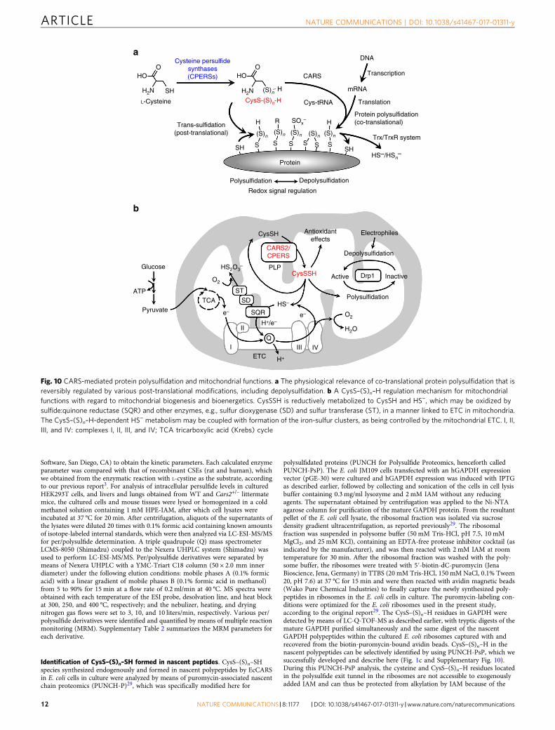
Fig. 10 CARS-mediated protein polysulfidation and mitochondrial functions. a The physiological relevance of co-translational protein polysulfidation that isreversibly regulated by various post-translational modifications, including depolysulfidation. b A CysS–(S)n–H regulation mechanism for mitochondrialfunctions with regard to mitochondrial biogenesis and bioenergetics. CysSSH is reductively metabolized to CysSH and HS−, which may be oxidized bysulfide:quinone reductase (SQR) and other enzymes, e.g., sulfur dioxygenase (SD) and sulfur transferase (ST), in a manner linked to ETC in mitochondria.The CysS–(S)n–H-dependent HS− metabolism may be coupled with formation of the iron-sulfur clusters, as being controlled by the mitochondrial ETC. I, II,III, and IV: complexes I, II, III, and IV; TCA tricarboxylic acid (Krebs) cycle
ARTICLE NATURE COMMUNICATIONS | DOI: 10.1038/s41467-017-01311-y
12 NATURE COMMUNICATIONS |8: 1177 |DOI: 10.1038/s41467-017-01311-y |www.nature.com/naturecommunications
unique physicochemical properties of the interior structure of the polypeptide exittunnel in the ribosome45–47, which allowed us to obtain specific and selectiveidentification of the intact forms of CysS–(S)n–H residues in the nascent peptidespresent only within the ribosomes, as Supplementary Fig. 10a shows. As soon asthe mature GAPDH isolated from E. coli. with the Ni-NTA agarose was treated byquick digestion with 10 μg/ml trypsin at 37 °C for 30 min, which was promptlysubjected to the LC-ESI-Q-TOF analysis, in a similar manner as shown for thePUNCH-PsP method.
Preparation and purification of recombinant CARS proteins. To generaterecombinant CARSs, open-reading frames of these genes were transferred intoAG1 (Agilent Technologies, Santa Clara, CA) competent cells. RecombinantEcCARS, mouse CARS1, and human CARS2 proteins were purified by using thefollowing standard procedure. Briefly, these proteins were produced in AG1, andthey were purified by using nickel nitrilotriacetic acid agarose; resultant purifiedproteins were extensively dialyzed against phosphate buffer and stored at −80 °Cuntil use. Protein concentration was determined by using the Protein Assay CBBSolution (Nacalai Tesque, Kyoto, Japan), and protein purity was confirmed viaSDS-PAGE.
Generation of CARS2 KO cell lines. The genome editing CRISPR/Cas9 systemwas used to generate human CARS2 KO cell lines. To obtain gRNA, which is highlyspecific for the first exon of the human CARS2 locus and has fewer off-target siteswithin the human genome, we based an optimal gRNA design on the softwareprogram CRISPRdirect48. To express Cas9 and gRNA in HEK293T cells, the pX459V2.0-CARS2 gRNA vector was created by inserting annealed oligonucleotide pairs(5′-caccTGGGCCTTGGGCGGGCTGGG-3′ and 5′-aaacCCC AGCCCGCC-CAAGGCCCA-3′) into the BpiI sites of pX459 V2.0. pX459 V2.0 vector, whichenables expression of a gRNA (directed to the CARS2 exon 1; SupplementaryFig. 15), SpCas9, and a puromycin resistance gene from a single vector, wasobtained from the Zhang laboratory via Addgene plasmid 6298849. HEK293T cellswere plated in 6-well plates (1.0 × 10⁵ cells per well) 24 h before transfection.Cultured cells were transfected with 2 μg of pX459 V2.0-CARS2 gRNA by usingLipofectamine 2000 (Invitrogen, Carlsbad, CA). The medium was changed 24 hafter transfection. After another 24 h of incubation, the cells were replated on 10-cm dishes and cultured for various time periods at 37 °C with a selection mediumcontaining 2.0 μg/ml puromycin (Invitrogen). Puromycin-resistant clones werearbitrarily selected and used for screening CARS2 KO cell lines to finally obtainstable CARS2 KO cell lines. Disruption of the CARS2 gene was verified by loss ofCARS2 protein expression as determined by western blotting.
Construction of mammalian hCARS2 expression vectors. To produce anhCARS2 expression vector (pPyCAGIP-FLAG-hCARS2), the XhoI fragment ofpET-15b-hCARS2 was cloned into the XhoI site of pPyCAGIP-FLAG. The samevectors containing various mutant hCARS2 genes were obtained via site-directedmutagenesis by using inverse PCR with pPyCAGIP-FLAG-hCARS2 as a templateand primer sets for generation of pPyCAGIP-FLAG-hCARS2 C78/257D, K124/127A, and K317/320A.
Transfection of various CARS2 genes and knockdown of CARS1/2. WT andvarious mutant CARS2 genes were transfected into HEK293T WT and mutant cells asreported recently3 by using expression plasmids such as pPyCAGIP-FLAG-hCARS2and CARS2 mutant vectors. Transfection of the expression plasmid was performed byusing Lipofectamine 2000 according to the manufacturer’s instructions. In brief, weincubated WT and CARS2 KO HEK293T cells seeded in 24-well plates (6 × 10⁵ cellsper well) and 8-well culture slides (2 × 10⁵ cells per well) for 12 h at 37 °C. Fortransfection, we mixed 1.5 μg per well of the expression plasmid with 50 μl of Opti-MEM (Invitrogen) in a tube. Before plasmid DNA and transfection reagent solutionswere added to the cells, solutions were mixed together and incubated for 5min atroom temperature and then added to the cells, after which incubation proceeded for30 h or 3 days. Also, knockdown of CARS1 and CARS2 was performed as reportedrecently3 by using the following small interfering RNAs (siRNAs): CARS1,CARSHSS101368 (Invitrogen), and CARS2, CARS2HSS128464 (Invitrogen). siRNAtransfection was performed by using Lipofectamine RNAiMAX (Invitrogen)according to the manufacturer’s instructions. The siRNA was introduced into WTand CARS2 KO cells, as described above for CARS2 gene transfection.
Generation of Cars2-deficient mice. All experimental procedures conformed to“Regulations for Animal Experiments And Related Activities at Tohoku Uni-versity”, and were reviewed by the Institutional Laboratory Animal Care and UseCommittee of Tohoku University, and finally approved by the President of Uni-versity. We generated two lines of Cars2-deficient mice as follows. Cars2 gRNAsvectors were constructed with use of a pT7-sgRNA and pT7-hCas9 plasmid (a giftfrom Dr. M. Ikawa, Osaka University)50. After digestion of pT7-hCas9 plasmidwith EcoRI, hCas9 mRNA was synthesized by using an in vitro RNA transcriptionkit (mMESSAGE mMACHINE T7 Ultra kit; Ambion, Austin, TX), according tothe manufacturer’s instructions. A pair of oligonucleotides targeting Cars2 wasannealed and inserted into the BbsI site of the pT7-sgRNA vector. The sequences ofthe gRNAs were designed as follows: 5′-GGACAGATCCAGCGAACAGG-3′ and
5′-AATAATCAAGAGAGCTAACG-3′, located at exons 1 and 3 of Cars2 gene, togenerate CARS2-deficient lines 1 and 2 mice, respectively. After digestion of pT7-sgRNA with XbaI, gRNAs were synthesized by using the MEGAshortscript kit(Ambion). We used C57BL/6N female mice (purchased from Crea-Japan Inc.,Tokyo, Japan) to obtain C57BL/6N eggs, and we performed in vitro fertilizationwith these eggs. In brief, Cas9 mRNA and gRNA were introduced into fertilizedeggs by injecting using a Leica Micromanipulator System, according to the pro-tocols reported previously50, after which we transferred the eggs to the oviducts ofpseudo-pregnant females on the day of the vaginal plug. A founder mouse har-boring the Cars2 mutant alleles was crossed with WT mice to obtain Cars2 het-erozygous mice. After segregating the Cars2 mutant alleles, heterozygous mice witha 200-bp deletion in exon 1 (line 1) and with a 1-bp insertion in exon 3 wereselected for additional analyses (Figs. 5–7; Supplementary Figs. 20 and 21).
MitoTracker Red staining for mitochondrial morphology. To analyze mito-chondrial morphogenesis under several experimental conditions in cells, mito-chondria were imaged by using the fluorescent probe MitoTracker Red CM-H2Xros (Invitrogen). In brief, culture slides were coated with 0.5% polyethyleneimine for more than 1 h and washed twice with PBS. CARS2 KO cells weretransfected with expression plasmids for WT and individual mutants of humanCARS2 via Lipofectamine 2000. At 3 days after transfection, cultured cells werewashed with Hank’s buffer, incubated with 1 μM MitoTracker Red CM-H2Xros at37 °C for 30 min, rinsed twice with Hank’s buffer, and examined with a Nikon EZ-C1 confocal laser microscope (Tokyo, Japan). We used ImageJ and Prism softwarefor image processing and quantification of mitochondrial dimensions includingtheir length.
Mitochondrial bioenergetic functions. To determine the membrane potential(ΔΨm) of mitochondria under several experimental conditions, tetra-ethylbenzimidazolyl carbocyanine iodide (JC-1) staining was performed accordingto the manufacturer’s protocol. Accumulation of the cell-permeable JC-1 probe(Abcam) in mitochondria depends on the membrane potential, associated with afluorescence emission shift from green to red. Briefly, WT and CARS2 KOHEK293T cells, cultured in 8-well multichamber Millicell slides coated with PEI,were treated with various CARS2 vectors or were untreated, as described above. ForJC-1 staining, cultured cells were washed with HKRB buffer (20 mM HEPES,103 mM NaCl, 4.77 mM KCl, 0.5 mM CaCl2, 1.2 mM MgCl2, 1.2 mM KH2PO4,25 mM NaHCO3 and 15 mM glucose, pH 7.3), incubated with 20 μM JC-1 at 37 °Cfor 30 min, rinsed twice with HKRB buffer, and examined with a Nikon EZ-C1confocal laser microscope. ImageJ software was used for image processing andquantification of the JC-1 fluorescent responses.
Mitochondrial bioenergetic functions. Mitochondrial function was investigated,according to a previous report with a slight modification51, by measuring the basalOCR of the mitochondria under various experimental conditions in WT andCARS2 KO cells, using the XF96 Extracellular Flux Analyzer (Seahorse Bioscience,Agilent). At the end of the experiment, rotenone and antimycin A (2.4 μM each)were added to inhibit complexes I and III of the mitochondrial electron transportchain, respectively, to determine the remaining mitochondria-independent OCR.Net OCR was normalized to the cell number determined at the end of theexperiments by means of sulforhodamine B staining (Sigma-Aldrich, St. Louis,MO). To obtain the mitochondria-specific OCR, only the rotenone/antimycin-sensitive part of cell respiration was used.
Effect of suppression of ETC on metabolic profiles of CysSSH. The mito-chondrial ETC in HEK293T cells was inhibited either by a complex III inhibitor,antimycin A, or by elimination of mitochondrial DNA (mtDNA) induced byethidium bromide. For the direct but partial ETC (complex III) inhibition, WT andCARS2 KO cells were treated with various concentrations of antimycin A for 1 h,followed by methanol extraction for measurement of CysSSH and its related sulfidederivatives by HPE-IAM labeling LC-ESI-MS/MS analysis as described earlier. Toindirectly suppress all ETC components (complexes), mtDNA from WT andCARS2 KO HEK293T cells was eliminated specifically by treatment with ethidiumbromide (50 ng/ml, 127 nM) for 12 days under standard cell culture conditions(37 °C, humidified, 5% CO2/95% air) with DMEM containing 10% FBS, 1%penicillin-streptomycin, sodium pyruvate (1 mM), nonessential amino acids (1%),and uridine (50 μg/ml), according to a previous method with a slight modifica-tion52. The cells without mtDNA were then subjected to HPE-IAM labeling LC-MS/MS analysis for persulfide metabolic profiling, similar to antimycin-treatedcells. The efficacy of the present mtDNA elimination and the resultant ETC sup-pression were assessed by measuring mtDNA as described below (SupplementaryFig. 24b), and these results were confirmed by substantial suppression of mito-chondrial cytochrome c oxidase subunit 1 (MTCO1: encoded by mtDNA), asidentified by western blotting. In contrast, MitoTracker Red staining showed noappreciable altered morphology of mitochondria in HEK293T cells with or withoutethidium bromide treatment, at least under the present experimental conditions.The quantity of each sulfide produced from CARS2 in the cells was determined bysubtracting the amount of each sulfide in CARS2 KO HEK293T cells from that inthe WT cells, after quantification of each metabolite via HPE-IAM labeling LC-MS/
NATURE COMMUNICATIONS | DOI: 10.1038/s41467-017-01311-y ARTICLE
NATURE COMMUNICATIONS |8: 1177 |DOI: 10.1038/s41467-017-01311-y |www.nature.com/naturecommunications 13
MS analysis. Changes in the amounts of CysSSH (ΔCysSSH) and HS− (ΔHS−)induced by complex III inhibition by antimycin A or by mtDNA elimination inWT and CARS2 KO HEK293T cells were then calculated.
Statistical analysis. Results are presented as means± s.d. of at least three inde-pendent experiments unless otherwise specified. For statistical comparisons, weutilized two-tailed Student’s t test or two-way analysis of variance followed by theStudent–Newman–Keuls test, with significance set at P< 0.05.
Data availability. The data that support the findings of this study are availablefrom the corresponding author upon request.
Received: 23 September 2016 Accepted: 29 August 2017
References1. Toohey, J. I. Sulfur signaling: is the agent sulfide or sulfane? Anal. Biochem. 413,
1–7 (2011).2. Fukuto, J. M. et al. Small molecule signaling agents: the integrated chemistry
and biochemistry of nitrogen oxides, oxides of carbon, dioxygen, hydrogensulfide, and their derived species. Chem. Res. Toxicol. 25, 769–793 (2012).
3. Ida, T. et al. Reactive cysteine persulfides and S-polythiolation regulateoxidative stress and redox signaling. Proc. Natl Acad. Sci. USA 111, 7606–7611(2014).
4. Dóka, É. et al. A novel persulfide detection method reveals protein persulfide-and polysulfide-reducing functions of thioredoxin and glutathione systems. Sci.Adv. 2, e1500968 (2016).
5. Kunikata, H. et al. Metabolomic profiling of reactive persulfides andpolysulfides in the aqueous and vitreous humors. Sci. Rep. 7, 41984 (2017).
6. Numakura, T. et al. Production of reactive persulfide species in chronicobstructive pulmonary disease. Thorax doi:10.1136/thoraxjnl-2016-209359(2017).
7. Ono, K. et al. Redox chemistry and chemical biology of H2S, hydropersulfides,and derived species: implications of their possible biological activity and utility.Free Radic. Biol. Med. 77, 82–94 (2014).
8. Saund, S. S. et al. The chemical biology of hydropersulfides (RSSH): chemicalstability, reactivity and redox roles. Arch. Biochem. Biophys. 588, 15–24 (2015).
9. Millikin, R. et al. The chemical biology of protein hydropersulfides: studies of apossible protective function of biological hydropersulfide generation. FreeRadic. Biol. Med. 97, 136–147 (2016).
10. Fujii, S. et al. Redox signaling regulated by an electrophilic cyclic nucleotide andreactive cysteine persulfides. Arch. Biochem. Biophys. 595, 140–146 (2016).
11. Cavallini, D., Federici, G. & Barboni, E. Interaction of proteins with sulfide. Eur.J. Biochem. 14, 169–174 (1970).
12. Massey, V. & Edmondson, D. On the mechanism of inactivation of xanthineoxidase by cyanide. J. Biol. Chem. 245, 6595–6598 (1970).
13. Branzoli, U. & Massey, V. Evidence for an active site persulfide residue in rabbitliver aldehyde oxidase. J. Biol. Chem. 249, 4346–4349 (1974).
14. Takahashi, N. et al. Reactive sulfur species regulate tRNA methylthiolation andcontribute to insulin secretion. Nucleic Acids Res. 45, 435–445 (2016).
15. Jung, M. et al. Protein polysulfidation-dependent persulfide dioxygenaseactivity of ethylmalonic encephalopathy protein 1. Biochem. Biophys. Res.Commun. 480, 180–186 (2016).
16. Wróbel, M. Sulfurtransferase activity and sulfur compound content in Ranatemporaria brain following hibernation. Acta Neurobiol. Exp. 61, 69–72 (2001).
17. Cortese-Krott, M. M. et al. Key bioactive reaction products of the NO/H2Sinteraction are S/N-hybrid species, polysulfides, and nitroxyl. Proc. Natl Acad.Sci. USA 112, E4651–E4660 (2015).
18. Yamanishi, T. & Tuboi, S. The mechanism of the L-cystine cleavage reactioncatalyzed by rat liver γ-cystathionase. J. Biochem. 89, 1913–1921 (1981).
19. Chen, W. et al. The development of fluorescent probes for visualizingintracellular hydrogen polysulfides. Angew. Chem. Int. Ed. Engl. 54,13961–13965 (2015).
20. Nishida, M. et al. Hydrogen sulfide anion regulates redox signaling viaelectrophile sulfhydration. Nat. Chem. Biol. 8, 714–724 (2012).
21. Yadav, P. K. et al. Biosynthesis and reactivity of cysteine persulfides insignaling. J. Am. Chem. Soc. 138, 289–299 (2016).
22. Morikawa, T. et al. Hypoxic regulation of the cerebral microcirculation ismediated by a carbon monoxide-sensitive hydrogen sulfide pathway. Proc. NatlAcad. Sci. USA 109, 1293–1298 (2012).
23. Shirozu, K. et al. Cystathionine γ-lyase deficiency protects mice fromgalactosamine/lipopolysaccharide-induced acute liver failure. Antioxid. Redox.Signal. 20, 204–216 (2014).
24. Nakano, S. et al. Hyperhomocysteinemia abrogates fasting-inducedcardioprotection against ischemia/reperfusion by limiting bioavailability ofhydrogen sulfide anions. J. Mol. Med. 93, 879–889 (2015).
25. Fletcher, J. C. & Robson, A. The occurrence of bis-(2-amino-2-carboxyethyl)trisulfide in hydrolysates of wool and other proteins. Biochem. J. 87, 553–559(1963).
26. Parker, A. J. & Kharasch, N. The scission of the sulfur-sulfur bond. Chem. Rev.59, 583–628 (1959).
27. Abdolrasulnia, R. & Wood, J. L. Persulfide properties of thiocystine and relatedtrisulfides. Bioorg. Chem. 9, 253–260 (1980).
28. Kice, J. L. Electrophilic and nucleophilic catalysis of the scission of the sulfur-sulfur bond. Acc. Chem. Res. 1, 58–64 (1968).
29. Aviner, R., Geiger, T. & Elroy-Stein, O. Genome-wide identification andquantification of protein synthesis in cultured cells and whole tissues bypuromycin-associated nascent chain proteomics (PUNCH-P). Nat. Protoc. 9,751–760 (2014).
30. Hallmann, K. et al. A homozygous splice-site mutation in CARS2 is associatedwith progressive myoclonic epilepsy. Neurology 83, 2183–2187 (2014).
31. Coughlin, C. R. II. et al. Mutations in the mitochondrial cysteinyl-tRNAsynthase gene, CARS2, lead to a severe epileptic encephalopathy and complexmovement disorder. J. Med. Genet. 52, 532−–540 (2015).
32. Eidenschenk C. et al. Record for madcow. Updated 13th May 2016. Center forthe Genetics of Host Defense, UT Southwestern, Dallas, TX, USA Available athttps://mutagenetix.utsouthwestern.edu/phenotypic/phenotypic_rec.cfm?pk=316. (Accessed 30th January 2017)
33. Akhtar, M. W. et al. Elevated glucose and oligomeric β-amyloid disruptsynapses via a common pathway of aberrant protein S−nitrosylation. Nat.Commun. 7, 10242 (2016).
34. Griesbeck, C. et al. Mechanism of sulfide-quinone reductase investigated using site-directed mutagenesis and sulfur analysis. Biochemistry 41, 11552–11565 (2002).
35. Grieshaber, M. K. & Völkel, S. Animal adaptations for tolerance andexploitation of poisonous sulfide. Annu. Rev. Physiol. 60, 33–53 (1998).
36. Goubern, M., Andriamihaja, M., Nübel, T., Blachier, F. & Bouillaud, F. Sulfide,the first inorganic substrate for human cells. FASEB J. 21, 1699–1706 (2007).
37. Hine, C. et al. Endogenous hydrogen sulfide production is essential for dietaryrestriction benefits. Cell 160, 132–144 (2015).
38. Szabo, C. et al. Regulation of mitochondrial bioenergetic function by hydrogensulfide. Part I. Biochemical and physiological mechanisms. Br. J. Pharmacol.171, 2099–2122 (2014).
39. Stehling, O. & Lill, R. The role of mitochondria in cellular iron–sulfur proteinbiogenesis: mechanisms, connected processes, and diseases. Cold Spring Harb.Perspect. Biol. 5, a011312 (2013).
40. Sawa, T. et al. Protein S−guanylation by the biological signal 8-nitroguanosine3′,5′-cyclic monophosphate. Nat. Chem. Biol. 3, 727–735 (2007).
41. Abiko, Y. et al. Involvement of reactive persulfides in biologicalbismethylmercury sulfide formation. Chem. Res. Toxicol. 28, 1301–1306 (2015).
42. Nishida, M. et al. Redox regulation of electrophilic signaling by reactivepersulfides in cardiac cells. Free Radic. Biol. Med. 109, 132–140 (2017).
43. Wakasugi, K. & Schimmel, P. Two distinct cytokines released from a humanaminoacyl-tRNA synthetase. Science 284, 147–151 (1999).
44. Suzuki, T., Ueda, T. & Watanabe, K. A new method for identifying the aminoacid attached to a particular RNA in the cell. FEBS Lett. 381, 195–198 (1996).
45. Nissen, P., Hansen, J., Ban, N. P. B. & Steitz, A. The structural basis of ribosomeactivity in peptide bond synthesis. Science 289, 920–930 (2000).
46. Voss, N. R., Gerstein, M., Steitz, T. A. & Moore, P. B. The geometry of theribosomal polypeptide exit tunnel. J. Mol. Biol. 360, 893–906 (2006).
47. Kramer, G., Boehringer, D., Ban, N. & Bukau, B. The ribosome as a platform forco-translational processing, folding and targeting of newly synthesized proteins.Nat. Struct. Mol. Biol. 16, 589–597 (2009).
48. Naito, Y., Hino, K., Bono, H. & Ui-Tei, K. CRISPRdirect: software for designingCRISPR/Cas guide RNA with reduced off-target sites. Bioinformatics 31,1120–1123 (2015).
49. Ran, F. A. et al. Double nicking by RNA-guided CRISPR Cas9 for enhancedgenome editing specificity. Cell 154, 1380–1389 (2013).
50. Mashiko, D. et al. Generation of mutant mice by pronuclear injection of circularplasmid expressing Cas9 and single guided RNA. Sci. Rep. 3, 3355 (2013).
51. Kaplon, J. et al. A key role for mitochondrial gatekeeper pyruvatedehydrogenase in oncogene-induced senescence. Nature 498, 109–112 (2013).
52. King, M. P. & Attardi, G. Isolation of human cell lines lacking mitochondrialDNA. Methods Enzymol. 264, 304–313 (1996).
AcknowledgementsWe thank J.B. Gandy for her editing of the manuscript and evaluating concepts andterminology of the paper with regard to the understanding by non-specialist readers.Thanks are also due to S. Akashi, H. Abdul Hamid, Y. Kakihana, and M. Matsubara fortheir technical assistance in our study. P.N. is a János Bolyai Research Scholar of theHungarian Academy of Sciences. M.F. is supported by the National Institute for HealthResearch through the NIHR Southampton Biomedical Research Centre. This work wassupported in part by Grants-in-Aid for Scientific Research ((S), (A), (B), ChallengingExploratory Research, and Innovative Areas “Oxygen Biology: a new criterion for inte-grated understanding of life”) from the Ministry of Education, Sciences, Sports, and
ARTICLE NATURE COMMUNICATIONS | DOI: 10.1038/s41467-017-01311-y
14 NATURE COMMUNICATIONS |8: 1177 |DOI: 10.1038/s41467-017-01311-y |www.nature.com/naturecommunications
Technology (MEXT), Japan, to T.A. (26111008, 26111001, 15K21759, 25253020,16K15208), Y.K. (25220103), H.M. (15H04692), K.I. (26116005), T.S. (15H03115), andM.N. (15K14959); a grant from the JST PRESTO program to T.S. (10104025) and M.N.(13417243); grants from The Hungarian National Science Foundation (K 109843) andfrom the National Institutes of Health (R21AG055022-01) to P.N.; and grants from theNational Institutes of Health (HL106598) and the National Science Foundation (CHE-1148641) to J.M.F.
Author contributionsT.A. and H.M., experiment design, biochemistry and cell biology, data analysis, animalstudies, and writing the paper; T.I., M.M.A., H.I., T.S., M.J. and T.M., MS analysis,biochemistry and molecular biology, cell biology, and cell imaging; S.W., K.I., proteinstructural analysis; F.-Y.W., S.K., Akir.N., H.S., N.T. and K.T., cell biology and mito-chondria study; M.M. and M.O., CRISPR/Cas9 technology, cell biology, and animalstudies; M.N., Akiy.N., S.F., K.Y. and Y.W., cell signaling, cell biology, and data analysis;P.N., M.F., J.M.F., chemical analysis and editing the paper.
Additional informationSupplementary Information accompanies this paper at doi: 10.1038/s41467-017-01311-y.
Competing interests: The authors declare no competing financial interests.
Reprints and permission information is available online at http://npg.nature.com/reprintsandpermissions/
Publisher's note: Springer Nature remains neutral with regard to jurisdictional claims inpublished maps and institutional affiliations.
Open Access This article is licensed under a Creative CommonsAttribution 4.0 International License, which permits use, sharing,
adaptation, distribution and reproduction in any medium or format, as long as you giveappropriate credit to the original author(s) and the source, provide a link to the CreativeCommons license, and indicate if changes were made. The images or other third partymaterial in this article are included in the article’s Creative Commons license, unlessindicated otherwise in a credit line to the material. If material is not included in thearticle’s Creative Commons license and your intended use is not permitted by statutoryregulation or exceeds the permitted use, you will need to obtain permission directly fromthe copyright holder. To view a copy of this license, visit http://creativecommons.org/licenses/by/4.0/.
© The Author(s) 2017
NATURE COMMUNICATIONS | DOI: 10.1038/s41467-017-01311-y ARTICLE
NATURE COMMUNICATIONS |8: 1177 |DOI: 10.1038/s41467-017-01311-y |www.nature.com/naturecommunications 15