Embed Size (px)
Citation preview

Title: Does the grass snake (Natrix natrix) (Squamata: Serpentes: Natricinae) fit the amniotes-specific model of myogenesis?
Author: Damian Lewandowski, Magda Dubińska-Magiera, Ewelina Posyniak, Weronika Rupik, Małgorzata Daczewska
Citation style: Lewandowski Damian, Dubińska-Magiera Magda, Posyniak Ewelina, Rupik Weronika, Daczewska Małgorzata. (2017). Does the grass snake (Natrix natrix) (Squamata: Serpentes: Natricinae) fit the amniotes-specific model of myogenesis?. "Protoplasma" (Vol. 254, iss. 4 (2017), s. 1507-1516), doi 10.1007/s00709-016-1040-5

ORIGINAL ARTICLE
Does the grass snake (Natrix natrix) (Squamata: Serpentes:Natricinae) fit the amniotes-specific model of myogenesis?
Damian Lewandowski1 & Magda Dubińska-Magiera1 & Ewelina Posyniak1&
Weronika Rupik2& Małgorzata Daczewska1
Received: 23 June 2016 /Accepted: 27 October 2016 /Published online: 10 November 2016# The Author(s) 2016. This article is published with open access at Springerlink.com
Abstract In the grass snake (Natrix natrix), the newly devel-oped somites form vesicles that are located on both sides ofthe neural tube. The walls of the vesicles are composed oftightly connected epithelial cells surrounding the cavity (thesomitocoel). Also, in the newly formed somites, the Pax3 pro-tein can be observed in the somite wall cells. Subsequently, thesomite splits into three compartments: the sclerotome,dermomyotome (with the dorsomedial [DM] and the ventrolat-eral [VL] lips) and the myotome. At this stage, the Pax3 proteinis detected in both the DM and VL lips of the dermomyotomeand in the mononucleated cells of the myotome, whereas thePax7 protein is observed in the medial part of thedermomyotome and in some of the mononucleated cells ofthe myotome. The mononucleated cells then become elongatedand form myotubes. As myogenesis proceeds, the myotome isfilled with multinucleated myotubes accompanied by mononu-cleated, Pax7-positive cells (satellite cells) that are involved inmuscle growth. The Pax3-positive progenitor muscle cells areno longer observed. Moreover, we have observed unique
features in the differentiation of the muscles in these snakes.Specifically, our studies have revealed the presence of twoclasses of muscles in the myotomes. The first class ischaracterised by fast muscle fibres, with myofibrils equally dis-tributed throughout the sarcoplasm. In the second class, com-posed of slow muscle fibres, the sarcoplasm is filled with lipiddroplets. We assume that their storage could play a crucial roleduring hibernation in the adult snakes. We suggest that themodel of myotomal myogenesis in reptiles, birds and mammalsshows the same morphological and molecular character. Wetherefore believe that the grass snake, in spite of the uniquefeatures of its myogenesis, fits into the amniotes-specific modelof trunk muscle development.
Keywords Amniotes . Myotomalmyogenesis . Pax3/7proteins . Satellite cells . Reptiles . Snake
Introduction
Reptiles are a very heterogeneous group of vertebrates (Olmo2008). They were the first vertebrates to settle in the terrestrialenvironment. This shaped their musculoskeletal, respiratoryand circulatory systems and their method of reproduction ina manner independent of the conditions of an aquatic environ-ment. However, reptiles are still ectothermic organisms, whichare strongly dependent on the environmental temperature.From an evolutionarily viewpoint, reptiles were the firstterrestrial vertebrates to display a great diversity of musclemodels, which correspond to their various modes of locomo-tion (crocodiles, turtles, lizards and snakes) (McNeill 2012).This great anatomical-physiological diversity of the muscularsystem renders it difficult to select a characteristic species thatis representative of the whole taxon (Schilling 2011). As aresult, the differentiations of the reptilian skeletal and limb
Handling Editor: Douglas Chandler
Damian Lewandowski and Magda Dubińska-Magiera contributedequally to this work.
Electronic supplementary material The online version of this article(doi:10.1007/s00709-016-1040-5) contains supplementary material,which is available to authorized users.
* Małgorzata [email protected]
1 Department of Animal Developmental Biology, Institute ofExperimental Biology, University of Wroclaw, Sienkiewicza 21,50-335 Wroclaw, Poland
2 Department of Animal Histology and Embryology, University ofSilesia, Bankowa 9, 40-007 Katowice, Poland
Protoplasma (2017) 254:1507–1516DOI 10.1007/s00709-016-1040-5

muscles are still poorly understood, even though reptiles arecrucial organisms in vertebrate evolution.
The muscle fibres of vertebrates are cylindrical, multinu-cleated cells that are divided into two main types: red (slow)and white (fast) muscle fibres (Yablonka-Reuveni 2011).White muscle fibres are characterised by a fast contraction,an anaerobic metabolism for energy, lower amounts ofmyoglobin and fewer mitochondria. They perform fast andintensive work, but only for a short period of time; therefore,they quickly experience fatigue. In contrast, the red musclefibres are rich in mitochondria and myoglobin. They aredependent on an aerobic metabolism and can perform slowand sustained contractions for a prolonged period of timewithout fatigue (Sänger and Stoiber 2001). The proportionof white to red muscles in a vertebrate organism depends onits lifestyle. In fish, the following phenomena are frequentlyobserved: in the eel, metamorphosis is accompanied by anincrease in the amount of red muscles, as preparation for thespawningmigration (Lewander et al. 1974); in the tuna, whichis the most active species of fish, an extra band of red musclescan be observed near to the vertebral column (as reviewed byKatz 2002); and in salmonids, as migrant fish, extra redmuscles are found as individual fibres distributed throughoutthe white muscles (Webb 1971).
It has been observed in lizards that, as terrestrial animals,the axial muscles stabilise the trunk during locomotion (Ritter1996). A body of evidence has revealed that lizards possessboth slow and fast muscles, whereas in snakes, white musclefibres represent the prominent group of muscles in themyotomes (Gleeson et al. 1980; Guthe 1981; Moritz andSchilling 2013). Crow and Stockdale (1986) revealed that, inavians, there is no single program of fast myosin heavy chains(MyHC) isoform expression during development. Furthermore,recent studies on mammals have revealed significant species-dependent diversity in the distribution of muscle fibres(Schiaffino and Reggiani 2011).
In all vertebrates, the myotomal muscles have a similarstructure and originate from the paraxial mesoderm, whichundergoes segmentation into repetitive units called somites(Mok and Sweetman 2011; Bentzinger et al. 2012).However, the mechanism of somite differentiation is differentin non-amniotes than in amniotes. For instance, the musclefibre precursors of Xenopus laevis and Danio rerio undergoa 90° rotation (Bryson-Richardson and Currie 2010). Theother characteristic feature of non-amniote myogenesis is thetrans-myotomal migration of the red muscle precursors to theperipheral part of the myotome. In amniotes, as in the non-amniotes, the somites differentiate into the following: theepithelial dermomyotome (the source of skin connectivetissues and the myotomal muscles) with characteristicdorsomedial (DM) and ventrolateral (VL) lips and the mesen-chymal sclerotome (the source of the connective tissues of theaxial skeleton) (Cinnamon et al. 2001; Ordahl et al. 2001;
Steinbacher et al. 2006; 2007). The main part of the somiteis occupied by the myotome, where the myotomal (trunk)muscles are differentiated in situ. It has been clearlyestablished that in vertebrates, the dermomyotome is anancient, conserved structure (Onai et al. 2015). Also, manydifferent lines of evidence (from investigations on fish,amphibian, birds and mammals) have shown that thedermomyotome is the main source of the muscle progenitorcells (Gros et al. 2005; Relaix et al. 2005; Kassar-Ducchossoyet al. 2005; Kalcheim and Ben-Yair 2005; Manceau et al.2008; Rossi and Messina 2014). The muscle progenitor cellsexpress the transcription factors Pax3 and Pax7 (Tajbakhshet al. 1997; Pownall et al. 2003; Halevy et al. 2004;Buckingham and Relaix 2007). The muscles then growthrough the fusion of the muscle precursors with theexisting muscle fibres (hypertrophy), resulting in an in-crease in the number of nuclei in the growing muscles.Additionally, during hyperplasia, the muscle precursorsfuse with each other to form new muscle fibres (Greer-Walker 1970; Koumans et al. 1993; Stickland 1983).
Although a number of good studies on the anatomicdescription of somites and myotome in reptiles have beenpublished, little work has been done in this class of animalsthat examines in parallel the immune-histological characteri-sation of those structures (Eckalbar et al. 2012; Rupik et al.2012). Therefore, the key aim of our studies was to fill the gapin knowledge on muscle differentiation in this taxon by inves-tigating muscle growth and its differentiation in the grasssnake (Natrix natrix).
Materials and methods
Study animals
Fertilised females of the grass snake, N. natrix Stejneger,1907, were caught in Poland in the vicinity of Wrocław atthe beginning of May 2014. All of the specimens used in theexperiments were captured according to the Polish regulationsconcerning the protection of wild species (Journal of Laws1991, No. 114 Item 492; Journal of Laws 2000, No. 66 Item802; Journal of Laws 2004, No. 112 Item 1183; and Journal ofLaws 2015, No. 133 Item 266). The Department of AnimalDevelopmental Biology of the University of Wroclawobtained approvals from the Local Ethics Commission inWroclaw (77/2013) and the Polish Ministry of Environmentto perform studies on a protected species (Ref. No:WPN.6401.59.2014.IW and DZP-WG.6401.02.3.2014.JRO).The animals were kept in vivaria in an open farm area, inconditions similar to those in the wild until the eggs were laid,and then, they were released into their native area. The eggs ofthe grass snakes (n = 128) after oviposition were carefullycollected and were placed inside plastic boxes filled with
1508 D. Lewandowski et al.

moistened perlite (at 100% humidity) with ventilation holes.The eggs were incubated at 30 °C, reflecting the seasonalambient temperatures in the wild. The embryos used for theexamination were isolated at regular intervals after the egglaying, until the 14th day after oviposition (stages I–VI). Thedevelopmental stages of the embryos were estimated using thedevelopmental table published by Rupik (2002).
The collected embryos were anaesthetised with tricainemethanesulfonate (MS-222; 500 μg per gram of bodyweight) (Conroy et al. 2009), before being decapitated anddissected for further analysis (Journal of Laws 2015, No.133 Item 266).
Light and transmission electron microscopy
For light and electron microscopic examination, small piecesof the embryonic body wall, including differentiated muscletissues, were fixed in a modified Karnovsky fixativeconsisting of 1% paraformaldehyde (PFA) and 1% glutaralde-hyde, in a 0.1 M phosphate buffer (pH 7.2) for 24 h at 4 °C.The material was then repeatedly rinsed with the same bufferand was postfixed for 2 h in a 1:1 mixture of osmiumtetroxide-potassium ferricyanide [OsO4-K3Fe(CN)6].Following rinsing in the phosphate buffer, the material wasdehydrated, first in a graded alcohol series and then in acetone,and was then embedded in epoxy resin Epon 812 (Sigma-Aldrich) (Luft 1961). The Epon blocks were cut on a LeicaUltracut UCT (Leica, Wetzlar, Germany). Semi-thin sections(0.6 μm) were collected on glass slides and were stained withmethylene blue in a 1% borax solution before being examinedunder an Olympus BX60 light microscope (Olympus).Additionally, ultra-thin sections were collected on 200-mesh copper grids and were stained with uranyl acetateand lead citrate according to the standard protocol(Reynolds 1963), before being examined under a trans-mission electron microscope, Zeiss EM 900 (Carl ZeissAG, Oberkochen, Germany; 80 kV).
Immunofluorescence analysis
After the dissection and fixation of the embryos in 4% PFA inphosphate buffered saline (PBS) for 45 min at room tempera-ture, the samples were transferred to 30% sucrose in PBS (foran overnight incubation at 4 °C). Next, samples were embed-ded in the optimal cutting temperature (OCT) medium andwere placed in a cryomold and frozen. The samples were cutinto 10-μm sections in a cryostat at −20 °C and were placed onSuperFrost Plus slides and subjected to immunofluorescencestaining.
Standard immunofluorescence reactions were carried outon whole embryos or on tissue cryosections. These sampleswere blocked with 1% bovine serum albumin (BSA) in PBST(PBS with 0.1% Tween-20) for 60 min at room temperature.
All of the wash steps were done with PBST. An incubationwith primary antibodies was conducted overnight at 4 °C andwith secondary antibodies for 60 min at room temperature.The following primary antibodies were used: mouse mono-clonal anti-Pax3 antibody (Developmental StudiesHybridoma Bank) at a dilution of 1:50 in PBST, rabbit poly-clonal anti-phospho-histone H3 [pSer10] (Sigma-Aldrich) at adilution of 1:200 in PBST, mouse monoclonal anti-fast skele-tal myosin (Fast MyHC) antibody (Abcam) at a dilution of1:100 in PBST, mouse monoclonal anti-slow skeletal myosin(Slow MyHC) antibody (Abcam) at a dilution of 1:100 inPBST and mouse monoclonal F59 antibody (DevelopmentalStudies Hybridoma Bank) at a dilution of 1:100 in PBST.Additionally, the following secondary antibodies were used:goat anti-mouse IgG FITC conjugated (Sigma-Aldrich) at adilution of 1:50 in PBST, goat anti-rabbit IgG TRITCconjugated (Sigma-Aldrich) at a dilution of 1:50 in PBSTand donkey anti-mouse IgG Cy5 conjugated (JacksonImmunoResearch) at a dilution of 1:100 in PBST. For the F-actin identification, Alexa Fluor 546-conjugated phalloidinand Alexa Fluor 488-conjugated phalloidin were used(Molecular Probes) at a dilution of 1:80 in PBS. The DNAwas stained with 4,6-diamidino-2-phenylindole (DAPI;0.2 μg/ml), and the neutral lipids were stained withBODIPY® 493/503 (Thermo Fisher Scientific) at a dilution1:1000. The samples were mounted in a fluorescent mountingmedium (Dako). For the imaging, an Olympus FluoViewFV1000 confocal laser scanning microscope (Olympus) wasused. The images were recorded by employing the Plan-Apochromat ×10, ×20 or ×40 objectives. Any brightnessand contrast adjustments were performed in the FV10-ASW_Viewer or in ImageJ.
Gel electrophoresis and Western blotting
The decapitated embryo extracts underwent a Western blotanalysis. The proteins were separated on 12% SDS gels bypolyacrylamide gel electrophoresis (PAGE) and wereelectrotransferred onto nitrocellulose filters. The membraneswere then detected and documented with a chemiluminescentmethod, using the Bio-Rad ChemiDoc Imaging System. Theprotein content in the Pax3 and Pax7 bands was then normal-ised according to the α-actinin content in each lane.
The membranes were blocked for 60 min at room tempera-ture in a blocking solution (5% non-fat dry milk in PBS with0.05% Tween-20). The membranes were then incubated over-night at 4 °C with primary monoclonal antibodies specific forPax3 (mouse monoclonal anti-Pax3 antibody [DevelopmentalStudies Hybridoma Bank] at a dilution of 1:100), Pax7 (mousemonoclonal anti-Pax7 antibody [Developmental StudiesHybridoma Bank] at a dilution of 1:100) and α-actinin (ratmonoclonal anti-α-actinin antibody [Babraham BioscienceTechnologies] at a dilution of 1:100). Chemiluminescence
Does the grass snake fit amniote-specific model of myogenesis? 1509
was then used for the detection of Pax3, Pax7 and α-actinin, by the incubation of the membrane for 60 minat room temperature with the following secondary anti-bodies: donkey anti-mouse IgG peroxidase (HRP) conju-gated (Jackson ImmunoResearch) and donkey anti-rat IgGperoxidase (HRP) conjugated (Jackson ImmunoResearch)at a dilution of 1:10,000.
Results
Somitogenesis and myogenesis
In the studied species (N. natrix), selected stages (from stage Ito VII) of the skeletal muscle development were analysedusing the light, confocal and transmission electronmicroscopeand with the Western blot method.
The confocal analyses, performed using the Alexa-Phalloidin 546, revealed that in the studied species, the newlydeveloped somite formed vesicles located on both sides of theneural tube (stage I) at the posterior (tail) part of the embryo.The wall of the vesicles was composed of tightly connectedepithelial cells surrounding the somitocoel (Fig. 1a).Somitogenesis showed that in a more advanced developmentof the somites, the anterior-posterior gradient of muscle dif-ferentiation manifested in the anterior part of the embryo (supFig. 1). Therefore, in the anterior part of the embryo (stage II),the somites were already differentiated into the ventromediallylocated mesenchymal sclerotome, the laterally situated epithe-lial dermomyotome and the myotome between these (Fig. 1b).These observations were consistent with the anterior to poste-rior progression of somitogenesis typically seen in vertebrateembryos.
The dermomyotome was characterised by well-developeddorsomedial (DM) and ventrolateral (VL) lips (Fig. 1b). TheTEM analysis revealed that the muscle progenitor cells locat-ed near to the dermomyotome closely adhered to each other.Their ultrastructure showed large nuclei with a narrow rim ofcytoplasm, rich in mitochondria and rough endoplasmic retic-ulum (RER) and well-developed Golgi apparatus (AG)(Fig. 1b inset). During the subsequent developmental stages,some of the post-mitotic myotome cells became elongated.Also, some of the cells located in the myotome werecharacterised by proliferative activity, as was confirmed inthe TEM (Fig. 1c) and with the confocal microscope for theimmunocytochemical detection of phosphorylated histone H3(stage III) (Fig. 1c inset) to visualise mitotic activity. At thesame developmental stage, the elongated post-mitotic cellsdifferentiated into mononucleated myotubes, which wasconfirmed by the presence of an incompletely developedcontractile apparatus. Numerous organelles (such as RER,mitochondria and AG, surrounded by small vesicles andglycogen granules) were observed in the sarcoplasm of the
mononucleated myotubes. It should be noted that the elongat-ed nuclei of the myotubes had a homogeneous content(Fig. 1d). As myogenesis proceeded (stage IV), thedermomyotome cells were no longer observed (Fig. 1e). Atthe next developmental stage (stage V), for the first time in themyotomes, multinucleated myotubes occurred (Fig. 1f). TheTEM analysis showed that the myotube sarcoplasm was richin mitochondria, RER and glycogen granules. Furthermore,the myofibrils were regularly arranged, which is characteristicfor a mature contractile apparatus (Fig. 1g).
Unique features of myotomal myogenesis in the grasssnake
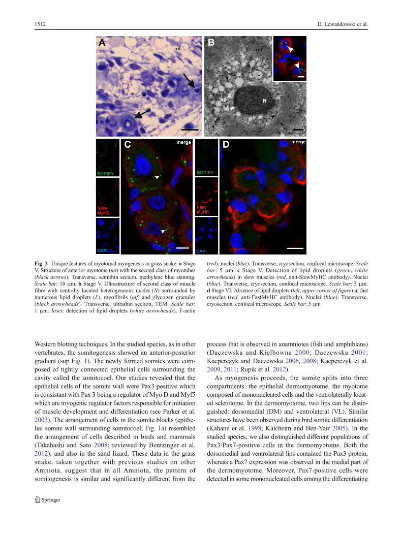
Studies using the light microscope and the TEM revealed thattwo classes of myotubes developed during the grass snakemyogenesis (stages V–VI). The first class was representedby typical muscle fibres with myofibrils that were located inthe whole sarcoplasm. In the second class of muscle fibres,myofibrils were located in the peripheral sarcoplasm, and itscentral region was filled with lipid droplets, as confirmed bythe histochemical staining with BODIPY (Fig. 2a, b). It isnoteworthy that in the second class of muscle fibre sarco-plasm, the myofibrils did not form a regular arrangement.Furthermore, in the nucleus, numerous patches of heterochro-matin were observed. Our studies showed that the lipid drop-lets were present only in those muscles which expressed aslow MyHC, whereas no lipid droplets were observed in thefast MyHC-positive fibres (Fig. 2c, d). These described fea-tures were not characteristic for the typical muscle fibres thatappear during myogenesis in other vertebrates.
Muscle growth
The immunocytochemical detection of the Pax3 protein (amarker of muscle progenitor cells that is upregulated duringmyogenesis) revealed that, in the newly formed somites, thisprotein was expressed in the somite wall cells (stage I)(Fig. 3a). As the somites became differentiated into three com-partments (the dermomyotome, myotome and sclerotome),this protein was detected in both DM and VL lips of thedermomyotome and in the mononucleated cells of the myo-tome (stage II) (Fig. 3b, c). Also, at the studied developmentalstage, the Pax7 protein (a marker of early proliferating myo-blasts and progenitors of the satellite cells) was observed inthe medial part of the dermomyotome and in some mononu-cleated cells of the myotome (Fig. 3c).
We also examined the expression levels of the Pax3/7 pro-teins during successive developmental stages, using theWestern blot method. Our analysis of the Pax3 expressionconfirmed that the highest level of that protein occurred instage I. We observed a slight decrease in the Pax3 level inlater stages (II, III), and a further reduction of the signal
1510 D. Lewandowski et al.

occurred in stages IVand V (the 7th day after oviposition). Theoldest analysed stages (V [the 9th day after oviposition], VI andVII) did not show detectable levels of the Pax3 protein(Fig. 3d). The immunoblot analysis of the Pax7 protein revealedits presence in all of the studied developmental stages (Fig. 3e).
The light and TEM studies revealed that the mononucleat-ed cells closely adhered to the surface of the multinucleatedmyotubes (stage V). The ultrastructure of these cells showed alarge nucleus that was rich in heterochromatin and with anarrow rim of cytoplasm (Fig. 3f, g). The presence of thePax7 protein in the nucleus of these cells was detected immu-nocytochemically (Fig. 3h). Furthermore, these cells showedmitotic activity (Fig. 3i). Our observations, based on the light,TEM and the confocal microscopes, suggested that these cellswere satellite cells involved in muscle growth due to theirfusion with the myotubes.
Discussion
Somitogenesis, muscle differentiation and growth
A few studies of the muscle differentiation in this highlydivergent group of animals have been carried out on the sandlizard (Lacerta agilis), the Egyptian cobra (Naja haje), theChinese soft-shelled turtle (Pelodiscus sinensis) and theAmerican alligator (Alligator mississippiensis), which repre-sent various modes of locomotion (Nagashima et al. 2005;Rupik et al. 2012; Kusumi et al. 2013; Khannoon et al.2016). Because the subject of reptilian trunk muscle develop-ment still remains elusive, we decided to shed more light onthis topic. We carried out our investigation on the muscledifferentiation and growth in the grass snake (N. natrix) bythe use of light, TEM, and a confocal microscope and with
Fig. 1 Somitogenesis and myogenesis. a Stage I. Structure of posteriorsomites (white arrows). Somites form vesicles (surrounded with dashedline) with centrally located somitocoel (SC). F-actin (red), anterior (A),posterior (P), dorsal (D), ventral (V). Whole mount staining, confocalmicroscope. Scale bar: 20 μm. b Stage II. Structure of anterior part ofembryo. Laterally located dermomyotome (dm; surrounded with dottedline) with ventrolateral (vll; highlighted in light yellow) and dorsomedial(dml; highlighted in light yellow) lips. NT neural tube, mt myotome(surrounded with dashed line). Transverse, semithin section, methyleneblue staining. Scale bar: 50 μm. Inset: ultrastructure of myotome.Nucleus (N), rough endoplasmic reticulum (black arrowhead),mononucleated cells (mc), Golgi apparatus (encircled). Transverse,ultrathin section, TEM. Scale bar: 2 μm. c Stage III. Ultrastructure ofposterior myotome (mt) with numerous organelles in mononucleated cells(mc) cytoplasm. Nucleus (N), rough endoplasmic reticulum (blackarrow), mitochondria (mit), myofibrils (mf), glycogen granules (blackarrowhead), divided cell in telophase stage (empty arrow).Longitudinal, ultrastructure section, TEM. Scale bar: 2 μm. Inset:
immunodetection of phosphorylated histone H3 (red) in the posteriormyotome. Nuclei (blue). Transverse, cryosection, confocal microscope.Scale bar: 5 μm. d Stage III. Ultrastructure of posterior myotome filledwith mononucleated myotubes (MT) with elongated nuclei (N).Numerous organelles visible in the cytoplasm: mitochondria (mit),Golgi apparatus (encircled), rough endoplasmic reticulum (RER),glycogen granules (black arrowheads), myofibrils (mf). Longitudinal,ultrathin section, TEM. Scale bar: 1 μm. e Stage IV. Structure ofposterior part of embryo. Neural tube (NT), myotome (mt). Transverse,semithin section, methylene blue staining. Scale bar: 50 μm. f Stage V.Structure of anterior part of embryo. Myotome (mt) is filled withmultinucleated myotubes (MT). Nuclei (N). Longitudinal semithinsection, methylene blue staining. Scale bar: 10 μm. g Stage V.Ultrastructure of posterior myotome. Sarcoplasm of multinucleatedmyotube (MT) filled with numerous organelles: mitochondria (mit),rough endoplasmic reticulum (RER), glycogen granules (blackarrowheads). Nucleus (N), myofibrils (mf). Transverse, ultrathinsection, TEM. Scale bar: 1 μm
Does the grass snake fit amniote-specific model of myogenesis? 1511
Western blotting techniques. In the studied species, as in othervertebrates, the somitogenesis showed an anterior-posteriorgradient (sup Fig. 1). The newly formed somites were com-posed of tightly connected epithelial cells surrounding thecavity called the somitocoel. Our studies revealed that theepithelial cells of the somite wall were Pax3-positive whichis consistant with Pax 3 being a regulator of Myo D and Myf5which are myogenic regulator factors responsible for initiationof muscle development and differentiation (see Parker et al.2003). The arrangement of cells in the somite blocks (epithe-lial somite wall surrounding somitocoel; Fig. 1a) resembledthe arrangement of cells described in birds and mammals(Takahashi and Sato 2008; reviewed by Bentzinger et al.2012), and also in the sand lizard. These data in the grasssnake, taken together with previous studies on otherAmniota, suggest that in all Amniota, the pattern ofsomitogenesis is similar and significantly different from the
process that is observed in anamniotes (fish and amphibians)(Daczewska and Kielbowna 2000; Daczewska 2001;Kacperczyk and Daczewska 2006, 2008; Kacperczyk et al.2009, 2011; Rupik et al. 2012).
As myogenesis proceeds, the somite splits into threecompartments: the epithelial dermomyotome, the myotomecomposed of mononucleated cells and the ventrolaterally locat-ed sclerotome. In the dermomyotome, two lips can be distin-guished: dorsomedial (DM) and ventrolateral (VL). Similarstructures have been observed during bird somite differentiation(Kahane et al. 1998; Kalcheim and Ben-Yair 2005). In thestudied species, we also distinguished different populations ofPax3/Pax7-positive cells in the dermomyotome. Both thedorsomedial and ventrolateral lips contained the Pax3 protein,whereas a Pax7 expression was observed in the medial part ofthe dermomyotome. Moreover, Pax7-positive cells weredetected in somemononucleated cells among the differentiating
Fig. 2 Unique features of myotomal myogenesis in grass snake. a StageV. Structure of anterior myotome (mt) with the second class of myotubes(black arrows). Transverse, semithin section, methylene blue staining.Scale bar: 10 μm. b Stage V. Ultrastructure of second class of musclefibre with centrally located heterogeneous nuclei (N) surrounded bynumerous lipid droplets (L), myofibrils (mf) and glycogen granules(black arrowheads). Transverse, ultrathin section, TEM. Scale bar:1 μm. Inset: detection of lipid droplets (white arrowheads). F-actin
(red), nuclei (blue). Transverse, cryosection, confocal microscope. Scalebar: 5 μm. c Stage V. Detection of lipid droplets (green, whitearrowheads) in slow muscles (red, anti-SlowMyHC antibody). Nuclei(blue). Transverse, cryosection, confocal microscope. Scale bar: 5 μm.d Stage VI. Absence of lipid droplets (left, upper corner of figure) in fastmuscles (red, anti-FastMyHC antibody). Nuclei (blue). Transverse,cryosection, confocal microscope. Scale bar: 5 μm
1512 D. Lewandowski et al.

myotubes. The time course of the Pax3/Pax7 expressiondescribed here is similar to what has been shown to occur inbirds (Galli et al. 2008). Both of the proteins were co-expressedduring the early somitogenesis. In chick, a high Pax3 expres-sion was observed in the DML and VLL, while Pax7 wassynthesized in the central dermomyotome (Ben-Yair andKalcheim 2005). Conversely, in mice, the Pax7 comes afterthe Pax3, and the early somites do not express Pax7 (Kassar-
Duchossoy et al. 2005). Previous studies conducted on chickand mice have revealed that the fate of Pax3/Pax7 cells isdifferent. Pax3-positive cells have been confirmed as muscleprogenitor cells, which are capable of differentiating into mus-cle fibres (Kassar-Duchossoy et al. 2005; Relaix et al. 2005;Galli et al. 2008; reviewed by Buckingham and Relaix 2007).On the other hand, the expression of the Pax7 protein ischaracteristic for those muscle stem cells known as satellite
Fig. 3 Muscle growth. a Stage I. Immunodetection of Pax3 protein(green) in posterior part of embryo. F-actin (red), nuclei (blue). Wholemount, confocal microscope. Scale bar: 50 μm. b Stage II.Immunodetection of Pax3 protein (green) in anterior part of embryo. F-actin (red), nuclei (blue), myotome (mt), ventrolateral lip ofdermomyotome (vll). Transverse, cryosection, confocal microscope.Scale bar: 20 μm. c Stage II. Immunodetection of Pax3 protein (green)and Pax7 protein (red) in anterior part of embryo. Nuclei (blue), myotome(mt), derso-medial (dml) and ventro-lateral (vll) lips. Transverse,cryosection, confocal microscope. Scale bar: 50 μm. d Western blotanalysis of Pax3 protein expression during successive developmentalstages. Pax3 is marked together with an α-actinin band used as aloading control. The highest level of Pax3 protein in stage I, a steadydecrease up to stages IV and V (7th day after oviposition) and itsdisappearance from stage V (ninth day after oviposition). e Western blot
analysis of Pax7 protein expression during successive developmentalstages. Pax7 is marked together with an α-actinin band used as aloading control. Approximately constant level in all studieddevelopmental stages (I–VI). f Stage V. Structure of myotome filledwith multinucleated myotubes (MT) with accompanying mononucleatedcells (black arrows). Nuclei (N). Longitudinal, semithin section,methylene blue staining. Scale bar: 10 μm. g Stage V. Ultrastructure ofmononucleated cell (black arrow) accompanying myotube (MT).Heterogeneous nuclei (N), myofibrils (mf). Transverse, ultrathin section,TEM. Scale bar: 1μm. h Stage V. Immunodetection of Pax7 protein (red)in anterior myotome. Nuclei (blue), F-actin (green). Transverse,cryosection, confocal microscope. Scale bar: 10 μm. i Stage V.Immunodetection of phosphorylated histone H3 (red). Nuclei (blue), β-tubulin (green). Transverse, cryosection, confocal microscope. Scale bar:5 μm
Does the grass snake fit amniote-specific model of myogenesis? 1513

cells (Gros et al. 2005; Relaix et al. 2005; Zammit 2006).Previous studies revealed that the Pax7 deficiency resultedin complete absence of satellite cells (Seale et al. 2000;Parker et al. 2003). It is worth noting that our analysis of thePax3 protein expression during successive developmentalstages showed a gradual decline, until its complete disappear-ance. In contrast, the Pax7 synthesis occurred at approximate-ly the same level in all of the studied developmental stages.Similar observations have been made during observations ofmouse and sand lizard myotomal muscle development (Horstet al. 2006; Rupik et al. 2012).
In the grass snake myotome, the progenitors of the musclefibres started to elongate and differentiate into mononucleatedmyotubes with an incompletely developed contractile appara-tus. At the next developmental stage, multinucleatedmyotubes appeared for the first time in the myotomes, accom-panied by mononucleated cells. Their location, ultrastructureand their Pax7 protein expression resembled those of satellitecells (as reviewed by Bentzinger et al. 2012; Yin et al. 2013).Thus, we are convinced that the cells described above areinvolved in muscle growth.
Unique features of grass snake myogenesis
During our investigation, we observed two classes of musclefibres. The first class was represented by typical muscle fibres,with myofibrils located throughout the sarcoplasm. In thesecond class of muscle fibres, myofibrils in an irregulararrangement were located in the peripheral sarcoplasm, andthe central region was filled with lipid droplets. Furthermore,during our studies, we confirmed that lipid droplets were onlypresent in the slow muscle fibres, whereas no lipid dropletswere observed in the fast muscles. The above-described fea-tures, confirmed by our immunocytochemical studies, are notcharacteristic for the typical muscle fibres that appear duringmyogenesis in other vertebrates. This observation is in agree-ment with the results obtained from a study of Naja hajemyogenesis (Khannoon et al. 2016) which indicated that themuscles capable of storing lipid droplets were slow muscles.According to these authors, lipid droplets are the mosteconomical form of storing energy and are used during hiber-nation. We therefore suggest that the fat-rich musclesappearing during snake myogenesis could be treated as anenergy source during hibernation even though this is not acommon feature in reptiles. Furthermore, our unpublisheddata revealed an absence of lipid droplets during sand lizard(Lacerta agilis) muscle differentiation. As such, we canhypothesise that lipid droplet storage during the trunk musclemyogenesis is a snake-specific feature. Further investigationsbased on physiological and biochemical methods are neces-sary to fully explain this phenomenon.
Our studies of muscle growth and differentiation in thegrass snake revealed similarities to the amniote model of
myogenesis. However, we also observed differences in theadvanced stages of muscle development. We believe that theunique features of snake myogenesis that have been describedmay depend on environmental conditions and the habitatsoccupied by this group of vertebrates. In conclusion, we alsosuggest that the model of myotomal myogenesis in reptiles,birds and mammals shows the consistent morphological andmolecular features. We strongly believe that the grass snake,in spite of the unique features of its myogenesis, fits into theamniote-specific model of trunk muscle development.
Acknowledgements The authors would like to thank Sylwia Nowakfrom the Laboratory of Microscopic Techniques (Faculty of BiologicalSciences, University of Wroclaw), Katarzyna Pajer from the Departmentof Animal Developmental Biology, the University of Wroclaw for hertechnical assistance and Anna Najbar (Department of Vertebrate Biologyand Conservation, University of Wroclaw) for her consultations in animalcare. We acknowledge the support of the Polish State Committee forScientific Research, Projects No. 1068/S/IBE/2015 and PSP/1S-0113-001-1-01-06/2015 and a grant for young researches and Ph. D. studentsof the Faculty of Biological Sciences, University of Wroclaw, founded bythe Ministry of Sciences and Higher Education (Grant No. 0420/1388/16).
Compliance with ethical standards
Conflict of interest The authors declare that they have no conflictof interest.
Open Access This article is distributed under the terms of the CreativeCommons At t r ibut ion 4 .0 In te rna t ional License (h t tp : / /creativecommons.org/licenses/by/4.0/), which permits unrestricted use,distribution, and reproduction in any medium, provided you giveappropriate credit to the original author(s) and the source, provide a linkto the Creative Commons license, and indicate if changes were made.
References
Ben-Yair R, Kalcheim C (2005) Lineage analysis of the aviandermomyotome sheet reveals the existence of single cells with bothdermal and muscle progenitor fates. Development 132(4):689–701.doi:10.1242/dev.01617
Bentzinger CF, Wang YX, Rudnicki MA (2012) Building muscle: mo-lecular regulation of myogenesis. Cold Spring Harb Perspect Biol 4:2. doi:10.1101/cshperspect.a008342
Bryson-Richardson RJ, Currie PD (2010) Optical projection tomographyfor spatio-temporal analysis in the zebrafish. Methods Cell BiolElsevier Acad Press 76:37–50
Buckingham M, Relaix F (2007) The role of Pax genes in the develop-ment of tissues and organs: Pax3 and Pax7 regulate muscle progen-itor cell functions. Annu Rev Cell Dev Biol 23:645–673.doi:10.1146/annurev.cellbio.23.090506.123438
Cinnamon Y, Kahane N, Bachelet I, Kalcheim C (2001) The sub-lipdomain—a distinct pathway for myotome precursors that demon-strate rostral-caudal migration. Development 128:341–351
Conroy CJ, Papenfuss T, Parker J, Hahn N (2009) Use of tricainemethanesulfonate (MS-222) for euthanasia of reptiles. J Am AssocLab Anim 48:28–32
1514 D. Lewandowski et al.

Crow MT, Stockdale FE (1986) The developmental program offast myosin heavy chain expression in avian skeletal muscles.Dev Biol 118(2):333–342
Daczewska M (2001) Mechanism of multinucleate myotomal musclefibre formation in Hymenochirus boettgeri (Anura, Pipidae).Zoomorphology 121:27–36. doi:10.1007/s004350100042
Daczewska M, Kiełbowna L (2000) Myotomal myogenesis in Triturusvulgaris L. (Urodela) with special reference to the role of mesenchy-mal cells. Folia Biol 48:37–42
Eckalbar WL, Lasku E, Infante CR, Elsey RM, Markov GJ, Allen AN,Corneveaux JJ, Losos JB, DeNardo DF, Huentelman MJ, Wilson-Rawls J, Rawls A, Kusumi K (2012) Somitogenesis in the anolelizard and alligator reveals evolutionary convergence anddiveregence in the amniote segmentation clock. Dev Biol 363(1):308–319
Galli LM, Barnes TL, Knight SR, Doak AK, Kadzik RS, Burrus LW(2008) Identification and characterization of subpopulations ofPax3 and Pax7 expressing cells in developing chick somites andlimb buds. Dev Dyn 237(7):1862–1874
Gleeson TT, Putnam RW, Bennett AF (1980) Histochemical, enzymatic,and contractile properties of skeletal muscle fibers in the lizardDipsosaurus dorsalis. J Exp Zool 214:293–302. doi:10.1002/jez.1402140307
Greer-Walker M (1970) Growth and development of the skeletal musclefibres of the cod (Gadus morhua L.). J Cons Int ExplorMer 33:228–244. doi:10.1093/icesjms/33.2.228
Gros J, Manceau M, Thomé V, Marcelle C (2005) A common somiticorigin for embryonic muscle progenitors and satellite cells. Nature435:954–958. doi:10.1038/nature03572
Guthe KF (1981) Reptilian muscle: fine structure and physiological pa-rameters. Biol Rep 11(2):265–354
Halevy O, Piestun Y, Allouh MZ, Rosser BWC, Rinkevich Y, Reshef R,Rozenboim I, Wleklinski-Lee M, Yablonka-Reuveni Z (2004)Pattern of Pax7 expression during myogenesis in the posthatchchicken establishes a model for satellite cell differentiation and re-newal. Dev Dyn 231:489–502. doi:10.1002/dvdy.20151
Horst D, Ustanina S, Sergi C,MikuzG, Juergens H, Braun T, Vorobyov E(2006) Comparative expression analysis of Pax3 and Pax7 duringmouse myogenesis. Int J Dev Biol 50:47–54. doi:10.1387/ijdb.052111dh
Kacperczyk A, Daczewska M (2008) The Australian lungfish(Neoceratodus forsteri)—fish or amphibian pattern of muscle devel-opment? Int J Dev Biol 52:279–286. doi:10.1387/ijdb.072323ak
Kacperczyk A, Daczewska M (2006) Mixed mesodermal and mesenchy-mal origin of myotomal muscles in pike (Esox lucius: Teleostei).Anat His tol Embryol 35:57–65. doi :10.1111/ j .1439-0264.2005.00665.x
Kacperczyk A, Jagla T, Daczewska M (2009) Pax-3 and Pax-7 labelmuscle progenitor cells during myotomal myogenesis inCoregonus lavaretus (Teleostei: Coregonidae). Anat HistolEmbryol 38:411–418. doi:10.1111/j.1439-0264.2009.00961.x
Kacperczyk A, Jędrzejowska I, Daczewska M (2011) Differentiation andgrowth of myotomal muscles in a non-model tropical fishPterophyllum scalare (Teleostei: Cichlidae). Anat Histol Embryol40:411–418. doi:10.1111/j.1439-0264.2011.01086.x
Kahane N, Cinnamon Y, Kalcheim C (1998) The cellular mechanism bywhich the dermomyotome contributes to the second wave of myo-tome development. Development 125:4259–4271
Kalcheim C, Ben-Yair R (2005) Cell rearrangements during developmentof the somite and its derivatives. Curr Opin Genet Dev 15:371–380.doi:10.1016/j.gde.2005.05.004
Kassar-Duchossoy L, Giacone E, Gayraud-Morel B, Jory A, Gomès D,Tajbakhsh S (2005) Pax3/Pax7mark a novel population of primitivemyogenic cells during development. Genes Dev 19:1426–1431.doi:10.1101/gad.345505
Katz SL (2002) Design of heterothermic muscle in fish. J Exp Biol 205:2251–2266
Khannoon ER, Rupik W, Lewandowski D, Dubińska-Magiera M,Swadźba E, Daczewska M (2016) Unique features of myogenesisin Egyptian cobra (Naja haje) (Squamata: Serpentes: Elapidae).Protoplasma 253:625–633. doi:10.1007/s00709-015-0840-3
Koumans JTM, Akster HA, Booms GHR, Osse JWM (1993) Growth ofcarp (Cyprinus carpio) white axial muscle; hyperplasia and hyper-trophy in relation to the myonucleus/sarcoplasm ratio and the occur-rence of different subclasses of myogenic cells. J Fish Biol 43:69–80. doi:10.1111/j.1095-8649.1993.tb00411.x
Kusumi K, May CM, Eckalbar WL (2013) A large-scale view of theevolution of amniote development: insights from somitogenesis inreptiles. Curr Opin Genet Dev 23:491–497. doi:10.1016/j.gde.2013.02.011
Lewander K, Dave G, Johansson ML, Larsson A, Lidman U (1974)Metabolic and hematological studies on the yellow and silver phasesof the European eel, Anguilla anguilla L. I. Carbohydrate, lipid,protein and inorganic ion metabolism. Comp Biochem Physiol B47:571–581
Luft JH (1961) Improvements in epoxy resin embedding methods. JBiophys Biochem Cytol 9(2):409–414
Manceau M, Gros J, Savage K, Thomé V, McPherron A, Paterson B,Marcelle C (2008) Myostatin promotes the terminal differentiationof embryonic muscle progenitors. Genes Dev 22:668–681.doi:10.1101/gad.454408
Mok GF, Sweetman D (2011) Many routes to the same destination: les-sons from skeletal muscle development. Reprod 141:301–312.doi:10.1530/REP-10-0394
Moritz S, Schilling N (2013) Fiber-type composition in the perivertebralmusculature of lizards: implications for the evolution of the diapsidtrunk muscles. J Morphol 274:294–306. doi:10.1002/jmor.20091
Nagashima H, Uchida K, Yamamoto K, Kuraku S, Usuda R, Kuratani S(2005) Turtle-chicken chimera: an experimental approach to under-standing evolutionary innovation in the turtle. Dev Dyn 232:149–161. doi:10.1002/dvdy.20235
McNeill AR (2012) Locomotion of reptiles. Herpetological Bull BrHerpetological Soc 121:1–5
Olmo E (2008) Trends in the evolution of reptilian chromosomes. IntegrComp Biol 48:486–493. doi:10.1093/icb/icn049
Onai T, Aramaki T, Inomata H, Hirai T, Kuratani S (2015) On the originof vertebrate somites. Zool Lett 1:33–43. doi:10.1186/s40851-015-0033-0
Ordahl CP, Berdougo E, Venters SJ, Denetclaw WF (2001) Thedermomyotome dorsomedial lip drives growth and morphogenesisof both the primary myotome and dermomyotome epithelium.Development 128(10):1731–1744
Parker MH, Seale P, Rudnicki MA (2003) Looking back to theembryo: transcriptional networks in adult myogenesis. NatRev Genet 4:497–507
Pownall ME, GustafssonMK, Emerson JCP (2003)Myogenic regulatoryfactors and the specification of muscle progenitors in vertebrateembryos. Annu Rev Cell Dev Biol 18:747–783. doi:10.1146/annurev.cellbio.18.012502.105758
Relaix F, Rocancourt D, Mansouri A, Buckingham M (2005) A Pax3/Pax7-dependent population of skeletal muscle progenitor cells.Nature 435:948–953. doi:10.1038/nature03594
Reynolds ES (1963) The use of lead citrate at high pH as an electron-opaque stain in electron microscopy. J Cell Biol 17(1):208–212
Ritter D (1996) Axial muscle function during lizard locomotion. J ExpBiol 199:2499–2510
Rossi G, Messina G (2014) Comparative myogenesis in teleosts andmammals. Cell Mol Life Sci 71:3081–3099. doi:10.1007/s00018-014-1604-5
Does the grass snake fit amniote-specific model of myogenesis? 1515

Rupik W (2002). Early development of the adrenal glands in the grasssnake Natrix natrix L. (Lepidosauria, Serpentes). Adv AnatEmbryol Cell Biol 164: I-XI, 1-102.
Rupik W, Swadźba E, Dubińska-Magiera M, Jędrzejowska I, DaczewskaM (2012) Reptilian myotomal myogenesis—lessons from the sandlizard Lacerta agilis L. (Reptilia, Lacertidae). Update Zool (Jena)115:330–338. doi:10.1016/j.zool.2012.04.002
Sänger AM, Stoiber W (2001) Muscle fiber diversity and plasticity. FishPhysiology. Acad Press 18:187–237
Schiaffino S, Reggiani C (2011) Fiber types in mammalian skel-etal muscles. Physiol Rev 91(4):1447–1531. doi:10.1016/0012-1606(86)90002-3
Schilling N (2011) Evolution of the axial system in craniates: morphologyand function of the perivertebral musculature. Front Zool 8:4.doi:10.1186/1742-9994-8-4
Seale P, Sabourin LA, Girgis-Gabardo A, Mansouri A, Gruss P, RudnickiMA (2000) Pax7 is required for the specification of myogenic sat-ellite cells. Cell 102:777–786
Steinbacher P, Haslett JR, Six M, Gollmann HP, Sänger AM, Stoiber W(2006) Phases of myogenic cell activation and possible role ofdermomyotome cells in teleost muscle formation. Dev Dynam235(11):3132–3143. doi:10.1002/dvdy.20950
Steinbacher P, Haslett JR, Obermayer A, Marschallinger J, BauerHC, Sänger AM, Stoiber W (2007) MyoD and myogenin
expression during myogenic phases in brown trout: a preco-cious onset of mosaic hyperplasia is a prerequisite for fastsomatic growth. Dev Dyn 236(4):1106–1114. doi:10.1002/dvdy.21103
Stickland NC (1983) Growth and development of muscle fibres in therainbow trout (Salmo gairdneri). J Anat 137(Pt 2):323–333
Tajbakhsh S, Rocancourt D, Cossu G, BuckinghamM (1997) Redefiningthe genetic hierarchies controlling skeletal myogenesis: Pax-3 andMyf-5 act upstream of MyoD. Cell 89:127–138. doi:10.1016/S0092-8674(00)80189-0
Takahashi Y, Sato Y (2008). Somitogenesis as a model to study theformation of morphological boundaries and cell epithelialization.Dev Growth Differ 50s1: S149-S155. doi:10.1111/j.1440-169X.2008.01018.x.
Webb PW (1971) The swimming energetics of trout: I. thrust and poweroutput at cruising speeds. J Exp Biol 55:489–520
Yablonka-Reuveni Z (2011) The skeletal muscle satellite cell: still youngand fascinating at 50. J Histochem Cytochem 59:1041–1059.doi:10.1369/0022155411426780
Yin H, Price F, Rudnicki MA (2013) Satellite cells and the muscle stemcell niche. Physiol Rev 93:23–67. doi:10.1152/physrev.00043.2011
Zammit PS (2006) Pax7 and myogenic progression in skeletal musclesatellite cells. J Cell Sci 119:1824–1832. doi:10.1242/jcs.02908
1516 D. Lewandowski et al.


![Klasyczny Masaż Leczniczy [Leszek Magiera]](https://img.pdfslide.tips/doc/110x75/5452152cb1af9f72248b4d22/klasyczny-masaz-leczniczy-leszek-magiera.jpg)